JOURNALOFVIROLOGY, May 1986,P. 331-338
0022-538X/86/050331-08$02.00/0
Copyright © 1986,American Society for Microbiology
Regulation of Herpes Simplex
Virus-Specific
Cell-Mediated
Immunity
by
a
Specific
Suppressor
Factor
DAVID W. HOROHOV,t JOHN H. WYCKOFF III, ROBERT N. MOORE, ANDBARRY T. ROUSE* Department of Microbiology, CollegesofVeterinaryMedicine and LiberalArts, University ofTennessee, Knoxville,
Tennessee 37996-0845
Received 1November1985/Accepted 27 January 1986
Our study was designed to investigate the nature ofan antigen-specific suppressor factor generated by
antigen-stimulated herpes simplex virus (HSV)-immune splenocytes. Factor SF-200, a 90,000- to 100,000-dalton fraction obtained after Sephacryl gel filtration, suppressed the generation of HSV-specific cytotoxic T-lymphocyte andlymphoproliferative responses. Sodium dodecyl sulfate-polyacrylamide gel electrophoresis
and Western blotanalysis of SF-200 indicatedthat itcontainedanI-J+, anti-idiotypic protein.Itwaspossible toadsorb thesuppressoractivity of SF-200toananti-I-Jimmunoaffinity column. Thesuppressoractivitycould be eluted from theimmunoaffinity column withalow-pH buffer. The acid-eluted materialwasdeterminedto
be both I-J+ and reactive with anti-HSV antiserum by Western blot analysis. Both SF-200 and the I-J+ suppressoractivity suppressedonlyHSV-specific cell-mediated immunityresponses. However, itwaspossible
togeneratenonspecific suppressoractivity by incubatingtheI-J+ suppressorfactorwith Lyt1+ splenocytes fromHSV-immune mice. The implication of these results withrespect tothe modelforasuppressorcellcircuit regulatingHSV-specific cell-mediated immunityresponses isdiscussed.
Immune responsiveness is regulated by suppressor cell circuits composed of various populations of macrophages
and Tlymphocytes (2, 12, 17, 18). Soluble factors transmit thesuppressogenicsignals between the T-cell subsets (1, 41) and mediate the ultimate suppressor effect (34, 42, 55). These factorsmaybe eitherantigen specificornonspecific in theiraction. Whereas the nonspecific factors contain neither genetic restriction elements norreceptor structures (4, 28), theantigen-specific factors possesseitheridiotypic antigen-binding sitesoranti-idiotypicsitesaswellasexpressvarious genetic restriction elements (3, 16, 51, 52). The antigen-specificsuppressorfactors appearto be composed of sepa-rate subunitsjoined by disulfide bonds. In suchcases, the idiotypic or anti-idiotypic site is present on one subunit, whereas the genetically restricted element (such as I-J) is
foundonanother subunit(3, 15,16, 31, 52). However, some
suppressor factors consist ofa single chain that expresses both I-J determinants andantigen-binding activity (24, 26).
The aboveinformationregarding the characterization and biological activity of suppressor cascades and suppressor factors has resulted from studies of immune responses to
haptens, alloantigens, and other noninfectious antigens. Nevertheless, suppressor mechanisms apparently regulate the immune responses to a variety of infectious agents, including bacteria(10, 36, 40), fungi (38), protozoans(8, 37, 46, 47), helminths (14, 19, 44), and viruses (7, 9, 20, 21, 23, 32, 33, 35, 39, 43, 45). The role that these suppressor cells have inthe disease process remains unclear. Forexample, both suppressor cells and factors were shown to influence variousaspectsof immuneresponsivenesstoherpessimplex virus (HSV) (21, 23, 39, 45), but the implication of such suppression in the pathogenesis of herpesvirus infections remains tobe established. One attractiveproposalwas that suppressor cell activity played an important role in
recrudescent herpetic disease (23, 48, 49). Further
investi-* Correspondingauthor.
tPresentaddress: Division ofVirology, FederalDrug
Adminis-tration,Bethesda, MD20892.
gation of thisconceptwillrequire better-definedsuppressor cellpopulations and their soluble factors. In thisreport, we furtherinvestigate thenatureof thesuppressorfactors which regulate HSV-specific responses and describe an antigen-specific suppressorfactor which binds both I-J- and HSV-specific antibodies. Incubation of this HSV-specific factor with
HSV-stimulated, HSV-immune Lyt 1+ splenocytes resulted intheproduction of nonspecific suppressoractivity.
MATERIALS AND METHODS
Viruspreparations. HSVtype1strainKOSwaspropagated
inHEp-2 cells asdescribedpreviously (30). The viral stock
hadan infectivity titer of 4 x 10⁸ PFU/ml. UV-inactivated viruswaspreparedby exposing 0.5 ml of the viral stocktoa
germicidallamp (Sylvania ElectricProducts, Danver, Mass.)
atadistance of3 cmfor 2 min. This resulted inareduction
of the viral titerto fewer than 102 PFU/ml. Influenza virus strainA/PR8/34, provided by J. Bennick (Wistar Institute of Anatomy and Biology, Philadelphia, Pa.),waspropagated in
embryonated chickeneggs.
Cells. Strain L929 cells (H-2k) were obtained from the American Type Culture Collection (Rockville, Md.), 3T3 strain A31 cells (H-2d) were obtained from Ray Tennant
(Biology Division, Oak Ridge National Laboratory, Oak Ridge, Tenn.), and EL4 thymoma cells (H-2b)wereobtained from M. Hilfiker (Cleveland ClinicFoundation, Cleveland, Ohio). Allcellswerecultured inMcCoy SA medium supple-mented with 5% donor calf serum (GIBCO Laboratories,
Grand Island, N.Y.). The cells and viral stocks were
rou-tinely tested for mycoplasmal contamination by the method ofKaplanetal.(25).
Mouse immunization and splenocyte cultures. C3H/HeJ mice (4 to 6 weeks old) were obtained from the breeding
colony of the University ofTennessee Memorial Research Center Hospital, Knoxville. C57BL/6 (H-2b) mice were
purchased from Cumberland View Farms, Clinton, Tenn. Themice receivedasingle 0.1-ml intraperitonealinjection of
106PFU ofHSV or 160hemagglutinatingunits of influenza virus4 weeks priorto use. In some experiments, the mice
331
Vol.58, No. 2
on November 10, 2019 by guest
http://jvi.asm.org/
receivedanintravenousinjection of 0.1 mlofa1/5 dilution of anti-asialo GM1 antiserum (WakoChemicals, Dallas, Tex.) 18 hpriortosacrifice. Thistreatmenteffectively eliminated natural killer cellactivity in the splenocyte cultures (6).
The preparation of single-cell suspensions ofsplenocytes has been described elsewhere (30). The splenocytes were
cultured in RPMI-1640 (GIBCO) containing 5% heat-inactivatedfetalcalfserum,2 mMglutamine, penicillin (100
UI/ml), streptomycin (100,ug/ml), gentamicin (50 ,ug/ml), and 5 x
10-5
M 2-mercaptoethanol (complete medium). Bulk culturesconsistedof107cellsin5mlofmediumperwellofa six-well cluster plate (Costar, Cambridge, Mass.).
Microculture consisted of5 x 105cells in0.2 mlof medium perwellofa96-wellflat-bottomed microtiterplate(Costar). HSV-stimulated cultures were incubated with HSV at a
multiplicity of infection of 1.0 PFUpercell calculated before inactivation. Influenza virus-stimulated cultures were
incu-bated with40hemagglutinating units in serum-free medium for 15 min and werethenadded tomediumcontainingfetal calfserum.
Measurement oflymphocyte proliferation. The incorpora-tion of tritiatedthymidine into cellular DNAwas used as a
measure oflymphocyte proliferation. Typically, 0.5 p.Ci of tritiated thymidine (New England Nuclear Corp., Boston, Mass.)wasaddedtothe wells of the 96-wellplateduring the final 6 h of incubation. The cells were then harvested onto
glass fiber filters (Skatron Inc., Steding, Va.) with a
semiautomatedcell harvester(FlowLaboratories, Inc., Mc-Lean, Va.). The filter papers were immersed in 0.5 ml of
ScintiVerse E(Fisher Scientific Co., Fair Lawn, N.J.) and counted in a Beckman LS7000 liquid scintillation
spectro-photometer. Results are expressed as the mean value ob-tained fromfourreplicate wells.
CTLactivity. ThecytotoxicT-lymphocyte (CTL) activity of bulkcultures andmicrocultureswasassessedasdescribed
previously (21, 30). A total of
104
51Cr-labeled, virus-infected, syngeneicorallogeneic cellswereaddedtoeach of the four replicate wells. The targets were prepared as described by Lawman et al. (30). The plates were centri-fuged (200 xg;2min)and incubatedfor 3 hat37°C. The 51Cr releasedfrom the lysedcells into the supernatantfluidwas then measured. The percentspecific cytotoxic activitywasdetermined as [(experimental release - spontaneous
release)/(totalrelease - spontaneousrelease)] x 100. Each
determinationwasperformedinquadruplicate.The sponta-neousrelease did notexceed 15% of the totalrelease. The
effector cell/target cell ratio in the microculture wells ap-proximated 12:1(data notshown).
Cellseparation.Murine-derivedanti-Thy 1.2, anti-Lyt 1.1, anti-Lyt 2.1, anti-I_Jk, anti-I-Ak, and anti-I-Ekantiseraand complement were purchased from Cedarlane Laboratories (Accurate Chetnicaland ScientificCorp., Westbury, N.Y.). Cell separation was achieved by negative selection of sple-nocytesasdescribed elsewhere(29).Briefly,107splenocytes
were suspended in specificantiserum and incubated for 45 minat4°C.Thecellswerewashed withcytotoxicity medium andsuspendedincomplement. Afterbeingincubatedat37°C for 30 min, the cells were washed several times with cytotoxicity medium and then suspended in complete
me-dium.Theviabilityof thetreated cultureswasdeterminedby
trypanblueexclusion. The number of cellspermilliliterwas
adjustedafterviability determinations wereperformed.
Preparationofsuppressor factor. Supernatantfluids from HSV-immune cultures were prepared as described
previ-ously(21). Briefly, supernatantfluids fromHSV-stimulated, HSV-immunesplenocyte cultureswereharvestedonday 5,
dialyzed overnight against RPMI-1640, filter sterilized
(0.2-[im-pore
Acrodisc; Gelman Sciences, Inc., Ann Arbor,Mich.), and stored frozen at -70°C. Serum-free superna-tants were prepared for biochemical fractionationby
incu-bating the splenocytes for 3 days in complete
medium,
pelletingthecells, and then suspending them in serum-free RPMI-1640. Followinganadditional incubation for 48h,the serum-free conditioned medium was concentrated by ultra-filtration on an Amicon PM-10 membrane and
dialyzed
overnight against phosphate-buffered saline (PBS) contain-ing 0.05% polyethylene glycol. Gel filtration chromatogra-phywas performed with Sephacryl S-200 (Pharmacia Fine
Chemicals,Piscataway, N.J.). The column dimensionswere 100 by 1.5 cm, and the column was equlibrated with PBS
containing0.05%polyethyleneglycolat aflow rate of 8mlIh
at 4°C. Molecularweight standards of 150,000, 65,000, and 120,000 were used to calibrate the column. A 1-ml portionof
the concentrated serum-free suppressive supernatant fluid
was applied to the column and eluted with PBS containing
0.05% polyethylene glycol. Fractions (2 ml) werecollected, dialyzedovernight against RPMI-1640, filtersterilized, and assayed at final concentrations of 2.5 to 25% by the microculture method.
Immunoaffinity columns were prepared by published pro-cedures(5). Immunoglobulins were purified from antiserum samples by affinity chromatography with protein A
conju-gated to Sepharose 4B (Pharmacia). Antibodies were then attached to Sepharose support beads with cyanogen bro-mide-activated Sepharose 4B (Pharmacia). Samples to be applied to the immunoaffinity columns were first dialyzed
against PBS containing 0.5 M NaCl. The samples were mixedwith the columngel overnight on arocking platform at 4°C. The mixture was then poured into a 10-ml disposable
column(Bio-RadLaboratories,RockvilleCentre, N.Y.) and washed with PBScontaining0.5 MNaCl until no detectable
proteinwaseluted from thecolumn.Thebound materialwas removed from theimmunoaffinitycolumnby the application of a low-pH glycine buffer. The eluted fractions were col-lecteddirectly into 0.2 MTrisbase (pH 8.2). The collected fractions were dialyzed against PBS and then RPMI-1640 prior to the assay.
Dialyzed suppressor factor from the anti-I-J
immunoaf-finity column (HSV-SF) was added to HSV-stimulated, HSV-immune, and normal splenocyte cultures. In some
experiments,the cultures weredepletedofvarious subpop-ulations of splenocytes before being incubated with virus and suppressor factor. To test for the presence of suppres-sor-acceptorcells, we pretreatedsome mice with 20 mg of
cyclophosphamide per kg 48 h prior to incubating their
splenocytes with virus and suppressor factor. Supernatant fluidsfrom these varioussplenocyte cultureswere collected onday3 and later tested fornonspecific suppressoractivity
by assaying againstboth HSV-specific and influenza
virus-specificCTL induction.
SDS-PAGE and Western blotting. The methods used for sodium dodecyl sulfate-polyacrylamide gel electrophoresis
(SDS-PAGE)
have been described by Compton andCourtney (11). All runninggelswere 7.5%acrylamide with 4% stacking gels. Approximately equal amounts ofprotein
(40 ,g)inasamplevolume of 50 ,ulwereaddedtoeach well. Proteins were resolved by SDS-PAGE, electrophoretically
transfered to nitrocellulose paper (Schleicher & Schuell, Inc., Keene, N.H.), and immunoblottedby published pro-cedures (54). As the primary antiserum, we used anti-I-Jk,
anti-I-Ak, or anti-I-Ek an-tisera (Cedarlane Laboratories),
rabbit anti-HSV antiserum, or normal rabbitserum (kindly
on November 10, 2019 by guest
http://jvi.asm.org/
REGULATION OF ANTI-HSV RESPONSES BY SUPPRESSOR FACTOR
provided by Janet Lathey, Department of Microbiology,
Universityof Tennessee, Knoxville). Specific antibody
bind-ing after subsequent washing was detected with
125I-labeled
protein A. Images of the resultant immunoblots were then developed through autoradiography. For molecular weight
determinations, HSV-SF and molecular weight standards were submitted to SDS-PAGE, and the gel was stained
directly withanultrasensitive silverstain(57).
Statistical analysis. The results presented are
representa-tive of experimentsthat wereperformed at least four times. In vitro assays were always performed in quadruplicate. Data wereanalyzed byStudent's t testand byananalysis of
variance.
RESULTS
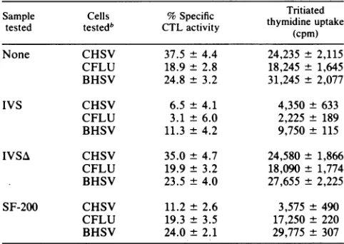
Generation and demonstration of the antigen-specific sup-pressor factor. Supernatant fluids from HSV-stimulated,
HSV-immune T-lymphocyte cultures significantly inhibited HSV-specific CTL induction and lymphoproliferation when
addedto testcultures. Whereas the unfractionated
suppres-sive supernatant also inhibited responses to influenza virus
(Table 1), gel filtration on Sephacryl S-200 revealed apeak
with an apparent molecular weight of 90,000 to 100,000
(SF-200) which inhibited both CTL induction and
lymphoproliferation in an antigen-specific and genetically restricted fashion. Thus, the cell-mediated immunity re-sponses to HSV weresuppressed,but the same responses to influenza virus were barely affected. Likewise, the
HSV-TABLE 1. SuppressionofHSV-specific cell-mediatedimmunity byasuppressorfactorproduced by HSV-stimulated,
HSV-immuneTlymphocytesa
Sample Cells %Specific Tritiated
tested testedb CTLactivity thymidine uptake
(cpm)
None CHSV 37.5 ±4.4 24,235± 2,115
CFLU 18.9 ± 2.8 18,245 ± 1,645 BHSV 24.8 ± 3.2 31,245 ± 2,077
IVS CHSV 6.5 ± 4.1 4,350 ±633
CFLU 3.1 ± 6.0 2,225 ± 189
BHSV 11.3 ± 4.2 9,750 ± 115
IVSA CHSV 35.0 ± 4.7 24,580 ± 1,866
CFLU 19.9± 3.2 18,090 ± 1,774 BHSV 23.5 ± 4.0 27,655 ± 2,225
SF-200 CHSV 11.2± 2.6 3,575 ±490
CFLU 19.3 ± 3.5 17,250± 220 BHSV 24.0± 2.1 29,775 ± 307 aSplenocytesfrom HSV-immunemice wereincubatedwithHSVantigens
for3days and then depleted of macrophages and B cellsbypassage over nylon wool columns. The nonadherent cellsweredeterminedtobegreater than95%Thy 1.2+ by antibody and complement-mediated cytotoxicity. Thy 1+ cells were then cultured an additional 3 days in serum-free medium. Afterwards, the cells werepelleted by centrifugation, and thesupernatant
(IVS)wasassayed forsuppressoractivity.In someexperiments, the super-natant washeated at60'Cfor15minIVSA. Serum-freesupernatantswere concentrated1,000-fold andappliedtoaSephacrylS-200gel filtration column. Those fractions which suppressed HSV-specific but not influenza virus-specific CTL induction were pooled (SF-200). All supernatant fluids and columnfractionswereassayedat afinal concentrationof20%o(vol/vol)after
dialysisovernight againstserum-freeRPMI-1640. Data areexpressedasthe mean+ standarddeviation.
IC4SV, HSV-stimulated,HSV-immunesplenocytesfromC3HIHeJmice;
CFLU,influenzavirus-stimulated,influenza virus-immunesplenocytes from
C314/HeJ mice; BHSV, HSV-stimulated, HSV-immune splenocytes from C57BL/6mice.
TABLE 2. Antigen-specific suppressionofHSV-specific
cell-mediatedimmunity by the I-J+ suppressoractivityof SF-200a Sampletested
Cellsb
%Specific Tritiatedthymidinetested CTL activity uptake (cpm)
None HSV 37.5 + 5.2 24,235 ± 2,135
FLU 18.9 ± 2.2 18,245± 1,260
I-J- HSV 43.3 ± 6.5 22,755 ± 2,305
FLU 18.5 ± 2.2 18,880± 1,655
I-J+ HSV 3.2 ± 2.2 6,445 ± 702
FLU 19.0 ± 2.4 18,555± 225 Immunoglobulin- HSV 2.4 ± 2.4 7,257± 577 FLU 19.7 ± 2.1 17,987± 1,855
Immunoglobulin+ HSV 42.1 ± 6.0 24,895 ± 2,366 FLU 21.5 ± 3.2 18,535± 966 aSF-200 fractions were concentrated and applied toanti-I-Jk and
anti-immunoglobulin immunoaffinity columns. The unadsorbed fractions (I-J-,
immunoglobulin-)and thosefractions collected after elution withapHbuffer (I-J+,immunoglobulin+)wereconcentrated 15-foldanddialyzed against PBS and then RPMI-1640before the assay. All samples weretested at afinal concentration of10%(voL/vol). Each determination wasperformedatleast fourtimes. Data areexpressedasthe mean +standarddeviation.
bHSV-immune, HSV-stimulated (HSV)orinfluenzavirus-immune, influ-enzavirus-stimulated (FLU) C3H/HeJ splenocytes were incubated in vitro with thecolumn eluates for5days. Both the CTL andlymphoproliferative responses ofthe cultures were assessed as described in Materials and Methods. The resultsreportedarefromarepresentative experiment.
specific responses ofC57BL/6
(H-2b)
micewere unaffected by C3H/HeJ(H-2k)-generated
SF-200. Furthermore, this suppressor activity was heat labile, suggesting a protein composition. The suppressoractivitywasnotgenerated by antigen-stimulated normal splenocytes or by unstimulated immune cells (21).Characterization of the antigen-specific suppressor factor.
Antigen-specific suppressorfactors frequently possess
de-terminants which react with anti-I-J antibodies (56). The exposure of SF-200 to an anti-IlJk immunoaffinity column resulted in the removal of the suppressoractivity (Table 2). Incontrast, exposure to an anti-immunoglobulin immunoaf-finity column failed to reduce the suppressor activity in SF-200. Thematerial which boundtotheanti-I-J columnwas elutedby application ofa low-pH glycine buffer, dialyzed,
and tested for suppressor activity against HSV- and
influ-enza
virus-specific
CTLinduction. Theacid eluate fromtheanti-I-J column contained the HSV-specific suppressor
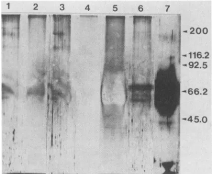
ac-tivity (HSV-SF) (Table 2). Furthermore, SDS-PAGE and Western blotting demonstratedthatthis eluate also reacted withtheanti-I-J antiserumbut notwith eithertheanti-I-Aor theanti-I-E antiserum(Fig. 1A, B, and D). Silver staining of
the gel used to determine molecular weight demonstrated four
protein
bands inthe55,000-to70,000-molecular-weight region contained within HSV-SF(Fig. 2, lane 6).The high-est-molecular-weight band corresponded to the single I-J+band in the Western blots. The other bands in the silver-stainedgelwerepresumedtorepresentdegradation products
as aresultoftheacid elution fromthe anti-I-J columnprior
to SDS-PAGE because they no longer bound I-J anti-bodies in subsequent Western blotting. HSV-SF samples
treated with 2-mercaptoethanol wereincompletely reduced
(Fig. 2, lane 7), precluding further interpretation of the
polypeptidenature of this factor.
InadditiontoexpressingI-Jdeterminants,
antigen-specific
suppressor factors often bear
anti-idiotypic
determinantsVOL.58, 1986 333
on November 10, 2019 by guest
http://jvi.asm.org/
[image:3.612.314.556.98.251.2] [image:3.612.56.297.414.585.2]334 HOROHOV ET AL.
A ANTIBODY
LANE
B
I-Ek
C D
NRS
I_Jk
E
HSV
12 1 2 1 2 12 1 2
'
d-"
.
'-...':..,:...~~~~~~~~~~~~~'iE
.''"MI;ag'aaLv
l ...u .!.
Iffi~~~~~
-,685KD
FIG. 1. Western blot analysis of the HSV-specific suppressor factor. Anti-I-J immunoaffinity column fractions were submitted to SDS-PAGE. Following electrophoresis, proteins were electroeluted from the gels onto nitrocellulose and then incubated with anti-I-A antiserum (panelA),anti-I-E antiserum (panelB),normal rabbitserum(panelC),anti-I-J antiserum (panelD), oranti-HSV antiserum(panel E). Lane1is the anti-I-J+immunoaffinity column-purified material, and lane2is the drop-through from theanti-I-J immunoaffinity column. KD, Kilodaltons.
which react with specific antibodies against the inducing
antigen (3, 51). We nextdetermined whether I-J+ HSV-SF alsoreacted with aspecificrabbitanti-HSVantiserumwhich
has been shown to share idiotypes with both human and
murine anti-HSV antisera(J. R. Lathey,B. T. Rouse, D. E.
Wiley,and R. J.Courtney, Immunology, inpress). Through Westernblotting,asingle band in
I-J'
HSV-SFwasdetectedwith the anti-HSV antiserum but not with normal rabbit
serum(Fig. 1C and D). This banddirectly corresponded to boththe
I-J'
bandin theanti-I-JWesternblot(Fig. 1D) and the68,500-molecular-weight
band in the silver-stained gel (Fig.2,lane 6). Together, theseresultsindicatethat theI-J'
suppressor activity reacted with the HSV-specific anti-serum.
Induction of nonspecific suppressor activity in
HSV-immune, HSV-stimulated splenocyte cultures by
I-J',
HSV-specificsuppressor activity.Anti-idiotypicsuppressorfactors in the hapten models are not themselves responsible for suppression but instead induce other cells to produce the eventualsuppressormolecule(2, 34).Wewishedtoestablishif suppression induced
by
concentrated eluates from theanti-I-J immunoaffinity column (HSV-SF) was mediated
directly
or depended on the activity of some auxiliary mechanism. To test this, we treated virus-stimulated orunstimulated HSV- or influenza virus-immune splenocyte populations (acceptorpopulation) with HSV-SFfor3 days,
after which the culture supernatants were collected and
assayed againstboth HSV- and influenzavirus-specific CTL
induction (Table 3). As reported elsewhere, low levels of suppressor activity were detectable after 3 days of HSV
stimulationoftheHSV-immune splenocytes(21). However,
the addition of HSV-SF to the stimulated, HSV-immune acceptor population resulted in an increase in the
nonspecific suppressor activityin the supernatantfluids. In the absence of HSV stimulation, HSV-SF failed to induce
theproduction of thenonspecific suppressoractivity by the acceptorpopulation (Table 3, group F). Furthermore, such
factorswere notgeneratedbyantigen-stimulated,
HSV-SF-treated normal acceptor populations orby influenza
virus-primed splenocytes.
The ilature of the cell type responding to the HSV-SF
stimulus was further investigated. Levels of suppression
weremarkedlyreduced iftheacceptorpopulationwastaken
from mice treated 2days previouslywith
cyclophosphamide
(Table 4). Furthermore,ifLyt 1+ or I-J+ but not Lyt 2+cells
were removed from the acceptor population prior to the addition of HSV-SF and antigenic stimulation, the
nonspe-1 2 3
r
1p
44 b
.:
,...
B....-..
.+,,.,+,
. 3,. at
F
1t
.'T}m: 4
S )B.
.:
#
.§
4 5 6 7
.116.2
-92.5
FIG. 2. SDS-PAGE ofbulk suppressive supernatant and frac-tionated components. Protein samples (40 ,ug) were submitted to SDS-PAGE, and the gelwassilver stained. Lanes: 1, bulk superna-tant; 2, S-200fraction; 3 and 4, drop-through and eluate, respec-tively, from the anti-immunoglobulin immunoaffinity column;5and 6,drop-through and eluate (HSV-SF), respectively, from the anti-I-J immunoaffinity column; 7, HSV-SF after treatment with 2-mercaptoethanol. Themigration ofthemolecularweightmarkersis indicated, andthe values are expressedinkilodaltons.
J.VIROL.
I-Ak
on November 10, 2019 by guest
http://jvi.asm.org/
[image:4.612.136.487.74.273.2] [image:4.612.335.546.467.641.2]REGULATION OF ANTI-HSV RESPONSES BY SUPPRESSOR FACTOR TABLE 3. Production of nonspecific suppressor factors by HSV-SF-treated splenocytesa
Acceptor population Virus added HSV-SF CTLinduction %SpecificCTL %Suppression
added culture tested activity
A. None None - HSV specific 33.6± 6.6 b
B. HSV immune HSV - HSV specific 34.0 ± 6.0 0
C. HSV immune HSV + HSV specific 21.0± 3.0 37.5c
D. FLU immune HSV + HSVspecific 31.2± 5.0 7.1
E. None None - FLU specific 25.8± 5.1
F. HSV immune FLU + FLU specific 26.2± 4.2 0
G. HSVimmune HSV + FLU specific 15.8± 2.9 38.8c
H.FLU immune FLU + FLUspecific 23.3 ± 4.7 9.7
aHSV-SFwasaddedtovarious splenocyte cultures.Afterincubationfor3 days, thesupematantfluidswerecollected and assayed forsuppressoractivity with
HSV-specific C3H/HeJ and influenza virus (FLU)-specific C57BL/6 CTL induction cultures. The supernatant fluids were addedto the culturesat afinal concentration of25%(vol/vol).Thepercentspecific CTL activity of the test cultureswascalculatedasdescribed in Materials and Methods. Percentsuppression = [1 - (specific CTL activity of HSV-SF-treated induction cultures/specific CTLactivity of untreated CTL induction cultures)] x 100. Data representfour experiments and areexpressed asthe mean±standarddeviation.
b_,Untreated controls for eachCTL induction culturetested.
cSignificantly different from the untreated controlsatP<0.01.
cific suppressor factor was not produced. These results
indicate that the acceptor cell necessaryfor the production
ofthenonspecific suppressor activityfollowingexposure to HSV and HSV-SF was I-J+, Lyt 1+2-, and
cyclophospha-mide sensitive.
DISCUSSION
Theinvitro stimulation of HSV-immunesplenocyteswith HSV antigens results in the production of a suppressive
supernatant (21). This suppressive supernatant contained both nonspecific and HSV-specific soluble suppressor
fac-TABLE 4. Production ofnonspecific suppressorfactorsby Lyt 1+ splenocytes treated with the I-J+, HSV-specific
suppressor
factor-Supematant
from CTL induction oSpecific
HSV-SF-treated CTL tsd CTL %Suppression
acceptor atvt
population
None HIV 33.6 ± 5.9 b
HI HIV 34.0 + 5.0 0.0
HIV HIV 21.0 ± 4.1 37.5C
HIVLyt 1- HIV 30.0 ± 5.5 10.7
HIVLyt2- HIV 22.2 ± 3.8 33.9c
HIV I-J- HIV 32.4 ± 6.2 3.6
HIVCY HIV 29.6 ± 4.5 11.9
None FIV 25.8 ± 4.1
HI FIV 24.0 ± 3.9 7.0
HIV FIV 15.8 ± 4.8 38.8c
HIVLyt1- FIV 23.3± 3.5 9.7
HIVLyt 2- FIV 16.6 ± 4.1 35.7C
HIV I-J- FIV 23.3± 3.1 9.7
HIVCY FIV 21.2 ± 2.8 17.8
aI-J+ effluents from immunoaffinity column-purified SF-200 (HSV-SF)
wereaddedtovarious splenocytecultures. Some of thesplenocyte popula-tions were pretreated withanti-Lyt 1.1,anti-Lyt 2.1,oranti-I-Jkantiserum andcomplement priortothe addition of theHSV-specific suppressor factor. Thissuppressorfactor was added to these cultures at afinal concentration of
2.5%(vol/vol). Onday3ofculturing,the supernatantfluidswereharvested andtested for suppressoractivity againstCTL induction as described in Table 3, footnotea. Data areexpressedasthe mean ± standarddeviation. HIV, HSV-stimulated, HSV-immunesplenocytes; HI, HSV-immune splenocytes; FIV, influenza virus-stimulated, influenza virus-immune splenocytes; HIV CY, splenocytes from HSV-immune mice pretreatedwithcyclophosphamide (50mg/kg)48 hpriortosacrifice.
b-, Untreated controlsforeachCTLinductionculture tested.
cSignificantly different fromthe untreated controlsatP<0.01.
tors which could be separated from each other by gel filtration. In this report, we further characterized this HSV-specific suppressoractivity and determined its relationship tothenonspecific suppressoractivity present in the suppres-sive supernatant.
Antigen-specific suppressor factors frequently bear I-J determinants (56) and thus can be bound by anti-I-J antibod-ies, allowing the selective adsorption of these molecules. Since it was possible to selectively adsorb out as well as recoverthe HSV-specific suppressor activity from an anti-I-Jimmunoaffinity column, it appears that the HSV-specific suppressorfactor bears I-Jdeterminants. Western blot anal-ysis of this immunoaffinity-purified suppressor material (HSV-SF) demonstrated a component which reacts with both I-J- andHSV-specific antisera (Fig. 1D and E) and has an apparent molecular weight of 68,500. The other bands observed in Fig. 2, lane 6, are most likely HSV-specific suppressor factor(s), either of the same species as the 68,500-dalton, I-J+ HSV-SForof otherspecies which have
acid-labileI-Jdeterminantsand presumably donotfunction afterimmunoaffinitypurification.
It is noteworthy that although HSV-SF was murine de-rived and a T-cell factor, we were able to detect this
molecule with a rabbit anti-HSV
antiserum,
thereforeindi-cating a sharing of idiotype between mice and rabbits.
Idiotypic cross-reactions between different animal species, including mice and rabbits, have been reported for a viral
antigen (27). Furthermore, it has beendemonstrated in our
laboratory that a murine-derived monoclonal antibody against glycoprotein D of HSV bears an idiotype which is also found in rabbit, murine, and human antisera against HSV (Lathey et al., in press). Such is the case with the rabbitanti-HSV antiserum used in this communication.
The reactivityof the suppressorfactorwiththe anti-HSV
antiserum indicates severalpossibilities regarding the
com-position of HSV-SF and its mechanism ofantigenic speci-ficity. Two possibilities are that the suppressor activity
represents either a viral protein or a host-derived protein
which binds viral proteins. Alternatively, the reactivity of the factor with the anti-HSV antiserum could be aresult of the presence ofanti-idiotypic determinants on the suppres-sor factor. We favorthe latter hypothesis for several rea-sons. Itseemsunlikelythatthe suppressoractivity is medi-atedbyavirus-encodedprotein, since HSV doesnotusually infect and persist in lymphocytes and since none of its proteinshave been showntobe immunosuppressive.
Like-VOL.58,1986 335
on November 10, 2019 by guest
http://jvi.asm.org/
[image:5.612.59.299.437.619.2]wise,
supernatants from HSV-stimulated normalspleno-cytes didnotcontain the suppressor
factor,
thusindicating
that infectionwith thevirus was aninsufficient stimulusfor suppressor induction.
Finally,
the presence ofI-Jdetermi-nantsalsomakes
unlikely
thepossibility
that thesuppressormolecule was of viral
origin.
Anotherpossibility
is that ahost-derived factor
directly
boundaviralprotein. However,we suggest that unless the suppressor factor covalently bound the viral
antigen (which
isa remotepossibility),
sucha
complex
would not be intactfollowing
elution from theanti-I-J
immunoaffinity column;
this wouldresult inmultiple
bands
being
detected on silver-stained HSV-SF.Multiple
bandswere
observed,
but thesingle
band which was visu-alizedon boththeanti-HSV and anti-I-JWestern blotswasofthe same relative molecular
weight,
whereas no otherbands reacted with the
polyclonal
anti-HSV antiserum.Therefore,
it isunlikely
thatHSV-SF isanI-J+ factor which bindsantigen,
because bands which reactedonly
with theanti-HSV antiserumandnotwiththe anti-I-Jantiserumwere not observed. In contrast,
antigen-specific I-J+
suppressorfactors whichexpress
anti-idiotypic
determinantshave beendemonstrated in othersystems
(13,
50, 53).
Suchfactorsplay
an
important
role in the transmission ofsuppressogenic
signals
inhapten-specific
suppressor cellcircuits(12,17, 18,
22).
A characteristic of these factors is that they arefre-quently composed
oftwosubunits heldtogether
by
disulfide bonds(3, 15, 31),
although
single-chain
factors have also been described(24,
26).
Atthistime,
itisuncertain whether the suppressorfactor described in this communication is oftheone- or
two-polypeptide-chain variety.
Thistopic
is still underinvestigation.
At this time we can
only
speculate
on how theHSV-specific
suppressorfactor(HSV-SF)
maybeinvolvedin theregulation
ofHSV-specific
CTL andlymphoproliferative
responses.
By
analogy
withhapten
systems,wesuppose thatthefactor forms part ofa
regulatory
cell circuitthat modu-lates immuneresponsiveness
to HSV. In support ofthis,
elsewherewehave demonstratedtheinvolvement ofatleast
three cell types and twofactors in thecircuit that controls
HSV-specific
CTL andlymphoproliferative
responses in vitro(21,
22).Assuch,
weconsider HSV-SFtobeaproduct of a second-order suppressor(Ts2)
cell which we haveidentified as a
Lyt
2+, I-J+,
HSV-immune Tlymphocyte,
presumably anti-idiotypic
in itsantigenic specificity.
Asobserved in
hapten
systems(2,
12, 13), Ts2
cellsproduce
ananti-idiotypic
suppressor factor which is also I-J+. Thisantigen-specific
factor fails tomediatesuppression
directly.
Instead,
it isrecognized
by
anantigen-activated
acceptor(Ts3)
cellthatresponds
by
producing nonspecific
suppressorfactors that
apparently
act as the actual suppressors(34).
Our results showed that
spleen
cells from HSV-immune micegenerated
nonspecific
suppressoractivity
whenex-posed
toHSV-SFalong
withantigen.
Theproduction
of thenonspecific
suppressoractivity
was shown torequire
cyclophosphamide-sensitive,
HSV-primed splenocytes
which
expressed
the Lyt 1+2-, I-J+phenotype.
Accord-ingly,
the cell resembles the suppressor-acceptor cellde-scribed
by
Asherson et al. thatregulates
hapten-specific
contact
sensitivity
(2, 34). However,
in thepicryl
chloridemodel,
thecyclophosphamide-sensitive,
suppressor-acceptor cellwasidentifiedas aLyt 2+
cell andwaspresent in micewhich were contactsensitized, although
notneces-sarily
withpicryl
chloride.Thus,
thesystemwedescribe is similaralthough
notidenticaltothehapten
model.However,
final
comparisons
must await thedevelopment
of morepurified
cellpopulations
andsuppressor T-cellhybridomas.
It is important to understand how suppressor cells and factors interact with the immune system invitro,sinceclues which will prove of value in vivo may emerge on how to
manipulatethe system. Forexample,suppressorcellscould serve to inhibit the protectiveaspects ofimmunitypriorto the development of recrudescent disease. There is some evidence that helper/suppressor T-lymphocyte ratios do
change around the time ofrecrudescence in humans
(48);
recently,ithas been shown in theguineapigmodel ofHSV type2recrudescence that suppressor cellsarepresentinthe spleen at the time of recrudescent disease(23). If suppressor cells andtheir products are indeed involved in modulating immunity to HSV in humans, they could provide auseful targetfor treatment aimed at breaking the cycle. Whether theoperation of a similar suppressor cell circuit accountsfor the variation of host responsiveness that accompanies the development of herpesvirus recrudescent disease requires
furtherinvestigation.
ACKNOWLEDGMENTS
Wegreatlyappreciatetheenthusiastic technical support of Linda Miller.
This workwassupportedbyPublic Health Service grants A114981 and A118960from theNational Institutes ofHealth.
LITERATURE CITED
1. Aoki, I., M. Minami, and M. E. Dorf. 1983. A mechanism responsible for the induction of H-2 restricted second order suppressorTcells. J. Exp. Med. 157:1726-1735.
2. Asherson, G. L., V. Colizzi, and M. A. Zembala. 1985. The
structureandbiologicalaction ofantigen-specificT suppressor factor,p.79-95.InM. J.Taussig(ed.),Tcellhybridomas.CRC Press, Inc., Boca Raton, Fla.
3. Asherson, G. L., M. C. Watkins, M. A. Zembala, and V. C. Colizzi. 1984. Two chain structure of T-suppressor factor: antigen-specific T-suppressor factoroccurs as asinglemolecule andasseparateantigenbindingandI-J'parts,both of whichare
requiredfor biologicalactivity. Cell. Immunol. 86:448-459. 4. Aune, T. M., C. M. Sorensen, and C. W. Pierce. 1982.
Non-antigen specific suppressor T cell mediators: structure and action, p. 387-410. In M. Feldmann and M. H. Schrier (ed.), Lymphokines. Academic Press, Inc.,NewYork.
5. Axen, T.,T.Porath, and S. Ernbach.1967.Chemical coupling of peptides and proteinstopolysaccharides bymeansofcyanogen halides. Nature(London) 214:1302-1304.
6. Bukowski, J. F., S. Woda, K. Habu,K. Okamura, and R. M. Welsh.1983. Natural killer celldepletion enhances viral synthe-sis and virus-induced hepatitis in vivo. J. Immunol. 131:1531-1538.
7. Chan, M., D. Clark, andW. E. Rawls. 1983. Pichinde virus-specificcell-associatedsuppressionofprimary footpadswelling inaninbred strain ofSyrianhamsters. J. Immunol. 130:925-931. 8. Charoenvit, Y., G.H.Campbell,andS. Tokuda. 1981. Suppres-sion ofparasiteantigen-specificlymphoid blastogenesis in Afri-cantrypanosomiasis. J. Immunol. 127:2350-2354.
9. Chong,K.T.,andC. A.Mims. 1983.Antigen-specific suppres-sion ofdelayed-type hypersensitivityto murine
cytomegalovi-rusin MCMV-infected mice.J. Gen. Virol.64:2433-2439. 10. Collins,F. M.,andS. R. Watson. 1979. Suppressor T-cells in
BCG-infected mice. Infect. Immun. 25:491-496.
11. Compton, T., and R. J. Courtney. 1984. Evidence for post-translationalglycosylationofanonglycosylatedprecursor pro-tein ofherpes simplex virustype 1. J.Virol. 52:630-637. 12. Dorf, M. E., and B. Benacerraf. 1984. Suppressor cells in
immunoregulation. Annu.Rev.Immunol. 2:127-158.
13. Dorf, M. E., K. Okuda, and M. Minami. 1985. UseofTcell hybridomastoanalyzeasuppressor cellcascade,p. 97-109.In M.J.Taussig (ed.),Tcellhybridomas. CRC Press, Inc., Boca Raton,Fla.
on November 10, 2019 by guest
http://jvi.asm.org/
REGULATION OF ANTI-HSV RESPONSES BY SUPPRESSOR FACTOR 14. Doughty, B. L., and S. M.Phillips. 1982. Delayed
hypersensi-tivity granuloma formation and modulation around Schistosoma mansoni eggs in vitro. II. Regulatory Tcell subsets. J.Immunol. 128:37-41.
15. Flood, P. M., A. Lowy, A. Tominaga, B. Chue, M.I. Greene, and R. K. Gershon. 1983. Igh variable region-restricted Tcell interactions. Genetic restriction of an antigen specific suppres-sor inducer factor is imported by an I-J' antigen-nonspecific molecule. J. Exp. Med. 158:1938-1947.
16. Flood, P. M., K. Yamanchi, andR.K.Gershon. 1982. Analysis of the interactions betweentwomolecules that are required for the expression ofLy 2 suppressorcell activity. Three different types of focusingevents may be needed todeliverthe suppres-sive signal. J. Exp. Med. 156:361-371.
17. Germain, R. N., and B. Benacerraf. 1981. A single major pathway ofT lymphocyte interactions in antigen-specific im-mune suppression. Scand. J. Immunol. 13:1-10.
18. Green, D. R., P. M. Flood, and R. K. Gershon. 1983. Immuno-regulatory T-cell pathways. Annu. Rev. Immunol. 1:439-464. 19. Green, W. F., and D. G. Colley. 1982. Modulation of
Schistosoma mansoniegg-inducedgranulomaformation. II. I-J restriction of Tcell-mediated suppression in a chronic parasite infection. Proc. Natl. Acad. Sci. USA78:1152-1156.
20. Greene, M.I., and H. L. Weiner. 1980. Delayedhypersensitivity in mice infected with reovirus. II. Induction oftolerance and suppressor T cells to viral specificgene products. J. Immunol. 125:283-287.
21. Horohov, D. W., R. N. Moore, and B.T. Rouse. 1985. Produc-tion of soluble suppressor factors by herpes simplex virus-stimulated splenocytes from herpes simplex virus-immune mice. J. Virol. 54:798-803.
22. Horohov, D. W., R. N. Moore, and B. T. Rouse. 1985. Regula-tion of herpes simplex virus-specific lymphoproliferation by suppressor cells. J. Virol. 56:1-6.
23. Iwasaka, T., J. F.Sheridan,andL.Aurelian. 1983. Immunityto herpes simplex virus type 2: recurrent lesions are associated with the induction ofsuppressor cells and soluble suppressor factors. Infect. Immun. 42:955-964.
24. Jayaraman, S., and C. J. Belione. 1985. Hapten-specific re-sponses to phenyltrimethylamino hapten. V. A single chain antigen-binding I-J'first-order suppressorfactor requires anti-gen to induceanti-idiotypic second-order suppressorT cells. J. Immunol. 134:1010-1018.
25. Kaplan, D. R., T. J. Henkel, V. Braciale, and T. J. Braciale. 1984.Mycoplasma infectionofcell cultures: thymidine incorpo-ration ofculture supernatants as ascreening test. J. Immunol. 132:9-11.
26. Kapp, J. A., and B. A. Araneo. 1982. Structureand function of monoclonal antigen-specific suppressor T cell factor, p. 187-195. In M. Feldmann and M. H. Schrier (ed.), Lymphokines. Academic Press, Inc., New York.
27. Kennedy, R. C., I. Sonescu-Matin, Y. Sanchez, and G. R. Dressman. 1983. Detection of interspecies idiotypic cross-reactions associated with antibodies to hepatitis B surface antigen. Eur. J. Immunol. 13:232-235.
28. Kramer, M., and U. Koszinowski. 1982. Tcell-specific suppres-sorfactor(s) withregulatory influence oninterleukin 2 produc-tion. J. Immunol. 128:784-790.
29. Larsen, H. S.,M.-F.Feng, D. W. Horohov, R. N.Moore,and B. T.Rouse. 1984. Role ofT-lymphocyte subsets inrecoveryfrom herpes simplex virus infection. J. Virol. 50:56-59.
30. Lawman, M. J. P., B. T. Rouse, R. J. Courtney, and R. D. Walker. 1980. Cell-mediated immunity against herpes simplex induction of cytotoxic T lymphocytes. Infect. Immun. 27:133-139.
31. Lei, H.-Y., and C. Waltenbaugh. 1984. Regulation ofimmune responses by I-J gene products. IV. Distinct suppressorfactors derived from "nonsuppressor" A strain mice. J. Immunol. 133:1723-1729.
32. Leung, K. N., R. B.Ashman,H.C. J. Ertl, and G. L. Ada. 1980. Selective suppression ofthe cytotoxic T cell response to influ-enza virus inmice. Eur. J. Immunol. 10:803-810.
33. Liew, F. Y., and S. M. Russell. 1980. Delayed-type
hypersensi-tivityto influenzavirus. Induction of antigen-specific suppres-sorT cells for delayed-type hypersensitivity to hemagglutinin during influenza virus infection in mice. J. Exp. Med. 151:799-814.
34. Malkovsky, M., G. L. Asherson, P. Chandler, V. Colizzi, M. C. Watkins, and M.Zembala. 1983.Non-specific inhibition ofDNA synthesis elaborated by T acceptor cells. I. Specifichapten- and I-J-drivenliberation of an inhibitor ofcellproliferationbyLyt 1-2+ cyclophosphamide sensitiveTacceptor cells armed with aproduct of Lyt 1+ 2+ cells.J. Immunol. 130:785-790. 35. Mathur, A., S. Rawat, andU. C.Chaturvedi.1984. Suppressor
Tcells fordelayed-type hypersensitivitytoJapanese encepha-litis virus. Immunology 52:395-402.
36. Mehra, V., J. Convit, A. Rubenstein, and B. R. Bloom. 1982. Activated suppressor T cells in leprosy. J. Immunol. 129:1946-1951.
37. Mitchell, G.F.1984. Host-protectiveimmunity and its suppres-sion in a parasitic disease: murine cutaneous leishmaniasis. Immunol. Today 5:224-226.
38. Murphy, J. W., and J. W. Moorhead. 1982. Regulation of cell-mediated immunityin cryptococcosis. I.Induction of spe-cific afferent suppressor cells by cryptococcal antigen.J. Immu-nol. 128:276-283.
39. Nash, A. A., P. G. H. Gell, and P. Wildy. 1981. Tolerance and immunity inmice infected with herpes simplex virus. Simulta-neous induction ofprotective immunity and tolerance to de-layed-type hypersensitivity. Immunology 43:153-159.
40. Petit, J.-C., G. Richard, B. Albert, and G.-L. Daguet. 1982. DepressionbyPseudomonas aeruginosa oftwoT-cell-mediated responses, anti-Listeria immunityanddelayed-type hypersensi-tivity to sheeperythrocytes. Infect. Immun. 35:900-908. 41. Ptak, W., M.Zembala, and R. K. Gershon. 1978. Intermediary
role of macrophages in the passage of suppressor signals be-tween T cell subsets. J. Exp. Med. 148:424-434.
42. Rich, R. R., and C. W. Pierce. 1974. Biological expressions of lymphocyte activation. III. Suppression ofplaque-forming cell responses in vitro by supernatant fluids from concanavalin-A activated spleencellcultures. J. Immunol. 112:1360-1368. 43. Rinaldo, C. R., Jr., W. P. Carney, B. S. Richter, P. H.Black,
and M. S. Hirsch. 1980. Mechanism ofimmunosuppression in cytomegalovirus mononucleosis. J. Infect. Dis. 141:488-495. 44. Rocklin, R. E.,J. W. Tracy, and A. E. Kholy. 1981. Activation
ofantigen-specific suppressor cells in human schistosomiasis mansoni by fractions of soluble egg antigens nonadherent to Con A Sepharose.J. Immunol. 127:2314-2318.
45. Schrier, R. D.,L. I. Pizer, and J. W. Moorhead. 1983. Tolerance andsuppressionofimmunitytoherpes simplexvirus: different presentations of antigens induce different types ofsuppressor cells. Infect. Immun. 40:514-522.
46. Schwab, J. H. 1975. Suppression ofthe immune response by microorganisms. Bacteriol. Rev. 39:121-143.
47. Scott, P. A., and J. P. Farrell. 1981. Experimental cutaneous leishmaniasis. I. Nonspecificimmunodepression in Balb/c mice infected withLeishmania tropica. J. Immunol. 127:2395-2400. 48. Sheridan, J. F., and L. Aurelian. 1983. Immunity to herpes
simplex virus type 2. V. Risk of recurrent disease following primary infection: modulationofT cellsubsets andlymphokine (LIF) production. Diagn. Immunol. 1:245-252.
49. Sheridan, J. F., A. D. Donnenberg, L. Aurelian, and D. J. Elpern. 1982. Immunity to herpes simplex virus type 2. IV. Impaired lymphokine production during recrudescence corre-lates with animbalance inT lymphocyte subsets. J. Immunol. 129:326-331.
50. Sherr, D. H., and M. E. Dorf. 1984. Characterization of anti-idiotypic suppressor T cells (Tsid) induced after antigen prim-ing. J. Immunol. 133:1142-1150.
51. Sumida, T., I. Takei, and M. Taniguchi. 1984. Activation of acceptor-suppressor hybridoma with antigen-specific suppres-sor T cellfactor oftwochain type: requirement ofthe antigen andthe I-J restricting specificity. J. Immunol. 133:1131-1136. 52. Taniguchi, M., I. Takei, and T. Tada. 1980. Functional and
molecularorganization of anantigen-specific suppressor factor from a T-cell hybridoma. Nature(London) 283:227-228. VOL. 58,1986
337
on November 10, 2019 by guest
http://jvi.asm.org/
53. Taniguchi, M., T. Tokuhisa, T. Itoh, and M. Kanno. 1984. Functional roles of two polypeptide chains that compose an
antigen-specific suppressor T cell factor. J. Exp. Med.
159:1096-1104.
54. Towbin, H., T. Staehelin, and J. Gordon. 1979.Electrophoretic transferofproteins from polyacrylamide gels tonitrocellulose sheets: procedureandsomeapplications.Proc. Natl.Acad.Sci. USA 76:4350-4354.
55. Truitt, G. A., R. R. Rich,andS. S. Rich.1978.Suppression of
cytotoxicTlymphocyte responsesin vitroby solubleproducts of alloantigen-activated spleen cells. J. Immunol. 121: 1045-1051.
56. Waltenbaugh,C. 1981.Regulation of immuneresponsesby I-J
gene products. I. Production and characterization of anti-I-J
monoclonal antibodies. J. Exp. Med. 154:1570-1583.
57. Wray, W., T. Boulikas, V. P. Wray, and R. Hancock. 1981. Silver staining of proteins in polyacrylamide gels. Anal. Biochem. 118:197-203.