RESEARCH
Immunological effects
of the intraparenchymal administration
of allogeneic and autologous adipose-derived
mesenchymal stem cells after the acute phase
of middle cerebral artery occlusion in rats
Zhang Yu
1,2†, Tang Wenyan
1†, Su Xuewen
1†, Dong Baixiang
3†, Wang Qian
1, Wang Zhaoyan
1, Yang Yinxiang
1,
Qu Suqing
1and Luan Zuo
1*Abstract
Background: Adipose-derived mesenchymal stem cell (ADMSC) therapy can promote recovery from cerebral ischemia; however, more information regarding appropriate sources of ADMSCs is required. This study was aimed at analyzing the immunogenicity of rat ADMSCs by comparing the immunological effects of intraparenchymal adminis-tration of allogeneic ADMSCs (allo-ADMSCs) and autologous ADMSCs (auto-ADMSCs) after the acute phase of middle cerebral artery occlusion (MCAO) in rats.
Methods: Allo- or auto-ADMSCs from rats (1 × 10⁶ cells) were transplanted into Lewis rats 8 days post MCAO. The
immunogenicity of ADMSCs was analyzed using coculture with T lymphocytes. The in vivo immune response induced by rat ADMSCs and the viability, migration, and differentiation of transplanted ADMSCs were detected using immu-nohistochemistry. Apoptosis within the populations of transplanted cells were detected using a TUNEL assay. Infarct volume was detected by 2,3,5-triphenyltetrazolium chloride staining. Post-treatment neurological function was evaluated using a modified neurological severity score and rotarod test. Data were analyzed using Kruskal–Wallis and Mann–Whitney U tests.
Results: Compared with allo-ADMSCs, auto-ADMSCs showed lower immunogenicity and evoked weaker immu-nological responses. Allo-ADMSCs evoked significantly stronger protein expression of interleukin-2 and interferon-gamma, as well as the local accumulation of CD4+ T lymphocytes, CD8+ T lymphocytes, and microglial cells. This indicates that auto-ADMSCs may contribute to higher survival rates, longer survival time, wider migratory scope, and fewer apoptotic cells. In addition, a small number of transplanted auto-ADMSCs expressed astrocyte-like and neuron-like markers 28 days after transplantation. We did not observe surviving transplanted allo-ADMSCs at this time point. We also found that auto-ADMSCs induced a greater degree of functional recovery and a greater reduction in infarct volume than allo-ADMSCs 28 days after transplantation.
Conclusions: Auto-ADMSCs were more effective than allo-ADMSCs in promoting recovery and reducing the infarct volume of MCAO rats. This could be associated with better viability, migratory ability, and differentiation potential, as
© The Author(s) 2018. This article is distributed under the terms of the Creative Commons Attribution 4.0 International License (http://creat iveco mmons .org/licen ses/by/4.0/), which permits unrestricted use, distribution, and reproduction in any medium, provided you give appropriate credit to the original author(s) and the source, provide a link to the Creative Commons license, and indicate if changes were made. The Creative Commons Public Domain Dedication waiver (http://creat iveco mmons .org/ publi cdoma in/zero/1.0/) applies to the data made available in this article, unless otherwise stated.
Open Access
*Correspondence: luanzuo@aliyun.com
†Zhang Yu, Tang Wenyan, Su Xuewen and Dong Baixiang contributed
equally to this work and should be considered co-first authors
1 Department of Pediatrics, Navy General Hospital, No. 6, Fucheng Road,
Haidian District, Beijing 100048, China
Background
Cerebral infarct or ischemic stroke is widely regarded as a major cause of disability and mortality [1]. No ideal treat-ment of cerebral infarct exists, and clinical outcomes are often poor. Many patients are left with permanent dis-abilities, including hemiplegic aphasia [2, 3]. Cell-based therapy is an interesting and emerging area of treatment, and is being explored for the treatment of strokes using embryonic stem cells [4], marrow-derived mesenchymal stem cells (BMSCs) [5, 6], and adipose-derived mesen-chymal stem cells (ADMSCs) [7].
Compared with embryonic stems cells and BMSCs, ADMSCs are advantageous because they are abundant, easily obtainable with minimal invasiveness, and readily culturable to a sufficient number for autologous trans-plantation without raising ethical concerns. Furthermore, ADMSCs have a shorter proliferation cycle than BMSCs and produce higher amounts of vascular endothelial cell growth factor and hepatocyte growth factor. A previous study demonstrated the therapeutic superiority of ADM-SCs over BMADM-SCs in an animal model of ischemic stroke, and particularly in improving brain function and reduc-ing infarct size [8]. In addition, a recent in vitro study showed that human allogeneic ADMSCs (allo-ADMSCs) have a lower immunogenicity than allogeneic BMSCs and evoke a weaker induction of cytotoxic T-lymphocytes [9]. However, a direct comparison of the immune responses elicited by and therapeutic effects of autologous (auto-) versus allo-ADMSCs has not been made to date.
Therefore, in this study, we aimed to clarify the supe-riority of auto-ADMSC transplantation and to analyze the underlying mechanisms in rats according to the per-formance of auto-ADMSCs in terms of the induction of the immune response, migration, survival, apoptosis, and differentiation; the improvement of neurological func-tion; and the reduction of infarct size. Ultimately, these analyses were aimed at providing a theoretical basis for the establishment of a more effective method for treating patients with cerebral infarction.
Methods
Experimental groups
Ninety pathogen-free adult male Lewis rats and 10 adult male Brown Norway (BN) rats (Beijing Vital River Labo-ratory Animal Technology Co., animal license number SCXK, Beijing 2012-0001) with a body weight range of 260–280 g were housed at 23 ± 2 °C, under a relative
humidity of 45 ± 15%, and a light/dark cycle of 12 h (lights on: 07:00 to 19:00), with free access to food and water. All Lewis rats underwent surgery to remove adipose tis-sue (see below) for the preparation of auto-ADMSCs and to induce middle cerebral artery occlusion (MCAO). All BN rats underwent surgery to remove adipose tissue (see below) for preparing allo-ADMSCs; MCAO was not induced. After establishing MCAO, the Lewis rats were randomly assigned to three experimental groups with 30 animals in each study group: an auto-ADMSC group, receiving autologous ADMSC transplantation; an allo-ADMSC group, receiving allogeneic allo-ADMSC transplan-tation; and a control group receiving only sterile saline. All experiments were designed to minimize animal suf-fering in compliance with Beijing’s Navy General Hospi-tal’s Ethical Committee guidelines for the Care and Use of Animals in Research. The researchers responsible for functional evaluation and molecular and histological studies were blinded to the treatment groups.
Isolation of ADMSCs from rats
All 90 Lewis rats and 10 BN rats were anesthetized via an intraperitoneal injection of 6% chloral hydrate at 6 mL/kg body weight. Inguinal fat pads were care-fully excised under sterile conditions, cut into < 1-mm3
pieces, and incubated with 0.125% type I collagenase solution (Sigma-Aldrich, St. Louis, MO, USA) at 37 °C for 80 min under constant agitation. After three washes with sterile saline, cell suspensions were prepared by gentle homogenization through a 100-µm cell strainer. Cells were collected via centrifugation, and a red blood cell lysing reagent was subsequently added (three times the volume of cells) (Noble Ryder Technology Co. Ltd., Beijing, China, Beijing, China). After pyrolysis for 5 min at room temperature, two sterilized saline washes were performed. Then, the cells were cultured in a serum-free medium (Sigma-Aldrich) for ADMSCs. Third-passage ADMSCs were used in subsequent experiments.
Cells were incubated with the appropriate antibodies in the dark at 4 °C for 30 min to confirm the presence or absence of MSC surface markers and major histocom-patibility complex (MHC) classes I and II by flow cytom-etry before transplantation. Antibodies used to detect MSC surface markers were PE-conjugated anti-rat CD29 (Biolegend, Santiago, CA, USA) and CD90 (Biolegend). Antibodies used to detect MHC class I and II were the anti-MHC class I RT Ia antibody [OX-18] (Biolegend)
well as a lower rate of apoptosis. Confirmation of the superiority of auto-ADMSCs and clarification of the underlying mechanisms will provide a theoretical basis for the improved clinical treatment of cerebral infarction.
and the anti-MHC class II antibody [MRC OX-6] (Biole-gend). The antibody PE-IgG1 (Biolegend) was used as an isotype control.
Coculture of ADMSCs and spleen lymphocytes in vitro
ADMSC immunogenicity was analyzed using co-culture with T lymphocytes. T lymphocytes were extracted and purified from the spleens of the Lewis rats and were cocultured for 3 days with auto- or allo-ADMSCs at a proportion of 1:10 in serum-free medium for ADMSCs. Then, the expression levels of IL-2 (eBioscience, San Diego, CA, USA) and IFN-γ (R&D Systems, Minneapolis, MN, USA) were detected using ELISA kits according to the manufacturers’ instructions.
ADMSC labeling before autologous transplantation
In order to track the transplanted ADMSCs in vivo, third-passage ADMSCs were plated at a density of 1 × 10⁶
cells per flask (T25) and then transduced with lentivi-rus expressing enhanced green fluorescence protein (EGFP) (Shanghai Genechem Co. Ltd., Shanghai, China) 3 days prior to transplantation. The optimal multiplica-tion of infecmultiplica-tion (MOI) in transducmultiplica-tion medium contain-ing 8 mg/L polybrene was 20. The medium was replaced 24 h after transduction. The efficiency of transduction was assessed by monitoring EGFP gene expression by fluorescence microscopy. Cell viability was determined prior to transplantation by trypan blue staining. Labeled cells were used for transplantation at a concentration of 1 × 10⁵ cells/µL.
Animal model of acute ischemic stroke
After ADMSCs were cultured for 2–3 weeks, acute stroke was induced in the Lewis rats that had undergone adipose-removal surgery. A transverse neck incision was made to expose the right common carotid artery, where a small incision was made to permit the insertion of a 0.28-mm diameter nylon filament. The filament was advanced into the distal right internal carotid artery to occlude the right middle cerebral artery, inducing brain infarction at the blood supply region. Two hours after occlusion, the nylon filament was removed, followed by closure of the muscle and skin layers. Neural function was evaluated using the Zea Longa 5-grade scale [10]. Seventy-five ani-mals with a score of 1–3 points were used in subsequent transplantation experiments.
Transplantation of ADMSCs
Eight days after the induction of ischemia, the animals were anesthetized and fixed to a stereotactic apparatus. The skull was exposed, and a burr hole was made 3 mm posterior and 1.9 mm lateral from the bregma using a small dental drill. A microinjector was inserted 2.9 mm
into the brain parenchyma from the surface of the dura mater, and 10 µL of cell suspension (approximately 1 × 10⁶ ADMSCs) or an equal volume of
physiologi-cal saline was injected into the brain parenchyma over a period of 20 min.
Apoptosis of transplanted ADMSCs
Apoptosis within populations of transplanted cells was detected using the TdT-mediated dUTP nick-end labe-ling (TUNEL) assay. One day after transplantation, the apoptosis of transplanted ADMSCs was detected in the peri-infarct zone using a TUNEL assay kit according to the manufacturer’s instructions (Beyotime, Shanghai, China). Cells were counted in one brain tissue section of each animal (n = 5 per group). The number of double-staining-positive (red and green fluorescence) cells was counted in a minimum of 10 microscopic fields based on their nuclear morphology, and dark color was quanti-fied using a 40× objective and Image-Pro image analysis software.
Viability, migration, differentiation, and immunological effects
The viability, migration, and differentiation of trans-planted ADMSCs, and the in vivo local accumulation of CD4+ T lymphocytes, CD8+ T lymphocytes, and micro-glial cells induced by rat ADMSCs, were detected using immunohistochemistry. The determination of concen-trations of IL-2 and IFN-γ in the brain induced by trans-planted ADMSCs was determined using an ELISA assay.
Five rats from each group were anesthetized at 1, 7, and 28 days after transplantation. After perfusion with nor-mal saline and 4% parafornor-maldehyde and dehydration with 15 and 30% glucose, brain tissues were harvested to produce cryosections. The localization of transplanted cells was observed and photographed under an inverted fluorescence microscope (Olympus IX51, Olympus Corporation, Tokyo, Japan). After EGFP markers were assessed quantitatively with the image analysis software Image-Pro Plus 6.0 (Media Cybernetics, Rockville, MD, USA), the survival rate of the transplanted cells in each group was calculated.
overnight, Abcam) to analyze the differentiation of trans-planted ADMSCs.
The distribution of CD4+ and CD8+ T lymphocytes and CD68+ and Iba-1+ microglias around transplanted ADMSCs was also investigated using immunohistochem-istry. To this end, these cells were labeled using anti-CD4 antibody (1:200 dilution, incubation at 4 °C overnight, Abcam), anti-CD8 antibody (1:200 dilution, incubation at 4 °C overnight, AbD Serotec, Kidlington, UK), anti-CD68 antibody (1:100 dilution, incubation at 4 °C overnight, Abcam), and the anti-Iba1 antibody (1:100 dilution, incu-bation at 4 °C overnight, Abcam), respectively. Red fluo-rescence-labeled (Alexa Fluor 594) secondary antibodies (Jackson ImmunoResearch, Hamburg, Germany) were diluted to 1:100 prior to use. Double-staining-positive (red and green fluorescence) cells were counted as previ-ously described.
To determine the concentrations of IL-2 and IFN-γ in the brain as induced by transplanted ADMSCs, ELISA assays were performed 7 days after transplantation. Five rats from each group were euthanized. Brain tissues from the hippocampus of the graft side were separated and ground on ice and then centrifuged at 5000×g for 5 min at 4 °C. Total protein in the supernatant was quantified using a BCA assay. Concentrations of IL-2 and IFN-γ were then determined using ELISA kits according to the manufacturer’s instructions. The protein expression lev-els of these factors in the brain tissue were expressed as nanogram per milligram total protein.
Assessment of motor function
Motor function was assessed at 14 and 28 days post transplantation using the modified Neurological Sever-ity Score (mNSS) and rotarod test by two investigators blinded to the experimental groups. The mNSS test is a composite of motor, sensory, balance, and reflex tests. Neurological function was graded on a scale of 0–18, where 0 is normal and 18 represents maximum deficit. In the rotarod test, the 10-cm diameter rotarod cylinder was accelerated from 5 to 40 rpm over 5 min. The time remaining on the rotarod was measured at 0, 14, and 28 days post transplantation, and the data were presented as the mean duration from three trials.
Assessment of relative cerebral infarction volume
In order to assess the relative cerebral infarction volume at 28 days post transplantation, 2,3,5-triphenyltetrazo-lium chloride (TTC; Sigma-Aldrich) staining was per-formed. Five animals from each group were anesthetized using 6% chloral hydrate. After the left ventricle was perfused with 200 mL physiological saline, the brain was removed, sliced into 2-mm sections, stained with 2% TTC for 30 min at 37 °C in the dark, and then fixed
with 4% paraformaldehyde. Under these conditions, live tissues retain a red color, while infarcted areas appear white. The ischemic area of the brain was quantified using ImageJ software (NIH, Bethesda, MA, USA), and ischemic volume was expressed as the percentage relative to the ipsilateral hemisphere.
Statistical analysis
Statistical analysis was performed using SPSS 17 for Win-dows (IBM, Chicago, IL, USA). Quantitative data are shown as mean values ± SDs. The Kruskal–Wallis test followed by the Mann–Whitney U test were used to com-pare functional evaluation scores, survival rates of trans-planted ADMSCs, numbers of apoptotic transtrans-planted ADMSCs, cerebral infarction volumes, and the expres-sion of IFN-γ and IL-2 in each group. Values of p < 0.05 were considered significant at 95% confidence.
Results
Characterization of ADMSCs from Lewis and BN rats
The third-passage cultured ADMSCs waiting for trans-plantation showed typical, fibroblast-like cell morphol-ogy (Fig. 1). Cultured cells from Lewis (96.78%) and BN (92.28%) rats were positive for the CD90-PE sur-face marker. Cultured cells from Lewis (96.76%) and BN (94.45%) rats were also positive for the CD29-PE surface marker. Non-MSC surface markers (CD34 and CD45) were expressed in less than 6% of cultured cells from Lewis and BN rats (Fig. 2). The MHC class I marker was expressed in 47.86% of the cultured cells from Lewis rats and in 28.82% of the cultured cells from BN rats, while the MHC class II marker was expressed in 5.36% of the cultured cells from Lewis rats and 2.76% of the cultured cells from BN rats (Fig. 3).
Immunogenicity of ADMSCs in vitro
To characterize the in vitro immunogenicity of ADM-SCs, we evaluated the levels of IL-2 and IFN-γ in mixed T-lymphocyte cultures prepared with either allo- or auto-ADMSCs. There were no significant differences in the production of IFN-γ and IL-2 between cultures with auto-ADMSCs and cultures without ADMSCs.
[image:4.595.306.540.615.697.2]Fig. 2 Expression of mesenchymal (CD90, CD29) and non-mesenchymal (CD34, CD45) stem-cell surface markers in transplanted cells from Lewis (a) and BN (b) rats as measured by flow cytometry
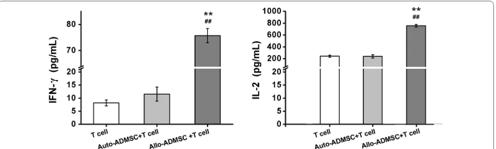
[image:5.595.61.539.87.335.2] [image:5.595.57.540.392.706.2]However, allo-ADMSCs induced a 6.56-fold increase in the production of IFN-γ and a 3.16-fold increase in the production of IL-2 when compared with auto-ADMSCs (p < 0.01, Fig. 4).
Apoptosis and the survival rate of transplanted ADMSCs in ischemic brain tissues
TUNEL staining was performed 1 day after transplanta-tion to detect the apoptosis of ADMSCs. ADMSCs were tracked by transfection with EGFP (green fluorescence), and the nuclei of the apoptotic cells were labeled by red fluorescence (TUNEL+) such that EGFP- and TUNEL-positive cells represented apoptotic-transplanted ADM-SCs (Fig. 5a, b). TUNEL staining showed that the number of apoptotic transplanted allo-ADMSCs per cubic mil-limeter of brain tissue near the infarct area was 3.19 times higher than that of apoptotic transplanted auto-ADMSCs (1323.02 ± 278.71 vs. 415.06 ± 68.79, p < 0.01; Fig. 5c). One day after cell transplantation, the survival rate of auto-ADMSCs was significantly higher than that of allo-ADMSCs (9.45 ± 0.34% vs. 4.19 ± 0.11%, p < 0.01; Fig. 5d). The survival rate of auto-ADMSCs was also sig-nificantly higher than that of allo-ADMSCs 7 days after transplantation (5.44 ± 0.25% vs. 1.33 ± 0.16%, p < 0.01; Fig. 5d). Twenty-eight days after cell transplantation, the survival rate of auto-ADMSCs was 3.35 ± 0.16% (Fig. 5d); however allo-ADMSCs were not observed in ischemic brain tissues at this time point.
Migration and expression of astrocyte or neuron markers of ADMSCs in ischemic brain tissues
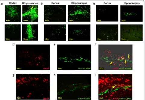
ADMSCs labeled with EGFP were transplanted into the CA1 region of the hippocampus. One day after cell transplantation, the migration of auto-ADMSCs was observed in the penumbra cortex and hippocampus, while the migration of allo-ADMSCs was observed only
in the penumbra cortex (Fig. 6a). The migration of allo-ADMSCs was observed in the hippocampus 7 days after transplantation. Overall, the migratory scope of auto-ADMSCs was wider than that of allo-auto-ADMSCs (Fig. 6b). Twenty-eight days after cell transplantation, auto-ADM-SCs were distributed mainly in the hippocampus, and to a lesser extent in the cortical injury area (Fig. 6c). How-ever, allo-ADMSCs were not observed in either the corti-cal injury area or the hippocampus (Fig. 6c).
Twenty-eight days after transplantation, cells posi-tive for EGFP (green fluorescence) and GFAP (red fluorescence) were found in the brain sections of the auto-ADMSC treatment group, indicating that the EGFP-labeled auto-ADMSCs expressed astrocyte markers. The percentage of EGFP- and GFAP-positive cells among all surviving auto-ADMSCs was 8.73 ± 0.92% (Fig. 6d–f). We also observed cells positive for EGFP and NeuN (red fluorescence) in the brain sections of the auto-ADMSC treatment group. The percentage of these immature neu-ron-like cells with spindle or round shapes that exhibited the expression of the apophyse marker was 2.14 ± 0.69% among all surviving auto-ADMSCs (Fig. 6g–i). Because surviving allo-ADMSCs were not observed on day 28 post transplantation, we did not examine the expres-sion of astrocyte or neuron markers for the administered allo-ADMSCs.
Immune response evoked by transplanted auto‑and allo‑ADMSCs in ischemic brain tissues
Immunohistofluorescence analysis demonstrated that allo-ADMSCs evoked a local immunological response, characterized by an accumulation of CD4+ and CD8+
T cells, CD68+ activated microglial cells, and IBA-1+
resting microglial cells in the brain tissue near the trans-planted cells at 7 days post transplantation (Fig. 7a–d), as well as a significant upregulation of the pro-inflammatory
[image:6.595.59.541.552.697.2]factor IFN-γ (3.12-fold, Fig. 8a) and the inflammatory cytokine IL-2 (2.53-fold, Fig. 8b) in comparison to con-trols (p < 0.01). However, these local immune responses were not observed around transplanted auto-ADMSCs.
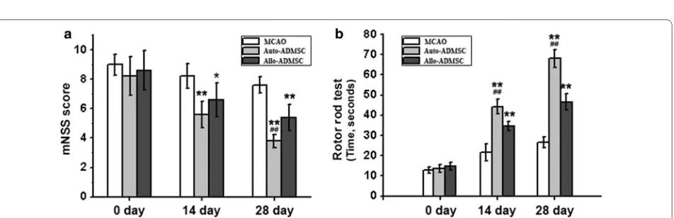
Auto‑ and allo‑ADMSCs improve functional recovery after transplantation
Both auto- and allo-ADMSCs improved functional recovery after MCAO in animals at 14 and 28 days post transplantation (Fig. 9). There were no significant differ-ences in mNSS scores between any groups prior to cell
[image:7.595.60.539.86.510.2]transplantation (p < 0.01; Fig. 9a). In the rotarod test, there were no significant differences in the mean hold-ing time between any groups prior to cell transplantation. Allo-ADMSC (34.6 ± 2.3 s, p < 0.01; 46.6 ± 3.8 s, p < 0.01) and auto-ADMSC (44.2 ± 3.7 s, p < 0.01; 68 ± 4.3 s, p < 0.01) transplantation groups showed significantly improved functional recovery in comparison to the con-trols (21.6 ± 4.0 s; 26.6 ± 2.8 s) at 14 days and at 28 days, respectively. Moreover, auto-ADMSC transplantation yielded a significantly longer holding time in comparison to auto-ADMSC transplantation at 14 days (p < 0.01) and 28 days (p < 0.01) post transplantation (Fig. 9b).
Auto‑ and allo‑ADMSC transplantation reduces infarct size
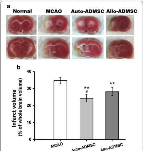
Staining with TTC 28 days after transplantation (Fig. 10a) showed that both allo-ADMSC (28.22 ± 2.38%, p < 0.01) and auto-ADMSC transplantation (24.35 ± 2.14%, p < 0.01) significantly reduced relative lesion volume with respect to the MCAO model group (34.69 ± 1.84%;
Fig. 10b). Moreover, auto-ADMSC transplantation reduced lesion volume to a significantly greater extent than allo-ADMSC transplantation (p < 0.05; Fig. 10b).
Discussion
We investigated the immunogenicity of rat ADMSCs in vitro and compared the immunological responses to and treatment effects of the intraparenchymal adminis-tration of allo- and auto-ADMSCs after the acute phase of MCAO in rats. We found that allo-ADMSCs exhibited greater immunogenicity and evoked stronger immuno-logical responses than ADMSCs. However, auto-ADMSCs displayed a higher survival rate, longer survival time, wider migratory scope, and fewer apoptotic cells. Moreover, a small number of transplanted auto-ADMSCs expressed astrocyte-like cells and neuron-like markers 28 days after transplantation, while no surviving allo-ADMSCs were found at this time point. In addition, we Fig. 6 Localization and expression of astrocyte or neuron markers of transplanted ADMSCs in the injured brain tissues. Transplanted ADMSCs were detected in the cortical injury area and hippocampus 1 day (a), 7 days (b), and 28 days (c) after transplantation. Yellow arrows indicated that the expression of astrocyte or neuron markers in the administered auto-ADMSCs on day 28 post transplantation in the injured brain tissues.
[image:8.595.59.541.88.422.2]found that the intraparenchymal administration of auto-ADMSCs yielded better functional recovery and reduced the infarct volume to a greater extent than allo-ADMSCs. We suggest that this effect might be associated with bet-ter viability, and migratory ability of auto-ADMSCs.
ADMSCs have been used in a number of animal mod-els for stroke and have been demonstrated to improve functional scores and reduce infarct size [7, 11]. In recent years, ADMSCs have been identified as especially interesting for clinical applications [12, 13]. It has been reported that ADMSCs have low MHC class I marker expression and lack MHC class II markers, suggesting that they are not likely to induce rejection [9]. How-ever, the present study shows that the expression of the MHC class I marker on ADMSCs from Lewis rats or BN rats was not low, and that MHC class II marker expres-sion could be found on the surface of these ADMSCs. The protein expression levels of IL-2 and IFN-γ were significantly greater in the T-lymphocyte cultures with allo-ADMSCs than in those with auto-ADMSCs. These
in vitro results suggest that allo-ADMSCs possess a cer-tain level of immunogenicity and might induce rejec-tion when transplanted to stroke model animals. The results of animal experiments in the present study have confirmed this hypothesis. When transplanted into the brains of MCAO rats, allo-ADMSCs were associated with a significant increase in the protein expression of IL-2 and IFN-γ, which are involved in inducing the inflam-matory reaction and promoting allograft rejection [14]. Allo-ADMSCs were also associated with significantly increased accumulation of CD4+ and CD8+ T lympho-cytes and microglial cells. CD8+ T lymphocytes can rec-ognize MHC-I molecules, and CD4+ T lymphocytes can recognize MHC-II molecules, where the latter plays a key role in initiating immune responses. Microglial cells are the resident macrophages of the brain. The degree of activation of microglial cells represents the degree of inflammation in the brain. In addition, activated micro-glia secrete a variety of proinflammatory cytokines, such as interleukin-1β and tumor necrosis factor-α, which lead Fig. 8 Expression of allograft rejection-related inflammatory cytokines in the brain tissue near the transplanted ADMSCs on day 7
post-transplantation was detected by ELISA. a IFN-γ. b IL-2. **p < 0.01 vs. MCAO model. ##p < 0.01 vs. auto-ADMSC transplantation group
Fig. 9 Motor function was assessed at 14 and 28 days post transplantation using the mNSS and rotarod test. Auto- and allo-ADMSCs improve functional recovery 14 and 28 days after transplantation. a mNSS score. b Rotarod test. *p < 0.05, **p < 0.01 vs. MCAO model group. #p < 0.01 vs.
[image:10.595.58.540.87.236.2] [image:10.595.64.540.275.429.2]to further activation of microglia, resulting in a gradual inflammatory response [15]. Inhibiting the activity of microglia after cerebral ischemia can reduce the volume of cerebral infarction and protect brain tissues [16]. How-ever, the aforementioned immune responses in the brain tissues of MCAO rats were not observed in response to auto-ADMSC transplantation.
Previous studies have shown that migration and/or implantation of xenogeneic (human) or allogeneic (rat) ADMSCs administered by intravenous infusion in the acute phase of MCAO were not detected in the injured brain at either 24 h or 14 days, but were found in vari-ous peripheral organs such as the spleen, lungs, and liver [17, 18]. The authors of these studies hypothesized that the administered xenogeneic or allogeneic ADM-SCs may act indirectly on the brain by secreting sev-eral growth factors that can act to enhance endogenous repair mechanisms normally activated in the brain after a stroke. However, in the present study, the migration and implantation of intraparenchymally-administered allo- and auto-ADMSCs were observed in the brain 1 and 7 days after MCAO, while the migration and implantation of auto-ADMSCs could even be observed at 28 days after MCAO. Furthermore, we found that
the migratory scope of auto-ADMSCs was wider than that of allo-ADMSCs. In addition, the survival rate of auto-ADMSCs was significantly higher than that of allo-ADMSCs on days 1, 7, and 28 after MCAO. The differences observed between the findings in the pre-sent study and previous studies could be explained by the fact that our route and timing of administration and cell sources (xenogeneic, allogeneic, or autologous) were different from those of the previous studies.
The most exciting observation in our study was that a small number of auto-ADMSCs were observed to express astrocyte or neuron markers 28 days after transplanta-tion, while surviving transplanted allo-ADMSCs could not be found. In agreement with this finding, several previous studies have shown that rat ADSCs were able to differentiate into floating neurospheres, where in addi-tion to being able to differentiate into neuronal- and glial-like cells, neurospheres could be induced to differentiate into Schwann cell-like cells. These Schwann cell-like cells were even able to form myelin structures with rat PC12 (pheochromocytoma cell) neurites in vitro [19, 20]. These results suggest that, aside from the growth factors secreted by surviving cells, the improved outcome owing to auto-ADMSC transplantation could have been a result of the cell replacement effect. Future studies are needed to explore the functional capacities of transplanted ADMSCs in vivo.
The majority of previous studies have focused on the effect that transplanted ADMSCs have on preserving brain tissue through the reduction of endogenous cell apoptosis [7, 21]. In the present study, we found that the number of apoptotic auto-ADMSCs was significantly lower than that of apoptotic allo-ADMSCs 1 day after transplantation, indicating that auto-ADMSCs likely elicit improved treatment outcomes.
[image:11.595.56.291.87.334.2]The administration of xenogeneic or allogeneic ADM-SCs has been shown to improve functional recovery independently of infarct volume in animal models of stroke [18]. Likewise, the present study demonstrated that administering auto-ADMSCs yielded better results in terms of improving functional recovery and reduc-ing infarct volume than allo-ADMSCs. The improved effects of auto-ADMSCs are likely related to decreased immunological responses, which thereby result in higher survival rates, longer survival times, a wider migratory scope, and a lower rate of apoptosis. The differences in the observed effects on infarct volume between our study and previous studies could be explained by the fact that the route and timing of administration were different. In addition, because only two evaluations of motor function were used here, future studies are needed to compare the effects of auto- and allo-ADMSC administration on Fig. 10 Auto-ADMSCs yielded better results in terms of reducing
the infarct volume than allo-ADMSCs on day 28 post transplantation.
a Brain sections with TTC staining (ischemic brain tissue is white).
b Quantitative data of infarct volume from brain sections with TTC staining. **p < 0.01 vs. MCAO model group. #p < 0.05 vs. auto-ADMSC
coordination, cognition, and memory impairment after stroke.
Conclusions
Auto-ADMSCs are more effective than allo-ADMSCs in promoting recovery and reducing infarct volume in MCAO rats. This may be associated with better viabil-ity, migratory abilviabil-ity, and differentiation potential, and the lower degree of apoptosis in auto-ADMSCs. Future research should focus on confirming the superiority of auto-ADMSCs in the treatment of MCAO models and clarifying relevant mechanisms to provide a solid foun-dation for the improved clinical treatment of cerebral infarction.
Abbreviations
ADMSCs: adipose-derived mesenchymal stem cells; Allo: allogeneic; Auto: autologous; BMSCs: bone marrow mesenchymal stem cells; EGFP: enhanced green fluorescence protein; GFAP: glial fibrillary acid protein; IFN-γ: interferon gamma; IL-2: interleukin-2; MCAO: middle cerebral artery occlusion; MHC: major histocompatibility complex; mNSS: modified neurological severity score; TTC : 2,3,5-triphenyltetrazolium chloride.
Authors’ contributions
ZY drafted/revised the manuscript; participated in the study concept and design, analysis, and interpretation of data; and carried out the experimental studies. TWY, SXW, DBX, WQ, and WZY helped to conduct the experiments; YYX and QSQ participated in the study concept and design; LZ revised the manuscript and participated in the study concept and design. All authors read and approved the final manuscript.
Author details
1 Department of Pediatrics, Navy General Hospital, No. 6, Fucheng Road,
Haid-ian District, Beijing 100048, China. 2 Department of Neonatal Intensive Care
Unit, Beijing Obstetrics and Gynecology Hospital, Capital Medical University, No. 251, Yaojiayuan Road, Chaoyang District, Beijing 100026, China. 3 Beijing
Yinfeng Dingcheng Bioengineering Technology Co., Ltd., No. 14, Zhonghe Street, Yizhuang Economic and Technological Development Zone, Daxing District, Beijing 100176, China.
Acknowledgements
We thank Dr. Yang Weili for assistance with the language of this manuscript.
Competing interests
The authors declare that they have no competing interests.
Availability of data and materials
All data generated or analyzed during this study are included in this published article.
Consent for publication Not applicable.
Ethics approval and consent to participate
All investigations were conducted after the approval of the Local Ethics Com-mittee (# 2015013) of Navy General Hospital, China.
Funding None.
Publisher’s Note
Springer Nature remains neutral with regard to jurisdictional claims in pub-lished maps and institutional affiliations.
Received: 19 August 2018 Accepted: 24 November 2018
References
1. Zhang JJ, Liu X. Aspirin plus dipyridamole has the highest surface under the cumulative ranking curves (SUCRA) values in terms of mortal-ity, intracranial hemorrhage, and adverse event rate among 7 drug therapies in the treatment of cerebral infarction. Medicine (Baltimore). 2018;97(13):e0123.
2. Gialanella B, Prometti P, Vanoglio F, Comini L, Santoro R. Aphasia and activities of daily living in stroke patients. Eur J Phys Rehabil Med. 2016;52(6):782–90.
3. Andres RH, Choi R, Steinberg GK, Guzman R. Potential of adult neural stem cells in stroke therapy. Regen Med. 2008;3(6):893–905.
4. Kim SU. Stem cell-based cell therapy in neurological diseases: a review. J Neurosci Res. 2009;87(10):2183–200.
5. Marei HE, Hasan A, Rizzi R, Althani A, Afifi N, Cenciarelli C, Caceci T, Shuaib A. Potential of stem cell-based therapy for ischemic stroke. Front Neurol. 2018;9:34.
6. Shen LH, Li Y, Chen J, Zhang J, Vanguri P, Borneman J, Chopp M. Intraca-rotid transplantation of bone marrow stromal cells increases axon-myelin remodeling after stroke. Neuroscience. 2006;137(2):393–9.
7. Leu S, Lin YC, Yuen CM, Yen CH, Kao YH, Sun CK, Yip HK. Adipose-derived mesenchymal stem cells markedly attenuate brain infarct size and improve neurological function in rats. J Transl Med. 2010;8(1):1–16. 8. Ikegame Y, Yamashita K, Hayashi S, Mizuno H, Tawada M, You F, Yamada
K, Tanaka Y, Egashira Y, Nakashima S, Yoshimura S, Iwama T. Comparison of mesenchymal stem cells from adipose tissue and bone marrow for ischemic stroke therapy. Cytotherapy. 2011;13(6):675–85.
9. Roemelingvan MR, Reinders ME, Franquesa M, Engela AU, Korevaar SS, Roelofs H, Genever PG, Ijzermans JN, Betjes MG, Baan CC, Weimar W, Hoogduijn MJ. Human allogeneic bone marrow and adipose tissue derived mesenchymal stromal cells induce CD8+ cytotoxic t cell reactiv-ity. J Stem Cell Res Ther. 2013;3(Suppl 6):004.
10. Longa EZ, Weinstein PR, Carlson S, Cummins R. Reversible middle cerebral artery occlusion without craniectomy in rats. Stroke. 1989;20(1):84. 11. Grudzenski S, Baier S, Ebert A, Pullens P, Lemke A, Bieback K, Dijkhuizen
RM, Schad LR, Alonso A, Hennerici MG, Fatar M. The effect of adipose tis-sue-derived stem cells in a middle cerebral artery occlusion stroke model depends on their engraftment rate. Stem Cell Res Ther. 2017;8(1):96. 12. Díeztejedor E, Gutiérrezfernández M, Martínezsánchez P, Rodríguezfrutos
B, Ruizares G, Lara ML, Gimeno BF. Reparative therapy for acute ischemic stroke with allogeneic mesenchymal stem cells from adipose tissue: a safety assessment: a phase II randomized, double-blind, placebo-controlled, single-center, pilot clinical trial. J Stroke Cerebrovasc Dis. 2014;23(10):2694.
13. Wen YD, Zhang HL, Qin ZH. Inflammatory mechanism in ischemic neu-ronal injury. Neurosci Bull. 2006;22(3):171–82.
14. Ekdahl CT, Kokaia Z, Lindvall O. Brain inflammation and adult neurogen-esis: the dual role of microglia. Neuroscience. 2009;158(3):1021–9. 15. Gutiérrezfernández M, Rodríguezfrutos B, Oteroortega L, Ramoscejudo J,
Fuentes B, Díeztejedor E. Adipose tissue-derived stem cells in stroke treat-ment: from bench to bedside. Discov Med. 2013;16(86):37.
16. Gutiérrezfernández M, Rodríguezfrutos B, Ramoscejudo J, Oteroortega L, Fuentes B, Díeztejedor E. Stem cells for brain repair and recovery after stroke. Expert Opin Biol Ther. 2013;13(11):1479–83.
•fast, convenient online submission •
thorough peer review by experienced researchers in your field • rapid publication on acceptance
• support for research data, including large and complex data types •
gold Open Access which fosters wider collaboration and increased citations maximum visibility for your research: over 100M website views per year •
At BMC, research is always in progress.
Learn more biomedcentral.com/submissions
Ready to submit your research? Choose BMC and benefit from: mesenchymal stem cells on functional recovery and brain repair markers
in experimental ischemic stroke. Stem Cell Res Ther. 2013;4(1):1–12. 18. Gutiérrezfernández M, Rodríguezfrutos B, Ramoscejudo J, Oteroortega L,
Fuentes B, Vallejocremades MT, Sanzcuesta BE, Díeztejedor E. Comparison between xenogeneic and allogeneic adipose mesenchymal stem cells in the treatment of acute cerebral infarct: proof of concept in rats. J Transl Med. 2015;13(1):1–10.
19. Xu Y, Liu L, Li Y, Zhou C, Xiong F, Liu Z, Gu R, Hou X, Zhang C. Myelin-form-ing ability of Schwann cell-like cells induced from rat adipose-derived stem cells in vitro. Brain Res. 2008;1239(1):49–55.
20. Xu Y, Liu Z, Liu L, Zhao C, Xiong F, Zhou C, Li Y, Shan Y, Peng F, Zhang C. Neurospheres from rat adipose-derived stem cells could be induced into functional Schwann cell-like cells in vitro. BMC Neurosci. 2008;9(1):1–10. 21. Yang YC, Liu BS, Shen CC, Lin CH, Chiao MT, Cheng HC. Transplantation of