C A S E R E P O R T
Open Access
Sudden cardiac death diagnosed with dilated
cardiomyopathy in a Kuwaiti family: a case report
Bassam Bulbanat
1, Dinu Antony
2, Kazem Behbehani
3, Osama Alsmadi
2, Daisy Thomas
2and Maisa Mahmoud Kamkar
2*Abstract
Background:Dilated cardiomyopathy is myocardial disease characterized by dilatation and impaired contraction of the left ventricle or both left and right ventricle. The majority of these cases are secondary to coronary artery disease, hypertension and valvular cardiomyopathy. Patients diagnosed with dilated cardiomyopathy are further clinically evaluated for evidence of familial history of the disease. Those families have shown to have genetic predisposition to dilated cardiomyopathy; thus, currently there is no available single genetic test that allows comprehensive testing of all causative genes. We report a Kuwaiti case of dilated cardiomyopathy that was diagnosed at young age. The patient clinical presentation pointed out to the fact that this was a familial disease. This case is the first reported in Kuwait clinically presented with familial dilated cardiomyopathy implying a genetic susceptibility factor to be further investigated within the at-risk family members.
Case presentation:23-year-old Arab ethnicity Kuwaiti male with strong family history of dilated cardiomyopathy was admitted witnessed with sudden cardiac death. The patient presented with sudden arrhythmic death and survived with permanent anoxic brain injury. Transthoracic echocardiography revealed dilated cardiomyopathy with severe global left ventricular systolic dysfunction. After thorough investigation, the patient shown to have strong family history of dilated cardiomyopathy.
Conclusion:Familial dilated cardiomyopathy is poorly documented in Kuwait. We present this case with future plan to study the genetic map of his family.
Keywords:Cardiomyopathies, Familial dilated cardiomyopathy, Sudden arrhythmic death
Background
According to the World Health Organization (WHO), cardiovascular diseases are the number one cause of death accounting for 30 percent (17.5 million) of all deaths worldwide [1]. Cardiovascular diseases are caused by disorders of the heart and blood vessels, they includes coronary heart disease (heart attacks), cerebrovascular disease (stroke), raised blood pressure (hypertension), peripheral artery disease, rheumatic heart disease, con-genital heart disease and heart failure. The WHO defined cardiomyopathies as diseases of the heart muscle. They classified them due to their etiology into, cardiomyopa-thies with unknown causes, and cardiomyopacardiomyopa-thies from cardiac dysfunction (sudden cardiac arrest) due to known
cardiovascular disorders such as hypertension, ischemic heart disease, or valvular disease [2] (See Tables 1 and 2).
Cardiac deaths are classified as sudden or non-sudden, sudden cardiac death is defined as death occurring sud-denly and unexpectedly in a patient who is otherwise stable prior to the event [3,4]. Sudden cardiac death is caused by ventricular tachycardia or fibrillation and may be aborted with an implantable cardioverter defibrillator. Among young adults (18–35 years), sudden cardiac death most commonly results from a previously undiagnosed congenital or hereditary condition such as coronary artery anomalies and inherited cardiomyopathies [5]. They are classified into (i) witnessed deaths, if death occurred within 1 hour after the onset of new symptoms or as (ii) un-witnessed deaths if the patient was seen alive and stable during the previous 24-hours. On the other hand, if the sequence of events is consistent with sudden death but a specific cause of death other than arrhythmia is confirmed; the death is classified as non-sudden. A careful * Correspondence:maisa.mahmoud@dasmaninstitute.org
2
Genetics and Genomics Unit/Dasman Genome Center, Biomedical Research Department, Dasman Diabetes Institute, Kuwait City, Kuwait
Full list of author information is available at the end of the article
history and physical examination, in addition to electro-cardiography and cardiac imaging, are essential to diag-nose conditions associated with sudden cardiac death [6].
Case presentation
In March 2003, a 23-year-old Arab ethnicity Kuwaiti male with a strong family history of dilated cardiomyopathy presented to a specialized neurology hospital with dizzy spells and feet parasthesia. He was given intravenous methylprednisolone for 3 days for probable multiple scler-osis and was booked electively for a magnetic resonance imaging (MRI) of the brain. On March 23rd2003, he had a witnessed sudden cardiac arrest and suddenly collapsed when he was at an automated banking teller machine. The paramedic personnel were at the scene within 5 minutes of the call, endotracheal intubation was carried out as well as an intra-tracheal atropine and adrenalin was given at the scene. A cardiac monitor was attached and revealed ventricular fibrillation (Figure 1), and thus was electrically cardioverted 3 times (200 J, 300 J and 360 J) until reverted to sinus rhythm. The resuscitation procedure including the transit time to hospital was around 30 minutes. On arrival to intensive care unit (ICU), he had a heart rate of 150 beats per minute and a blood pressure of 110/70 mm/ Hg. Electrocardiography (ECG) revealed sinus rhythm with incomplete Left bundle branch block (LBBB) and non-specific ST-T wave changes (Figure 2). A transtho-racic echocardiography showed a mildly dilated left ventricular (LV) with severe global LV systolic dysfunction with an estimated Left Ventricular Ejection Fraction (LVEF) of 20 to 25% (Figure 3). An echocardiogram was
[image:2.595.56.292.123.320.2]done 6 months prior to the hospital admission and LV systolic function reported to be normal. During his ICU stay, he had recurrent convulsions, which were controlled with epanutin and clonazepam. Computed tomography scan (CTS) of the head showed a small infarct (probably embolic) in the right centrum semiovale adjacent to the body of the right lateral ventricle. An electroencephalo-graph (EEG) was performed and this revealed diffuse
Table 1 Classification criteria of primary cardiomyopathy according to the American heart association and the European society of cardiology
Primary cardiomyopathy
Genetic Mixed* Acquired
•HCM* •DCM* •Inflammatory
(myocarditis) •ARVC/D* •Restrictive
(nonhypertrophid and nondilated)
•Stress-provoked (*takotsubo)
•LV/NC* •Peripartum
•Glycogen storage
(PRKAG2, Danon) •
Tachycardia-induced
•Conduction defects •Infants of insulin-dependent diabetic mothers
•Mitochondrial myopathies •Ion channel Disorders
(LQTS, Brugada, SQTS, CVPT, Asian SUNDS)
*HCM: Hypertrophic Cardiomyopathy, *DCM: Dilated Cardiomyopathy, *ARVC/D: Arrhythmogenic Right Ventricular Cardiomyopathy/Dysplasia, *LV/NC: Left Ventricular Noncompaction Cardiomyopathy.
Table 2 Classification criteria of secondary cardiomyopathy according to the American heart association and the European society of cardiology
Secondary cardiomyopathy
Infiltrative •Amyloidosis (primary familial autosomal dominant, senile, secondary forms) •Hurler’s disease
•Hunter’s disease Storage •Hemochromatosis
•Fabry’s disease
•Glycogen storage disease (type II, Pompe) Toxicity •Drugs, heavy metals, alcohol
Endomyocardial •Hyperesinophilic syndrome (Loeffler’s endocarditis)
Inflammatory •Sarcoidosis
Endocrine •Diabetes Mellitus
•Hyperthyroidism •Hypothyroidism •Hyperparathyroidism •Pheochromocytoma •Acromegaly Cardiofacial •Noonan syndrome
•Lentiginosis Neuromuscular/
neurological •
Friedreich’s ataxia
•Duchenne-Becher muscular dystrophy •Emery-Dreifuss muscular dystrophy •Myotonic dystrophy
•Neurofibromatosis •Tuberous sclerosis
Nutritional deficiencies •Beriberi (thiamine), pellagra, scurvy, selenium, carnitine, kwashiorkor
Autoimmune/collagen •Systemic lupus erythematosis •Dermatomyositis
•Rheumatoid arthritis •Scleroderma •Polyarteritis nodosa Consequence of
cancer therapy •
Anthracyclines: doxorubicin (adriamycin), daunorubicin
•Cyclophosphamide •Radiation
Bulbanatet al. BMC Research Notes2014,7:914 Page 2 of 8
[image:2.595.304.535.133.620.2]cortical dysfunction with no epileptic discharge. He had a battery of laboratory investigations including liver function test, renal function test, complete blood count and thyroid function test and these were all within normal limits. Later on, he had a transient rise in liver enzymes and this was found to be secondary to hepatic hypo-perfusion (hepatitis viral screening, auto-immune screening and serum copper tests were all normal). Eventually a tracheostomy was performed and a naso-gastric tube fed him. His neurological condition showed a slow but steady improvement over a course of 3 months and regained consciousness, with spontaneous breathing and thus the tracheostomy was closed. However, he was left mentally handicapped with sphincteric incontinence and was referred to the rehabilitation hospital for
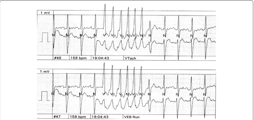
[image:3.595.57.541.89.269.2]aggressive physiotherapy. During his follow up, he was treated medically with anti-failure and antiepileptic drugs (digoxin, revotril, epanutin, zestril, lasix, zantac and amiodarone). However, he continued to show non-sustained ventricular tachycardia on a 24-hour holter monitor (Figure 4). Eventually, he had an automated implantable cardioverter-defibrillator (AICD) inserted in 2004 and his functional status significantly improved and he is still alive. He developed amiodarone induced thyroiditis with hyperthyroidism, Thyroid test, FT4 = 15.29 (Normal = 5.69-13.44) and Thyroid stimulating hormone, TSH = 0.03 (Normal = 0.43-4.1) which was proven by a positive thyroid scan. His thyroid function normalized following the discontinuation of amiodarone. Investigations of his family history revealed that his father Figure 1Ventricular fibrillation of the patient using a cardiac monitor.
[image:3.595.59.538.499.716.2]was known to have dilated cardiomyopathy (DCM) and was treated with AICD, but had a sudden cardiac death a few years ago while he was waiting for cardiac transplant-ation. In addition, two of his sisters had pacemakers inserted, and one of his paternal uncles died suddenly at the age of 25 years. Family pedigree is shown in (Figure 5).
Discussion
Though the American heart association (AHA) and the European society of cardiology (ESC) defined and classified cardiomyopathies differently [7,8]; however, both organiza-tions have agreed on general classification systems into primary and secondary cardiomyopathies (Figures 6 and 7). While the AHA considers ion channelopathies as one of the cardiomyopathies, the ESC does not consider ion
[image:4.595.59.539.90.243.2]channelopathies as an accepted cardiomyopathy because genes encoding for ion channels might not result in mor-phofunctional phenotypes. Thus, in 2013, the American college of cardiology (ACC) and AHA defined cardiomy-opathies as disorders characterized by morphologically and functionally abnormal myocardium in the absence of any other disease that is sufficient, by itself, to cause the observed phenotype [9]. The majority of these cases are secondary to coronary artery disease, hypertension and valvular cardiomyopathy. The number and severity of each of the traditional cardiovascular risk factors should be evaluated. These risk factors must evaluate as continuous rather than discrete variables that exert a dose-dependent effect on coronary artery disease. Moreover, there are novel biomarkers reflecting thrombosis, inflammation and Figure 3A transthoracic echocardiography showed a mildly dilated left ventricular with severe global left ventricular systolic dysfunction with an estimated left ventricular ejection fraction of 20 to 25%.
Figure 4The patient still showing non sustained ventricular tachycardia on 24 hour holter monitor.
Bulbanatet al. BMC Research Notes2014,7:914 Page 4 of 8
[image:4.595.57.540.487.716.2]oxidative stress that contribute to the pathophysiological process of atherthrombosis in acute ischemic heart disease [10]. A fourth of the cases of congestive cardiac failure are secondary to idiopathic cardiomyopathy [11]. It is possible that idiopathic dilated cardiomyopathy (IDC) represents a common expression of myocardial damage that has been produced by un-established myocardial insults [12]. The incidence of a familial form of dilated cardiomyopathy (is
up to 20% of all cases of DCM [13,14]. Identifying these individuals will help the treating physician in pursuing major changes in the clinical management or counseling of these patients with the following recommendations:
1. Avoidance of competitive sport activity.
2. Instituting early medical treatment such as ACEi
[image:5.595.56.540.89.248.2]andβblockers.
Figure 5Pedigrees of a Kuwaiti family diagnosed with familial dilated cardiomyopathy.Squares indicate men; circles, women; crossed symbols, deceased; highlighted symbols, diagnosed with dilated cardiomyopathy and has peacemaker; clear symbol, number of unaffected/ asymptomatic members.
[image:5.595.60.538.424.708.2]3. Refraining from the use of injurious agents: A. Succinyl choline or volatile anesthetics
(halothane and isoflurane) in emerinopathies and laminopathies.
B. Statins in genetic cardiomyopathies with possible involvement of the skeletal muscle, even when markers of myopathy are negative.
C. Patients with mitochondrial cardiomyopathy and epilepsy should not receive valproate because it may cause pseudo atrophy of brain.
Here we differentiate a patient with ‘true’ IDC who had his first-degree family members clinically screened (history, physical examination, echocardiogram, and ECG) to rule out familial dilated cardiomyopathy (FDC) versus a‘presumptive’ IDC – one who is negative for familial disease by a careful 3–4 generation family history but has not had family members screened beyond the family history [15]. Our patient fulfilled the criteria for idiopathic cardiomyopathy that had first-degree family members with a definite diagnosis of idiopathic dilated cardiomyopathy (IDC). Despite the evidence supporting a genetic basis of IDC/FDC, physicians have poorly complied with the implementation of guidelines [16]. Adherence to such guidelines would require a shift in focus from strictly therapeutic measures for a single patient presenting with advanced disease to the consid-eration and assessment of DCM risk for an entire family. This represents a major health problem in the Kuwaiti society and the middle-eastern communities in general. In this regard, close relative marriages are common in these societies, which results in the cluster of pathological genes in certain families. This is also seen in inherited hematological disorders. Usually families affected by these disorders end up having to live with the potential tragedy of losing their off springs or close relatives before their eyes. Familial cardiomyopathy is a serious disease when the phenotypic expression leads to severe cardiomyopathy, and our patient represents an example of this serious dis-ease. The patient became handicapped and lost his father,
who was his caregiver. Therefore, a better understanding the genetic basis of FDC disease in Kuwait will help med-ical staff convince families to change the pattern of fa-milial marriage. This change will save lives and prevent families from going through such a devastating experience and will help them enjoy having healthy generations. We sought to adhere to the guidelines in pursuing the genetic work up of the family of our patient according to the flow diagram shown in (Figure 8). Although, numerous genes have been reported in association with non-syndrome di-lated cardiomyopathy [9,16], there have been no reports of genes in association with DCM in Kuwait.
Current and future prospective
Genetic polymorphisms associated with cardiovascular responses were reported in number of studies. Three main genes were shown as key players in the regulation of cardiac responses causing electrophysiology and cardiac myocyte hypertrophy. These genes are calcium/calmodu-lin-dependent kinase IV (CaMKIV), the G-protein-coupled receptor kinases (GRKs) and the heptahelical G-protein-coupled receptors (GPCRs) [17-19]. The family history of the patient was taken and clinical screening of first-degree relatives (who are at risk) was performed. A total of 26 blood samples from all first-degree family members were collected, shown in the pedigree (Figure 5). The mode of inheritance in this family appears to act through an autosomal dominant type. Whole exome sequencing using Hiseq technology was our method of choice to unravel the causative genes, through a series of phenotype-genotype filtration processes. A thorough evaluation of the patients’ relatives who are at risk will be performed to detect the absence or presence of pathologic mutation(s).
Study limitations
[image:6.595.57.540.89.216.2]The patient is assumed to have had “idiopathic cardio-myopathy” based on the absence of secondary causes; however, this assumption may be inaccurate. The patient had an echocardiogram 6 months prior to hospital admission and the left ventricular function was reported Figure 7General classification systems into secondary cardiomyopathies according to the American heart association and the European society of cardiology.
Bulbanatet al. BMC Research Notes2014,7:914 Page 6 of 8
to be normal. Inherited DCM is a slow progressive disease in which the left ventricular systolic function deteriorates over a number of years rather than over few months. Hence, this case lacks the documentation of the progres-sive nature of the disease. Moreover, he had an arrhythmic sudden cardiac death, which required prolonged cardio-pulmonary resuscitation. Actually, it took the paramedics 5 minutes to arrive at the scene and therefore, this pro-longed ischemic insult may have caused myocyte necrosis resulting in permanent left ventricular systolic dysfunc-tion. Finally, rare causes of DCM cannot be ruled out, as myocardial biopsy was not available in Kuwait at that time.
Conclusions
Familial cardiomyopathies are genetic diseases and are becoming widely recognized worldwide. The phenotypic expression is linked to the specific causative genotype in
a small percentage of dilated cardiomyopathy of unknown etiology. To better understand the phenotypic expression, treating physicians need to study the genetic pedigree of affected patients with suspected inherited cardiomy-opathy. This testing may affect the management of the patients carrying the defective gene as well as their first-degree relatives. However, the results of the genetic testing must be correlated with the clinical presentation, as it is too early to take clinical decisions based on the genetic study alone.
Consent
[image:7.595.57.543.91.478.2]and any accompanying images. A copy of the written consent is available for review by the Editor-in-Chief of this journal.
Abbreviations
ACC:American college of cardiology; AHA: American heart association; AICD: Automated implantable cardioverter-defibrillator; CaMKIV: Calcium/ calmodulin-dependent kinase IV; CTS: Computed tomography scan; DCM: Dilated cardiomyopathy; ECG: Electrocardiography; EEG: Electroencephalograph; ESC: European society of cardiology; FDC: Familial dilated cardiomyopathy; FT4: Thyroid test; GPCRs: G-protein-coupled receptors; GRKs: G-protein-coupled receptor kinases; ICU: Intensive care unit; IDC: Idiopathic dilated cardiomyopathy; LBBB: Left bundle branch block; LV: Left ventricular; LVEF: Left ventricular ejection fraction; MRI: Magnetic resonance imaging; TSH: Thyroid stimulating hormone; WHO: World Health Organization.
Competing interests
The authors declare that they have no competing interests.
Authors’contributions
BB prepared the clinical section of the case review. MK and DA carried out the molecular genetic studies of the index case and corresponding family members. DT helped in the coordination and patients sample collection. OA and KB participated in drafting the manuscript. All authors read and approved the final manuscript.
Acknowledgments
Dasman Diabetes Institute funded this study; project number RA-2011-007.
Author details
1Division of Cardiology, Al-Amiri Hospital, Ministry of Health, P.O. Box 1180, Dasman 15462, Kuwait.2Genetics and Genomics Unit/Dasman Genome Center, Biomedical Research Department, Dasman Diabetes Institute, Kuwait City, Kuwait.3Dasman Diabetes Institute, Kuwait City, Kuwait.
Received: 6 May 2014 Accepted: 11 December 2014 Published: 16 December 2014
References
1. The current epidemiology of CV disease:Epidemiology of Cardiovascular Disease in the 21stCentury: Updated Numbers and Updated Facts.Jived 2013,1:1–2.
2. Report of the WHO/ISFC task force on the definition and classification of cardiomyopathies.Br Heart J1980,44:672.
3. The Cardiac Arrhythmia Suppression Trial (CAST) Investigators:Preliminary report: effect of encainide and flecainide on mortality in a randomized trial of arrhythmia suppression after myocardial infarction.N Engl J Med 1989,321:406–412.
4. Chugh SS, Jui J, Gunson K, Stecker EC, John BT, Thompson B, Ilias N, Vickers C, Dogra V, Daya M, Kron J, Zheng ZJ, Mensah G, McAnulty J:Current burden of sudden cardiac death: multiple source surveillance versus retrospective death certificate-based review in a large U.S. community.J Am Coll Cardiol 2004,44:1268–1275.
5. Stojanovska J, Garg A, Patel S, Melville D, Kazerooni E, Mueller G:Congenital and hereditary causes of sudden cardiac death in young adults: diagnosis, differential diagnosis, and risk stratification.Radiographics 2013,33(7):1977–2001.
6. Groh WJ, Groh MR, Saha C, Kincaid J, Simmons Z, Ciafaloni E, Pourmand R, Otten R, Bhakta D, Nair G, Marashdeh M, Zipes D, Pascuzzi R:
Electrocardiographic Abnormalities and Sudden Death in Myotonic Dystrophy Type 1.N Engl J Med2008,358:2688–2697.
7. Maron BJ, Towbin JA, Thiene G, Antzelevitch C, Corrado D, Arnett D, Moss A, Seidman C, Young J:Contemporary Definitions and Classification of the Cardiomyopathies: An American Heart Association Scientific Statement from the Council on Clinical Cardiology, Heart Failure and Transplantation Committee; Quality of Care and Outcomes Research and Functional Genomics and Translational Biology Interdisciplinary Working Groups; and Council on Epidemiology and Prevention.Circulation2006,113:1807–1816. 8. Elliott P, Andersson B, Arbustini E, Bilinska Z, Cecchi F, Charron P, Dubourg O, Kühl U, Maisch B, McKenna WJ, Monserrat L, Pankuweit S, Rapezzi C, Seferovic C, Tavazzi L, Keren A:Classification of the cardiomyopathies: a position
statement from the European society of cardiology working group on myocardial and pericardial diseases.Eur Heart J2008,29:270–276. 9. Arbustini E, Narula N, Dec GW, Reddy KS, Greenberg B, Kushwaha S,
Marwick T, Pinney S, Bellazzi R, Favalli V, Kramer C, Roberts R, Zoghbi WA, Bonow R, Tavazzi L, Fuster V, Narula J:The MOGE(S) Classification for a Phenotype–Genotype Nomenclature of Cardiomyopathy: Endorsed by the World Heart Federation.J Am Coll Cardiol2013,62(22):2046–2072. 10. Santulli G:The role of cardiovascular risks in determining coronary artery
disease (Coronary heart disease risk factors and mortality).JAMA2012, 307(11):1137.
11. Brown CA, O’Connell JB:Myocarditis and idiopathic dilated cardiomyopathy.Am J Med1995,99:309.
12. Braunwald E, Zipes DP, Libby P:Heart Disease: A text book of cardiovascular medicine.6th edition. Philadelphia: W.B. Saunders Company; 2001:1775. 13. Manolio TA, Baughman KL, Rodeheffer R, Pearson TA, Bristow JD, Michels
VV, Abelmann WH, Harlan WR:Prevalence and etiology of idiopathic dilated cardiomyopathy (Summary of a National Heart, Lung, and Blood Institute Workshop).Am J Cardiol1992,69:1458. 12.
14. Durand JB, Bachinski LL, Bieling LC, Czernuszewicz G, Abchee A, Yu Q, Tapscott T, Hill R, Ifegwu J, Marian AJ, Brugada R, Daiger S, Gregoritch J, Anderson J, Quiñones M, Towbin J, Roberts R:Localization of a gene responsible for familial dilated cardiomyopathy to chromosome 1q32. Circulation1995,92:3387. 1995.
15. Hershberger RE, Siegfried JD:Update 2011: Clinical and Genetic Issues in Familial Dilated Cardiomyopathy.J Am Coll Cardiol2011,57(16):1641–1649. 16. Hershberger RE, Lindenfeld J, Mestroni L, Seidman CE, Taylor MR, Towbin JA, Heart Failure Society of America:Genetic Evaluation of cardiomyopathy a Heart Failure Society of America practice guideline.J Card Fail2009, 15:83–97.
17. Santulli G, Cipolletta E, Sorriento D, Giudice C, Anastasio A, Monaco S, Maione AS, Condorelli G, Puca A, Trimarco B, Illario M, Iaccarino G:CaMK4 Gene Deletion Induces Hypertension.J Am Heart Assoc2012,4:e001081. 18. Lobmeyer MT, Wang L, Zineh I, Turner S, Gums J, Chapman A, Cooper-DeHoff
R, Beitelshees A, Bailey K, Boerwinkle E, Pepine C, Johnson J:Polymorphisms in genes coding for GRK2 and GRK5 and response differences in
antihypertensive-treated patients.Pharmacogenet Genomics2011,21:42–49. 19. Santulli G, Trimarco B, Iaccarino G:G-protein-coupled receptor kinase 2
and hypertension: molecular insights and pathophysiological mechanisms.High Blood Press Cardiovasc Prev2013,20:5–12.
doi:10.1186/1756-0500-7-914
Cite this article as:Bulbanatet al.:Sudden cardiac death diagnosed with dilated cardiomyopathy in a Kuwaiti family: a case report.BMC Research Notes20147:914.
Submit your next manuscript to BioMed Central and take full advantage of:
• Convenient online submission
• Thorough peer review
• No space constraints or color figure charges
• Immediate publication on acceptance
• Inclusion in PubMed, CAS, Scopus and Google Scholar
• Research which is freely available for redistribution
Submit your manuscript at www.biomedcentral.com/submit
Bulbanatet al. BMC Research Notes2014,7:914 Page 8 of 8