The development of RNA extraction protocols to examine the effects of early exercise on gene expression in the articular cartilage and subchondral bone of Perendale sheep : a thesis presented in partial fulfilment of the requirements for the degree of Mas
161
0
0
Full text
(2) The development of RNA extraction protocols to examine the effects of early exercise on gene expression in the articular cartilage and subchondral bone of Perendale sheep. A thesis presented in partial fulfilment of the requirements for the degree of Master of Science. at. Massey University Palmerston North New Zealand Renée Marie Pedley 2010.
(3) ii.
(4) TABLE OF CONTENTS LIST OF ILLUSTRATIONS.......................................................................... VII LIST OF TABLES...................................................................................... VIIII ABBREVIATIONS ........................................................................................ IX ACKNOWLEDGEMENTS ............................................................................ X ABSTRACT ............................................................................................... XIII. CHAPTER 1 .................................................................................................... 1 INTRODUCTION .............................................................................................. 1 1.1 BACKGROUND ....................................................................................... 1 1.2 LITERATURE REVIEW ............................................................................. 4 1.2.1. Synovial joint structure & function................................................. 4. 1.2.2. Osteochondral tissues .................................................................. 8. 1.2.3. The ovine hock joint ................................................................... 19. 1.2.4. Joint growth, modeling & remodeling .......................................... 23. 1.2.5. Degenerative joint disease: Osteoarthritis .................................. 26. 1.2.6. Effects of exercise on articular cartilage & subchondral bone ..... 29. 1.2.7. Gene expression ........................................................................ 34. 1.2.8. Methods in assessing articular cartilage & subchondral bone ..... 44. 1.2.9. Polymerase chain reaction (PCR) .............................................. 48. 1.2.10. Animal models ........................................................................ 49. 1.3 SUMMARY ........................................................................................... 50 1.4 HYPOTHESES ...................................................................................... 52 1.4.1. Objectives of this study .............................................................. 52. CHAPTER 2 .................................................................................................. 54 OPTIMISATION OF RNA EXTRACTION PROTOCOLS FROM ARTICULAR CARTILAGE AND SUBCHONDRAL BONE FOR GENE EXPRESSION ANALYSIS IN JOINT DISEASE. 54. 2.1 ABSTRACT ........................................................................................... 54 2.2 INTRODUCTION .................................................................................... 55 2.3 MATERIALS AND METHODS ................................................................... 57 2.3.1. Tissue preparation...................................................................... 57. 2.3.2. Limb dissection .......................................................................... 58. 2.3.3. RNA Extraction ........................................................................... 60. iii.
(5) 2.3.4. RNA quality evaluation ............................................................... 62. 2.3.5. Reverse transcription polymerase chain reaction (RT-PCR) ....... 62. 2.3.6. RT-PCR amplicon quantification ................................................. 64. 2.3.7. Statistical analysis ...................................................................... 65. 2.4.. RESULTS ......................................................................................... 65. 2.4.1. Comparison between tissue homogenization methods ............... 65. 2.4.2. Comparison of RNA quality between extraction methods............ 67. 2.4.3. Comparison of RNA quantity between extraction methods ......... 68. 2.4.4. Effectiveness of manual method to isolate RNA ......................... 69. 2.4.5. Gene expression analysis ........................................................... 71. 2.5.. DISCUSSION..................................................................................... 73. 2.6 CONCLUSIONS ..................................................................................... 76. CHAPTER 3 .................................................................................................. 78 THE EFFECTS OF EXERCISE ON GENE EXPRESSION IN ARTICULAR CARTILAGE AND SUBCHONDRAL BONE. ................................................................................... 78. 3.1 ABSTRACT ........................................................................................... 78 3.2 INTRODUCTION..................................................................................... 79 3.3 MATERIALS & METHODS........................................................................ 82 3.3.1. Animals....................................................................................... 82. 3.3.2. Exercise regimen ........................................................................ 82. 3.3.3. Timeline ...................................................................................... 83. 3.3.4. Tomography scan ....................................................................... 85. 3.3.5. Tissue preparation ...................................................................... 85. 3.3.6. Limb dissection ........................................................................... 85. 3.3.7. RNA Extraction ........................................................................... 87. .3.8. RNA Analysis (RT-PCR) ............................................................. 87. 3.3.9. Statistical analysis ...................................................................... 88. 3.4 RESULTS ............................................................................................. 89 3.4.1. Gross morphology ...................................................................... 89. 3.4.2. Gene expression ........................................................................ 89. 3.5 DISCUSSION ........................................................................................ 91 3.6 CONCLUSIONS ..................................................................................... 98. iv.
(6) CHAPTER 4 .................................................................................................. 99 GENERAL DISCUSSION ................................................................................. 99 4.1 INTRODUCTION .................................................................................... 99 4.2 RNA EXTRACTION METHOD .................................................................100 4.3 THE EFFECTS OF EXERCISE ON GENE EXPRESSION IN ARTICULAR CARTILAGE AND SUBCHONDRAL BONE ...........................................................................101. 4.4 LIMITATIONS OF THIS STUDY ................................................................103 4.5 FUTURE STUDIES ................................................................................104. APPENDICES ..............................................................................................107. REFERENCES .............................................................................................126. v.
(7) LIST OF ILLUSTRATIONS Figure 1.1. Diagramatic representation of a synovial joint ........................... 5. Figure 1.2. Squeeze film lubrication between two loaded surfaces ............. 8. Figure 1.3. The osteochondral tissues ........................................................ 9. Figure 1.4. Collagen. fibre. orientation. and. the. etiopathogenesis. of. osteoarthritis in the Beagle dog. .............................................. 10 Figure 1.5. Proteoglycan: structure of repeating disaccharide units .......... 14. Figure 1.6. Proteoglycan attachment along collagen fibril ......................... 14. Figure 1.7. Lateral view of the hock joint. .................................................. 21. Figure 1.8. The left distal aspect of the tibia. ............................................ 22. Figure 1.9. Endochondral bone formation ................................................ 25. Figure 1.10. Factors involved in the initiation of cartilage degradation and onset of osteoarthritis.............................................................. 26. Figure 1.11. Enzymatic factors involved in cartilage degradation and onset of osteoarthritis ........................................................................... 27. Figure 1.12. The mechanical signals of ex exercise .................................... 34. Figure 1.13. Collagen fibril formation .......................................................... 39. Figure 1.14. The zinc dependent endopeptidase families ........................... 42. Figure 2.1. The distal tibia from a two year old ewe following gallop exercise. ................................................................................. 59. Figure 2.2. Normality plot of quality of RNA extracted from ovine liver, articular cartilage and bone. .................................................... 67. Figure 2.3. Normality plot of the quantity of RNA extracted from ovine liver, articular cartilage and bone. .................................................... 69. Figure 2.4. Gene. expression. signals. confirming. GAPDH. (120. bp),. β-Actin (193 bp) and MMP2 (159 bp) in ovine liver and uterine tissue. ..................................................................................... 71 Figure 2.5. Gene expression signals for GAPDH (120 bp) and collagen type I (155 bp) for subchondral bone in skeletally mature Perendale ewes (2 years of age). ............................................................ 72. Figure 2.6. Gene expression signals for GAPDH (120 bp) and collagen type II (141 bp) for articular cartilage from skeletally mature Perendale ewes (2 years of age). ........................................... 72. vi.
(8) Figure 3.1. Osteo-inductive exercise training program timeline 2005-2004. ............................................................................................... 84. Figure 3.2. The distal tibia from exercise trained and unexercised normal two year old ewes ............................................................................................. 86. Figure 3.3. The relative amount of mRNA gene expression...................... 91. vii.
(9) LIST OF TABLES Table 1.1. Different collagen types in normal articular cartilage and their known functions. ..................................................................... 16. Table 1.2. Molecular. mechanisms. regulating. endochondral. bone. development. .......................................................................... 29 Table1.3. The role of peptide regulatory factors involved in cartilage metabolism. ............................................................................ 35. Table 1.4. Technologies to measure bone mineral density (BMD) and bone strength in large animals. ........................................................ 45. Table 5. Primer sequence for ovine, GAPDH, β-Actin , collagen, and collagenase genes .................................................................. 63. Table 6. Comparison between mechanical disruption methods used to extract total RNA from osteochondral tissues from juvenile and skeletally mature sheep using a commercially available kit and adaptation of a manual method. .............................................. 66. Table 7. Mean values for total RNA quality and quantity from ovine fibrous and osteochondral tissues using an adaptation of a manual RNA isolation method ................................................. 70. Table 8. Effectiveness of RNA extraction methods reported in literature ............................................................................................... 77. Table 9. Osteo-inductive exercise regimen .......................................... 83. viii.
(10) ABBREVIATIONS AC. Articular cartilage. ADAM TS. Disintegrin matrix metalloproteinases with thrombospondin repeats. ADAMS. Disintegrin matrix metalloproteinases. BMD. Bone mineral density. BMP. Bone morphogenic protein. CT. Computed tomography. DJD. Degenerative joint disease. DXA. X-ray absorptiometry dual energy. GAPDH. Glyceraldehyde phosphate dehydrogenase. IL-1β. Interleukin-1-beta. MMP. Matrix metalloproteinase. MRI. Magnetic resonance imaging. OA. Osteoarthritis. OCD. Osteochondrosis dissecans. PCR. Polymerase chain reaction. PRF. Peptide regulatory factors. RA. Rheumatoid arthritis. RER. Rough endoplasmic reticulum. RT. Reverse transcription. Rt. Real time. SCB. Subchondral bone. TIMP. Tissue inhibitor metalloproteinase. TNF-α. Tumour necrosis factor alpha. US. Ultrasonography. VEGF. Vascular endothelial growth factors. β-Actin. Beta actin. ix.
(11) ACKNOWLEDGEMENTS I am very grateful for the encouragment and support I have received from my family, friends and colleagues during the course of this thesis. In particular, I would like to thank my supervisors, Dr Laryssa Howe, Dr Chris Rogers, and Dr Alastair Smith. The contribution of their time, guidance and patience has been invaluable and initiated an increasing interest in molecular biology and musckuloskeletal research.. I would like to thank the The Guardian Trust for their generosity in providing financial assistance towards this project and for allowing me the opportunity to be recipient of the Norman Cunningham Fellowship during 2006 and 2007. I am especially grateful to Massey University for allowing me the opportunity to be recipient of a Massey Masterate Scholarship. Thanks also go to the IVABS Post-Graduate Fund whom supplied funds for my research.. Special thanks are extended to friends and colleagues within IVABS and the College of Science. In particular Mike Hogan, Cathy Davidson, Keren Dittmer, and Dr Alastair Johnstone who kindly assisted with samples and sample collection. I would like to thank Sue Winter for the generous use of -80oC freezer space.. Special thanks must be extended to Errol Kwan for his. generosity and providing advice and valuable feedback throughout my laboratory work.. To my parents Jean and Matthew, thank you for your encouragement and support. To my son Aidan, thank you for being the wonderful person you are and to Clive, thank you. To my sister Nikki, thank you for always being there for me.. x.
(12) xi.
(13) xii.
(14) ABSTRACT To examine gene expression in vivo, total RNA was extracted from articular cartilage and subchondral bone. As limited methodology existed for ovine, RNA extraction was performed by optimization of previously published protocols used for other species and in vitro studies (Chomczynski & Mackey, 1995; Chomczynski & Sacchi, 1987; Heinrichs et al., 1997).. Reverse. transcription polymerase chain reaction (RT-PCR) was used to amplify the extracted RNA to evaluate the gene expression of inflammatory cytokines, collagens, collagenase and housekeeping genes glyceraldehyde phosphate dehydrogenase and Beta-Actin. We observed changes in the expression of inflammatory cytokines, collagen and collagenase genes between exercised and unexercised sheep.. The results of this research are consistent with. clinical imaging and microscopy studies which suggest that moderate exercise during early life can stimulate an adaptive response in articular cartilage and subchondral bone (Brama, Tekoppele, Bank, Barneveld, & van Weeren, 2000; Firth, 2006; Firth & Rogers, 2005b; Lammi et al., 1993). These changes can have a chondro-protective effect (Jones, Bennell, & Cicuttini, 2003; Otterness et al., 1998) and may reduce susceptibility to athletic injury in later life. Future research using fluorescent probes and polymerase chain reaction may permit quantification of gene expression in real-time to determine the anabolic and catabolic response of articular cartilage with developmental age and exercise in vivo.. xiii.
(15) xiv.
(16) Chapter 1 _____________________________________ Introduction 1.1. Background. Osteoarthritis (OA) is the most common degenerative joint disease (DJD) affecting terrestrial vertebrates (Bhosale & Richards, 2008).. It is a. heterogeneous group of conditions that culminate in defective articular cartilage (AC) and bone loss. OA is characterized by slow and progressive cartilage deterioration, subchondral bone (SCB) remodeling, and inflammation of the synovium, pain and increasing disability (Pelletier, Martel-Pelletier, & Abramson, 2001; Schlueter & Orth, 2004). By 65 years of age more than 50% of people are afflicted with OA (M. B. Goldring, 2006). With an increasingly aging population, DJD is a growing epidemiological problem (Schaller et al., 2005).. Many mammals develop OA spontaneously (Huebner, Otterness, Freund, Caterson, & Kraus, 1998) and this causes significant financial and welfare cost to animal products (Arican, Coughlan, Clegg, & Carter, 2000; Hernandez & Hawkins, 2001). In sport and racing horses OA and lameness are the most important causes of poor performance and early retirement of the equine athlete (Jeffcott, Rossdale, Freestone, Frank, & Towers-Clark, 1982; Pool, 1996; Schlueter & Orth, 2004; R. J. Todhunter, 1996).. The tarsus (hock) is. the most commonly affected hind limb region associated with lameness in the horse. (Vanderperren,. Raes,. Bree,. &. Saunders,. 2009).. 1.
(17) Chapter 1 – Literature review. Injury as a result of cyclic trauma, normal and abnormal stressors, genetics and age can initiate degenerative changes within the synovial joint (McIlwraith, 1996). In horses, these changes can include inflammation of the synovium, degradation of AC and sclerosis of the underlying SCB (Cantley, Firth, Delahunt, Pfeiffer, & Thompson, 1999).. The resilience of AC to withstand mechanical strain is dependent on the structural organization of extracellular matrix (ECM) macromolecules and chondrocyte viability (Brama, Tekoppele, Bank, Barneveld, Firth, et al., 2000). Damage to the collagen network can cause chondrocyte injury, disrupt ECM metabolism and failure to maintain proteoglycan (PG) content. An increase in bone turnover and sclerosis of SCB are important features of DJD (Hayami et al., 2006). However, the involvement of inflammatory mediators originating in AC have been observed in the absence of SCB loss (McIlwraith, 1996).. Osteoarthritic lesions are often localized to weight bearing cartilage and sites of trauma. The local actions of interleukinL-1beta (IL-1β) and tumour-necrosisfactor-alpha (TNF-α) have been associated with cartilage degradation and the initiation of inflammatory processes. A catabolic response to injury outweighs synthetic processes and results in progressive ECM degeneration and cartilage loss (M. B. Goldring, 2000a).. Matrix metalloproteinases (MMPs) play an important role in the turnover of cartilage ECM under normal and pathological conditions (Nguyen, Murphy, Hughes, Mort, & Roughley, 1993). During growth and skeletal development, the degradative process is balanced by the expression of proteinases and their biological inhibitors (tissue inhibitor metalloproteinase, TIMP) (M. B. Goldring, 2000a). In normal adult tissue, MMP activity is very low and the ECM is in a state of dynamic equilibrium. This is in contrast to the upregulated/activated state of MMP’s observed during physiological remodeling or inflammation (Johnson, Dyer, & Hupe, 1998). Proinflammatory cytokines, growth factors and ECM interactions (mechanical stress and pericellular environment) are key initiators in these processes.. 2.
(18) Chapter 1 – Literature review. Exercise is necessary for the maintenance of healthy AC (Otterness et al., 1998). effects.. Mechanical strain within physiological range has chondroprotective Exercise of a moderate intensity and cyclic load rate cause an. increase in PG synthesis and content, and increase in stiffness of the collagen fibrillar network. Joint disuse and exercise intensity that exceeds physiological constraint result in cartilage fibrillation, pitting, fissuring and gradual deterioration of the matrix and collagen network (Otterness et al., 1998).. An adaptive response to exercise has been observed in AC and SCB in various animal models including the horse, dog, cow, sheep, rabbit, rodents and non-human primates (Brandt, 2003).. These studies identify areas of. ongoing and future research regarding the pathology of joint disease. However, anatomical differences, lack of age matched controls, differences in the joint examined, individuals and exercise regime make it difficult to generalize data across species.. Few studies have reported on the. interrelationship between exercise, ageing and cartilage (Arokoski, Jurvelin, Vaatainen, & Helminen, 2000).. This research will be the first to examine gene expression in AC and SCB in vivo in exercised and unexercised pasture maintained sheep. This research will contribute to the limited knowledge of the signaling events that regulate the adaptation and growth of AC and SCB under the influence of exercise and developmental age.. 3.
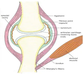
(19) Chapter 1 – Literature review. 1.2. Literature Review. 1.2.1 Synovial joint structure & function The joints of the vertebrate skeleton allow the connection of opposing bones, movement and mechanical support. Joint organs consist of bone, AC and soft periarticular tissues (R. J. Todhunter, 1996).. Joint morphology determines. structural and functional properties.. Three classifications of joints are observed, based on connective tissues and range of articulation.. Synarthrodal joints are immoveable, amphiarthrodal. joints allow a small degree of movement, diathroidal or synovial joints are freely moveable. Synovial joints are the most common and complex joints in the body and a variety of types exist.. The synovial joint (Figure 1.1) provides lubricated contact between the surface of two moving bones and functions to facilitate movement and reassign load between bones (R. J. Todhunter, 1996).. Characteristic properties of the. synovial joint include separation of the articulating bones by a joint cavity and a thin layer of hyaline cartilage enveloping the articulating surfaces.. Near. frictionless movement can be achieved by separation of the joint surfaces with synovial fluid.. The joint capsule and ligament tissue protect and stabilize the joint.. The. synovium provides nutrition and lubrication. Normal function is dependent on the integrity of the joint structures and the specialized properties of AC. i). Joint capsule & ligaments. The articular capsule and ligaments govern range of motion, maintain joint function and prevent dislocation (Caron, 1999). The articular capsule consists of two separate layers: a thick outer stratum fibrosum and thin inner stratum synoviale. The stratum synoviale secretes and absorbs synovial fluid. Local thickening of the stratum fibrosum in certain areas form the ligaments which connect adjacent bones, stabilize the joint and can be intracapsular and or extracapsular. 4.
(20) Chapter 1 – Literature review. Figure 1.1. Diagramatic representation of a synovial joint. The ends of the articulating bones are covered with articular cartilage and separated by the joint cavity. A fibrous capsule connects the two bones and is lined by synovium which secretes the synovial fluid that invests the joint and provides for lubrication. Sourced: Stevens. and. Lowe,. Color. Histology,. p.. 249,. Figure. 13.28. www.mc.vanderbilt.edu/histology/labmanual2002.... 5.
(21) Chapter 1 – Literature review. Ligaments are composed predominantly of water (70%), collagen type I, elastin and small amounts of PGs and other proteins. The capsular and ligament tissues are innervated; receive blood supply and lymphatic drainage. ii). Synovial membrane. The synovium is a thin tissue, which lines the joint space and defines the limits of the synovial cavity. The membrane originates from the AC boundaries of adjacent bones (Frandson, Wilke, & Fails, 2003) and consists of synovial intima and subsynovial fibrous, areolar and fatty tissues which function to stabilize the joint, connect adjacent bones and increase surface area (plicae synoviales). Villi (villi synoviales) project into the joint cavity (Caron, 1999). During inflammatory conditions, the synovium contributes to joint degradation (Smith, 1999).. The synovium produces three specialized types of cells (synoviocytes) which produce a number of macromolecules (Caron, 1999). Type A cells are macrophagic and synthesize hyaluronan.. Type B cells have secretory,. fibroblastic and phagocytic ability and produce collagen, hyaluronic acid, proinflammatory mediators, cytokines, eicosanoids and proteases.. Type C. cells provide both functional and phagocytic properties and are considered a transitional cell type. iii). Joint innervation & blood supply. The synovial joint is a highly innervated and vascularized organ equipped with proprio- and nocioreceptors (Bonnet & Walsh, 2005; Dowd, McQueen, Chessell, & Humphrey, 1998). Afferent output from mechanoreceptors within the joint can alert the central nervous system of impending injury which may be avoided through reflex systems. Innervation of the synovium may stimulate an inflammatory response to pain and protect the joint from injury or disease (Mapp, 1995). iv). Synovial fluid & lubrication. Articular cartilage is without vascular, neural or lymphatic supply and dependent on nutrient diffusion from surrounding tissues (synovial fluid in mature subjects as well as SCB in immature subjects) (Maroudas, Bullough,. 6.
(22) Chapter 1 – Literature review. Swanson, & Freeman, 1968). According to Ficks Law, the movement of a solute into a membrane is dependent on the concentration gradient of the solute across the membrane and the diffusion coefficient. As the permeability of a solute is increased by agitation, joint movement and weight bearing play an important role in the nutrition of AC (Maroudas et al., 1968).. Hyaluronic acid (HA) is a major component of synovial fluid (C.W Archer, Dowthwaite, & Francis-West, 2003). HA and other low molecular weight proteins provide viscosity, soft tissue lubrication and shock absorption (Caron, 1999; Moreland, 2003). HA covers the surface of AC, is between collagen fibrils and sulfated PGs and protects chondrocytes and the collagen network from mechanical stress and deformation (Balazs, 1974). HA may protect the cartilage matrix from PG loss into the synovial space as well as prevent the invasion of inflammatory cells (Moreland, 2003).. The lubricating and deformable properties of articular surfaces are due to their separation by synovial fluid under hydrodynamic and elastohydrodynamic pressure and boundary lubrication (Wright & Dowson, 1976). Normal surface motion draws in the entrapped synovial fluid film and gives rise to a squeeze film or hydrostatic lubrication (Figure 1.2). Damage to the articular surface through mechanical or biochemical means, disrupts tissue integrity and lead to loss of fluid pressurization and increased stress on the collagen-PG network (Ateshian & Wang, 1995). v). The extracellular matrix. The connective tissues of the joint capsule, ligaments, and menisci are composed of collagen, PGs, non-collagenous proteins and water. Collagen type I predominates in these connective tissues with smaller amounts of collagen types III and V.. 7.
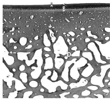
(23) Chapter 1 – Literature review. Figure 1.2. Squeeze film lubrication between two loaded surfaces. Both surfaces are completely separated by a fluid film i.e. synovial fluid. Under hydrodynamic pressure, surface motion draws fluid between the two surfaces. Articular cartilage deforms under hydrodynamic pressure and elastohydrodynamic lubrication. When the two surfaces approach each other in a normal direction the entrapped fluid gives rise to squeeze film lubrication. Sourced: (Wright & Dowson, 1976) adapted.. 1.2.2 Osteochondral tissues The osteochondral tissues of the synovial joint include AC and subchondral mineralized tissues: calcified cartilage, subchondral cortical bone and subchondral trabecular bone (Figure 1.3) (Burr, 2004).. Collectively, these. structures support the joint surface and function (Holopainen et al., 2008; R. J. Todhunter, 1996).. The most superficial tissue is AC; a viscoelastic connective tissue designed to absorb shock and sustain normal joint function.. Beneath the AC and. separated from it by the tidemark is the calcified cartilage, an intermediary zone between the compliant AC and stiffer underlying SCB designed to reduce shear stress (Mente & Lewis, 1994). Unlike AC, calcified cartilage undergoes continued endochondral ossification throughout life (Burr, 2004). The junction between calcified and non-calcified cartilage becomes thin with aging and injury evident by reduplication of the tidemark (Burr, 2004; Secombe, Firth, Perkins, & Anderson, 2002). Deep to the calcified cartilage is the cortical SCB plate which demarcates the trabecular bone. SCB provides structural support to the superficial cartilage layers.. 8.
(24) Chapter 1 – Literature review. Figure 1.3. The osteochondral tissues. A histological section of the osteochondral tissues of the articular joint. (A) articular cartilage, (B) calcified cartilage, (C) subchondral bone plate, (D) trabecular bone. Sourced: Kawcak et. al. (2001).. i). Articular cartilage. Articular cartilage is derived from the hyaline cartilage template formed during embryogenesis. This specialized connective tissue persists throughout adult life and is equipped with unique hydrodynamic properties.. During. development, the mechanical environment determines cartilage surface geometry, contour and topological variation in thickness (Wong & Carter, 2003). Collagen fibrils and PG aggregates enable a low-friction gliding surface and smooth articulation of the joint (M. B. Goldring, 2000a; Hamerman & Schubert, 1962; McIlwraith, 1996; C. A. Poole, 2000; Stockwell, 1991).. Articular cartilage covers the ends of long bones and protects underlying subchondral mineralized tissues from peak mechanical, shearing and compressive forces generated during locomotion and weight bearing (Kawcak, McIlwraith, Nordin, Park, & James, 2001; Stockwell, 1991). The physical properties of AC are dependent on the organization and interaction of matrix macromolecules: water, collagen PGs, and non-collagenous glycoproteins (Johnston, 1997; C. A. Poole, 2000; Smith, 1999). Articular cartilage is divided into four horizontal zones distinguished by chondrocyte morphology and orientation of collagen type II fibres (Figure 1.4). 9.
(25) Chapter 1 – Literature review. (Clarke, 1971; McIlwraith, 2005; Torzilli, Deng, & Ramcharan, 2006; van Turnhout et al., 2008). Fibre orientation varies with tissue depth, age, species and adaptation to withstand derangement (C.W Archer et al., 2003; Clarke, 1971). AC thickness is greatest across convex surfaces, less on concave surfaces and decreases in thickness with age (Serafini-Fracassini & Smith, 1974; Wong & Carter, 2003).. Figure 1.4. Collagen fibre orientation and the etiopathogenesis of. osteoarthritis in the Beagle dog. Oval structures represent chondrocytes, dots represent glycosaminoglycans (GAGs) and curved lines represent collagen fibrils. (I) Normal histology: articular surface is intact and collagen fibrils are tightly bound and orientated parallel with, and perpendicular to, the surface extending from superficial to deep zones. A high content of GAGs is evident. (II) Disease onset: a loss of GAG and exposure of superficial collagen fibrils results is disorganization of the collagen network. An increase in water content, thickening and softening of the cartilage layer ensues. In addition, thickening of calcified cartilage and subchondral bone plate occurs with increased trabecular bone remodeling. (III) With disease progression, increased GAG content loss, advancing tidemark and subchondral bone loss. Pathology is irreversible. Sourced: Arosoki (2000) adapted.. 10.
(26) Chapter 1 – Literature review. a). Chondrocytes. Chondrocytes are the singular cells of AC and comprise 1-12% of the ECM (M. B. Goldring, 2000b; McIlwraith, 1996). Chondrocytes are arranged in clusters or singly within spaces referred to as lacunae and regulate matrix metabolism in a highly organized and efficient manner (Stockwell, 1979a; Tyler, Bird, & Giller, 1990). Chondrocytes synthesize matrix components during growth and development and maintain tissue homeostasis during adult life responding to stimuli with increased activity (Hedbom & Hauselmann, 2002). Chondrocytes are embedded within the ECM and stabilized by the interactions within matrix macromolecules.. The pericellular environment immediately surrounding the chondrocytes is composed mainly of collagen type VI. This environment functions to support chondrocyte viability, providing structural, functional and metabolic stability (Bhosale & Richards, 2008; C. A. Poole, 2000). With increasing distance from cells, there is a decrease in matrix metabolism and distant interterritorial regions may be susceptible to deterioration, especially in mature adult tissue (Jadin et al., 2005). During growth and maturation a high density of cells are observed in the superficial zone.. Chondrocytes have high individual metabolic activity.. However, AC is. hypocellular and over matrix volume metabolic activity is low (Bhosale & Richards, 2008).. Due to the low oxygen concentration of the matrix,. chondrocytes are dependent on anaerobic metabolism (M. B. Goldring, 2006; Platt, 1996). As a result, cartilage matrix proteins have a low turnover rate in healthy adult tissue (Byers & Brown, 1990).. Chondrocytes influence the homeostatic balance of the ECM in response to mechanical stimuli, growth factors, and cytokines (M. B. Goldring, 2006; Platt, 1996). Regional differences in chondrocyte activity exist, with an increase in ECM remodeling in the immediate pericellular zones. Cartilage composition and function are intimately related (C. A. Poole, 2000) and disruption to chondrocyte morphology can alter collagen synthesis and initiate release and. 11.
(27) Chapter 1 – Literature review. activation of enzymes capable of ECM degradation (Smith, 1999; Woessner, 1991).. Chondrocytes have little ability to repair or reorganize the structural architecture of the matrix when it has reached its mature adult state (Barbero et al., 2004). Damage to collagen type II at the superficial layer can progress to mid and deeper zones with age and during OA (Hollander et al., 1995). Consequently, cartilage degeneration can lead to irreversible damage and DJD (O'Rear. et. al.,. 2005).. During. tissue. turnover. fragmented. matrix. macromolecules can be found in the synovial fluid (Saxne & Heinegard, 1995). b). The extracellular matrix. The extracellular matrix consists of collagen fibrils, PGs, water and chondrocytes.. The collagen network gives the matrix tensile strength and. integrity, PGs and high osmotic pressure maintain tissue hydration and chondrocytes maintain matrix metabolism.. The ECM reflects the loading. history of AC and functions to protect chondrocytes from mechanical load, store cytokines and growth factors, and nutrient diffusion to chondrocytes (Bhosale & Richards, 2008).. The matrix acts as a signal transducer and when it deforms under load, produces mechanical, electrical and chemical signals which affect chondrocyte function. The ability of AC to resist compressive, shear and tensile forces is dependent on the integrity of the ECM and chondrocyte morphology (Grodzinsky, Levenston, Moonsoo, & Frank, 2000).. Any defect in matrix. organization can result in structural malfunction and cartilage degradation (Figure 1.4) (Stockwell, 1991). c). Water content. The water content of AC comprises 65-80% of wet weight (Bhosale & Richards, 2008) and is dependent on interaction with large hydrophilic negatively charged aggregating PGs (Smith, 1999).. Water allows the. deformation of the cartilage dependent on load and provides nutrition and medium for lubrication (Maroudas et al., 1968). Water content declines with age from 80-90% in fetal tissue to 70% in adult (Bhosale & Richards, 2008). 12.
(28) Chapter 1 – Literature review. Following injury, water content is observed to increase due to increased permeability and disruption of the cartilage matrix. d). Proteoglycans & aggregan. Proteoglycans form 10-20% wet weight of the ECM and consist of one or more glycosaminoglycan (GAG) chains covalently attached to a protein core (aggrecan) (R. J. Todhunter, 1996). The GAG chains are linear polymers of repeating disaccharides and contain a hexosamine and carboxylate or sulfate ester, or both.. Link protein stabilizes a non-covalent interaction between. aggrecan and HA which maintain fluid and electrolyte balance within the ECM by a Donnan effect (Bhosale & Richards, 2008).. Proteoglycans are produced by chondrocytes and secreted into the matrix as large aggregating monomers (aggrecans) or small non-aggregating PGs (decorin, biglycan and fibromodulin) (Bhosale & Richards, 2008). PGs provide compressive strength and the ability to resist compression due to their high osmotic attraction of water and sodium.. The stiff collagen network limits. swelling tendency and induces tensile stress (Bank, Soudry, Maroudas, Mizrahi, & TeKoppele, 2000). Aggrecans make up the greatest percentage of PGs in AC and provide resistance to proteolytic attack (Smith, 1999). Aggrecans contain chondroitin sulphate and in most species keratan sulphate (Figure 1.5).. The non-. aggregating PGs in AC are characterised by chondroitin sulphate-dermatan sulphate PGs or keratan sulphate PGs. The role of these small leucine-rich PGs (SLRPG) in vivo is not fully understood (R. J. Todhunter, 1996). In vitro studies suggest SLRPs interact with collagen and regulate fibril diameter and matrix assembly (Iozzo, 1999; Scott, 1988).. Hyaluronic acid is the major GAG of AC and differs from other PGs as it is not covalently bound to a core protein when synthesized and is not sulphated (C.W Archer et al., 2003). It is a high molecular weight protein composed of units of glucoronate linked to N-acetyl glucosamine. In AC, HA forms a ‘backbone’ to which PGs attach via a hyaluronan binding region stabilized by a link protein. 13.
(29) Chapter 1 – Literature review. (Figure 1.6). HA and PGs form macromolecular aggregates several milliion KDa in molecular weight. A number of non-collagenous proteins have been described in AC including matrix proteins and link protein (P. G. Todhunter et al., 1996; Upholt & Olsen, 1991).. Figure 1.5. Proteoglycan: structure of repeating disaccharide units. The structures of repeating disaccharide units of glycan in proteoglycans which associate orthogonally along the collagen fibril at specific sites. Key: (1) chondroitin sulphate (as the 4-sulphate), (2) dermatan sulphate, (3) keratan sulphate (monosulphate). (a)-(c) diagrammatic representation of the (a) small (b) large (c) very large proteoglycans. Sourced: Moreland (2003) adapted.. Figure 1.6. Proteoglycan attachment along collagen fibril. Proteoglycans are attached to a backbone of hyaluronic acid that is intertwined among collagen fibrils. Proteoglycans can have chondroitin-sulphate and keratan-sulphate rich regions. Link proteins assist the binding of aggrecan to hyaluronic acid. Sourced: Moreland (2003) adapted.. 14.
(30) Chapter 1 – Literature review. Proteoglycan turnover, unlike collagen, is rapid and percentage content within AC varies with age and tissue depth (Serafini-Fracassini & Smith, 1974). Fetal tissue contains little or no keratan sulphate.. However, following birth the. content of keratan sulphate increases in linear fashion throughout life. Similarly, chondroitin-6-sulphate remains constant during post natal life whilst the content of chondroitin-4-sulphate declines (Bailey & Mansell, 1997). GAG content in the superficial layers constitutes as little as 3% of dry weight and increases in content towards the cartilage-bone junction (Bailey & Mansell, 1997). e). Articular cartilage collagen. Collagen type II and other minor collagen components (collagen types VI, IX, X, XI, XII, XIV) form a dense fibrillar network in which a high concentration of large chondroitin sulphate and keratan sulphate substituted PGs are embedded. The integrity of the collagen network determines AC stiffness and dynamic shear modulus as well as PG content (Oakley et al., 2004). Up to 95% of the collagen in AC is collagen type II which is most concentrated in the superficial zone. The tensile strength of AC can be attributed to the triple helix formation of collagen fibril polypeptide α-chains (Eyre, Wu, Niyibizi, & Chun, 1990) and cross-linking bonds formed with hydroxylysine-aldehyde residues (Eyre et al., 1990).. Hydroxylysyl pyridinoline is the predominant cross-linking structure. found in mature cartilage (Eyre et al., 1990). The cross-linking bonds between hydroxylysylpyridinoline in cartilage collagen types II, IX and XI are dominantly formed by an interaction between telopeptide domains of collagen type II (Eyre et al., 1990). Table 1.1 identifies the different collagen types in AC and their known functions and chain composition.. 15.
(31) Chapter 1 – Literature review. Table 1.1. Different collagen types in normal articular cartilage and their. known functions. Sourced: Bhosale and Richardson (2008), Todhunter (1996), Eyre (2001).. Collagen Type. Chain Composition. Known Functions. II. [α1(II)]3. Most abundant (90-95%) collagen in hyaline cartilage forming the principal component of collagen macrofibrils providing tensile strength. Also found in developing cornea, vitreous humor. VI. α1(VI) α2(VI) α3(VI). Present in mammalian articular cartilage in the pericellular matrix but not avian. Forms microfibrils and may link larger collagen fibrils and chondrocyte attachment within matrix. Located in pericellular matrix and polymerizes to form filamentous network providing multiple adhesion domains for cells and matrix components. IX. α1(IX) α2(IX) α3(IX). Involved in fibril formation forming critical heteropolymer cross-linked with collagen II and XI. Located on surface of major collagen fibrils. Limits collagen fibril diameter and interacts with other matrix components. Provides tensile properties and cross-links to surface of macrofibrils. Mutations of collagen IX gene result in chondrodysplasia precocious osteoarthritis.. X. [α1(XI)]3. Related to hypertrophied cells during development and in deep calcified cartilage layer in adult, interfaces articular cartilage with bone. Provides structural support associated with mineralization.. XI. α1(XI) α2(XI) α3(XI). Located within or on macrofibrils of type II collagen. Believed to be involved in fibril formation forming critical heteropolymer crosslinked with collagen II and IX. Mutations of collagen XI gene result in chondrodysplasia precocious osteoarthritis.. XII. [α1(XII)]3. A minor collagen present in mammalian articular cartilage and connective tissues. Bound to fibril surfaces but not covalently attached. XIV. [α1(XIV)]3. Function not well understood. Structurally related to type IX collagen. Bound to fibril surfaces but not covalently attached. 16.
(32) Chapter 1 – Literature review. ii). Subchondral bone. Long bones originate from a hyaline cartilage template, rich in collagen type II and PGs, by endochondral ossification during the fetal stage of development. With formation, collagen type II is progressively replaced with fibrillar collagen type I essential for the deposition of bone mineral (C.W Archer, Morrison, Bayliss, & Ferguson, 1996).. Bone is composed of organic (30%) and inorganic (70%) compounds. Organic bone is made up of collagen (95%) and non-collagenous proteins (5%) including osteocalcin and bone sialoprotein. Mineral hydroxyapatite comprises the inorganic component and includes mainly calcium and phosphate. Collectively collagen type I and mineral density govern the biomechanical properties and functional integrity of bone. Collagen fibres contribute to bone structure, elasticity and ability to absorb energy. The mineral phase provides strength and stiffness.. Bone is an anisotropic and elastic material which continually adapts to physiological and mechanical stimuli through modeling and remodeling processes (Kawcak et al., 2001). Bone modeling, proposed by Wolff in 1892, is based on mathematical theory governed by mechanical and biological signals at the organ (macromodeling) or trabecular level (micromodeling) (Coelho, Fernandes, Rodrigues, Cardoso, & Guedes, 2009). Bone growth, modeling and remodeling are continuous throughout life and active to different degrees in different mineralized tissues (Burr, 2004).. Subchondral bone remodels rapidly and can change the shape and congruity of the joint. Fatigue and micro-damage can initiate this process and result in normal remodeling, excessive remodeling and sclerosis, or accumulation of micro-damage.. This can lead to fracture or traumatic osteochondritis. dissecans lesions (Schlueter & Orth, 2004).. 17.
(33) Chapter 1 – Literature review. Bone adapts in order to satisfy functional demand and metabolic cost (Coelho et al., 2009). During bone growth, formation results in increased bone mass. During bone modeling, the process of activation-formation or activationresorption results in a net increase in mass and change in structural geometry. During bone remodeling activation-resorption-formation are coupled and contribute to bone maintenance (Burr, 2004). When mechanical stimulus is low, bone remodeling removes bone.. When mechanical stimulus is. excessively high, remodeling adds bone.. Bone remodeling is kept at a. relatively low level due to inhibitory signals produced through physiologic loading (R. B. Martin, 2000). In the normal joint, endochondral ossification and subchondral remodeling are in balance (Burr, 2004).. Subchondral bone acts as a shock absorber between AC and epiphyseal bone (Firth & Rogers, 2005b; Viguet-Carrin, Garnero, & Delmas, 2006) and maintains joint shape (Kawcak et al., 2001).. SCB absorbs ground impact. forces transmitted through the limb during locomotion and minimizes pathology at the articular surface (Secombe et al., 2002).. The functional adaptive. response of SCB, reflects the loading history of the articular surface (Eckstein, Muller-Gerbl, Steinlechner, Kierse, & Putz, 1995).. In horses, the basic collagen fibril network of SCB is established by 5 months of age (P.A.J. Brama, J.M. Tekoppele, R.A. Bank, A. Barneveld, & P.R. van Weeren, 2002; Holopainen et al., 2008).. SCB mineral content and volume. increase significantly with growth and maturation (Holopainen et al., 2008). Variation in SCB thickness can be observed across the joint surface consistent with the mechanical properties and imposed stressors at each site (Kawcak et al., 2001).. The ability of bone to resist fracture is dependent on bone quantity and quality and can be described in terms of architecture, turnover and mineral and organic properties of the matrix (Viguet-Carrin et al., 2006). During remodeling activation, resorption and formation can take days to weeks to complete. Following formation, 65-70% of new bone is quickly mineralized but total mineralization may take 12 months (Burr & Schaffler, 1997). Bone mineral. 18.
(34) Chapter 1 – Literature review. mass density has a strong inverse correlation with fracture risk (Lepage, Carstanjen, & Uebelhart, 2001).. Extensive bone remodeling may underlie overall SCB bone loss (Arokoski et al., 2000; Burr & Schaffler, 1997). The increased rate of bone turnover may result in newly formed bone not being well mineralized and overall material stiffness and elastic modulus reduced despite an increase in structural stiffness. Increased density in calcified cartilage in proximity to the tidemark may increase stress in the deep AC layer and contribute to cartilage loss. An increase in calcified cartilage mineral content and thickness with age and OA likely has greater effect on cartilage loss than does SCB (Burr, 2004).. Endochondral ossification and tidemark advancement occur throughout life and contribute to thickening of calcified cartilage depending on the modeling processes in SCB (Schlueter & Orth, 2004).. However, remodeling at the. osteochondral junction can cause calcified cartilage thinning. In normal joints remodeling and endochondral ossification are in balance.. 1.2.3 The ovine hock joint Cloven hoofed ungulates walk on their third and fourth digits and are in the order Artiodactyl. The sheep is a domestic ungulate in the suborder Ruminatia which includes the cow (Bos Taurus, Bos indicus), sheep (Ovis aries) and goat (Capra hircus).. The horse (Equus caballus) is an odd-toed ungulate of the Perissodactyla, suborder Hipppomorpha and Equidae family.. Equids characteristically have. limb muscle close to the trunk with tendons extending over the long third metacarpal and metatarsal bones to the digits.. This provides significant. leverage and the ability to maintain rapid locomotion.. The skeleton of terrestrial vertebrates is designed to withstand and transmit loads experienced during locomotion.. Maximum strain rate can be observed. to increase linearly with speed but peak strain is dependent on gait. The. 19.
(35) Chapter 1 – Literature review. relationship between peak functional strain and failure of cortical bone in the radius and tibia of quadrupeds during vigorous activity is uniform (C. T. Rubin & Lanyon, 1982).. In quadrupeds the distal tibia and tarsal bones of the hind limb collectively form the hock joint (articulations pedis) and correspond to the human ankle. The hock joint is a complex series of joints formed of the tibio-tarsal, intertarsal, and tarso-metatarsal articulations (Figure 1.7). Differences between the talus of the sheep and horse exist.. In sheep, the bones which collectively comprise the hock joint include the: (i) lateral malleolus (distal end of fibula), (ii) calcaneus, (iii) talus (iv) fused central and fourth tarsal, (iv) first tarsal, (v) fused second and third tarsal bones. The weight of the hind limb is carried by the tibia.. In horses, the bones which collectively comprise the hock joint include: (i) lateral malleolus (distal end of fibula), (ii) calcaneus, (iii) talus, (iv) central tarsal bone, (v) fourth tarsal bone, (vi) fused third tarsal bone. The tarsus is the most commonly affected site of lameness in the hindlimb in horses (Vanderperren et al., 2009).. Due to the anatomical differences between the equine and ovine hock joint, articulation between the tibial cochlea and the trochlea of the talus are directed laterally in the horse where the hindfoot moves outside the forefoot when the horse gallops. In the sheep the talocural articulation is almost vertical(Skerritt & McLelland, 1984). In the horse the proximal intertasal joint is formed between the talus and calcaneus proximally and the central and fourth tarsals distally. In contrast, that of the sheep articulates with the central tarsal bone via the trochlea (Skerritt & McLelland, 1984). The lateral malleolus is not fused and articulates with the tibia and the tibial and fibular tarsal bones (Figure 1.8).. 20.
(36) Chapter 1 – Literature review. A B. C C D. Figure 1.7. Lateral view of the hock joint.. In quadrupeds the distal tibia and bones of the hind limb collectively form the hock joint. The hock joint is a complex series of joints formed of the tibio-tarsal, intertarsal, and tarso-metatarsal articulations. The weight of the hind limb is carried by the tibia. The distal tibia articulates with the bones of the talus, see insert. Insert: Bones of the right talus: (A) Fibular tarsal; (B) Tibial tarsal; (C) Central-fourth tarsal; (D) Secondthird tarsal. Sourced: www.thehorse.com May (1995) adapted.. 21.
(37) Chapter 1 – Literature review. 2. 8 4. 1. 3. 1 5. 2. 4. 3 5. 7. 6. (A). Figure 1.8. 6. 7. (B). The left distal aspect of the tibia.. (A) Left distal tibia (dorsal view), (B) Left distal tibia (caudal. view).. 1.. Tibial. tuberosity; 2. Spines; 3. Tibial crest; 4. Lateral condylee; 5. Proximal end of fibula; 6.. Medial malleolus;. 7.. Lateral malleolus;. 8.. Intercondyleoid fossa.. Sourced:. www.thehorse.com May (1995) adapted.. 22.
(38) Chapter 1 – Literature review. 1.2.4 Joint growth, modeling & remodeling Vertebrate long bones are formed by a highly coordinated and complex process of endochondral ossification (Provot & Schipani, 2005). Systemic and local factors regulate the gradual replacement of the embryonic cartilage skeleton with bone through intracellular signaling, and feedback systems (Malemud, 2006b). Systemic factors regulating chondrocyte behaviour include growth hormone and thyroid hormone. Intrinsic factors regulating chondrocyte gene expression and differentiation include: Sox proteins, fibroblast growth factors, bone morphogenic proteins (BMPs), parathyroid hormone and related peptides, Indian hedgehog and runt transcription factors, hypoxia-inducible factors and vascular endothelial growth factors (VEGF) (Table 1.2) (Figure 1.10) (Mackie, Ahmed, Tatarczuch, Chen, & Mirams, 2008; Provot & Schipani, 2005).. Synovial joints arise in long bone as a consequence of cartilaginous differentiation at the prospective joint site (C.W Archer et al., 2003).. Key. initiators are the production of hyaluronan by cells of the interzone and presumptive synovial cells and mechanical stimulis (C.W Archer et al., 2003; C.W. Archer, Morrison, & Pitsillides, 1994). Limb movement is necessary for joint cavitation and AC development (Arokoski et al., 2000).. Growth. differentiating factor-5, BMPs, BMP antagonist noggin, and Wnt-14 are also involved (Table 1.2) (C.W Archer et al., 2003). AC development is driven by appositional growth of progenitor/stem cell populations within the articular surface. AC has escaped endochondral ossification and is permanent at each end of the long bone.. Matrix metalloproteinases (MMPs) are critical factors involved in long bone growth, maturation and joint formation. MMPs induce cartilage growth plate angiogenesis and apoptosis of hypertrophic chondrocytes in conjunction with VEGF (Malemud, 2006b). The expression of MMPs 1, -2, -3, -9, -13, and -14 are temporally and spatially related to the stage of limb development in the rat (Figure. 1.9). (Malemud,. 2006b).. 23.
(39)
(40) Molecular mechanisms regulating endochondral bone development.. Actions: Chondrocyte differentiation, proliferation & hypertrophy. Regulate expression of cartilage matrix proteins: collagen types II, IX, XI and aggrecan FGFR3 expressed in round & columnar proliferating chondrocytes. FGF signaling hastens late stage chondrocyte hypertrophy. Mutation in FGFR3 gene results in human skeletal dysplasias including achondroplasia. Growth & differentiation factors. Induce cartilage & bone formation. Crucial role in mesenchymal condensation and joint formation, induces apoptosis through the actions of Noggin. Noggin null mutations result in poor limb development & absence of peripheral phalangeal joints.. PTHrP binds its receptor & is a key regulator of calcium/phosphate metabolism & bone remodeling. Found in fetal & adult: cartilage, kidney, heart, hair follicle, placenta, breast, lung, epithelial tissues. Functions as an autocrine, paracrine factor. Mutation or absence PTH/PTHrP receptor results in chondrodyspolasias,: prenatal lethality, premature & abnormal bone mineralization & ossification, shortened limbs: Blomstrand’s lethal chondrodysplasia, and Jansen’s metaphyseal chondrodysplasia Ihh proteins expressed in the interzone between proliferating and hypertrophic chondrocytes, feedback loops with PTHrP, BMPs, FGFs & Runx2. Ihh inhibits hypertrophic chondrocyte differentiation & delays mineralization of cartilage matrix through negative feedback loop with PTHrP. Ihh is necessary for PTHrP expression. Ihh induces BMPs in a positive feedback loop. FGFs reduce Ihh expression. Runx2 upregulates Ihh expression & induces chondrocyte hypertrophy Runx2 essential to osteoblast differentiation. Runx2 expressed in chondrocytes & drives maturation & expression of hypertrophic markers & collagen X. Runx2 feedback activates Ihh & PTHrP expression which determines chondrocyte maturation & proliferation. Runx2 plays critical role in vascular invasion of cartilage through Runx2-dependent regulation of blood vessel invasion meditated by vascular endothelial growth factor.. Early chondrogenesis in the limb. Sox proteins: Sox9, L-Sox5,Sox6. Inhibitor of growth & chondrocyte proliferation Fibroblast growth factor (FGF) receptor3: FGF1, -2, -4, -8, -9, -18. Endochondral bone development Bone morphogenic proteins (BMP): BMP2, -3, -4, -5, -6, -7, Growth differentiation factor5, Noggin (BMP-inhibitor). Mineral ion homeostasis & bone development: Parathyroid hormone-related peptide (PTHrP) & its receptor. Central coordinator of endochondral bone development: Indian hedgehog (Ihh), Sonic hedgehog (Shh), Smoothened (Smo), Patch (Ptc). Chondrocyte hypertrophy, Runt transcription factors: Runx2, Runx3. Sourced: Archer et. al., (2003), Mackle et. al., (2008), Malemud, (2005b), Provot & Schipani, (2005).. Table 1.2.
(41)
(42) Chapter 1 – Literature review. Figure 1.9. Endochondral bone formation. Schematic representation of a mouse tibia during late stage fetal development. The cartilaginous template is progressively calcified with the deposition of bone. Characteristic markers of bone formation, and chondrocyte differentiation are identified. Chondrocyte differentiation is under the regulation of MMPs, Sox proteins, fibroblast growth factor-3 (FGF3), Indian hedgehog (Ihh), Runx2/3, parathyroid hormone and related peptide (PTHrP) and its receptor (PPR).. Collagen type I is. characteristic of bone matrix, collagen types II and X are characteristic of articular cartilage. Sourced: Provot & Schipani (2005) adapted.. 25.
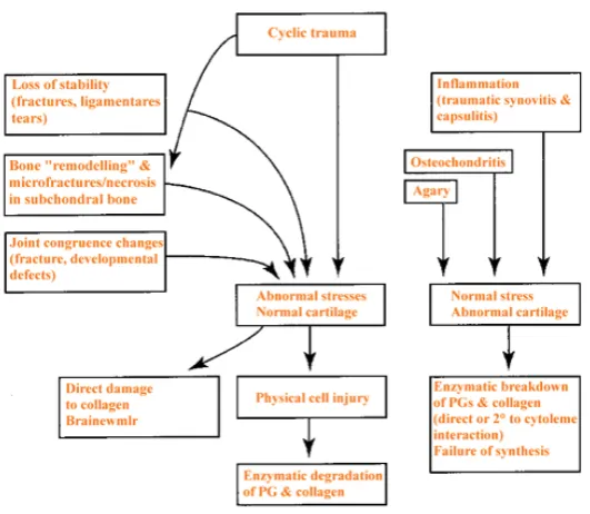
(43) Chapter 1 – Literature review. 1.2.5. Degenerative joint disease: Osteoarthritis. The etiology of OA is multifactorial involving a complex interaction of inflammatory, metabolic and mechanical signals. Repetitive minor and acute trauma, immobilization, ligament or muscular instability, bony misalignment, joint sepsis, obesity, genetics and age can contribute to OA (Arokoski et al., 2000; Bhosale & Richards, 2008; McIlwraith, 1996). In horses, high intensity training, biomechanics, farrier treatment, and environmental factors are also associated with DJD (Schlueter & Orth, 2004). It is widely held that once the integrity of AC in the superficial zone is compromised, the underlying cartilage is subjected to abnormally high strain (Arokoski et al., 2000) (Fig.1.10). Disease progression results in all tissues surrounding the joint becoming involved (Brandt, 2003; Oegema & Visco, 1999).. Figure 1.10. Factors involved in the initiation of cartilage degradation and. onset of osteoarthritis Sourced: McIlwraith (1996) adapted.. 26.
(44) Chapter 1 – Literature review. The homeostatic balance in cartilage regulated by several factors produced by the synovium and chondrocytes including: cytokines, inflammatory mediators and MMPs (Brandt, 1997; M. B. Goldring, 2000b; Hollander et al., 1995; Johnston, 1997; Schlueter & Orth, 2004; Smith, 1999; R. J. Todhunter, 1996) (Figure 1.11). In normal cartilage, the balance between matrix synthesis and degradation is maintained. In OA, disruption to cartilage homeostasis and PG synthesis occurs. Disorganization of the type II collagen network and cartilage softening are early indicators of pathology (Hollander et al., 1995).. Figure 1.11. Enzymatic factors involved in cartilage degradation and onset of. osteoarthritis Sourced: McIlwraith (1995).. Defects in AC can arise secondary to either trauma or osteochondritis dissecans (OCD) and include partial and full thickness chondral and osteochondral lesions. Depth and size of the defect affect the repair process (Bhosale & Richards, 2008; Mankin, 1982). Partial thickness defects in adult AC do not heal spontaneously and enlarge over time. However, full chondral defects may heal transiently (Hunziker, 1999). A feature of the repair response. 27.
(45) Chapter 1 – Literature review. is renewed cell division and upregulated matrix synthesis which resemble some aspects of development (C.W. Archer et al., 1994).. Cartilage lesions are not characterised by pain unless they extend into SCB or are accompanied by synovitis (McIlwraith, 2005). Exposure of bone marrow mesenchymal progenitor cells and growth factors, initiate an intrinsic repair response (Redman, Oldfield, & Archer, 2005).. However, the fibrocartilage. repair tissue is inferior to hyaline cartilage and biomechanically cannot endure repetitive loading (Bhosale & Richards, 2008). A shift from collagen type I fibrous tissue synthesis to collagen type II articular cartilage synthesis within 56 weeks of subchondral injury has been observed (Bhosale & Richards, 2008).. Upregulation of collagen type II and aggrecan gene expression are observed in early OA, consistent with reported increases in cartilage thickness in the ovine OA model. (Appleyard et al., 2003).. However, the thicker cartilage is. biomechanically inferior and undergoes degenerative change over time. This is a result of a loss and failure of articular chondrocytes to replace the water binding PGs. Chondrocytes are unable to maintain homeostatic balance and cartilage degradation exceeds synthesis (Stockwell, 1991). By the time clinical signs of disease are evident irreversible degenerative damage has occurred (Johnston, 1997).. Bone is intimately involved in the initiation and progression of OA (AndersonMacKenzie et al., 2005; Burr, 2004; Fuller, Barr, Sharif, & Dieppe, 2001). However, changes in AC and SCB are closely related making it difficult to determine a single initiating factor in either tissue (Kawcak et al., 2001). This has led to disagreement as to whether changes in SCB precede, are simultaneous to, or follow changes in AC.. The process of endochondral ossification and subchondral remodeling are in a state of homeostasis in the normal joint. During OA, hypermineralisation in hyaline cartilage, calcified cartilage, and SCB has been reported in human femoral heads (Ferguson, Bushby, & Boyde, 2003). The highly mineralized fragments may contribute to wear of the articular surfaces, alter loading. 28.
(46) Chapter 1 – Literature review. patterns and contribute to joint pathology.. Bovine in vitro studies have. revealed that the extent of matrix damage and chondrocyte death are correlated to rate of mechanical load (Blain et al., 2001; Ewers, DvoracekDriksna, Orth, & Haut, 2001).. 1.2.6. Effects of exercise on articular cartilage & subchondral bone. Studies in young animals demonstrate that exercise is important in the adaptation, maintenance and integrity of osteochondral tissues (Arokoski et al., 2000; Brama, Tekoppele, Bank, Barneveld, & van Weeren, 2000; Deschner, Hofman, Nicholas, & Sudha, 2003; Otterness et al., 1998). Exercise shapes the biological properties and mechanical behaviour of AC and SCB which respond to mechanical load in a site-specific way (Appleyard et al., 2003; Arokoski et al., 2000; Brama, Bank, Tekoppele, & Weeren, 2001; de Grauw et al., 2006; Murray, Smith, Henson, & Goodship, 2001; Murray, Vedi, Birch, Lakhani, & Goodship, 2001; Young et al., 2005).. Joint movement and. mechanical load are necessary for postnatal development, the prevention of joint disease, and rehabilitation following injury (Little & Ghosh, 1997; R. J. Todhunter, 1996). i). Articular cartilage. At birth, the composition of AC is homogenous (Brama, Tekoppele, Bank, Barneveld, & van Weeren, 2000). With development and rapid growth there are significant biochemical and biomechanical changes.. Genetic factors,. environment, exercise type and intensity influence the final quality of the musculoskeletal system (Arokoski et al., 2000; Little, Gosh, & Rose, 1997; Mienaltowski, Huang, Stromberg, & MacLeod, 2008; van Weeren, Brama, & Barneveld, 2000).. In young Beagles, adaptive changes in AC have been observed following moderate and long term exercise (Lammi et al., 1993).. Vigorous physical. activity during prepubertal and pubertal years in children has been associated with cartilage acquisition in the knee (Jones et al., 2003). In horses, functional adaptation of AC at different sites within the joint have been observed in response to weight bearing and exercise during the first 6 months of postnatal 29.
(47) Chapter 1 – Literature review. life (Brama, Tekoppele, Bank, Barneveld, & van Weeren, 2000; van Weeren et al., 2000).. These studies suggest that the adaptive response of AC to. mechanical load may take place prior to attainment of skeletal maturity.. Daily exercise protects AC from degeneration and maintains homeostatic balance within the tissue (Otterness et al., 1998).. This is in contrast to the. negative effects of withholding exercise and mechanical load during early development (Otterness et al., 1998; van Weeren et al., 2000). A sedentary lifestyle in hamsters resulted in decreased PG content in AC and SF, cartilage fibrillation, pitting and fissuring (Otterness et al., 1998).. In dogs, joint immobilization caused a rapid reduction in AC thickness, PG synthesis and content (Brandt, 2003) and long-term immobilization caused long-lasting matrix changes in immobilized and contralateral joints despite the introduction of moderate exercise (Jortikka et al., 1997).. However, these. changes may be reversed if not prolonged (Haapala et al., 2001). Studies in the rat knee demonstrate that these changes may be associated with decreased mechanical stress and joint fluid circulation (Hagiwara et al., 2007).. Mechanical load influences chondrocyte biosynthesis and gene expression (Jones et al., 2003) and shapes the structural and functional properties of AC (Arokoski et al., 2000).. In vitro studies demonstrate this is dependent on. magnitude and duration of load. An increase in collagen type II and aggregan mRNA has been observed in human chondrocytes subjected to increasing intermittent hydrostatic pressure (Ikenoue et al., 2003). Moderate (Parkkinen, Lammi, Helminen, & Tammi, 1992) and intermittent cyclic compression (Burton-Wurster, Vernier-Singer, Farquhar, & Lust, 1993) caused an increase in sulphate incorporation and PG synthesis in bovine and canine chondrocytes.. Arokoski demonstrated that in adult AC and SCB loading with physiological mechanical forces was necessary to maintain tissue homeostasis (Arokoski et al., 2000). Regular cyclic loading of AC increased PG synthesis and cartilage stiffness while continuous compression suppressed anabolic metabolism and. 30.
(48) Chapter 1 – Literature review. lead to tissue necrosis (Arokoski et al., 2000). Normal, moderate joint loading maintains joint health and function (Wong & Carter, 2003). Microdamage to the collagen network and degenerative changes in AC and SCB are evident following strenuous exercise and cumulative stress (Appleyard et al., 2003; Brama, Tekoppele, Bank, Barneveld, Firth, et al., 2000; McIlwraith, 1996). Horses subjected to high intensity exercise regimens had cartilage lesions, fibrillation, PG loss and reduced tissue stiffness compared to the cartilage of horses subjected to low intensity exercise (Murray et al., 1999). High intensity training may lead to deterioration of AC and injury to SCB (Murray, Smith, et al., 2001; Murray et al., 1999).. Mechanical stimulus is an important contributor to the repair and regeneration of damaged cartilage. An increase in chondrocyte metabolism, collagen type II and PG synthesis was observed in experimentally induced OA in the canine model (Matyas, Huang, Chung, & Adams, 2002). In vitro synthesis of collagen type II was observed in mature rabbit AC and associated with tissue healing (Cheung, 1978).. The hypertrophic response of femoral head AC in dogs subjected to strenuous treadmill exercise was associated with an early repair response to injury (Vasan, 1983).. However, the collagen component of mature AC has little. capacity for remodeling due to its extremely low turnover rate (Stockwell, 1979b).. In addition, early repair tissue resembling AC deteriorates. progressively with time (Hunziker, 1999).. Deschner et. Al. (2003) demonstrated that the mechanical signals of exercise can promote degradation or repair of cartilage, dependent on the intensity of strain (Deschner et al., 2003). During exercise, signals of high mechanical strain stimulate cartilage destruction, whilst signals of low mechanical strain stimulate cartilage adaptation, synthesis and repair (Agarwal et al., 2004; Deschner et al., 2003). The effect of these signals can be observed in young growing horses where the exercise regimes to which they are subjected has lifelong repercussions for joint health and resistance to injury (van der Harst, 2005).. 31.
(49) Chapter 1 – Literature review. Determining the physiological capacity of joint adaptation to mechanical load during skeletal development, may assist in the design of training and exercise regimes which prevent DJD and promote tissue healing following injury. However, the response of cartilage or tendon to exercise in vivo is difficult to measure (Firth, 2006) and the intensity of strain required to bring about adaptive response in these tissues is unknown (Firth, 2006; McIlwraith, 2005). ii). Subchondral bone. Exercise has a significant influence on skeletal development (P.A.J Brama, J.M Tekoppele, R.A Bank, A Barneveld, & P.R van Weeren, 2002; P.A.J. Brama et al., 2002; C. H. Turner, 1998). The adaptive response of bone to mechanical load can be observed by an increase or decrease in mass and geometry (Firth, 2004).. Bone formation or resorption at a given site is determined by. mechanical strain in terms of magnitude, rate, duration and direction (Lotz, Hashimoto, & Kühn, 1999; Woo et al., 1981) and is influenced by age, peak bone strain, gait and cyclic frequency (Nunamaker, Butterweck, & Provost, 1990).. Moderate long-term exercise causes an increase in bone quantity but not quality. For example, prolonged moderate exercise (65-80% maximum heart rate) in immature swine caused an increase in femur cortical thickness and volume without change in bone mechanical properties or composition (Woo et al., 1981). This adaptive response of bone increased load carrying capacity and energy absorption before failure. However, high intensity training in young growing animals inhibits growth (Woo et al., 1981).. Studies in human athletes reveal strain magnitude may be more important than the number of cyclic loads in controlling bone adaptation to load (Bennell et al., 1997). Osteogenesis is optimized when high-magnitude strain is applied at a high rate with few strain cycles. Activities such as weight training, sprinting and jumping have greater effect on bone mass and BMD than does endurance running. For example, Thoroughbred horses train at a pace much slower than racing speed and over time this may lead to low strain fatigue and loss of stiffness. With infrequent high speed training which loads bone differently, high. 32.
(50) Chapter 1 – Literature review. strain cyclic fatigue results in an increased incidence of fracture in 2 year old horses (Nunamaker et al., 1990).. Horses subjected to high intensity exercise had an increase in bone formation and SCB thickness and structural stiffness at loaded sites compared to low intensity exercised or unexercised horses (Firth et al., 2005; Murray, Vedi, et al., 2001). With high intensity exercise, osteoblasts lining the spongiosa recruit and up-regulate their activity and thickening of trabecular at the expense of the intra-trabecular spaces which become completely replaced with bone (Murray, Vedi, et al., 2001; Secombe et al., 2002). However, high intensity bone strain can cause bone matrix microdamage and initiate bone remodeling (Kawcak et al., 2001; R. B. Martin, 2000). Two year old thoroughbred horses subjected to high-speed treadmill exercise had an increase in SCB density which was also associated with lameness (Kawcak, McIlwraith, Nordin, Park, & Steyn, 2000).. Vascular penetration of the calcified cartilage through to hyaline AC has been observed in the distal third metacarpal of trained and untrained 2 year old thoroughbred horses (A. Boyde & Firth, 2004).. The vascular canals may. provide a pathway between bone and AC for extracellular fluid and the matrix degrading enzymes produced by osteoclasts. Repair of micro-cracks in the calcified cartilage layer with vascular invasion from the SCB may play a role in OA (Arokoski et al., 2000; Kawcak et al., 2001). Remodeling processes in SCB and calcified cartilage can cause biological and mechanical alteration to AC (Burr & Schaffler, 1997).. The response of AC and SCB to exercise, involves several pathways at the molecular level dependent on the intensity of strain. The expression of numerous genes involved in inflammation, tissue adaptation and remodeling (Figure 1.12). An understanding of the balance between joint adaptation and injury during high intensity training is necessary to increase understanding of exercise related DJD (Murray, Vedi, et al., 2001). The intimate relationship between AC and SCB and the influence of exercise during development has not yet been fully examined at the molecular level.. 33.
(51) Chapter 1 – Literature review. Figure 1.12. The mechanical signals of ex exercise. The mechanical signals of exercise affect all the osteochondral tissue and are mediated at the molecular level.. In articular cartilage these signals induce a. phenotypic change in articular cartilage chondrocytes, cellular proliferation, apoptosis, and the production of matrix catabolic and metabolic components including degradative enzymes and inflammatory cytokines.. An adaptive response in bone. leads to remodeling in subchondral mineralized tissues.. 1.2.7 Gene expression Trauma & inflammation Recent cell culture studies suggest that the conversion of mechanical signals of exercise may be mediated through a common pathway with the activation of either cartilage destruction or repair (Deschner et al., 2003). The mechanical signals of exercise can promote degradation or repair of cartilage, dependent on the intensity of strain (Deschner et al., 2003). These signals are likely mediated by inflammatory cytokines; interleukin-1beta (IL-1β) and tumour necrosis factor-alpha (TNF-α) and may be involved in the synthesis or degradation of collagen acting via a common pathway (Agarwal et al., 2004; Deschner et al., 2003; Perkins, Rogers, Firth, & Anderson, 2004).. During. exercise, signals of high mechanical strain stimulate cartilage destruction,. 34.
Figure


+7


Related documents
19% serve a county. Fourteen per cent of the centers provide service for adjoining states in addition to the states in which they are located; usually these adjoining states have
Methods: A follow-up study 5.5 years after the explosion, 330 persons aged 18 – 67 years, compared lung function, lung function decline and airway symptoms among exposed
SP-A, surfactant protein A; SP-D, surfactant protein D; BALF, bronchoalveolar lavage fluid; HS, healthy subjects; IPF, idiopathic pulmonary fibrosis patients; SAR,
It was decided that with the presence of such significant red flag signs that she should undergo advanced imaging, in this case an MRI, that revealed an underlying malignancy, which
Assessing the Impact of Biodiversity Conservation in the Management of Maize Stalk Borer (Busseola f
Field experiments were conducted at Ebonyi State University Research Farm during 2009 and 2010 farming seasons to evaluate the effect of intercropping maize with
As middleboxes can be located between two routers with consecutive ICMP-triggering TTLs (i.e., there is no uncertainty zone), we arbitrarily choose to label a middlebox with the
The CMM model asks us to consider “what spe- cific meanings are (people) making in given situations, how are they making those meanings, and how those meanings affect the social
