CARE CENTRE, CHENNAI,
TAMIL NADU
Dissertation Submitted for
MD DEGREE EXAMINATION
BRANCH VII - PAEDIATRI C MEDICINE
INSTITUTE OF CHILD HEALTH AND HOSPITAL
FOR CHILDREN
MADRAS MEDICAL COLLEGE
THE TAMIL NADU DR.M.G.R. MEDICAL UNIVERSITY
CHENNAI.
Certified that this dissertation entitled "PROFILE OF NEONATAL SEPSIS IN A TERTIARY CARE CENTRE, CHENNAI, TAMIL NADU" is a bonafide work done by Dr. D. Velmurugan, Post Graduate Student of Pediatric Medicine, Institute of Child Health and Hospital for Children Egmore, Chennai - 600 008, during the academic years 2003 - 2006.
Prof.Dr.Saradha Suresh, Prof.Dr.Meer Mustafa Hussain,
M.D.,Ph.D. M.D.,D.C.H.,Ph.D.
Additiona l Professor of Paediatrics, Professor, Dept of Neonatology, Institute of Child Healt h and Institute of Child Health and Hospital for Children, Hospital for Children, Madras Medical College, Madras Medical College, Chennai. Chennai.
Prof.Dr.Mangayarkarasi Senguttuvan Prof.Dr.Kalavathi Ponniraivan
M.D., D.C.H., B.Sc.,M.D.,
Director and Superintendent, Dean, Madras Medical College Institute of Child Healt h and Chennai.
I declare that this dissertation entitled "PROFILE OF NEONATAL SEPSIS IN A TERTIARY CARE CENTRE, CHENNAI, TAMIL NADU" has been conducted by me at the Institute of Child Health and Hospital for Children. It is submitted in part of fulfillment of the award of the degree of M.D (Paediatrics) for the September 2006 examination to be held under the Tamil Nadu Dr.M.G.R Medical University, Chennai. This has not been submitted previously by me for the award of any degree or diploma from any other university.
My sincere thanks to Prof. Dr. Kalavathi Ponniraivan, Bsc., M.D.
ACKNOWLEDGEMENTS
I would like to express my sincere gratitude to Prof. Mangayarkarasi
Senguttuvan M.D.D.C.H., Professor and Head of the department of Paediatrics and Director and
Superintendent of Institute of Child Health and Hospital for Children for permitting me to undertake
this study.
I am extremely thankful to my unit Chief Prof.Dr.Saradha Suresh, M.D., D.C.H., Ph.D., for
her invaluable help, guidance, encouragement and support throughout the study.
I am greatly indebted to Prof. Dr. Meer Mustafa Hussain. MD. D.C.H. Ph.D., for the unstinted guidance given by him throughout my study.
I thank the assistant professors of my unit Dr.Luke Ravi Chellaiah, M.D.,(Paediatrics ), Dr. Guna Singh M.D., D.C.H., Dr. C. Subbulakshmi, M.D., D.C.H., and former assistant Professor,
Dr. R. Kandaswamy., M.D., D.C.H., for their guidance and support.
I thank the assistant professors of new born unit Dr.A.Paramanandam, M.D. D.C.H., Dr. J.Kumudha, M.D., D.C.H., Dr. V. Vijayakumar, M.D.,D.C.H., for their guidance and support.
I express my sincere thanks to Prof.Mohamed Meeran, M.D., (Microbiology) M.D.,
(Venerology) D.V., Head of Department of Microbiology and Dr.U.Umadevi, M.D., (Microbiology)
Assistant Professor, Department of Microbiology for their valuable guidance, supervision and
I extend my sincere thanks to registrars, Dr.Kothai Nayaki, M.D., D.C.H and Dr. P.
Ramachandran, M.D., D.C.H.,for their valuable suggestions in doing this work.
I sincerely thank all the neonates and their parents who had submitted themselves for
CONTENTS
CHAPTER TITLE PAGE NO.
1. INTRODUCTION 1
2. EPIDEMIOLOGY AND PATHOGENESIS 4
3. REVIEW OF LITERATURE 21
4. RATIONALE 29
5. OBJECTIVES 30
6. METHODOLOGY 31
7. RESULTS 34
8. DISCUSSION 54
9. CONCLUSION 62
10. REFERENCES
1.
INTRODUCTION
Of the 130 million babies born every year, about 4million die in the rst 4 weeks of life, the neonatal period1. Most neonatal deaths (99%) arise in low-income and middle- income countries, and about half occur at home. In poor communities, many babies who die are unnamed and unrecorded, indicating the perceived inevitability of their deaths. The three major causes of neonatal deaths worldwide are infections, including sepsis, pneumonia, tetanus, and diarrhea (36 per- cent); prematurity (28 percent); and birth asphyxia or problems related to childbirth complications1.
(Figure 1)
Figure 1
Other 6% Congenital 7%
S epsis/
p n e u m o n i a 26%
As phyxia
23% Diarrhea 3%
Tetanus 7%
Preterm 28%
have tended to focus on pneumonia, diarrhea, malaria, and vaccine-preventable conditions, which are important causes of death after the rst month of life. Between 1980 and 2000, child mortality after the rst month of life i.e., from month 2 to age 5 years fell by a third, whereas the neonatal mortality rate (NMR) was reduced by only about a quarter. Hence, an increasing proportion of deaths are now in the neonatal period.
Infections are the major cause of death after the first week of life. Most of these infection-related deaths could be prevented if all mothers and their babies had access to simple preventive measures and treatments. A quarter of a million babies continue to die each year from neonatal tetanus a condition that can be prevented by giving a pregnant woman two injections of tetanus toxoid.
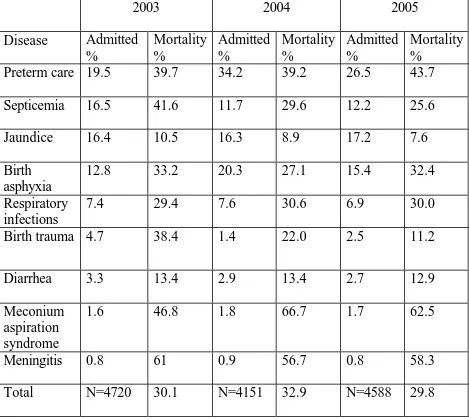
In the Institute of Child Health and Hospital for Children, which is a tertiary care centre, attached to Madras Medical College, admits approximately 4000 neonates for various causes in its extramural new born ward. The common causes of admissions were preterm care, septicemia, jaundice, birth asphyxia etc.
Table 1: INSTITUTE OF CHILD HEALTH NEW BORN STATISTICS
2003 2004 2005
Disease Admitted % Mortality % Admitted % Mortality % Admitted % Mortality %
Preterm care 19.5 39.7 34.2 39.2 26.5 43.7
Septicemia 16.5 41.6 11.7 29.6 12.2 25.6
Jaundice 16.4 10.5 16.3 8.9 17.2 7.6
Birth asphyxia
12.8 33.2 20.3 27.1 15.4 32.4
Respiratory infections
7.4 29.4 7.6 30.6 6.9 30.0
Birth trauma 4.7 38.4 1.4 22.0 2.5 11.2
Diarrhea 3.3 13.4 2.9 13.4 2.7 12.9
Meconium aspiration syndrome
1.6 46.8 1.8 66.7 1.7 62.5
Meningitis 0.8 61 0.9 56.7 0.8 58.3
2. EPIDEMIOLOGY AND PATHOGENESIS
The uniqueness of neonatal infections is a result of a number of factors. There are diverse modes of transmission of infectious agents from mother to fetus or newborn infant 2.
1). Transplacental hematogenous spread may occur at different times during gestation. Vertical transmission of infection may take place in utero, just prior to delivery, or during the process of delivery. After birth, the newborn infant may be exposed to infectious diseases in the nursery or in the community. With the increasing complexity of neonatal intensive care, gestationally younger and lower birth weight newborns are surviving and remaining for a longer time with a high risk of infection.
2) The newborn infant may be less capable of responding to infection owing to one or more immunologic deficiencies involving the reticuloendothelial system, complement, polymorph nuclear leukocytes, cytokines, antibody, or cell-mediated immunity.
impairs functions of polymorph nuclear leukocytes.
4) The manifestations of infectious diseases in the newborn infant are extremely variable.
In addition, the comparative immunodeficiency of the neonate not only predisposes him to infection, but also means that when infection occurs it may disseminate very rapidly, with septicemia shock and death occurring within 12 hours of the first signs of illness. This dissemination which is particularly rapid has two major implications:
1. Early diagnosis is essential. Even very trivial clinical findings that suggest infection demand full laboratory evaluation.
2. Initial therapy must be started on the basis of clinical suspicion. There is no time to wait for the laboratory results like blood culture to come back after 48 – 72 hours later.
Although the onset of illness is often inconspicuous, the clinical course may be alarmingly fulminant, leading to septicemic shock, disseminated intravascular coagulation, and death within hours of the onset of clinical manifestations. Infected infants must therefore be promptly identified and differentiated from non-infected patients, and antibiotics started without delay. However, as microbiological culture results and antimicrobial susceptibility data are not usually available until at least 48 hours after the specimen reaches the laboratory, early identification of genuine sepsis is a major diagnostic problem. In addition, antimicrobial treatment based solely on risk factors and clinical grounds is likely to result in over treatment. Continuation of antibiotics for presumptive bacterial infection often leads to unnecessary and prolonged treatment.
the presence of infection at the time of birth.
Infection in the newborn infant may be limited to a single organ or may involve multiple organs (focal or systemic); it may be mild, moderate, or severe; acute, sub acute, or chronic; or it may be asymptomatic. Asymptomatic bacteremia has been demonstrated in infants born to women with risk factors. Infections with different microorganisms may have overlapping patterns; it is usually not possible to make a definitive diagnosis of a specific etiologic agent from the clinical features alone. Early manifestations of infection may be subtle and nonspecific such as inability to tolerate feeding, irritability, or lethargy. Signs consistent with infection in the newborn may also be caused by a variety of noninfectious disease processes involving different organs
although a focus of infection may not be apparent. In premature infants, hypothermia or temperature instability is more likely to be associated with infection, but some degree of temperature instability is not unusual in low-birth weight infants.
Cutaneous manifestations of infection provide useful clues. Impetigo, cellulitis, mastitis, omphalitis, and subcutaneous abscesses should be recognizable. Ecthyma gangrenosum is indicative of pseudomonal infection. The presence of small salmon-pink papules suggests Listeria monocytogenes infection. A vesicular rash is consistent with herpesvirus infection. The mucocutaneous lesions of Candida albicans are covered later. Petechiae and purpura may have an infectious cause.
(1) There are diverse modes of transmission of infectious agents from mother to fetus or newborn infant 2.
(2) The newborn infant may be less capable of responding to infection owing to one or more immunologic deficiencies involving the reticuloendothelial system, complement, polymorphonuclear leukocytes, cytokines, antibody, or cell-mediated immunity.
(3) Coexisting conditions of the newborn often complicate the diagnosis and management of neonatal infections. Respiratory disorders such as hyaline membrane disease may coexist with bacterial pneumonia. Acidosis impairs functions of polymorphonuclear leukocytes.
(4) The clinical manifestations of infectious diseases in the newborn infant are extremely variable. There may be subclinical infection, congenital malformations, focal disease, and poorly localized systemic infection. The timing of exposure, inoculum size, immune status, and virulence of the etiologic agent influence the expression of disease in the fetus or newborn infant.
asymptomatic or had nonspecific signs and symptoms at the time of presentation.
(6) A variety of etiologic agents infect the newborn, including bacteria, viruses, fungi, protozoa, and mycoplasma.
(7) Finally, with the advances in the neonatal care, increasingly immature, very low birth weight (VLBW) newborns are surviving and remain in the hospital for a longer time, an environment that puts them at ongoing high risk of infection2.
PATHOGENESIS OF ASCENDING BACTERIAL INFECTION
result in disease. Factors influencing which colonized infant will develop disease are not well understood but include prematurity, underlying illness, invasive procedures, inoculum size, virulence of the organism, and transplacental maternal antibodies.
Difficult or traumatic delivery and premature delivery are also associated with increased frequency of neonatal infection. Resuscitation at birth, particularly if it involves endotracheal intubation, insertion of an umbilical vassel catheter, or both, is associated with increased risk of bacterial infection.
The physical and chemical barriers to infection in the human body are present in the newborn but are functionally deficient. Skin and mucus membranes are broken down easily in the premature infant. Neonates who are ill and/or premature are additionally at risk because of the invasive procedures that breach their physical barriers to infection. Because of the interdependence of the immune response, these individual deficiencies of the various components of immune activity in the neonate conspire to create a hazardous situation for the neonate exposed to infectious threats.
apparent before delivery, at delivery, or after a latent period of few hours. Aspiration or ingestion of bacteria during the birth process may lead to infection after a period of 1-2 days
PATHOGENESIS OF LATE ONSET POSTNATAL INFECTIONS
Factors influencing the balance between health and disease in neonates exposed to potential pathogens
Gestation>37 weeks PROM < 12hr
No underlying illness Effective immunity Mucosal & skin barriers Transplacental antibody Less virulent organism Low inoculum
HEALTH DISEASE
IMMUNOLOGICAL STATUS OF A NEONATE
Exposure to Organism
Gestation < 37weeks ROM > 18hrs
Underlying illness Ineffective immunity Ineffective transplacental antibodies
There have been many studies that compare immunologic function of newborn infants to that in adults. Diminished concentrations of immunologic factors and decreased function are often demonstrated. Despite these defects in immunity in premature and full-term infants, the rate of invasive infectious diseases is low in the absence of obstetric and neonatal risk factors. It is important to maintain this perspective when evaluating immunologic prophylactic measures such as the use of intravenous immunoglobulin in the newborn.
Immunoglobulins.
Complement.
Complement mediates bactericidal activity against certain organisms such as E. coli and functions as an opsonin with antibody in optimal phagocytosis of bacteria such as GBS. There is essentially no transfer of complement from the maternal circulation. The fetus synthesizes complement components as early as the first trimester. Full-term newborn infants have slightly diminished classic pathway complement activity and moderately diminished alternative pathway activity. There is considerable variability in both concentration of complement components and activity. The alternative pathway components (B and P) are usually 35%–60% of normal. Premature infants have lower levels of complement components and less complement activity than full-term newborns. These deficiencies contribute to diminished complement-derived chemotactic activity and to diminished ability to opsonize certain organisms in the absence of antibody. In general, opsonization of Staphylococcus aureus is normal in neonatal sera, but varying degrees of impairment have been noted with GBS and E. coli.
Quantitative and qualitative deficiencies of the phagocytic system are important factors contributing to newborns’ increased susceptibility to infection. Chemotaxis is abnormal at birth both in term and in preterm infants. In addition, neonatal neutrophils have decreased adhesion, aggregation, and deformability, all of which may delay the response to infection.
The number of circulating neutrophils is elevated after birth in both full-term and premature infants, with a peak at 12 hr, returning to normal by 22 hr. Band neutrophils constitute less than 15% in the normal newborn and may increase in newborns with infection and other stress responses such as asphyxia.
Neutropenia is frequently observed in preterm infants and intrauterine growth restriction, and it increases the risk of sepsis. The neutrophil storage pool in newborn infants is 20—30% of that of adults and is more likely to be depleted in the face of infection.
Monocyte-Macrophage system.
monocytes is impaired, which affects the inflammatory response in tissues.
Clinical features: Early signs
Lethargy, tachypnea, refusal to suck hypotonia, listlessness,
irritability, pallor, mottled skin, are often the first, mild, non-specific signs that a neonate is unwell2.
Temperature change. A body temperature below 36°C or above 37.5°C sustained for more than an hour or two is due to infection until proved otherwise.
Vomiting, abdominal distention and constipationare features of sepsis particularly when there is an intra abdominal infection.
Late signs
These are usually specific to one organ system
Respiratory:Chest retractions, grunting and cyanosis.
Abdominal:Bilious vomiting, gross abdominal distention, absent bowel sounds.
CNS: High pitched cry, bulging fontanelle and convulsion. Hemorrhagic diathesis: Petechiae, ecchymoses.
Investigations:
1. Blood culture
At least 0.5ml (preferably 1mL) of blood should be taken in pediatric blood culture bottles containing 9 ml of brain heart infusion agar broth.
2. WBC count
A total WBC <5000, >15000 and IT Ratio >0.2 have all been correlated with presence of bacterial infection. IT Ratio is 0.16 at birth and declines to peak value of 0.12 after 72hrs of age. IT Ratio of >0.2 is suggestive of infection.
3. C-reactive protein
It should be done for all the cases unless there is an obvious extrapulmonary focus of infection.
5. Lumbar Puncture
Normal CSF WBC counts in term, noninfected infants are variable, with a mean of less that 30 cells/mm3 with ranges of up to 90 cells, and widely varying levels of polymorpho- nuclear cells on the differential. The normal CSF protein concentration is
0.6gms/L(range 0.4-1.0gms/L)in term and preterm neonates, with an upper limit of 1.5-2.0gms/L. The CSF should be gram-stained and cultured.(nrc Robert 246)
TREATMENT
Any neonate in whom it is remotely possible that an infection is responsible for the abnormal clinical and laboratory findings should be given antibiotics. In virtually all cases the antibiotic should be given intravenously and can be stopped in 5 days or less if the neonate’s condition rapidly improves and cultures are negatives.
Empirical antibiotic therapy2 includes broad spectrum antibiotic usually beta-lactum antibiotic (ampicillin) and an aminoglycoside (gentamycin).A third generation cephalosporin (cefotaxime or ceftazidime) is added to the empirical treatment based of the blood culture results.
REVIEW
OF
3. REVIE W OF LITERATURE
Literature review was done to understand the current status of the clinical profile, causative organisms and their sensitivity pattern, laboratory findings, neonatal and maternal risk factors of neonatal sepsis in similar hospitals in India and abroad.
CLINICAL PROFILE
Hajiehe Borna, M.D. et al 6 has shown that clinical signs such as tachypnea, apnea, cyanosis, tachycardia, abdominal distension and skin presentations were more common
R.S.Jaswal et al 8 have shown that the most common symptoms, by which 66% of these patients presented were refusal for feed, followed by lethargy and jaundice.
BLOOD CULTURE
Roy I et al 4 ., have shown that 47.5% had positive on blood culture.
Jain NK et al., 5 have shown that, in their study 28.3 % were culture positive
suspected sepsis had positive cultures.
R.S.Jaswal et al 8 has shown that the incidence of blood culture positivity was 42% in his study.
Mathur M et al 11 have shown that blood culture was positive in 24.88%.
A.S.M. Nawshad Uddin Ahmed et al 20 , have shown that the 34.9% had positive blood culture.
Das PK et al 23 have shown that in his 48.38% had positive blood culture.
Chacko Betty et al 24 have shown that culture proven EOS occurred in 41.6%.
CAUSATIVE ORGANISMS AND SENSITIVITY PATTERN
Roy I et al., 4 have shown that in his study the most frequent offender was Klebsiella (24.5%) followed by Enterobacter (22.8%). There was an overall predominance of gram negative organisms.
Jain NK et al., 5 in their study have shown an increased prevalence of gram negative septicemia, E-coli and Klebsiella were the common organisms.
Mathur M et al 11 have shown that gram negative septicemia was encountered in 87.1% of these neonates. Klebsiella and Enterobacter species were the predominant pathogens amongst Gram negative organisms. Of Gram positive isolates, Staphylococcus aureus was the predominant isolate (79.0%). Salmonella species was isolated in 2.4% of these cases.
Greenberg et al 14 have shown that out of the 229 cases studied, 57 of all isolates were Gram-negative organisms (mainly Klebsiella pneumoniae (20%) and Escherichia coli (16%)). Gram-positive organisms were isolated in 41% of cases.
A.S.M. Nawshad Uddin Ahmed et al 20 , have shown that majority (70%) of the cultures isolated gram negative bacilli, most commonly E.coli and Klebsiella. These isolates were most often sensitive to gentamicin, ciprofloxacin, and third generation cephalosporins.
Kurien Anil Kuruvilla et al 21 have shown that E. coli and E. fecalis were the predominant organisms causing E0S, while Klebsiella and E. fecalis were the predominant organisms in LOS.
the commonest (60%) isolate.
MORTALITY
Jain NK et al 5 ., in their study have shown that the incidence of neonatal deaths due to sepsis was 11.32%.
A.S.M. Naushad Uddin Ahmed et al 20 ., has shown that in his study, the incidence of neonatal deaths due to sepsis was 40%.
Das PK et al 23 has shown a overall case fatality was 17.1% in his study
Kurien Anil Kuruvilla et al 21 has shown in his study the case fatality rate was 14.4%.
RISK FACTORS
Abdul Hakeem Jokhio et al 12 has shown that training traditional birth attendants and integrating them into an improved health care system were achievable and effective in reducing perinatal mortality improving the perinatal and maternal health in developing countries.
Schuchat A, et al 19. have shown that an obstetric risk factor -preterm delivery, intrapartum fever, or membrane rupture > 18 hours - was found in 49% of GBS cases and 79% of other sepsis.
Kurien Anil Kuruvilla et al 21 have shown in his study that maternal factors significantly associated with EOS were meconium staining of liquor and multiple vaginal examinations.
Chacko Betty et al 24 has concluded in his study that screening for sepsis in an asymptomatic neonate is warranted only in the presence of a maternal risk factor even if the neonate is at high risk of developing sepsis due to associated problems of prematurity, low birth weight or asphyxia.
CRP had a high NPV (97%) but low PPV (36%) with the sensitivity and specificity of 79% and 85% respectively.
D K L Chan, L Y Ho et al 7 have shown that the sensitivity, specificity, positive and negative predictive values of CRP M0.7 mg/dL were 56%, 72%, 71% and 57% respectively. Only abnormal platelet counts had similar efficiency as CRP. Abnormal WCC had the lowest sensitivity and positive predictive value while abnormal ANC had the lowest specificity and negative predictive value among them.
R.S.Jaswal et al 8 have shown that the negative predictive value of serial serum CRP is 100% in deciding duration of antibiotics therapy in neonatal septicemia up to7 days. Newborn with suspected septicemia having raised CRP levels and positive blood culture need longer duration of antibiotics therapy (more than 7 days).
first hour); and latex haptoglobin (positive greater than 25 mg/100 ml). When applied early 93% of cases subsequently proven to have infection had two or more abnormal tests. When less than two tests were positive, the probability that sepsis was not present was 99%.
Bomela et al 13 have found in their study that serial CRP estimation correctly identified 99 of 100 infants in the study as not requiring further antibiotic therapy (negative predictive value, 99%; 95% confidence intervals, 95.6 to 99.97%) and they conclude that the use of serial CRP measurements to guide antibiotic therapy is a safe and practical approach in neonates with suspected sepsis in a developing country.
,specificity of 94% ,PPV and NPV of 91.6% and 100% respectively ,of CRP for detecting proven sepsis and localized infection at cut off point >or =5mg/l.
Manucha et al 26.A CRP level measured at the beginning of septic work-up has a sensitivity of 76% and negative predictive value (NPV) of 96%.
Garland SM et al 27 have shown that a CRP level measured at the beginning of septic work-up has a sensitivity of 67% and negative predictive value (NPV) of 87%.
4. RATIONALE
• Neonatal mortality remains high in our country in spite of the
decline in the infant mortality rate.
• In ICH, in the year 2003, of the 4720 admissions in the new born
ward, 781 ( 16.5%) were neonatal sepsis with a mortality of 325 ( 41.6%) compared to the total mortality in the unit which is 1421 (30.1%) deaths.
• One third of the neonatal mortality is reported to be due to sepsis
and related illness
• Hence this study was planned to understand the clinical parameters,
5. OBJECTIVES
• To describe the clinical presentation of community acquired sepsis in
neonates admitted in the extramural ward of our hospital.
• To analyze the causative organisms and their sensitivity pattern. • To identify the neonatal and the maternal risk factors in the causation
and outcome of neonatal sepsis.
• To identify modifiable risk factors in order to develop appropriate
strategies to address them.
6. METHODOLOGY
This descriptive study was done during the period of Feb 2005-Feb 2006 in the extramural ward, department of neonatology, Institute of Child Health and hospital for children. This hospital is a tertiary care centre which services predominantly low income community. The study population constituted all neonates admitted with history and clinical features suggestive of neonatal sepsis. With the expected culture positivity of 20% and 95% confidence interval for the point estimate, the sample size of 112 neonates with suspected sepsis need to be studied.
INCLUSION CRITERIA: All neonates with symptoms and clinical signs suggestive of sepsis with or without maternal and/or neonatal risk factors.
EXCLUSION CRITERIA: 1) Birth Asphyxia
2) MAS
3) Physiological jaundice
MANEUVER:
2) Probable sepsis
3) Proven sepsis
Case definitions
Clinical sepsis: Neonates in whom only clinical features are consistent with sepsis, without laboratory abnormalities or growth of organism in body fluid cultures.
Probable sepsis: Neonates in whom clinical and laboratory findings are consistent with sepsis but culture negative. They can be either CRP positive neonates or neonates who were positive for two hematological parameters.
Proven sepsis: Neonates with positive blood culture or positive cerebrospinal fluid culture or positive culture of other body fluids.
7. RESULTS
In this study 120 neonates with suspected sepsis admitted in the new born ward were enrolled. Sepsis workup was done for all the neonates, including blood culture, CSF culture( wherever required), tests for indirect evidence of infection like C-reactive protein (CRP), and hematological indicators such as peripheral smear studies for abnormal WBC counts, immature to mature leukocytes ratio (IT ratio) and the presence of toxic granules in neutrophils. Based on the sepsis screen results, the diagnosis was categorized into 3 groups.
[image:48.612.160.459.426.594.2]Classification of neonatal sepsis:
Table 2: Categories of Neonatal Sepsis
N = 120
n %
Clinical Sepsis 52 43.3
Probable Sepsis 43 35.8
Proven Sepsis 25 20.8
Figure 2: Classification of neonatal sepsis
43%
21%
36%
Proven Probable Clinical
Figure 2 shows that out of the 120 neonates with suspected sepsis 25(20.8%) are proven sepsis, 43(35.8) are probable sepsis and 52(43.3%) are clinical sepsis.
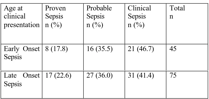
Table 3: Early onset vs. Late onset Sepsis N=120 Age at clinical presentation Proven Sepsis n (%) Probable Sepsis n (%) Clinical Sepsis n (%) Total n Early Onset Sepsis
8 (17.8) 16 (35.5) 21 (46.7) 45
Late Onset Sepsis
17 (22.6) 27 (36.0) 31 (41.4) 75
Clinical features
presence of these signs were of no significance. Chest x-ray showed bronchopneumonia in 8 neonates and pneumonitic changes in 14 neonates.
Table 4: Frequency of Clinical signs
Clinical Signs n %
Tachypnea (RR>60)
118 98.3
Lethargy 79 65.8
Refusal to suck 79 65.8
Fever 70 58.3
Chest Retractions 43 35.8 Poor weight gain 34 28.3
Incessant cry 28 23.3
Weak cry 23 19.2
Abdominal distension
23 19.2
Grunt 19 15.8
Seizure 13 10.8
Shock 12 10
Bulging fontanelle
5 4.2
Hypothermia 5 4.2
Apnea 3 2.5
fontanelle were not found in the clinical sepsis group.
Table 5: Clinical Symptoms and Signs
N=120
Proven (n-25) Probable (n-43) Clinical (n-52) Symptoms and signsn (%) n (%) n (%)
Total
Tachypnea 25(21.2) 42(35.6) 51(43.2) 118
Lethargy 20 (25.3) 32 (40.5) 27 (34.2) 79 Refusal to
suck
22 (27.8) 30 (38.0) 27 (34.2) 79
Fever 19 (24.1) 32 (40.5) 28 (35.4) 79
Chest retractions
15 (34.9) 21 (48.8) 7 (16.3) 43
Poor weight gain
7 (20.6) 12 (35.3) 15 (44.1) 34
Incessant cry
3 (10.7) 5 (17.9) 20 (71.4) 28
Weak cry 8 (34.8) 6 (26.1) 9 (39.1) 23
Abdominal distension
6 (26.1) 11 (47.8) 6 (26.1) 23
Grunt 5 (26.3) 12 (63.2) 2 (10.5) 19
Seizure 5 (38.5) 7 (53.8) 1 (7.7) 13
Shock 5 (41.7) 7 (58.3) 0 12
Bulging fontanelle
4 (80.0) 1 (20.0) 0 5
Hypothermia 2 (40.0) 2 (40.0) 1 (20.0) 5
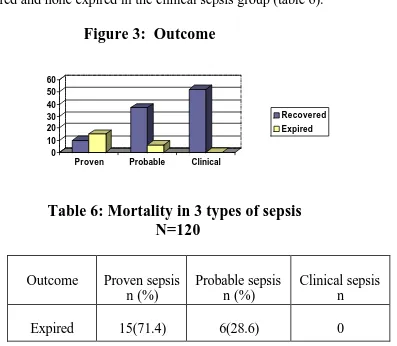
Outcome in neonatal sepsis
[image:53.612.122.519.374.720.2]At the time of admission, sepsis screen was done for all the neonates enrolled and empirical antibiotic therapy with Ampicillin and gentamycin were administered intravenously. The neonates were periodically reviewed clinically and with laboratory results. Based on the organism grown in culture the antibiotic regimen was changed according to the sensitivity pattern. Figure 3 shows that, 21(17.5%) neonates expired and 99(82.5%) neonates recovered. Among the expired group 71.4% were in the proven sepsis group. In the probable sepsis group 6(14%) out of the 43 neonates expired and none expired in the clinical sepsis group (table 6).
Figure 3: Outcome
0 10 20 30 40 50 60
Proven Probable Clinical
Recovered Expired
Table 6: Mortality in 3 types of sepsis
N=120
Outcome Proven sepsis n (%)
Probable sepsis n (%)
Clinical sepsis n
1. Causative organisms and their sensitivity pattern 2. Indicators of probable sepsis
3. Neonatal risk factors 4. Maternal risk factors
Causative organisms and their sensitivity pattern
[image:55.612.113.494.349.718.2]In this study, of the 120 neonates, blood culture was positive in 25 neonates. CSF culture was done in 18 neonates who had features of meningitis. Among them 7 neonates had positive CSF culture but these neonates were also blood culture positive and hence this did not add to the diagnosis. Majority of infections were caused by gram negative organisms. About 76% of infections were caused by gram negative organisms (Table 7) and E coli was the commonest organism (52%) of all (Table 8).
Table 7: Organisms causing sepsis
N =25
Proven sepsis n %
Gram positives 6 24
Gram negatives 19 76
Table 8: Organisms and frequency
N =25
Specific organism n %
E Coli 13 52
Klebsiella 6 24
Staphylococcus
Among the early onset sepsis, 87.5% were gram negative organisms and 12.5% were gram positive organisms. Table 9 shows that E coli was the most common organism (62.5%) followed by klebsiella (25%). Staph aureus was the only organism found in gram positive group (12.5%).
Table 9: Organisms in early and late onset sepsis
(N=25)
Among the late onset sepsis group also, gram negative organisms (70.6%) predominate. E.coli was the most common organism (47.1%) followed by Staph aureus (29.4%).
In this study the sensitivity pattern of various organisms to antibiotics are described as follows.
E coli was sensitive to amikacin in 76.9% neonates, and resistant to ampicillin and cefotaxime in 53.8% and 23.1% neonates respectively.
Klebsiella was sensitive to amikacin in 88.89% and ciprofloxacin in 83.3%. It was resistant to ampicillin in 33.3% and cefotaxime in 23.1%.
E coli n (%) Klebsiella n (%) Staph aureus n (%) Total n (%) Early onset sepsis
5 (62.5) 2 (25) 1 (12.5) 8
Late onset sepsis
neonates and to ceftriaxone in 58.33% neonates. It was resistant to ampicillin in 50% and ciprofloxacin in 66.7% neonates.
Indicators of probable sepsis
In
this study neonates who were CRP positive or positive for 2hematological parameters were classified as probable sepsis.
C-reactive protein (CRP):
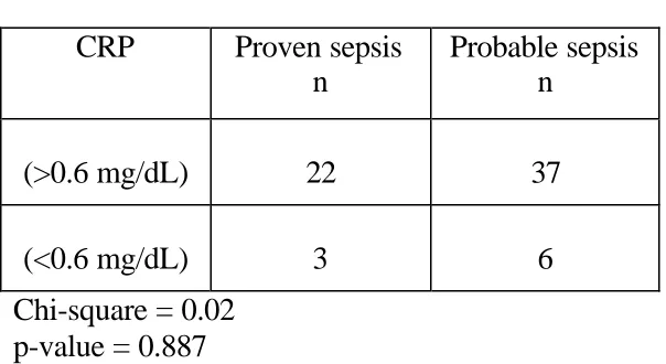
[image:58.612.156.458.530.695.2]The table below shows among the 43 probable sepsis neonates 37 were CRP positive of whom 5 neonates expired and 6 were CRP negative but classified as probable sepsis based on the hematological parameters of whom 1 neonate expired (Table 11) .
Table 11: C-reactive protein
N = 68
CRP Proven sepsis
n
Probable sepsis n
(>0.6 mg/dL) 22 37
(<0.6 mg/dL) 3 6
In this study C-reactive protein was qualitatively estimated and they were positive (>0.6mg/dL) in 59 neonates, 22 in proven sepsis, 37 in probable sepsis (Table 11).
[image:59.612.85.526.240.388.2]Hematological parameters:
Table 12: WBC counts(N =120)
WBC Count Proven sepsis n (%) Probable sepsis n (%) Clinical sepsis n (%) Total n
<5000 1 (6.3) 1 (6.3) 14 (87.5) 16
5000-15000 12 (18.8) 19 (29.7) 33 (51.6) 64
>15000 12 (30.0) 23 (57.5) 5 (12.5) 40
Chi-square = 30.181 p-value = 0.000
WBC Counts <5000 was found in 6.3% in both proven and probable sepsis. Counts >15000 was found in 30% and 53.5% in proven and probable sepsis respectively (Table 12). This was found to be statistically significant (p<0.05).
Table 13: IT Ratio (N =120)
IT Ratio Proven
sepsis n (%) Probable sepsis n (%) Clinical sepsis n (%) Total n
>0.2 16 (41.0) 22 (56.4) 1 (2.6) 39
< 0.2 9 (11.1) 21 (25.9) 51 (63.0) 81
[image:59.612.90.515.566.700.2]41.0% in proven sepsis, 56.4% in probable sepsis and 1% in clinical sepsis. IT Ratio <0.2 was found in 11.1% in proven sepsis group, 25.9% in probable sepsis group and 63% in clinical sepsis group (Table 13). This was found to be statistically significant (p < 0.05).
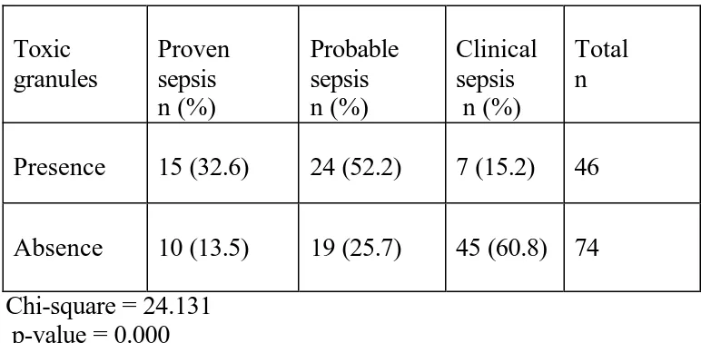
Table 14: Toxic granules
N =120
Toxic granules Proven sepsis n (%) Probable sepsis n (%) Clinical sepsis n (%) Total nPresence 15 (32.6) 24 (52.2) 7 (15.2) 46
Absence 10 (13.5) 19 (25.7) 45 (60.8) 74
Chi-square = 24.131 p-value = 0.000
Other Neonatal Risk Factors
[image:61.612.87.497.337.504.2]Of the 120 neonates who had undergone sepsis work-up during study period, the mean (±SD) age was 12.05 days (± 8.34) and range 1 to 30 days. The mean (±SD) of gestational age was 37.52 week (±1.0) with the range of 34-39 weeks. These parameters were 2534.83 (±189.87) gram (range: 1950-3580) respectively for their birth weight .The mean (±SD) duration of stay was 6.41(±2.91).
Table 15: Neonatal risk factors
Proven Sepsis ( ± 1 SD)
Probable Sepsis ( ± 1 SD)
Clinical Sepsis ( ± 1 SD)
Age (days) 12.92 ± 8.6 11.95 ± 8.35 11.75 ± 8.34
Gestational Age (weeks)
37.72 ± 0.84 37.56 ± 0.83 37.38 ± 1.17 Weight on
Admission
2506.4± 112.47 2583.72± 236.16 2508.08± 170.58
Duration of stay (days)
7.96 ± 3.71 7.12 ± 2.80 5.08 ± 1.83
weight on admission and duration of stay, but the mean age of admission was significantly (p=0.02) longer among female neonates.
[image:62.612.83.499.491.638.2]Table 16 shows that the occurrence of sepsis in exclusively breast fed neonates and in neonates who were not breast fed was 37 (30.8%) and 83 (69.2%) respectively. The percentage of neonates in proven and probable sepsis group was significantly low among exclusively breast fed infants. This is found to be statistically significant (p<0.05). It also shows that 56 neonates were given home remedies. The percentage of occurrence of sepsis is significant in proven and probable sepsis group. This is found to be statistically significant (p<0.05). Vasambu ingestion, nose blowing and oil bath were among the commonly found bad child rearing practices.
Table 16: Feeding pattern
Proven Sepsis n (%)
Probable sepsis n (%)
Clinical Sepsis n (%)
Total n Exclusive
breast fed
6 (16.2) 10 (27.0) 21 (56.8) 37
Other types of feeds
19 (22.9) 33 (39.7) 31 (37.4) 83
Home remedies
It was also found that the occurrence of sepsis was high in those neonates who were given prelacteal feeds but it is not statistically significant (p>0.05).
Maternal Risk Factors
(N=13)
Home deliveries Proven sepsis n (%) Probable sepsis n (%)Clinical sepsis n (%)
Total n By untrained
Dai
2 (25.0) 5 (62.5) 1 (12.5) 8
By trained Dai
0 2 (40.0) 3 (60.0) 5
p-value = 0.149
Intrapartum fever was present in 2 neonates of suspected sepsis of which one was in the probable sepsis group and other in the clinical sepsis group.
Table 18: PROM (N=45)
PROM Proven sepsis
n (%)
Probable sepsis n (%)
Clinical sepsis n (%)
Total n
>18hrs 3 (33.3) 3 (33.3) 3 (33.3) 9
<18hrs 5 (13.9) 13 (36.1) 18 (50.0) 36
Chi-square = 0.499 p-value = 0.778
The above table (Table 18) shows that in 9 neonates, the risk of premature rupture of membranes for >18 hours was present, in which a higher percentage was found in the proven sepsis group, but it is not statistically significant (p>0.05).
[image:64.612.84.531.126.235.2] [image:64.612.81.532.383.489.2]Table 19: Vaginal examinations during labour
(N=36)
>3 vaginal examination
Proven sepsis n (%)
Probable sepsis n (%)
Clinical sepsis n (%)
Total n
Yes 5 (17.2) 13 (44.8) 11 (37.9) 29
No 0 0 7 (100) 7
Chi-square = 8.69 p-value = 0.013
In this study, urinary tract infection in the last trimester was found in one neonate in the proven sepsis group.
The most common maternal illness complicating pregnancy was anemia (19 neonates) followed by pregnancy induced hypertension (2 neonates). There is no significant difference in the occurrence of sepsis in neonates born for mothers who had antenatal illnesses.
MODIFIABLE RISK FACTORS
Table 20: Validity of individual laboratory tests against blood
culture as gold standard test
Lab tests Sensitivity Specificity PPV NPV
CRP 88 61 37 95
WBC >15000 48 70.5 30 83.8
IT Ratio >0.2
64 75.8 41 88.9
Toxic Granules
60 67.4 32.6 86.5
The sensitivity, specificity, positive predictive value and negative predictive value were studied for CRP and hematological parameters like WBC count, IT Ratio and toxic granules against the gold standard (Table 20). The sensitivity and negative predictive value of C-reactive protein were more when compared to the other tests. The specificity and positive
Table 21: Validity of laboratory tests in combination against
blood culture as gold standard test
Lab tests Sensitivity Specificity PPV NPV
WBC count >15000 + IT ratio >0.2
36 87 43 84
IT Ratio >0.2 + Toxic granules
52 89 57 88
WBC count >15000 +
Toxic granules
36 84 38 83
WBC count >15000 + IT ratio >0.2
+ Toxic Granules
32 93 53 84
8.DISCUSSION
In this study, out of 120 neonates enrolled, blood culture was positive in 25(20.8%) neonates, C-reactive protein positive in 59(49.2%) neonates, abnormal WBC counts in 56(46.6%) neonates, IT ratio > 0.2 in 39(32.5%) neonates and neutrophil toxic granules in 46(38.3%) neonates.
[image:69.612.128.483.461.691.2]In this study, the rate of culture positive sepsis is 20.8%. This rate ranged from 20% to 48.38% in other studies. A low blood culture isolation rate in this study might be due to several reasons, e.g. different study population, administration of antibiotics before blood collection either to
Table 22: Culture positivity in various studies
Study Culture positive %
DKL Chan et al. 20
Present study 20.8
Mathur M et al 24.8
Roy I et al. 28.3
ASM Nawshad uddin et al 34.9
R.S Jaswal et al 42
cannot be ruled out. In this study 11 neonates were administered antibiotics before referral and 59 neonates were referred from institutions with referral slips. Moreover, negative blood cultures do not exclude sepsis
and neonates with negative blood culture have been reported with fatal illness and post-mortem evidence of infection.
In this study, the case fatality rate is 17.5% which is 14.4% with Kurien Anil Kuruvilla et al, 40% with A.S.M Nawshad uddin ahmed et al, 11.32% with Jain N.K et al and Das PK et al has shown a overall case fatality was 17.1% in his study
In this study, the common clinical presentations are lethargy (65.8%), refusal to suck (65.8%), tachypnea (98.3%) and fever (58.3%) which were comparable with studies done by A.S.M Nawshad uddin ahmed et al, Jain N.K et al and Chacko Betty et al.
This may be due to the fact that traditional birth attendants are not trained to conduct deliveries in a sterile manner.
In this study, the incidence of sepsis was shown to be higher among illiterate mothers, those with premature rupture of membranes (PROM) >18 hours and vaginal examination >3 times during delivery (statistically significant). Kurien Anil Kuruvilla et al have found that multiple vaginal examinations were significantly associated with early onset sepsis. Anne Schuchat et al have also shown that among the maternal risk factors,
PROM >18 hours and multiple vaginal examinations during delivery are significant risk factors. Premature rupture of membranes and multiple vaginal examinations during delivery are important for the organisms in the genital tract to gain access into the amniotic fluid to cause ascending infections.
In this study, the mean duration of hospitalization in proven sepsis group was significantly longer. The mean age of admission was significantly longer among female neonates which show the sex discrimination in the health seeking behavior of the family.
given native medicines and bad child rearing practices like vasambu ingestion, nose blowing, oil bath, etc.
The variations in the population studies with respect to age and gestational age, the definition of sepsis and different evaluation methods as well as different cut–off point setting and various methods of measuring CRP with respect to the number and timing of sample collection, lead to getting different results. There is not an established standard practice for the use of CRP in infants, and a variety of approaches are described in the literature. However, it can be emphasized that a single CRP level measured at the first contact with health care professional lacks sufficient sensitivity to be useful in identifying neonate with septicemia. In addition CRP can not be recommended as a sole indicator of neonatal sepsis, but it may be used as part of a sepsis workup and in combination with other laboratory tests.
leukocytes. Results of white cell counts and ratios varied widely across studies, with sensitivity and specificity ranging from 17% to 90% and 31% to 100% respectively. In general, studies have shown that I/T ratio >0.2, tend to have high sensitivity, whereas abnormal leukocyte counts, tend to have high specificity.
The varying microbiological pattern of neonatal septicemia warrants the need for an ongoing review of the causative organisms and their antibiotic sensitivity pattern. In this study, majority of infections were caused by gram negative organisms. About 76% of infections were caused by gram negative organisms whereas 24% by gram positive organisms. Ecoli was the commonest organism causing sepsis (52%). The pattern of organisms isolated was similar, regardless of time of onset of disease, birth weight or gestational age. A.S.M. Nawshad Uddin Ahmed et al have shown that of the total organisms isolated, nearly three-fourths (73%), were gram-negative bacilli; (27%) were gram-positive. Escherichia coli was the most common organism (30%), followed by Klebsiella pneumoniae (23%) and Staphylococcus aureus (17%) which is comparable to this study.
was the commonest organism in their study. The major causative organism in EOS was E. coli, similar to the findings of Kurien Anil Kuruvilla et al.
Group B Streptococcus (GBS) was not isolated in this study, unlike western, developed countries where it is the major agent of neonatal septicemia. The insignificance of GBS as a pathogen in many developing countries is supported by a number of other studies. This may be attributable to low prevalence of GBS colonization of pregnant mothers in this area, or, possibly, to the presence of strains with low virulence. The prevalence of E.coli may be due to the fact that it is commonly found as part of the intestinal and vaginal flora, and most deliveries were conducted at home, presumably under conditions of poor hygiene.
• Blood culture was positive in 25(20.8%) neonates.
• About 76% of infections were caused by gram negative organisms,
E coli being the commonest organism causing sepsis
• For most of the gram-negative organisms, gentamycin and
third-generation cephalosporins were effective.
• The common clinical presentations are lethargy (65.8%), refusal to
suck (65.8%), tachypnea (98.3%) and fever (58.3%).
• When clinical signs like chest retractions, grunt and bulging fontanelle
were present the likelihood of proven sepsis is high.
• The incidence of sepsis was shown to be higher among neonates with
maternal such as risk factors, premature rupture of membranes (PROM) >18 hours and multiple vaginal examination during labour. • The occurrence of sepsis was high (69.2%) in the neonates who were
not exclusively breast fed and when given home remedies.
• CRP has a high negative predictive value but low positive predictive
value with sensitivity and specificity of 88% and 61% respectively • The specificity of combinations of hematological parameters were
1. Lawn, J.E., Cousens, S., Zupan, J., “4 million neonatal deaths: When? Where? Why?” The Lancet.365:891-900.
2. In: Nelson Textbook of Pediatrics (17th Edition). Eds Behrman RE, Kleigman RM, Jenson HB. Philadelphia, WB Saunders Company, 2004; pp.623-630.
3. In: A manual of neonatal intensive care (4th Edition). Rennie J.M., Roberton N.R.C, 2002; pp.230-236.
4. Roy I et al in their article in Indian Journal of Medical Microbiology 2002 volume: 20 Issue: 3 Page: 156-159
5. Jain NK et al., in their article in the Kathmandu University Medical Journal (2003) Vol. 1, No. 2, 117- 120
6. Hajiehe Borna, M.D. et al in the Internet Journal of Pediatrics and Neonatology 2005. Volume 5 Number 2.
7. D K L Chan, L Y Ho et al in Singapore medical journal 1992; 33:119-22.
8. R.S.Jaswal et al in the journal Indian Pediatrics 2003; 40:880-883. 9. AG Philip, JR Hewitt et al in the journal Pediatrics Volume 65, Issue
10.Y Kumar et al in the journal Arch Dis Child Fetal Neonatal Ed 2001;85:F182-F186.
11.Mathur M et al in the journal of post graduate medicine (1994) volume: 40 Issue: 1 Page: 18-20.
12.Abdul Hakeem Jokhio et al in New England journal of medicine In 2005 Volume 352:2091-2099.
13.Bomela et al in Pediatric Infectious Disease Journal. 19(6):531-535, June 2000.
14.Greenberg et al in the Pediatric Infectious Disease Journal. 16(8):768-773.
15.Agarwal R et al in the Indian Journal of pediatrics, 2001-68(12). 16.S Vergnano et al in Archives of Disease in Childhood Fetal and
Neonatal Edition 2005; 90: F220-FF224.
17.Vinod Kumar CS et al Indian J Med Microbiol 2005;23:270-271. 18.Ghosh S et al, In Indian journal of medical sciences, 2000, 54,9. 19.Anne Schuchat et al in PEDIATRICS Vol. 105 No. 1 January 2000,
pp. 21-26.
35:851-858.
22.A Narang et al in the journal of Indian Pediatrics 1987 Jan; 24(1): 39-43.
23.Das PK et al in the journal of Indian medical association 1999 Jan: 97.
24.Chacko Betty et al in Indian journal of pediatrics 2005;72:23-26. 25.Nuntnarumit P et al . Predictive values of serial c-reactive protein in
neonatal sepsis Med Assoc Thai.2002 Nov; 85 suppl 4: S1151-8. 26.Manucha V, Russia U, SikaM, Faridi NM, Madan N.; "utility of
hematological parameters and C-reactive protein in the detection of neonatal sepsis." J pediatr child health. 2002 Oct; 38(5):459-64. 27.Garland SM, Bowman ED;" Reappraisal of C-reactive protein as a
PROFORMA FOR NEONATAL SEPSIS
SR.NO IP NO
NAME SEX: Male/Female TERM/PRE-TERM
GESTATIONAL AGE: ____ weeks AGE ON ADMISSION: ___days__hrs BIRTH WEIGHT: _____ gms
WEIGHT AT ADMISSION: ____ gms DATE OF BIRTH:
DATE OF ADMISSION: DATE OF DISCHARGE: DATE OF DEATH:
DURATION OF STAY: ____ days SELF REFERRAL: [Y/N]
REFFERED FROM FACLILITIES: 1. Home
2. HSC 3. PHC
4. Taluk HQ hospital 5. District HQ hospital 6. Tertiary referral unit 7. Private OP
ANTE-NATAL DATA 1. Total no of hospital visits
[0/1/2/3/4/5/more]
2. Total no of home visits by health worker [0/1/2/3/4/5/more]
3. IFA intake [Y/N]
4. Tetanus toxoid injections: [Y/N]
MATERNAL DELIVERY DATA
1. Mode of delivery: [1/2/3] [1-Vaginal/2-Assisted/3-caesarian] 2. Place of delivery: [1/2] [1-Home/2-Institution]
• Home: [1/2]
[1-Untrained/2-Trained personnel]
• Institutional: [1/2/3/4/5/6]
[1-HSC/2-PHC/3-Taluk HQ hospital/4-District HQ hospital/5-Tertiary referral unit/6-Private Nursing home]
• Conducted by: [1/2/3/4]
[1-Nurse/2-Doctor/3-Obstetrician/4-None] 3. Type of facility:
1. Birth order [1/2/3/4/5/more] 2. Multiple gestations [Y/N/NK] 3. Intrapartum fever [Y/N/NK] 4. PROM > 18hrs [Y/N/NK]
5. Vaginal examinations >3 in labour [Y/N/NK] 6. Meconium staining: [Y/N/NK]
7. Cloudy amniotic fluid: [Y/N/NK] 8. Foul smelling amniotic fluid: [Y/N/NK] 9. UTI in the last trimester: [Y/N/NK] 10.Maternal Illness: [1/2/3/4/5]
[1-PIH/2-Anaemia/3-Diabetes/4-Heart disease/5-Others]
11.Maternal education: _________Paternal education: __________ NEONATAL RISK FACTORS
1. Birth asphyxia: [Y/N/NK]
2. Cord status: [1/2/3] [1-Bandage/2-Ointment/3-Dry] 3. Feeding
a. Feeding pattern [1/2/3] [1-Exclusive/2-Not exclusive] b. Prelacteal feeds: [Y/N] If Yes specify ____________ 4. Clean clothes: [Y/N]
5. Bath to the baby: [Y/N]
1. Superficial infections
• Umbilical sepsis: [Y/N]
• Pyoderma: [Y/N]
• Conjunctivitis: [Y/N]
2. Apneic spells: [Y/N]
SYMPTOMATOLOGY General
Lethargy Refusal to suck Poor cry
Poor weight gain Incessant cry Respiratory System Respiratory rate ___/min Chest retractions
Grunt Apnea
Central nervous system ALOC
Seizures
Bulging Fontanels Poor neonatal reflexes Shock
Temperature Fever
Hypothermia
Gastro intestinal tract Abdominal distention Vomiting
Bleeding
LABORATORY FINDINGS
Blood culture: [Positive/Negative]
If positive- organism and sensitivity C-reactive protein: [Positive/Negative] Peripheral smear studies:
WBC Count: [1/2/3] [1-<5000/2-5000-15,000/3->15,000] IT Ratio>0.2: [Y/N]
Toxic granules: [Y/N] X-Ray chest and abdomen Lumbar puncture
Cells Protein Sugar
Culture & sensitivity
OUTCOME 1. Recovered 2. Expired
FINAL DIAGNOSIS
Annexure – 2
BLOOD CULTURE
RHELAX CRP Principle:
RHELAX CRP slide test is a qualitative test for detection of CRP which is based on the principle of agglutination. The test specimen (serum) is mixed with RHELAX CRP latex reagent and allowed to react. If CRP