A STUDY ON ATAXIA
Dissertation Submitted to The TamilNadu Dr.M.G.R
Medical University for M.D. Degree in General
Medicine
Branch I
The Tamil Nadu
Dr. M.G.R. Medical University
Chennai
CERTIFICATE
This is to certify that the dissertation titled “A Study on Ataxia” is a bonafide work done
by ‘Dr. A.K. SENTHIL KUMAARAN’. It is a regular systematic study done under my
guidance and supervision during the period of 18 months (July 2004 to Jan 2006) and
submitted for ensuring ‘M.D Branch I General Medicine Examination’ September
2006 of Tamilnadu Dr. M.G.R. Medical University, Chennai.
Place :
Date :
Prof. Dr. G. YASODHARA M.D.,
Prof.Dr.K.UMAKANTHAN
M.D.,
Prof & HOD of Medicine,
Prof & Unit Chief,
Coimbatore Medical College & Hospital,
Dept of Medicine,
Coimbatore.
Coimbatore Medical College.
Dean
COIMBATORE MEDICAL COLLEGE & HOSPITAL
ABBREVATION
1.
AT
: ATAXIA TELENGIECTASIA2. CAT : COMPUTRIZED AXIAL TOMOGRAPHY
3. CIDP : CHRONIC INFLAMMATORY
DEMYELINATING POLYNEUROPATHY
4. CP : CEREBELLO PONTINE
5. CVJ : CRANIO VERTEBRAL JUNCTION ANOMALY
6. DRPLA : DENTATO RUBRO PALLIDEO LUCEAN ATROPHY
7. EOCA : EARLY ONSET CEREBELLAR ATAXIA
8. FA : FREDRIECH’S ATAXIA
9. HCA : HEREDITARY CEREBELLAR ATAXIA
10.HMSN : HEREDITARY MOTOR SENSORY
NEURONOPATHY
11.OPCA : OLIVO PONTO CEREBELLAR ATROPHY
12.SCA : SPINO CEREBELLAR ATAXIA
INTRODUCTION
“Taxia” means co-ordination in Greek. Ataxia therefore is defined as in-coordination or
condition without order. They are mainly cerebellar, sensory and vertiginous ataxia.
They may be hereditary . congential or acquired. Hereditary ataxias are most often familial
Sporadic cases do occur which may represent recessive inheritance in small kindreds or
dominant inheritance with new mutations or variability of penetrance. It may also occur as a
manifestation of a pleotropic gene.14
HISTORICAL ASPECTS
Friedreich was the first person to give its clinical description. comprising ataxia of the
extremities, nystagmus and dysarthria appearing at puberty or earlier.
In 1891 Menzel described a family in which 4 members displayed ataxia of extremities
followed at a later stage by dysarthria and choreiform movements and called this syndrome
Olive Ponto Cerebellar Atrophy .
Ataxia telangiectasia was first described by Madam Louis Bar.
In 1972 Nakano et al described the proband Machado family residing in Massachussettes,
which descended from a native of the island of Sao Miguel in the Portuguese Azores. The
syndrome described included cerebellar signs, sensory loss and distal amyotrophy.
The hereditary ataxias may occur occasionally as one of the various phenotypic expressions
–syndrome of Olivo Ponto Cerebellar Atrophy (Olivo Ponto Cerebellar Atrophy with diabetes
Insipidus, diabetes Mellitus, Optic Atrophy and Deafness)34 Hereditary ataxias may be
dominant, recessive or sporadic.
Dominant Ataxias
Dominant ataxias are common. Intrafamilial variability is the rule rather than exception.
The disease can begin at any age between 18 months to 80 years. But typically begins between
adolescence and the forties. Ataxia involves the gait and extremities Ophthalmoplegia is often
present and usually involves saccadic more than pursuit eye movements. Combination of
pyramidal and extrapyramidal. dysfunction may be seen. Reflexes are typically increased early
in the disease However, motor neuron degeneration occurs later in some patients, producing
absence of tendon reflexes, atrophy and fasciculation. Dementia develops late in the course in
some patients (Stupmf68 1985)
Recessive Ataxias
There are many distinctive forms of recessive ataxia. The most clearly defined variety is
Friedreich ataxia. At least one third of recessively inherited ataxias do not fit in with the
current clinically or biochemically defined diagnostic categories”.
Sporadic Ataxias
Sporadic ataxias account for about one 3rd of the patients with chronic progressive ataxia8.
Specific metabolic abnormalities have already been detected in some of these diseases and
Acquired Ataxias
Acquired Ataxias67 can be produced by heterogeneous causes. Collectively they are the
major group of ataxias. Important among these are vascular, neoplastic, infective,
demyelinative and degenerative varieties.
Cerebrovascular diseases67 producing acute and sub-acute infarctions, and hemorrhages
may result in ataxic syndromes. Lesions of these types will result in cerebellar symptoms
ipsilateral to the injured cerebellum and may be associated with impaired levels of
consciousness due to increased intracranial pressure and ipsilateral pontine signs. Similarly,
cerebllar tumors, demyelinating plaques of multiple sclerosis and abscess formation
produceprogressive deficits ipsilateral to the cerebellar lesion. Two other important entities are
paraneoplastics disease due to neoplasms outside the brain and causing bilateral cerebellar
deficits by an antibody – mediated process and subacute cerbellar degeneration of the vermis
with gait ataxia, due to chronic alcoholism.
Patients with AIDS67 may develop an acute ataxia due to progressive multifocal
leukoencephalopathy, which causes a rapid demyelination process, as a result of the JC virus.
Progressive ataxia of gait and extremities may also caused by toxic or metabolic disorders,
including hypothyroidism, hyponatremia. Vitamin B1 defeciency, vitamin B12 deficiency,
toxic levels of phenytoin, lithium, bismuth, germanium, methyl mercury, and organic solvents
and treatment with cyotoxic chemotherapeutic drugs. Rarely, congenital lesions, such as the
Chiari type 1 malformation with cerebellar tonsillar compression of the brainstem, and
congential dilatation of the fourth ventricle into a large cyst owing to impaired drainage of CSF
(Dandy-Walker syndrome), present in adults as a progressive diffuse ataxic syndrome. Specific
Ataxias are an important problem of management with respect to diagnosis, prognosis and
genetic counseling. Department of anatomy and Neurobiology, Washington.University school
of medicine 59 in their study mechanism of ataxia (1997), they came to the conclusion that
ataxia may be due to inability to co-ordinate the relative activity of multiple muscles and
REVIEW OF ANATOMICAL AND PHYSIOLOGICAL ASPECTS
Many disorders present with ataxias but only three types of ataxias are of practical clinical
importance sensory, cerebellar and vertegenous Sensory ataxia refers to dysfunction of posterior
column, dorsal roots and peripheral nerves and a worsening of the ataxia once the sensory input is
blocked. Sensory input from labyrinth are also important in maintaining postures. Cerebellar ataxia
refers to incoordination due to cerebellar dysfunction. Because of the rich afferent and efferent
connections from and to the rest of the C.N.S and of the unique anatomic arrangement of the
organ, the cerebellum is well equipped to act as a servo system. Hence it is worthwile reviewing
the functional anatomy of the cerebellum.
The cerebellum (“Small Brain 30(a)11) is a dorsal portion of the metencephalon, lying in the
posterior fossa of the cranium. The cerebellar surface has a characteristic patterning of parallel and
curved furrows that separate the cortex into numerous laminae or folia. Each portion of the
cerebellum has an anatomical name.
The narrow, central part of the cortex is called the vermis, the two larger, lateral masses are
the right and left hemisphere. The several lobes and principal fissures of the cortex are identified as
follow, in a retro-caudal direction:
1. Anterior lobe 2. Middle lobe
3. Posterior lobe 4. Flocculonodular lobe
The anterior lobe +pyramid+ uvula is known as the paleocerebellum, the middle and
posterior lobes are the neocerebellum and the flocculonodular lobe+lingula is the archicerebellum.
Afferent Fibres:
a. Climbing Fibres: b.Mossy Fibres :
Cells of the Cerebellar cortex:
a. Granule Cells b. Golgi type II neurons
c. Basket cells and Stellate cells d. Purkinje Cells
Cerebellar Nuclei:
a. Nuclues Fastigi b. Nuclei globosus c. embolifomis d. Dentate nucleus
CEREBELLER CONNECTIONS:
A. Inferior peduncle (restiform body) : Predominantly afferent
B. Middle peduncle (brachium points):mainly afferent
C. Superior peduncle (branchium conjunctivum): both afferent and efferent.
Anatomy posterior Column
12aThe large fibres, which subserve tactile and position sense and kinaesthesia project
rostrally in posterior column on same side of the spinal cord and make their 1st synapse in the
gracile or cuneate nuclei of lower medulla. The second order neuron decussates and ascend on the
medial leminiscus located medially in the medulla and in the tegmentum of Thalamus. The third
order neuron projects to the parietal sensory cortex.
Anatomy and Physiology of labyrinthine system
30bReceptors for 2 sensory modalities hearing and equilibrium are housed in the ear. The semi
circular canals, the utricle, and the saccule of inner ear are concerned with equilibrium. Receptors
acceleration in the horizontal direction and receptors in the saccule detect acceleration in the
vertical direction. The receptors are hair cell and they are 6 groups in each inner ear one in each ‘3’
semi circular canal. one in urticle and one in saccule and one in cochlea (hearing).
Neural Pathyway
30bThe cell bodies of the 19000 neuron supplying the cristae and macule on each side are
located in thee vestibular ganglion. Each vestibular nerve terminates on the ipsilateral 4 part
vestibular nucleus and in the floculonodular lobe of the cerebellum. Second order neurons pass
down the spinal cord from the vestibular nuclei on the vestibulo spinal tract and ascend through the
MLF to the motor nuclei of the cranial nerves concerned with the control of eye movement. The
tracts that descend from the vestibular nuclei into the spinal cord are concerned primary with
postural adjustments, and the ascending connections to cranial nerve nuclei are largely concerned
with eye movements.
Pathogenesis of Unsteadiness
GAIT Imbalance 12b
Imbalance results from disorders of the spinocerebellar of vestibular sensory impur, the
integeration of these imputs in the brainstem or cerebellum, or the motor output to the spinal
neurons that control axial and proximal muscles.
The position of the head in space is normally detected by the inner car. Excitatory input
form the vestibular nerve and nuclei is to the fastigial nucleus deep in the midline of the
cerebellum and via glutaminergic mossy fibres to overlying ipsilateral granule cells of the
and proximal limb muscles. This sensory input is transmitted both along the posterior column and
medial lemniscal pathways to the cereberum and the spinocerebellar pathyways to the cerebellum.
Visual input, by way of the tectum of the midbrain, is transmitted to the cerebellum through
similar, excitatoty mossy fibres.
The midline cerebellar cortex and nuclei are of paramount importance in the integration of
these inputs and in control of appropriated motor responses required to maintain normal balance.
The major cerebellar output for balance is from the fastigeal nuclei to the vestibular nuclei
and reticular formation and, to a lesser extent, directly from the midline Purkinje cells to the
vestibular nuclei, Vestibular nuclei and reticular formation project descending vestibulospinal and
reticulospinal output via the ventromedial pathways to control the axial and proximal muscles of
REVIEW OF LITERATURE
Several studies have been conducted in India26,64,63 and abroad 8,49,65 on the ataxia.
HISTORICAL REVIEW
Nikolas Friedreich of Heodelberg in 1861 published as account of 9 cases of ataxia
occurring in 3 families. It was then regarded as a hereditary or juvenile form of tabes or a form of
multiple sclerosis. Again in 1876 Friedreich claimed it as an independent disorder and he called it
as hereditary ataxia.
INCIDENCE
The first comprehensive clinical study of 32 cases of hereditary ataxia in India was done by
Chuttani26 et al from Amritsar between 1947 to 60. This study included 27 cases of Friedreich’s
ataxias. He emphasised that it was not an uncommon disease in India as was thought. He clearly
described all the clinical features of the disease. He mentioned about a family of 8 siblings -3 with
clinical evidence of Friedreich’s Ataxia and 2” with forms fruste of Friendreich’s Ataxia. In
another family of 5 members, 3 were affected – 2 had features suggestive of Friedreich’s Ataxia
and one had Sanger Brown Marie Disease Jelly63 in 1961 analysed 200 cases of spinocerebellar
degeneration which included 9 cases of classical Friendreich’s Ataxia, 6 of cerebellospinal group
and 5 of cerebellar type. He included these cases under paraplegias because the patients primarily
presented with difficulty in walking . A similar study of paraplegias was conducted by Chaudhary
et al in 1968 and it showed a 9% incidence of spino cerebellar degeneration as compared to the
10% incidence in Jelly’s series.
The prevalence of hereditary ataxia in Western countries varies between 7 and 17/100000
Sahadevan73 from Calicut studied 21 cases of during a period of 14 months (1963-64). His
aim was to compare his findings with that of similar study conducted in a teaching hospital at
London. He conducted that our patients had an earlier onset of cerebellar ataxia than that reported
in the London series.
Wadia64 and Swami studied 9 families with spinocerbellar ataxia and noticed slow eye
movements in all of them.
Sarma and Vermani61 (1972) reported 65 cases of spinocerbellar ataxia which included 2
cases of ataxia telangiectasia and one case of Ramsay Hunt syndrome.
CLASSIFICATIONS
In the course of time many attempts have been made to classify the hereditary ataxias
clinically.
Holmles in 1910 classified hereditary ataxias into 3 groups
1. Predominant spinal form (Eg. Friedreich’s Ataxia)
2. Cerebellospinal form (Eg. Sanger Brown Marie Disease)
3. Predominant Cerebellar form (Eg. Olive Ponto cerebellar Degeneration)
Later on in 1954, Green field modified this classification into 2 major varieties
1. Spinal form and 2. Cerebellar form
An indigeneous variety of spinocerebellar ataxia was described form India by Wadia and
Swami64 in 1971. They noted a particularly striking abnormally in their patients consisting of a
severe loss of saccadic movement of the eyes. Skre and Loken60 (1970) investigated a large
Friedreich’s ataxia in which the emphasis is on spinal symptoms, cerebellar ataxia with emphasis
on cerebellar symptoms and spinocerebellar ataxia, a transient form between the other two. The
starting point of his classification was genetic and clinical.
Harding (193)49 studies about 274 cases of hereditary ataxias during 1966-80 and put forth
a classification
1. Ataxias with known metabolic cause
2. Ataxias with no known metabolic cause.
Thus, as Refsum and Skree (1978) started there are as many classification as there are
authors on the subject.Widely accepted classification at present are given below
Classification of Ataxia
1. Cerebeller ataxia 2. Sensory ataxia 3. Vertigenous ataxia
Classification of Cerebellar Ataxia (Hardings)56
1. Congenital Cerebellar Ataxia
a. Ganule cell hypoplasia - Cerebellar ataxia, Mental Retardation
b. Pontocerebellar hypoplasia - Cerebellar ataxia + MR + Spasticity
c. Paine syndrome - Ataxia + MR + Spasticity – X- linked
d. Gillespe syndrome - Ataxia + MR + Partial aniridia
e. Joubert’s syndrome - Ataxia + MR + Episode hyperpnoea
2. Congenital development anomaly
-CVJ anomly
-Basilar invagination
3. Acquired Cerebellar Ataxia71
Trauma
Tumours
Vascular Cerebellar haemorrhage
Cerebellar infarct
Vertebrobasilar TIAs
Infections Tuberculoma
Pyogenic / fungal abscess
Acute viral cerebellitis
Slow virus infections / prion diseases
Falciparum malaria
Enteric fever
Drugs Anticonvulsants
Antineoplastic agents
Lithium
Piperazine
Toxins Heavy metals
Solvents
Organochlorine compounds
Nutritional / Alcohol Vitamin B1
(Deficiency of ) Vitamin E
Vitamin B12
Metabolic / Endocrine disorders Hepatic encephalopathy
Hypothyrodism
Hypoglycaemia
Post-infectious cerebellars
Miller Fischer syndrome
Paraneoplastic disorders
Hyperthermia/Hypoxia
4 Spinocerebellar ataxia – Genotypic Calssification67
SCA 1 to 22 – Autosomal Dominant
SCA 1 – Ataxia + Pyramidal + Extra pyramidal + Opthalmoparesis
SCA 2 - Ataxia + Pyramidal + Extra pyramidal + Slow saccades
SCA 3 - Ataxia + Pyramidal + Extra pyramidal + Amyotrophy
SCA 4 - Ataxia + Pyramidal + Sensory neuropathy
SCA 5 - Ataxia + Dysarthria
SCA 6 - Ataxia + Dysarthria + Nystagmus
SCA 7 - Ataxia + Retinal Degeneration
SCA 8 - Ataxia + Dysarthria + Nystagmus + Leg spasticity + Reduced vibratory sensation
SCA 10 - Ataxia + Dysarthria + Nystagmus + Seizures + Polyneuropathy
SCA 11 - Ataxia + Dysarthria + Vertical Nystagmus + Hyper reflexia
SCA 12 – Ataxia + Tremor + Decreased movement + Dementia +Dysautonomia
SCA 13 - Mutation unknown
SCA 14 - Mutation unknown
SCA 15 - Ataxia + Dysarthria
SCA 16 - Ataxia + Dysarthria + Head tremor + Horizontal nystagmus
SCA 17 - Ataxia + Dementia + Seizures + Parkinsonism
SCA 18 - Ataxia + Sensory neuropathy
SCA 19 - Ataxia + Tremor + Cognitive impairment + Myoclonus
SCA 22 - Assigned not yet published
DRPLA – Ataxia + Dementia + Dystonia + Myclonus + Choreoathetosis+ Seizures
Episodic Ataxia (AD)
Type -I Ataxia for minutes Type –II Ataxia for days
Freidreich’s Ataxia (AR)
5. Ataxia Due to Mitochondrial Disorders67
1. MERRF syndrome – AR
Myoclonic + Epilepsy + Ragged red fiber myopathy + Ataxia
2. MELA’S syndrome – AR
Mitochondrial encephalopathy + Lactic Acidosis + Stroke + Ataxia
3. KEARN’S syndrome (AR)
-Opthalmoplegia + Hear Block + RP
4. LEIGH’S syndrome
- Hyptonia + Obtundation + Respiratoty failure
5. ATAXIA TELENGIECTASIA
- Telengiectasia + ataxia + Respiratory infection
6 Hereditary Ataxia due to metabolic cause71
Intermittent Ataxias
With hyperammonaemia
Urea cycle defects
Maple syrup urine disease
Hartnup’s disease
Isovaleric aciduria
Disorder of pyruvate / lactate metabolism
Pyruvate carboxylase deficiency
Multipile biotin dependent carboxylase deficiency
Mitochondrial disorders
Progressive Ataxias
Ataxia occurs as a major feature
Ataxia with isolated vitamin E deficiency (AVED)
Abetalipoproteinaemia
Cerebrotendinous xanthomatosis
Refsum’s disease
Hexosaminidase deficiency
Ataxia occurs as a minor feature
Wilson’s disease
Neuronal ceroid lipofuchsinosis
Leucodystrophies
ETIOLOGY OF CEREBELLAR ATAXIA67
Symmetrical and Progressive sign Focal and ipsilateral Cerebellar signs
Acute Subacute Chronic Acute Subacute Chronic
Alcohol, lithium Diphenylh ydantoin barbiturate s Intoxication: Mercury, Gasoline, Chemotherapeu tic drugs Paraneo plastic syndrome Vascular, Cerebellar infarction, hemorrhag e, or subdural hematoms Neoplastic cerebellar glioma or metastatic tumor Stable gliosis secondary to vascular lesion or demycoinati g plaque. Acute Viral Cerebelliti s Alcoholic-nutritional Hypothyroidism Infection s Cerebella r abscess Demyelinating Multiple sclerosis Aids related PML. Congenitial lesion Dandy- Walker of Arnold-Chiarimalfo rnations. Postinfecti on Syndrome
Lyme’s disease Inherited
CLINICAL FEATURES
The subject of Olivo-Ponto-Cerebellar atrophy has been extensively reviewed by
Konigsmark and Weiner 41 (1970) has been classified into 5 subgroups.
S.No Types Clinical Features
1 Type I Autosomal dominant – Menzel type
2 Type II Autosomal recessive – Fickler Winkler type
3 Type III Autosomal dominant – Olive Ponto Cerebellar
Atrophy with retinal degeneration
4 Type IV Autosomal dominant – Schut kindred
5 Type V Autosomal dominant – Ophthalmoplegia
Dementia and extrapyramidal manifestation
The essential features of Olivo-Ponto-Cerebellar Atrophy are cerebellar and extrapyramidal
dysfunction-Duvoisin,34 1986. The classical eye signs in Olivo-Ponto-Cerebellar Atrophy are
hypometric Saccades, impairment of upward gaze, loss of optokinetic nystagmus. (Duvoisin,
1986). Supranuclear ophthalmoplegia has also been reported in dominant ataxia. Slow saccades
were characteristically described in an indigenous variety of ataxia by Wadia Swami (1971).
Garcin and Man in 1958 described the characteristic slow eye movement seen in cerebellar and
spino cerebellar degeneration.
Rendent et al52 (1983) reported a case of Menzel’s ataxia with slow eye movements
myoclonus, facial dystonia and signs of spinal cord and peripheral nerve involvement
Neuropathological examination revealed Olive Ponto Cerebellar Atrophy associated with
degenerative changes of spinal cord characteristic of Menzel’s ataxia. Slow eye movement in the
One of the distinct variety of OPCA is familial, essentially autosomal dominant cerebellar
ataxia associated with slow saccadic eye movements. It is now commonly known as ‘Wadia type’
of OPCA. Wadia so far reported 60 members (38 males and 22 females ) and 23 families from
Maharashtra belonging to Hindu, Muslim and Christian religions. The south Indian family of Kini
and Venugopal also had similar oculomotor disorder with cerebellar ataxia and similar autosomal
dominant pattern of inheritance, although oculometic examination s and autopsy were not done in
these cases.
Neuroimaging42 with CT and MRI shows ballooning of fourth ventricle, because of
excavation of its floor together with atrophy of brachia pontis and conjuncitva give a characteristic
‘molar tooth’ appearance with its root projecting posteriorly (Wadia, Biswas, S.Singh – 1997)
In Wadia’s view “this is the most common variety of ataxia in India, not excluding
Friedreich’s ataxia”. However, all reported cases so far from India are restricted to Maharashtra
state except the family of Kini and Venugopal from South India.
A comparative study of familial and sporadic Olive Ponto Cerebellar Atrophy was done by
Bercanio72 (1982). He gathered 54 cases of familial Olivo Ponto cerebellar Atrophy from
literature. He found that the disease began earlier in familial Olivo Ponto Cerebellar Atrophy and
lasted longer. He stressed that the differences in the percentage of clinical manifestation and
associated lesions were also significant with regard to the greater frequency of abnormal
movements, ophthalmoplegia, spinal symptoms and lesions in the dentate nucleus and spinal cord
in familial cases.
Bercanio72 (1982) described urinary incontinence in advanced stage of Olive Ponto
had dementia and 27 had posterior column degeneration. Urinary retention was extremely
uncommon, but double incontinence was not rare.
Stumpf68 (1985) has reported the occurance of neurogenic – Spastic and atonic bladders in
several of their patients with Friedreich’s Ataxia.
Dysphagia had been described as an important symptom in the intermediate and advanced
stages of the Olivo Ponto Cerebellar Atrophy except from occasional early occurrence (Bercanio
1982).
These changes according to Duvoisin34 consistent with lesions of locus ceruleus and
pontine tegmentum. Sleep abnormalities well documented in OPCA by Duvoisin – 1986. These
disorders reflect neuronal degeneration in brainstem regions where hypnogenic and respiratory
control mechanisms are situated.
SCA2 Symptoms and sign
Another clinical phenotype, SCA2, has been described in Cubans. These Patients Probably
are descendants of a common ancestor, and the population may be the largest homogeneous group
of ataxic patients yet described. The age of onset ranges from 2 to 65 and there is considerable
clinical variability within various families.
Machado-Joseph Disease / SCA 3:
Machado- Joseph – Azorean Disease:
After the first description by Nakano et al (1972) the clinical and pathological features of
degeneration with nuclear opthalmoplegia, characterized by gait ataxia, ophthalmoplegia,
spasticity or rigidity in the limbs, nystagmus and limb ataxia.
Rosenbergy50 (1976) reported a Portuguese family in which clinical signs were different.
Spasticity, lurching gait, spastic dysarthira, loss of fast saccade eye movement, opthalmoparesis
for upward gaze, facio-lingual fasciculations and dystonia without signs of cerebellar dysfunction
were found. The neuropathologic features included degenerative changes of substantia nigra,
Clarke’s column, Anterior Horn cell and involvement to a lesser degree of Pontine nuclei and
brainstem cranial nerve nuclei, sparing the inferior olives. Decrease in Homo Vanilic Acid levels
in Cerebro Spinal fluid has been reported in Joseph’s disease and it is due to marked nigral
degeneration seen in this disease (Sakai51 et al, 1983). Roseenberg et al (1976) have studied protein
patterns in fibro blast and brain in patients with joseph disease and have reported increase in J or L.
proteins in frontal cortex. cerebellar cortex and putamen (1981).
Barucha et al15 (1986) described a syndrome very similar to Joseph’s disease from an
Indian family in which 3 generations had varying combinations of ophthalmo paresis cerebellar
ataxia, puramidal signs, amyotrophy and intentional facial fasciculation like movements, as the
salient clinical features.
MID has been found in families from Portugal, Australia, Brazil, Canada, China, England,
France, India, Italy, Japan, Spain, Taiwan and the United Stares.
Sakai et al51 in 1983 reported a family showing 2 types of neurological abnormality – one
was dominated by cerebellar and pyramidal signs, loss of fast saccades, horizontal and vertical
nystagmus, opthalmoplogia with negative oculocephalic rerlex and facio-lingual faciculations, all
characteristic of Machado-Joseph disease type II. The second patient had cerebellar signs
Autopsy of these cases showed marked degeneration of the substantia nigra, anterior horn cell,
Clarke’s column and dentate nucleus with involvement of pons and cranial nerves or their nuclei.
Abnormalities of saccadic eye movement were also described by Healton et al (1979) in 4
patients of a black family in 1979.
SCA 4
One family with progressive ataxia, pyramidal tract deficits, normal eye movements, and
prominent sensory axonal neuropathy is described in which the trait has autosomal dominant
transmission and is mapped to chromosome 16q24-ter.
SCA 5
Finally, another family is reported (which has two major branches, both descended from
the paternal grandparents of President Abraham Lincoln) in which dominantly inherited
spinocerebellar ataxia (SCA5) is mapped to chromosome 11.
SCA6
Yabe-1, Sasaki et al471998 (Department of Neurology, University school of medicine –
Hokkaido – Japan) analysed the initial symptoms and the mode of progression in this disorder on
25 genetically verified patients. The initial symptoms were recurrent episodes of transient vertigo
(72%) or unsteady gait (28%), gaze evoked nystagmus (92%) transient positional nystagmus
(82%) and periodic alternating nystagmus (4%) in addition to cerebellar ataxia. These fluctuating
symptoms at the initial stage of the illness were clearly different from those to other SCA. They
also stressed the point the clinical similarly between SCA 6 and Episode ataxia type II and
Genomic screening for CAG repeats in other families with autosomal dominant ataxia have
yielded another locus.
SCA 7
Retinopathy associated with familial cerebellar atrophy was published by Carpenter21 and
Schumacher (1996). Optic atrophy and opthalmopelgia were observed by Weiner and Kogniamark
(1970) in 27 affected individuals of 5 generations in a family. Only 14 of these patients were
examined and the remaining 13 were presumed to have ocular involvement by reliable history. 14
had pigmentary changes, 10 had optic atrophy and 5 had ophthalmoplegia.
SCA – 8 to 25
Studies are going on new types of spinocerebellar ataxias. Their current number is large
and includes SCA 1 through SCA 2575. SCA 12 is one of the recently identified SCA’s , first
described by Holmes, O’Hearn and colleagues in 1999. In a phenotype genotype study conducted
at Ranchi on 54 families with ADCA, 12 famalies with SCA 12 mutation was identified76.
Mutation produced expanded CAG repeats, smallest reported was 51 CAG repeats (Srivastava et al
2004)77
DRPLA
Sirichai and Walters in 1984 reported an autosomal Dominant syndrome of progressive
sensori – neural deafness, myoclonus and cerebellar ataxia in identical twins of a family and 3
other members of the family had hearing loss and 2 had seizure disorder. Autosomal dominant late
onset cerebellar ataxia with myoclonus, peripheral neuropathy and sensori-neural deafness was
Skre and loken (1970)60 also published a report of a family with 3 members having ataxia.
One member had epilepsy along with ataxia the other had dementia and yet another had
Schizophernia. On following them up one brother of the affected patient also later on developed
myoclonic epilepsy and progressive dementia along with ataxia.
Bird and Shaw (1984)17 also reported the association of progressive myoclonus and
epilepsy with dentatorubral degeneration. The pathological findings reported by the autors varied
considerably but the involvement of dentate nucleus was almost always invariable, with severe
neuronal loss and gliosis. (Baraitser et al, 1984).
Van Bagaert ans Martin (1984)18 studied a families with hereditary ataxias and came across
several findings like deafness, optic atrophy, albinism, mental disturbances, endocrine
abnormalities and pigmentary disorders apart from ataxia. They commented that in Hereditary
Ataxia, disturbances of the vestibular apparatus were more common in as many as 2/3rd of the
patients with Hereditary ataxia. Temporal bone pathology was reported in 2 cases of Friedreich’s
ataxia with vesibulo-cochlear-disorders by Spoendlin (1974). He observed selective degeneration
of the primary neurons with predominant damage to the cochlear nerve and greatest preservation
of the neurons of the macular branches of the vestibular nerve.
Ramsay Hunt Syndrome
Now Ramsay Hunt syndrome is considered to be a mitochondrial cytopathy (Petty et al,
198646 and Dimauro et al, 198533) May and White62 (1968) reported the occurrance of cerebellar
ataxia. deafness and myoclonic jerks in several members of a single family. It was thought to
Episode Ataxia Types 1 And 2
These are two rare dominantly inherited disorders that have been mapped to chromosomes
12p (a potassium channel gene ) for type I and to 19p for type 2.
Mitochondrial Ataxias
Spinocerebellar syndromes have been identified with mutations in mitochondrial DNA (mt
DNA). Thirty pathogenic mt DNA point mutations and over 60 different types of mt DNA
deletions are known, and several of these mutations cause or are associated with ataxia.
Ataxia Telangiectasia
The clinical syndrome of ataxia telangiectasia has attracted attention of various research
workers. In 1963 Cutman31 reported 2 cases of ataxia tenlangiectasia. Another study about this was
done by Mckusick and Cross45 in 1966. In a family 2 members had full blown disease and another
member had only Swiss type of agammalgobulinaemia. Cutaneous manifestation of this disease
were studied in detail by Reed44 in 1966. In 1965 Karpate and Isen reported 6 patients of 4
unrelated families 6 were alive and 2 dead. All of them had dysgammaglobulinaemia.
Millar43 (1969) reported 2 cases of ataxia telangiectasia among 28 cases of cerebellar
disorders of childhood. Hong and Amman (1970) demonstrated antinuclear antibody in this disease
and suggested that it was an auto-immune disease affecting multiple organs.
Amman in 1969 stressed that patients of ataxia telangiectasia wre more prone to respiratory
infection. They had in addition to lg G deficiency, lg E deficiency also. In 1969 Verma published a
report of Ataxia telangiectasia, emphasising the clinical features of the disease and the
Pathologically the Purkinje and granular cells of the cerebellum are selectively involved in
ataxia telangiectasia (Walton, 1986).
Martinez et al (1977) demonstrated abnormalities of sensory and mixed evoked potentials
with other evidence of peripheral nerve involvement in Ataxia telangiectasia. Teplitz (1978)
stressed that patients with ataxia telangiectasia showed marked deficiency of DNA repair and that
the condition was characterized by spontaneous chromosomal instability.
Friedreich’s Ataxia
Nanning40 in 1950 studied 5 patients with Friedreich’s ataxia – 3 patients showed
abnormal rhythym and extrasytoles. One had cardiac failure and one showed T wave
abnoramalities in the electro cardio gram.
The 1962 Boyer et al analysed electro-cardio graphic abnormalities in 38 cases of
Friedreich Ataxia. Among these, 12 had abnormal electro-cardio graphic findings such as T
inversion, low voltage complexes, multiple ectopics and diphasic T.
9 cases of Friedreich Ataxia with Cardiac manifestation in children were reported by
Adams and Anderson (1955). Cardiomyopathy, generally of the hypertrophic type was described
as a cardinal feature of Friedreich’s Ataxia by various other workers also (Cote28 et. Al., 1976).
ACQUIRED ATAXIA
Cerebellar Ataxia
Among the various causes producing cerebellar ataxia, cerebro vascular accident is the
CVA Affecting Cerebellum and Brain Stem
The northern Manhatton70 stroke study done by Gan R, Sacco RL, Kagman et al in 1997
categorised lacunar syndrome as pure motor hemiparesis (PMH), pure sensory syndrome (PSS)
sensory motor syndrome (SMS), ataxic- hemisphere (AH) and other lacunar syndromes and found
out that PMH was the commonest lacunar syndrome accounting for 45%, SMS 20%, AH 18% and
PSS 7%.
Among the 196 patients with lacunar syndromes they studied, atherosclerosis accounted for
17 (19%), Cardioembolism 10(15%), Cryptogenic 17(9%) and other usnusual causes 4(2%)
Atherosclerosis has a predilection for the origin and the distal segments of the verteberal
arteries, the proximal basilar artery, and the origin of the major and minor branches of the
vertebral, basilar, and posterior cerebral arteries. Predictably atheromatous disease at each site
produces its own clinical syndromes. Caplan LR69, Pressum MS and Mohr JP (1992) stressed the
point atherosclerosis is the commonest etiology of vertebro basilar occlusive disease and embolic
manifestations are rare but can occur.
Atheroma in the fourth segment of the vertebral artery can occur proximal or distal to the
origin of the posterior inferior cerebellar artery, as well as at the junction with the other vertebral
artery that forms the basilar artery. When it is proximal to the origin of the posterior inferior
cerebellar artery, a critical narrowing can threaten the lateral medulla and posteroinferior surface
of the cerebellum38.
Atheromatous occlusion of the penetrationg branches of the thalamic and
thalamogeniculate arteries )Castaiqe P, Lhermitte F, Buge et al - 198137) produces less extensive
thalamic and thalamocapsular lacunar syndromes. The thalamic syndrome of Dejerine and Roussy
(pain and temperature) and deep sensation (touch and proprioception). Occasionally, it may affect
only pain and temperature or vibration and joint position sense. After a few week or months, an
agonizing, searing or burning pain may develop in the affected areas. Weisberg LA (1986)36
re-analysed this syndrome by clinical and C.T. Correlation study of thalamic hemorrhage.
Vertebral and Posterior Inferior Cerebellar Arteries:
TIA’s resulting from vertebral artery insufficiency cause dizziness or vertigo, numbness of
the ipsilateral face and contralateral limbs, diplopia, hoarsenss, dysarthria, and dysphagia.
Hemiparesis is rare.
Superior Cerebellar Artery
73Occlusion of this artery results in severe ipsilateral cerebellar ataxia, nausea and vomiting
dysarthria, and contralateral loss of pain and temperature sensation over the extremities, body and
face.
Anterior Inferior Cerebellar Artery:
73Occlusion produces variable degrees of infarction because the size of the artery and the
territory it supplies vary inversely with those of the posterior inferior cerebellar artery. The
principal symptoms include ipsilateral deafness, facial weakness, true vertigo.
LacunarDisease
Amarenco O and Hauw JJ’s 27study (1990) of 20 cases of cerebellar infarction in the
On side opposite to lesion:
Impaired pain and thermal sense over half the body, sometimes face (Spinothalmic tract).
Total unilateral medullary syndrome (occlusion of vertebral artery). Combination of
median and lateral syndromes.
Lateral pontomedullary syndrome (occlusion of vertebral artery). Combination of lateral
medullary and lateral inferior pontine syndrome. The very rare case of lateral inferior pontine
syndrome and lateral medullary syndrome due to vertebral artery dissection was reported in 1977
by Hashimot Y et al29 from Department of Neurology, Kumamato city hospital.
Basilar artery syndrome (the syndrome of the lone vertebral artery is equivalent) A
combination of the various brainstem syndromes plus those arising in the posterior cerebellar
artery distribution.
a. Bilateral long tract signs (Sensory and motor; cerebellar and peripheral cranial nerve
abnormalities);
b. Bilateral long tact; cerebellar and peripheral cranial nerves.
c. Paralysis or weakness of all extremities, plus all bulbar musculature.
Corticobulbar and corticospinal tracts bilaterally.
Ataxic hemiparesis from vascular lesions of corona radiata also reported by sage JI et al in
1983. Masson C, Berhtelot et al (1997)55 reported case of ataxic crural hemiparesis caused by
posterior spinal artery infarction. Infarction in the territory of PSA is uncommon, giving rise to
Cerebellar Hemorrhage
This usually develops over a period of several hours and loss of consciousness at the onset
is usual. Repeated vomiting is a prominent feature, along with occipital headache vertigo, and
inability to sit, stand, or walk. Often these are the only abnormalities making it imperative to have
the patient stand and walk. Otherwise the examination may appear falsely normal. In the early
phase of the illness, other clinical signs of cerebellar disease may be minimal or lacking, only a
minority of cases show nystagmus or cerebellar ataxia of the limbs, although these sign must
always be sought. A mild ipsilateral facial weakness and a diminished corneal reflex are common.
Dysarthria and dyphagia may be prominent but certainly may be absent. Contralateral hemiplegia
and facial weakness do not occur unless there is horizontal displacement of the medulla against the
clivus. There is often paresis of conjugate lateral gaze to the side of the hemorrhage, forced
deviation of the eyes to the opposite side, or an ipsilateral sixth nerve weakness. Vertical eye
movements are retained other ocular signs include blepharospasm, involuntary closure of one eye
skew deviation. “Ocular bobbing,” and small but often unequal pupils that continue to react until
very late in the illness.
INFECTIONS
Cerebellar Abscess
Pathogenesis
:With the exception of a small proportion of cases (about 10 percent) in which infection
may be introduced from the outside (compound fractures of the skull, intracranial operation, bullet
wounds), brain abscess is always secondary to focus of suppuration elsewhere in the body.
Approximately 40 percent of all brain abscesses are secondary to disease of the paranasal sinuses,
Otogenic and rhinogenic abscesses reach the nervous system in one of two ways. One is by
direct extension, in which the bone of the middle ear or nasal sinuses becomes the seat of an
osteomyelitis,.
It is estimated that about 5 percent of cases of congenital heart disease are complicated by
brain abscess (Cohen25, Newton, 1966). In children, more than 60 percent of disease (Malson), the
tetralogy of Fallot is by far the most common anomaly associated with brain abscess but the latter
may occur with any right – to left shunt. Nearly half of the reported cases of pulmonary
arteriovenous fistulas also have Osler-Rendu-Weber telangiectasia and neurologic symptoms.
Without telangiectasia. Only 18 percent have neurologic symptoms of these 5 percent prove to
have brain abscesses.
A multicentre prospective study about bacteriology of abscess of CNS by DeLovois et al24
came to conclusion that streptococci are the commonest organism responsible for cerebellar
abscess.
Headache is the most frequent initial symptom of intracranial abscess. Other presenting
symptoms, roughly in order of their frequency, are drowsiness and confusion.
Acute Cerebellitis (Acute Ataxia):
Ataxia is a component of both infections and postinfectious encephalitis, but a special
comment should be made concerning the isolated acute ataxia due to meningocerebellitis. It was
described by Westphal in 1872, following smallpox and typhoid fever in adults, but Batten is
credited with defining the more common ataxic illiness that occurs after childhood infections of
measles, pertusis and scarlet fever. Currently acute ataxia is most often associated with chicken
pox (one quarter of 73 consecutive cases reported by Connolly er al), but it can occur after (or
Coxsakie)EBV, mycoplasma, cytomegalovirus, Q fever, vaccine, a number of vaccinations, rarely
with HSV, and also after nondescript respiratory infections (Weisis and Guuberman). In adults the
most common preceeding organisms are probably EBV and mycoplasma. The syndrome appears
relatively abruptly, over a day or so, and consists of limb and gait ataxia and less consistently,
dysarthia and nystagnus. Often there are additional minor signs such as increased limb tone,
Babinski’s sign or confusion. The spinal fluid shows a mild pleocytosis and the protein is elevated
but some cases have a normal CSF. MRI show no abnormality in the majority of cases. Most
patients make a slow recovery, but permanent residua are known to occur. Brownell23 and
oppenheimer reported an ataxic form of creutzfeldt – jakob disease in 1965.
Malaria with Ataxia
The study of residual neurologic sequelae after childhood malaria done by Vanhensbroek et
al,22 in Royal Victoria hospital, UK (1997) came to some important conclusion about cerebral
malaria. The prospective study in 624 patients, admitted with cerebral malaria in two hospital. By
one month the proportion had decreased to 86% and at 6 months only 4.4% of the survivors
sequelae. The most common form of neurologic sequelae were paresis, and ataxia often found in
combination with other neurologic abnormalities.
Neuroborreliosis
19Zafkowska et al (1998) reported a case of 47 yrs old female with progressive hearing loss,
tinnitus, ataxia and paraparesis following borrelial infection. Her CSF showed mononuclear
pleocytosis, protein concentration over 600 mg% and antibodies against borrelia burgodorgeri in
Brain Stem Tumor
Astrocytomas of the brain stem are relatively slow growing tumours that infiltrate tracts
and nuclei. They produce a variable clinical picture, depending on their location in the medulla,
pons or midbrain.
(A careful imaging and clinical study of 87 patients by Barkovich20 and coworkers (1993)
has emphasized the importance of distinguishing between diffusely infiltrating and focal nodular
tumors. The diffusely infiltrating tumours, usually showing an asymmetrical enlargement of the
pons, have a poorer prognosis than the focal or nodular tumors). With the conclusion of study, they
made a classification of Brainstem glioma based on MRI.
Acoustic Neuroma (Vestibular Schwannoma)
This tumor was first described as a percentage entity by Sandifort in 1777, first diagnosed
clinically by Openheim in 1890, and first recognized as a surgically treatable disease around the
turn of the century. Cushing’s monograph16 (1917) was a milestone, and the papers of House and
Hitselberger and of Ojemann and colleagues provide excellent description of the modern
diagnostic tests and surgical treatment as well as comprehensive bibliographies.
A detailed study about clinical feature of acoustic neuroma done by Harner13 S.G. They
noted, as the eigth nerve schwannoma grows, it extends into the posterior fossa to occupy the angle
between the cerebellum and pons (cerebellopontine angle). In this lateral position it is so situated
as to compress the seventh, fifth, and less often the ninth and tenth cranial nerves, which are
implicated in various combinations. Later it displaces and compresses the pons and lateral medulla
and obstructs the CSF circulation very rarely, it is a source of subarachnoid haemorrhage. (Harner
Cushing H.(1917) in his monograph stressed that there are so many space occupying
lesions other than acoustic neuroma to produce C.P angle syndrome like, meningioma,
cholesteatoma, secondaries, tuberculoma and even syphilitic gumma.
Degenerative Disorders
Neurodegenerative disorders, (Joseph.B, Martin et al11 1999) which are chronic and
progressive, are characterized by selective and symmetric loss of neurons in motor, sensory, or
congnitive systems. Delineation of the patterns of cell loss and the identification of
disease-specific cellular markers have aided in nosologic classification.
Important degenerative disease affecting cerebellum are hereditary cerebellar ataxia,
alcoholic cerebellar degeneration and paraneoplastic degeneration of cerebellum.
Alcoholic Cerebellar Degeneration
This terms refers to a common and uniform type of cerebellar degeneration in alcoholics.
This disorder is about twice as frequent as Wernicke disease, but unlike the later, it is considerably
more frequent in men than in women. According to Trovik et al (1982), it is characterized
clinically by a wide-based stance and gait. Varying degree of instability of the trunk, and ataxia of
the legs, the arm being affected to a lesser extent and often not at all. Nystagmus and dysarthria are
infrequent signs. (Torvik A. Lindboe- 1982)58
Behse F and Buchthal F (1977)10, in their biopsy study about alcoholic cerebellar
degeneration, demonstrated the pathologic changes which consists of a degeneration of all the
neurocellular elements of the cerebellar cortex, but particularly of the Punkinje cells, and are
There are two particular forms of this syndrome. In the chronic fixed form, the clinical
abnormalities are limited to an instability of station and gait. A second type is acute and transient
in nature: except for their reversibility, the cerebellar symptoms are identical to the ones that
characterize that chronic, fixed form of the disease.
Victor M and Mancall El (1959) mentioned a restricted form of cerebellar degeneration in
alcoholic partients in their study. The cerebellar ataxia of Wernicke disease and that referred to as
alcoholic cerebellar degeneration represent the same disease process. (Adams).
Paraneoplastic Cerebellar Degeneration
. In revieweing this subject in 1970 it was found that only 41 were pathologically verified
cases (Adams), and in a subsequent review (Henson and Urich 1982)9 only a few more case were
added. The actual incidence is obviously higher than these figures indicate. At the Cleveland
Metropolitan General Hospital, in a series of 1700 consequtive autopsies in adults, there were five
instances of cerebellar degeneration associated with neoplasm. In the experience of Henson and
Urich, about half of all the patients with nonfamilial, late onset cortical cerebellar degeneration
proved sooner of later to be harboring. In recent years, large series of cases been reported from
mayo clinic and the Memorial Sloan-Kettering Cancer Center (Hammock et ak, Anderson et al).
In approximately one-third (33%) of the cases, the under–lying neoplasm has been in the
lung (most often a small-cell carcinoma) this figure reflects the high incidence of this tumor.
Characteristically the cerebellar symptoms have an insidious onset and steady progression
over a period of weeks to months, in more than half the cases, the cerebellar signs are recognized
Anderson NE., Rosenblum and Posner6 have done a study about clinical and
immunological correalation in paraneoplastic cerebellar degeneration in 1988
CV Junction anomaly:
Def. craniovertebral anomalies 53 are developmental defects involving the bony and or
neural structure at the occipito cervical transition zone. This is one cause of cerebellar ataxic
syndrome; which is due to a congenital anomaly that later on leads to neurological manifestations.
This can be due to bony pathology or soft tissue pathology Bony pathologies are platybasia, basilar
invagination, Klipped fail syndrome, atlantoaxial dislocations and occpitalistation of atlas. Soft
tissue anomalies are Arnold chiarimalformation. Dandy-Walker syndrome, occipitocervical
meningomyelocele and cysts in the posterior cranial fossa. Combined anomaly can also be present.
Common clinical features are short neck (Height Neck Radio, > 13.86) low hair line, facial
asymmetry and restricted neck movements.
Secondary changes such as repeated trauma pressure effects and vascular occlusions
aggravate the neurological disability and lead to progression.
This syndrome can be proved radiologically by taking X-ray skull AP, lateral view and
open mouth view, X-ray neck in flexion, neutral position and in extension. Various radiological
lines used are MC Gregor’s line., Bimastoid line, digastric line and chamber lane’s line.
Sensory Ataxia
Preservation of upright position depends upon labyrinthine, cerebellar and visual postural
Sensory ataxia is differential from cerebellar and labyrinthine ataxia in that severe instability and
tendency to fall results only from severe impairement of position and joint sense in the lower
limbs. Patient show sensory ataxia while walking, being unaware of the position of his feet in
relation to the ground. He walks with high stepping gait.
Ataxia of sensory7 type may also be apparent in upper limbs if similarly affected the hands
are clumsy and fine movements cannot be performed particularly when the hands are out of sight
(useless hand syndrome).
1.Peripheral nerve lesions:
Along with general features of sensory ataxia, peripheral nerve lesions specifically show
some additional features. They are glove and stocking type of sensory disturbance, all modalities
of sensation, are impaired and associated signs of lowermotor neuron signs (wasting, hypotonia
and hyporeflexia).
2. Posterior Nerve Root Lesions
1. Post Infective Demyelination – GBS like syndrome (Tornero etal – 1997, case report).
2. Tabes Dorsalis
3. CIDP
Features – Sensory Ataxia, Depressed All Modalities of Sensation And Diminished or
Absent Tendon Jerk.
3. Posterior column in spinal cord.
1.Compressive myelopathy 2.SCD
3.Pellagra 4.Advanced syringomyelia
Brainstem lesion:
Sensory abnormalities can be easily interpreted on anatomical basis. Complete hemisensory
loss is present if the lesion is in the upper midbrain, while medullary lesion can give facial sensory
involvement on one side with hemianasthesia on the trunk and limbs on the opposite side. A
midbrain lesion involving the 3rd nerve palsy. With contralateral static tremor, hamianaesthesia
(Benedikt’s Syndrome).
Etiology - Brainstem - Tumor - Infarct / Haemorrahage.
- Demyelination - M.S.
Thalamic Lesion:
Contralateral hemianaesthesia and thalamic pain are the prominent features. Fisher (1965)
described pure sensory stroke in a patient with thalamic lacunar infarct, who had contralateral
hemianaesthesia.
Etiology – Infarct / hemorrhage - Tumor
Sensory cortex lesion:
If sensory cortical lesion is irritative in nature it can cause sensory jacksonian, epilepsy.
When there is destruction of part of post central gyrus, appreciation of position tactile
discrimination, localization of size, shape and texture are impaired.
VERTEGENOUS ATAXIA
Neurological Causes of Vertigo
Vertigo54 is a very specific condition in which the environment or the patient himself seems
of rotate. It is purely subjective, but may be associated with objective signs Acute Vertigo that
lasts for days results from a unilateral loss of vestibular function. The loss can be from a peripheral
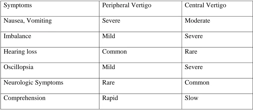
[image:41.612.79.510.292.480.2]lesion (lanyrinth or vestibular nerve) or from a central lesion (brain-stem or cerebellum).
Table -1
Difference Between Peripheral And Central Vertigo
54Symptoms Peripheral Vertigo Central Vertigo
Nausea, Vomiting Severe Moderate
Imbalance Mild Severe
Hearing loss Common Rare
Oscillopsia Mild Severe
Neurologic Symptoms Rare Common
Comprehension Rapid Slow
Spontaneous nystagmus of peripheral origin does not change in direction with gaze to
either side, although it increases in amplitude with gaze in the direction of the fast phase
(Alexander’s Law)54. In contrast, spontaneous nystagmus of central origin typically changes
direction when the patient looks away form the direction of the fast phase.
Infarction of the labyrinth, brain-stem or cerebellum typically occurs in older patient
population with known vascular risk factors. Since the circulation to the labyrinth originates from
the vertebro-basilar system, infarction of the labyrinth can be part of a larger brain stroke
1992)5 labyrinthine infarction is commonly associated with infarction of lateral pontomedullary
region and the anterior inferior cerebellum.
Vertebrobasilar insufficiency (VBI ) (Gomez CR, Cruz – Floreesa, 1996)4 is a common
cause of spontaneous attacks of vertigo in older patients. Vertigo occurs in approximately 25% of
AIM OF THE STUDY
1. To study the various aspects of acquired and hereditary ataxic syndromes
2. To study about the level of involvement of the neuraxis in various type of ataxic
syndromes.
3. Correlative study of CT scans finding and clinical features in acquired and hereditary
MATERIALS AND METHODS
70 Patients were studied who studied who presented with ataxia as the main complaints.
The period of study was from July 2004 to Jan 2006. All the cases were inpatients of medical and
neurology wards or who attended neurology or ENT OPD in Coimbatore Medical College
hospital. All the patients who had ataxia were chosen like cerebellar, sensory and vertiginous
ataxia. Ataxia of acute subacute and chronic type and ataxia of hereditary and acquired type were
also included in the study. Among the acquired type of ataxia due to stroke, infection, neoplasm,
demyelinations, degeneration, drugs, alcohol and paraneoplastic syndrome were included in the
study. Ataxia due to congenital bony anomalies like cranio vertebral junction anomaly was also
considered. All patients had a detailed clinical work up which included history general
examination and systemic examinations and relevant investigations to identify anatomical
locations and nature of pathology leading to ataxia.
Demographic informations such as age, sex, occupations and percapita income were
obtained. Details of social habits, such as alcohol consumption, smoking habits were enquired into.
Exposure to STD, TB, Toxins, vaccine were also searched. Apart from the main complaint ataxic
symptoms referable to cranial nerves, pyramidal system, extrapyramidal system, autonomic
nervous system and also symptoms of raised. ICT were enquired into. Note was made on trauma,
fever, exanthems, csom, drugs especially antiepileptics ATT and dapsone, etc.
Past history of SHT, DM, TB,RHD, STROKE were carefully enquired into Family history
of ataxia was also searched. Details of consangunity also was obtained.
In higher function examination, more stress were given for speech and tried to find out type
of abnormality like, scanning, staccato or spastic when it was present.
Individual cranial nerves were thoroughly examined and fundus was looked for optic
atrophy papilleodema and pigmentary change, Nystagmus was thoroughly examined to findout
whether it was due to central / peripheral lesions.
During motor system examinations, bulk, tone strength and deep tendon reflex,
co-ordination and involuntary movements were looked to find out associated pyramidal, LMN,
extrapyramidal and cerebellar lesion. When incoordination was present all possible clinical tests
were done to findout whether it was due to cerebellar, sensory or vestibular abnormality. If the
incoordination was due to cerebellar lesions, further clinical examinations was directed to find out
the ataxia is limb ataxia or gait ataxia or truncal ataxia.
Sensory systems also thoroughly examined to find out which tract was involved and the
site of lesion.Spine and neck examined to find out any feature of short neck and vertebral anomaly.
Other systems were also examined to findout any pathology for the patients problem eg.,
malignancy for paraneoplastic cerebellar degeneration and cardio vascular system to rule out
PATIENTS SELECTION
Patients were broadly divided into three groups.
1. Ataxia of hereditary cause and 2. Ataxia of acquired cause and 3. Ataxia due to
congenital bony anomaly where ataxia developed later on. Individual patients were
studied accordingly to anatomy of the disease, onset of illness and pathology of the
disease.
According to anatomy
1.Cerebellar ataxia 2.Sensory ataxia 3.Vertigenous ataxia
According to onset
Acute Subacute Chronic
According to Pathology
Vascular Infection
Demyelinations Degenerative
Tumor Paraneoplastic
Drugs, toxins, alcohols Trauma
Criteria for selecting patients with ataxia of cerebellar type
1. Limb, gait or truncal ataxia 2. Scanning or staccato speech.
3. Dysmetria Æ Intentions tremor 4. Dysdiadokokinesia
5. Coarse gaze evoked, horizontal 7. Broad based gait.
Criteria for selecting patients with ataxia of sensory type.
General Features:
1. History of pins and needle with tingling sensations
2. Cotton wool sensations during walking.
3. Ataxia – swaying forward and backward.
4. High stepping gait
5. Rombergism
Peripheral nerve lesion producing ataxia
6. Hypotonia
7. Hyporeflexia
8. Glove and stocking sensory disturbance
9. Peripheral nerve thickening
Posterior columns cervical cord level.
10.Lhermitte sign
Due to medial leminicus.
- Other long tract sign - Cranial nerves
Criteria for selecting patients with ataxia of vertiginous type.
1. Vertigo 2. Tinnitus
3 Deafness 4. Nystagmus
- Fine gaze evoked with fast component to opposite side of lesion with some rotatory
component. Werdelins in 1986 put forward some criteria for diagnosis of hereditary cerebellar
ataxia syndrome. The same criteria were applied in the present study for diagnosing hereditary
1. Signs of cerebellar dysfunctions
a. Ataxia of the extremities b. Dysarthira – Scanning staccato
independent of vision. c. Dysmetria (Intention tremor)
d.Nystagmus e. Hypotonia.
f. Broad based gait
2. Signs of pyramidal dysfunction
a. Paresis b. Babinski’s sign
3. Amyotrophy
4. Deformities, usually club foot
5. Involuntary chorea – like movements
6. Extrapyramidal signs
a. Rigidity b. Bradykinesia
7. Optic atrophy
8. Dementia
9. Epilepsy
10. Retinal pigmentary changes.
Hereditary ataxia are again divided into early onset (before 20 years ) and late onset. (After
20 years) Patients who are having a known etiology like infarct, haemorrhage, tumour, infection,
degeneration, demyelination, trauma were taken as acquired ataxia in the present study.
Investigations were done in detail for such patients and showed positively in most of the patients
eg.1. If ataxia in one patients is suspected due to cerebellar neoplasm CT scan of brain taken and
confirmed the diagnosis. 2. If ataxia is suspected due to demyelination of posterior nerve root
Routine urine and blood investigations were done to find out any evidence of diabetes,
infections and raised ESR. Special blood test like, VDRL thyroid functions test and liver functions
test were done whenever appropriate.
X-ray chest PA view taken to rule out any foci of pulmonary tuberculosis, to rule out
evidence of cardiology (RHD) and search for bronchogenic, pleural or mediastinal malignancy.
X-ray of skull and cervical spine and were taken in suspected cases of CVJ anomalies and cervical
cord compression.
CT scan of brain also was done (48 – cases) whenever appropriate to help the diagnosis.
MRI scan was done when it was very essential to pickup up brain stem pathology and CVJ
anomly. Lumbar Puncture and CSF analysis were done whenever appropriate like demyelination
(GBS, CIDP).
Audiogram was done when ataxia was due to vertiginous cause. Caloric test could not be
70 cases of various type of ataxia syndromes were studied. The various clinical syndrome
encountered in the study were. (Table-1) Cerebellar ataxia 60.(85.7%), sensory ataxia 8(11.4%)
and labyrinthine ataxia 2(2.8%)
Age Incidence
( Table- II) The age of the patients varied from 6 to 70 years. Lowest age noticed is 6 years who was suffered from Brain stem glioma. Highest age was 65 years who was having lateral
medullary syndrome. Between 0-10 years there were 3 cases (4.2%) between 11.20 years 2 cases
(2.8%) between 21-30 years 20 cases (28.5%) between 31-40 years, 17 cases (24.2%) between
41-50 years 13 cases (11.5%) between 51-60 years 10 cases (14.2%) and between 61-70 5 cases
(7.1%) . The least incidence at 11-20 years period and maximum incidence 21-30 years period.
SEX INCIDENCE
Out of 70 cases men were 43 and women were 27. The ratio is 1.59:1
Main anatomical site of pathology
:(Table – III) Various anatomical site of involvement could be made out which leads to
ataxic syndromes. They were pure cerebellum 26 cases (37.1%) leading the highest group, Then
brainstem, 18 cases (25.7%), combined spinal cord and cerebellum 15 cases (21.4%), pure spinal
cord 3 cases (4.2%), posterior root 3 cases (4.2%), labyrinth 2 cases (2.8%), peripheral nerve 2
cases (2.8%) and thalamus one case (1.4%)
Incidence of hereditary and acquired ataxia:
(Table IV) Out of 70 cases studied 50 (71.4%), were of acquired variety and 17 (24.2%),
Among the 50 cases of acquired males were 31 and female were 19 ratio is 1.6:1. Among the 17
cases of hereditary ataxia 12 and 5 females Ratio is 2.4:1
Incidence of Hereditary type of Cerebellar and hereditary type of sensory
ataxia:
(Table V) Hereditary cerebellar ataxia were 16 out of 17 hereditary ataxia and ataxia due to
hereditary sensory neuropathy was 1 out of 17.
Age incidence of hereditary ataxia
There were totally 17 hereditary ataxia comprising both hereditary cerebellar and
hereditary sensory ataxias. Between 0-10 years there was only one case. Between 21-30 there were
8 cases between 31-40 years, 5 cases, between 41-50 years 3 cases between 41-70 there were no
cases. The congenital anomaly later on leading to ataxia (CVJ) present in this study was at the age
interval of 31-60 years.
Incidence of various type of spinocerebellar ataxia
(Table – VI) In the present study there were 16 cases of spinocerebellar ataxia Among that
OPCA(SCAI) was the major group of spinocerebellar ataxia which were 8 out of 16 cases SCA –
II was 3, SCA – III – 1, there was no SCA 5 to 22. In the case of DRPLA, Friedriech’s ataxia and
ataxia telengiectasia one case of each was present.
Age and Sex incidence Hereditary Cerebellar Ataxia
(Table VII) There were 16 cases of Hereditary Cerebellar Ataxia in the present study.
Between 0-10 years age there were one case. Between 21-30 years 7 cases, between 31-40 years 4