Copyright © 1998, American Society for Microbiology. All Rights Reserved.
Complete Protein Linkage Map of Poliovirus P3 Proteins:
Interaction of Polymerase 3D
pol
with VPg and with
Genetic Variants of 3AB
WENKAI XIANG,1† ANDREA CUCONATI,1‡ DEBRA HOPE,2KARLA KIRKEGAARD,3
ANDECKARD WIMMER1*
Department of Molecular Genetics and Microbiology, School of Medicine, State University of New York at Stony Brook, Stony Brook, New York 11794-52221; Department of Molecular, Cellular and Developmental Biology,
University of Colorado, Boulder, Colorado 80309-03472; and Department of Microbiology
and Immunology, Stanford University, Stanford, California 94365-54023
Received 5 February 1998/Accepted 6 May 1998
Poliovirus has evolved to maximize its genomic information by producing multifunctional viral proteins. The P3 nonstructural proteins harbor various activities when paired with different binding partners. These viral polypeptides regulate host cell macromolecular synthesis and function as proteinases, as RNA binding pro-teins, or as RNA-dependent RNA polymerase. A cleavage product of the P3 region is the genome-linked protein VPg that is essential in the initiation of RNA synthesis. We have used an inducible yeast two-hybrid system to analyze directly protein-protein interactions among P3 proteins. Sixteen signals of homo- or heterodimer interactions have been observed and have been divided into three groups. Of interest is the newly discovered affinity of VPg to 3Dpol that suggests direct interaction between these molecules in genome replication. A battery of 3AB variants (eight clustered-charge-to-alanine changes and five single-amino-acid mutations) has been used to map the binding determinants of 3AB-3AB interaction which were found to differ from the amino acids critical for the 3AB-3Dpolinteraction. The viral proteinase 3Cprowas not found to interact with other 3Cpromolecules or with any other P3 polypeptide in yeast cells, a result confirmed by glutaraldehyde cross-linking. The weak apparent interaction between 3AB and 3CDproscored in the yeast two-hybrid system was in contrast to a strong signal by far-Western blotting. The results elucidate, in part, previous results of biochem-ical and genetic analyses. The role of the interactions in RNA replication is addressed.
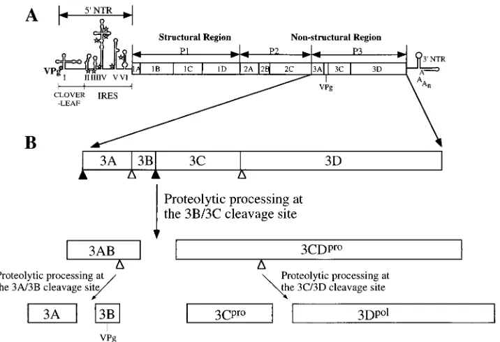
Specific interactions between virus-encoded polypeptides are likely to play a crucial role in poliovirus replication (25, 33, 37, 43, 44, 46, 56, 57). Compared to the average size of DNA virus genomes, the genomes of RNA viruses are small, a re-striction that can be related to the error-prone RNA replica-tion of RNA virus quasispecies (55). Poliovirus is no excepreplica-tion. Its genome of ca. 7,500 nucleotides has coding capacity for only a limited number of proteins, all of which are derived from a single translation product, the 247-kDa polyprotein (Fig. 1A) (29, 48). The polyprotein is processed by two virus-encoded proteinases into some 27 cleavage intermediates and end prod-ucts (23, 55). To maximize the information stored in the small viral genome, the cleavage end products and their precursors have, in many cases, distinct functions. This expands the menu of polypeptides useful for viral proliferation. The C-terminal region (P3) of the viral polyprotein may serve as typical exam-ple (Fig. 1B). The P3 precursor is generated through a very fast cleavage event at the amino terminus of the 3A coding region, and it undergoes further rapid processing to yield the two
multifunctional, relatively stable cleavage intermediates 3AB (an RNA binding protein involved in RNA polymerization and
the precursor to VPg) and 3CDpro(a sequence-specific RNA
binding protein that displays also proteinase activity).
Subse-quent slow processing of 3AB and 3CDprogenerates the four
cleavage end products, 3A, 3B (the genome-linked protein
VPg), 3Cpro (a proteinase), and 3Dpol (an RNA-dependent
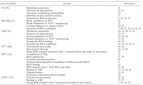
RNA polymerase). Table 1 summarizes the biochemical activ-ities and known functions of these polypeptides and provides appropriate references.
Some of the viral polypeptides obtain specific functions by acquiring different binding partners. 3AB is a membrane-asso-ciated protein that serves as precursor for VPg (50). Purified detergent-solubilized 3AB is also a nonspecific RNA binding
protein (44, 56) that, in association with 3CDpro(37), acquires
binding specificity for the 59-terminal cloverleaf in the pres-ence of a 1,000-fold excess of tRNA (25, 56, 57). Genetic and
biochemical data have argued that this complex ([3AB/3CDpro/
59-cloverleaf]) plays an essential role in the initiation of
plus-strand RNA synthesis (25, 56, 57). Association with 3CDpro, on
the other hand, leads to another biochemical event: 3AB
stim-ulates cleavage of the otherwise rather stable 3CDproprecursor
to 3Cproand 3Dpol(37, 56). Purified preparations of 3AB have
binding affinity for 3Dpol, and at low 3Dpolconcentrations, the
3AB/3Dpolcomplex possesses greatly increased polymerase
ac-tivity compared to that of 3Dpolalone (33, 44, 46). Moreover,
the 3AB/3Dpolcomplex appears to have binding specificity for
the 39-terminal nontranslated region (NTR) of the poliovirus genome, a phenomenon that would be useful to guide the polymerase to the proper template site in the initiation of minus-strand synthesis (44). The activity of 3AB is also
regu-* Corresponding author. Mailing address: Department of Molecular Genetics and Microbiology, School of Medicine, State University of New York at Stony Brook, Stony Brook, NY 11794-5222. Phone: (516) 632-8787. Fax: (516) 632-8891. E-mail: [email protected] .edu.
† Present address: Molecular Pathogenesis Program, Skirball Insti-tute of Biomolecular Medicine, New York University Medical Center, New York, NY 10016.
‡ Present address: Center for Advanced Biotechnology and Medi-cine, Department of Biological Sciences, Rutgers University, Piscat-away, NJ 08854.
6732
on November 9, 2019 by guest
http://jvi.asm.org/
lated by its association with 3CDpro, since membrane-bound
3AB can be cleaved by 3CDpro into 3A and 3B (33), two
cleavage products that have lost the functions of 3AB (see Fig.
5 for overview). Finally, 3Dprocan form homo-oligomers with
increased RNA binding and RNA polymerization activities in vitro (43).
The molecular nature of these exceedingly complex interac-tions of the poliovirus P3 proteins (summarized in Fig. 5) is unknown. Where do the binding sites of the binding partners map on the corresponding polypeptide chains? Is binding suf-ficient for function, or can binding and function be genetically
separated? Does VPg have binding affinity to 3Dpol? Is the
substrate for uridylylation by 3Dpolprecursor 3AB or cleavage
product VPg (31, 51, 52)? Have all possible interactions be-tween P3 proteins been detected biochemically?
The yeast two-hybrid system is a powerful tool to detect protein-protein interactions (8, 16). By using this method, a protein linkage map has been established in Escherichia coli bacteriophage T7, allowing tabulation of special interactions of the phage gene products (3). The yeast two-hybrid system can also be used to map protein domains that are responsible for protein interactions and to identify mutations that affect these interactions. Here, we report the linkage map of poliovirus proteins mapping to the P3 domain of the polyprotein. In the course of these experiments, we have tested three versions of the two-hybrid system of which only one, the semi-inducible
system (58), was adequate. Whereas the viral proteinase 3Cpro
yielded no interaction with other P3 proteins, we detected
interaction between VPg and the polymerase 3Dpol, a result
highly significant for the study of the mechanism of initiation of viral RNA synthesis. Moreover, interactions between genetic
variants of 3AB with wild-type (wt) 3AB or 3Dpol revealed
binding domains of 3AB to these proteins and a relationship between complex formation and function. Since the structure
of 3Dpol has been elucidated at the atomic level (22), the
results reported here may contribute to the elucidation of the multicomponent RNA replication complex of poliovirus.
MATERIALS AND METHODS
Strains and plasmids.The Saccharomyces cerevisiae strains and all plasmids for two-hybrid analysis are listed in Table 2. The yeast strains and parental plasmids used in two-hybrid systems I and II are generous gifts from Stanley Fields (University of Washington) (2, 16), while those in system III are from Roger Brent (Harvard Medical School) (58). Detailed information about the yeast strains and the plasmids can be found in the cited references. The DNA fragments encoding the six P3 proteins were amplified by PCR with XmaI and
SalI sites flanking the ends. The pGBT.9, pGAD.GH, and pBTM116 vectors
were digested with the same restriction enzymes and ligated with one of the above fragments. The new plasmids are named after the parental plasmids and the viral proteins (e.g., pGAD.GH3AB). Subsequently, the EcoRI and SalI fragments were cut out from the pGBT.9 fusion plasmids and ligated to pEG202 and pJG4-5 that had been digested with EcoRI/SalI and EcoRI/XhoI, respec-tively. Additionally, pRFHM1, which expresses LexA fused to a transcriptional inert fragment of the Drosophila Bicoid protein, served as a negative control.
The mutant 3AB genes, in which clustered charged residues were changed to alanines (see Fig. 3), were constructed previously (31, 56) except that M20 was a generous gift of Bert Semler (University of California, Irvine). In the M20 mutant, threonine 67 was replaced with lysine (19). DNA fragments that encode those mutant 3AB proteins were cloned into pJG4-5 as described above.
Two-hybrid analysis.In systems I and II (Table 2), each yeast strain was transformed with the corresponding DNA binding domain (BD)- and transcrip-tion activatranscrip-tion domain (AD)-viral peptide fusion plasmid by the lithium acetate method (2). The transformants were plated on synthetic medium lacking tryp-tophan and leucine. About 10 Trp1Leu1colonies from each plate were inoc-ulated into selective liquid medium, andb-galactosidase activity was measured in a liquid assay (2). To quantify theb-galactosidase activity in the liquid assay, Miller units were defined as 1,0003optical density at 574 nm (OD574) divided by the product of the volume (milliliters)3time (minutes)3O.D.600.
[image:2.612.124.482.68.313.2]In system III (Table 2), the yeast strain was first transformed with plasmid pSH18-34, which has a lacZ reporter gene preceded by an upstream LexA binding site (58). The transformed cells were amplified and further transformed with pEG202- and pJG4-5–peptide fusion plasmids and plated onto synthetic medium containing 2% glucose but lacking histidine, tryptophan, and uracil. About 10 His1Leu1Ura1colonies from each transformation were inoculated
FIG. 1. Genome organization and processing of the polyprotein P3 region. (A) The poliovirus genome is divided into the 59NTR, polyprotein, and 39NTR. The 59NTR consists of the cloverleaf and internal ribosome entry site (IRES) elements. Open stars, unused AUG initiation codons. The polyprotein is divided into the structural and nonstructural regions, whose major cleavage products are indicated. (B) Enlargement of the P3 nonstructural precursor and its processing products analyzed in this study. Closed and open triangles indicate rapidly and slowly processed Q/G cleavage sites, respectively. For functions of the cleavage products, see Table 1.
on November 9, 2019 by guest
http://jvi.asm.org/
in selective medium. When the OD600of the liquid culture was around 0.6 to 1.0, the expression of the AD fusion protein was induced with 2% galactose and the incubation was continued for 4 h. Theb-galactosidase activity was then assayed. Chemical cross-linking.One microgram of purified 3Cproor 3AB protein was incubated for 15 min at 30°C with different amounts of glutaraldehyde in a buffer containing 25 mM HEPES (pH 7.5) and 5 mM MgCl2. In some reactions, 0.5% sodium dodecyl sulfate (SDS) was also included. The samples were then sepa-rated by SDS–15% polyacrylamide gel electrophoresis, transferred to nitrocel-lulose membranes, and blotted with a specific antibody to each protein.
Far-Western blotting analysis.Two or 4mg of purified proteins was directly dot blotted on a nitrocellulose membrane (Schleicher & Schuell). The membrane was then incubated with 56 nM purified 3CDpro(a 3Cpro-3Dpolcleavage site mutant [20, 24]) in a buffer containing 25 mM Tris-HCl (pH 7.5), 5 mM MgCl2, 50 mM NaCl, 1 mM dithiothreitol, and 0.075% Tween 20 overnight at 4°C (12). Bound protein was detected by enhanced chemiluminescence immunoassay (Amersham) with anti-3Cproserum.
RESULTS
Analysis of three versions of the yeast two-hybrid system to study interactions among poliovirus-encoded P3 proteins.
There are three widely used yeast two-hybrid systems, referred to as systems I, II, and III in Table 2 (2, 58). In system I, yeast cells constitutively express the GAL4 BD and AD. The coding
regions for six poliovirus P3 peptides (3A, 3B, 3AB, 3Cpro,
3Dpol, and 3CDpro [Fig. 1B]) were cloned into plasmids to
produce fusion proteins with both domains. The only
interac-tion detectable was that between the BD-3Dpoland the
AD-3Dpolfusion proteins, and the signal was unexpectedly weak
(Table 3).
We then switched to system II, in which the sequence of E.
coli repressor LexA protein replaces that of GAL4 BD. The
signal for the 3Dpol-3Dpolinteraction was enhanced (Table 3).
Moreover, we were now able to observe interaction between BD-3D and AD-3AB. However, in both systems I and II, the growth rate of yeast transformants that expressed fusion pro-teins containing either 3C, 3A, or their precursors was notably low. The slow growth was pronounced in cells transformed
with BD-3Cpro/AD-3Cpro, BD-3A/AD-3A, and
BD-3AB/AD-3AB. Poliovirus protein 3Cpro has been reported to inhibit
transcription by RNA polymerases II and III in mammalian cells (10, 11). It may also damage the microtubule network (28). 3A and 3AB, in turn, have been reported to modify membrane permeability (32) and interfere with host protein secretion (15). Thus, the constitutive expression of these viral proteins could be deleterious to the normal metabolism of the yeast cell.
[image:3.612.56.554.76.373.2]To circumvent these obstacles, we chose system III, devel-oped in the laboratory of Roger Brent (58). This system uses
TABLE 1. Biochemical activities and known functions of proteins encoded in the poliovirus P3 region
Protein (size [kDa]) Activities Reference(s)
3A (10) Membrane association 32
Substrate for glycosylation 13, 14
Alteration of membrane permeability 32
Inhibition of host protein secretion 15
Essential for RNA replication 4, 18, 26, 54
3B (VPg) (2) Weak stimulation of 3Dpol 44
Weak stimulation of 3CDproautocleavage 37
Covalent linkage to 59genome RNA 29, 41
Initiation of RNA replication 7, 30, 31, 45, 51, 52, 53
3AB (12) Membrane association 18, 32, 50, 52, 54
Substrate for glycosylation 13, 14
Strong stimulation of 3Dpol 33, 44, 46
Strong stimulation of 3CDproautocleavage 37, 56
Nonspecific RNA binding 44, 56
Essential for RNA replication 18, 19, 25, 56, 57
3Cpro(20) Viral protein processing 20, 40
Host protein cleavage 10, 11, 28
Weak RNP complex formation with 59cloverleaf RNA and 3AB (or host factor) 25
3Dpol(52) Uridylylation of VPg 45
RNA polymerase 17, 39
Terminal adenylyltransferase 38
Polymerization-dependent unwinding of double-stranded RNA 9
RNA binding 43
RNP complex with 39NTR RNA and 3AB 25, 44
Binding to 3AB 27, 33, 44, 46
Oligomerization 22, 43
Interaction with human protein Sam68 36
3CDpro(72) Viral protein processing 24, 35
Binding to 3AB 25, 37
Strong RNP complex with 59cloverleaf and 3AB (or host factor) 1, 25, 56, 57
TABLE 2. Three two-hybrid systems used in this work
System Vector Yeast strain Expression ofAD fusion Reference
BD AD
I pGBT9 (GAL4 BD) pGAD.GH (GAL4 AD) Y153 Constitutive 2
II pBTM116 (LexA BD) pGAD.GH CTY-10d Constitutive 2
III pEG202 (LexA BD) pJG4-5 (B42) EGY48/pSH18-34 Inducible 58
on November 9, 2019 by guest
http://jvi.asm.org/
[image:3.612.49.551.664.727.2]the LexA DB domain as in system II. Whereas expression of the BD fusion protein is constitutive, that of the AD fusion is under the control of the inducible GAL1 promoter and con-tains the bacterial peptide B42. Since the promoter is induc-ible, the yeast cell can proliferate before synthesis of AD fusion protein is induced. Indeed, better growth of the transformants,
especially of the cells expressing AD-3Cpro and AD-3AB
fu-sion proteins, was observed. This semi-inducible expresfu-sion yielded the highest sensitivities and signal/noise ratios in de-tecting viral protein-protein interaction (Table 3). This was
true especially for 3Dpol-3AB interactions: theb-galactosidase
activity in system III was 104-fold higher than those in systems
I and II. Such high signal in the inducible system was reported recently also in a separate study (27).
Protein-protein interactions of P3 polypeptides.Six LexA
BD-poliovirus peptide fusions (Lex-3A, -3B, -3AB, -3Cpro,
-3Dpol, and -3CDpro [Fig. 2]) were constructed in plasmid
pEG202. For negative controls, plasmid pRFHM1, which ex-presses LexA-Bicoid fusion protein, was included in the anal-yses (58) (Materials and Methods). In addition, six B42
AD-viral peptide fusions (AD-3A, -3B, -3AB, -3Cpro, -3Dpol, and
-3CDpro) were constructed in vector pJG4-5. Forty-nine pairs
of plasmids, including every combination of the two groups, were transformed into yeast strain EGY48. If two viral
poly-peptides interact with each other, transcription of the
b-galac-tosidase gene is induced;b-galactosidase activity can then be
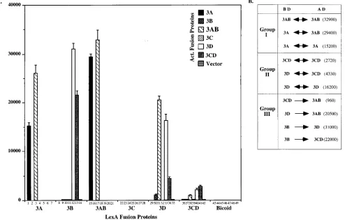
quantified by a simple colorimetric assay. For example, BD-3A was paired with AD-3A, AD-3B, AD-3AB, etc., and the col-orimetric signal is presented in Fig. 2 as bars 1, 2, 3, etc., respectively; or 3B was paired with AD-3A, AD-3B, AD-3AB, etc., and the signal was recorded as bars 8, 9, 10, etc.,
respec-tively. Theb-galactosidase activities of 12 transformants were
well above background (Fig. 2), an observation suggesting an affinity between corresponding poliovirus polypeptide pairs.
It should be noted that the failure to observe an interaction in the two-hybrid system does not necessarily mean that the two proteins do not interact in their normal environment.
[image:4.612.57.551.350.665.2]FIG. 2. Interactions of six proteins encoded in the P3 nonstructural region in the inducible yeast two-hybrid system. (A) Yeast strain EGY48, expressing the pair of indicated hybrid proteins, was analyzed forb-galactosidase activity (given in Miller units). Each bar presents the result of a pairing of a LexA fusion protein with an AD fusion protein. Thus, the interaction of LexA-3A (referred to also as BD-3A) with AD-3A, AD-3B, AD-3AB, AD-3C, etc., is given in bars 1, 2, 3, 4, etc. Lack of a bar indicates absence of a color reaction. The enzyme activity of each transformant was quantified in a liquid assay and presented as Miller units. The Bicoid segment served as negative control. (B) Detected protein interactions divided into three groups (see text). Theb-galactosidase activity from each interaction is indicated in parentheses. The double-headed arrows indicate that the interaction between two different viral proteins is bidirectional; the single-headed arrows indicate unidirectional interactions.
TABLE 3. Comparison of the sensitivity among the three two-hybrid systems
System
Meanb-galactosidase activity (Miller units)6SD
(BD fusion-AD fusion) Signal/noise ratioa
3D-3D 3D-3AB 3D-Vc
I 2.360.6 0 0
II 51.263.6 1.460.2 1.160.2 46.5 (1.27)
III 16,20061,600 20,5006900 152617 107 (135)
aDefined as 3D-3D/3D-Vc (or 3D-3AB/3D-Vc).
on November 9, 2019 by guest
http://jvi.asm.org/
Improper folding or poor expression of the hybrid proteins can influence whether an interaction is apparent. Often, the polar-ity in which two proteins are presented may play a crucial role. Likewise, in the absence of information about the relative expression levels (which can change from experiment to
exper-iment), the relative amounts ofb-galactosidase expression
be-tween unrelated hybrid pairs do not directly reflect differences in the affinities of the pairs of proteins. Nevertheless, the
ob-servation ofb-galactosidase activity in the presence of a pair of
hybrid proteins is a strong indication that two polypeptide chains expressed as fusion proteins do interact.
The 12 observed interactions can be divided into three groups (Fig. 2B), depending on the nature of the partners. Interactions between those polypeptides that map to the N terminus of P3 (3AB, 3A, and 3B) have been combined in group I. Homodimer pairs of 3A and 3AB strongly react with each other in this system (bars 1 and 17, respectively), the
latter giving the highest value ofb-galactosidase activity (;33
104Miller units) scored in any of these experiments. The
ex-traordinary homoaffinity of purified 3AB, even in the presence of strong detergent, was demonstrated also by a biochemical assay (see below). Experiments using vector pairs with a Bicoid
hybrid control or AD alone failed to yield b-galactosidase
activity (BD-3A/AD and BD/AD-3A [bars 7 and 43, respec-tively] and BD-3AB/AD and BD/AD-3AB [bars 21 and 45, respectively]), an observation showing that the interactions are due to affinities between the viral polypeptide chains. 3B, on the other hand, did not show significant binding to other 3B-containing fusion proteins in this system—3B (bar 9; BD-3B/AD in bar 14), 3A (bars 2 and 8), or 3AB (bars 10 and 16)—although the LexA-3B fusion protein was expressed, as
evident from strong signals obtained with 3Dpoland 3CDpro
(bars 12 and 13) (see also below). AD-3B was also expressed
since it interacted with Lex-3Dpol (bar 30); this interaction,
however, was weak and thus is not a good measure of AD-3B expression (see below).
Interactions between those polypeptides that map to the C
terminus of the P3 precursor (3CDpro, 3Cpro, and 3Dpol) have
been combined in group II. Only the 3Dpolhomopair displayed
strong interaction in this system (Fig. 2A, bar 33). In contrast,
the 3Dpol-3CDpro interactions were more difficult to detect,
regardless of the polarity of the partners (bars 34 and 40). The
signal for homopair interaction between 3CDpro chains was
also relatively weak (bar 41). Nob-galactosidase activity was
observed in attempts to pair 3Cpro with itself (bar 25), with
3AB (bars 18 and 24), or with 3CDpro(bars 27 and 39) as a
partner. The failure to obtain significant signals with the viral
3Cproand 3CDproproteinases as binding partners in some of
the pairings is unlikely to be the result of 3Cpro-specific
pro-teolytic activity that might interfere with the formation of, or destabilize, a protein-protein complex. This we conclude from
studies with mutant polypeptides 3Cpro(C147S), 3CDpro
(F83A), and 3CDpro(R87Q). Although a 3Cpro(C147S)
muta-tion constructed into the poliovirus polyprotein retained some 3C-related activity in cis cleavages (34), we have reported
pre-viously that the purified 3Cpro(C147S), 3CDpro(F83A), and
3CDpro(R87Q) polypeptides have lost proteolytic activity
be-low detectable levels in trans-cleavage assays (20, 21). When
cloned into pEG202, these 3Cpro(C147S), 3CDpro(F83A), and
3CDpro(R87Q) mutant polypeptides did not yield increased
b-galactosidase activity (data not shown). Moreover, as
men-tioned, the BD-3B/AD-3CDpro pair showed a strong signal
(bar 13).
Group III (Fig. 2B) depicts interactions between 3AB and
3CDpro and their cleavage products. Surprisingly, all
interac-tions in this group were found to be unidirectional. For
exam-ple, 3Dpolinteracted with 3AB only if presented as BD-3Dpol
(bar 31) (27). Since BD-3Dpol does not form a detectable
complex with 3A (bar 29), the binding between 3Dpoland 3AB
in this system may rely predominantly on sequences of 3B.
Whereas interaction between BD-3Dpoland AD-3B was more
difficult to detect (bar 30), that in the reverse direction,
be-tween BD-3B and AD-3Dpol, was strong (bar 12). As discussed
later, the observation of the affinity between 3B and 3Dpolis
highly significant in view of the mechanism of initiation of poliovirus minus-strand RNA synthesis (45). We have previ-ously suggested that both the 3A and 3B moieties in 3AB are necessary to form the superactive RNA polymerase [3AB/ 3D]super-pol(44, 56). It appears, therefore, that some
cooper-ativity of the two domains of 3AB may be important in the
binding of 3AB to 3Dpol.
As is the case for 3Dpol-3AB and 3B-3Dpol, the 3CDpro-3AB
interaction is also unidirectional (Fig. 2A; compare bar 38 with
bar 20). However, the interaction between 3CDpro and 3AB
was very weak, which was a big surprise since immune precip-itation experiments suggested that these polypeptides form a complex in solution (37). Therefore, the question of
3AB-3CDprointeraction was addressed again by another assay (see
below).
The molecular basis of the unidirectional nature of some of the two-hybrid interaction is obscure. It is not due to insuffi-cient expression of the corresponding fusion proteins because in homologous pairings, the viral polypeptides show strong signals (Fig. 1B, groups I and II). In addition, Western blotting of both 3B fusion proteins revealed adequate expression of the polypeptides (57a).
The interactions between mutants of 3AB and wt P3 pro-teins. Several lines of evidence argue that polypeptide 3AB plays multiple roles in the replication of poliovirus (4, 18, 19, 25, 30–33, 37, 44, 46, 50, 52, 54, 56, 57). We have provided previously biochemical evidence that the polypeptides 3AB,
3Dpol, and 3CDpro interact with each other in an as yet
un-known fashion to provide crucial functions for poliovirus ge-nome replication (25, 27, 33, 44, 56, 57). These studies included purified 3AB polypeptides that each carried clustered-charge-to-alanine mutations in the 3A portion of 3AB or point muta-tions in the 3B portion (Fig. 3B) (56). All of these mutamuta-tions, when constructed into the poliovirus genome, were deleterious to genome replication (18, 30, 31, 56). Moreover, Giachetti et al. (18) provided evidence that mutations in a hydrophobic region of 3AB influence poliovirus RNA synthesis. It was un-known, however, to what extent the phenotypes of these mu-tants were related to impaired protein-protein interaction. Therefore, we tested the effects of specific 3AB mutations (18,
56) on the formation of 3AB homodimers or 3Dpol/3AB
het-erodimers in the yeast two-hybrid system. Interaction of the
mutant 3AB proteins with 3CDprowas not analyzed because of
the weak signal scored between wt 3AB and 3CDpro fusion
proteins.
Eight clustered-charge-to-alanine mutations and four point mutations, all of which cause severe defects in RNA synthesis during poliovirus proliferation in tissue culture (18, 56), were introduced into AD-3AB and tested with wt BD-3AB or with
BD-3Dpol. Surprisingly, most mutant AD-3AB polypeptides
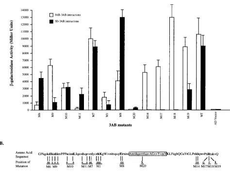
gave as strong an interaction signal with wt BD-3AB as did the wt BD-3AB/AD-3AB pair (Fig. 3; compare the open bars for M6 through M19 with that for WT). There were three excep-tions. M6 and M11 AD-3AB fusion proteins displayed 10- and
20-fold reductions in b-galactosidase activity, respectively.
More dramatically, the M20 mutant of 3AB showed a 500-fold
reduction inb-galactosidase accumulation in the presence of
wt 3AB-containing AD fusion protein (Fig. 3, open bar, M20).
on November 9, 2019 by guest
http://jvi.asm.org/
In this mutant polypeptide, a threonine residue located in the strongly hydrophobic region of 3AB (46) (boxed region in Fig. 3B) was exchanged with a positive amino acid (lysine) (T67K [18]). Finally, none of the mutations in the 3B domain affected binding between 3AB polypeptides (Fig. 3, open bars, M14 and M17 to M19). These results imply that the 3A moiety of 3AB, perhaps predominantly its hydrophobic region, is essential for homodimer formation.
It should be noted that all mutant AD-3AB fusion proteins were expressed in yeast, as judged by the strong signals scored with these hybrids either with BD-3AB (Fig. 3, open bars) or
with BD-3Dpol(closed bars) as a partner (see below).
Expres-sion of the M20 hybrid was further analyzed by Western blot-ting analysis using an anti-3AB antibody, and it was found to be in the range of the other mutant hybrids (data not shown).
A different picture emerged when the AD-3AB mutant
pro-teins were paired with wt BD-3Dpol. Whereas most mutations
in the hydrophilic domain of 3AB failed to significantly
influ-ence interaction with BD-3Dpol, the M14, M17, and M18
mu-tations in the 3B domain of 3AB reduced 3AB/3Dpolbinding to
barely detectable levels in this system (Fig. 3, closed bars). All three mutations reduce the positive charge in the 3B domain. Interestingly, the positive charge of the lysine residue in the 3B domain of 3AB in M19 is apparently adequate for the
3AB-3Dpolinteraction whereas glutamine or glutamic acid residues
in this position are not. Only the M1 and M20 mutations in the
3A domain reduced significantly the signal of the 3AB-3Dpol
interaction (Fig. 3). As discussed later, these data serve to explain, in part, our results previously reported for biochemical assays with 3AB mutants (56).
Binding of P3 proteins in vitro.The high score of protein-protein binding of the BD-3AB/AD-3AB fusion protein-proteins was contrasted by a total absence of interaction of corresponding
3Cpro fusion proteins in the yeast two-hybrid system (Fig. 2).
We tested the validity of this result by a chemical cross-linking experiment.
One microgram each of purified 3AB (33) and 3Cpro (40)
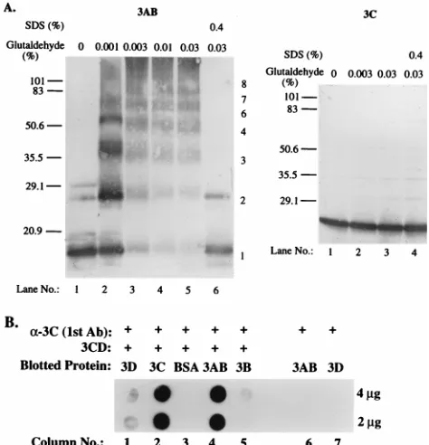
[image:6.612.66.540.69.424.2]was incubated with increasing amounts of glutaraldehyde. As is apparent in Fig. 4A, 3AB could be cross-linked, forming dimers and higher-order oligomers. Polypeptide 3AB is partic-ularly intriguing in that it reproducibly displayed a small num-ber of dimeric molecules even in the absence of glutaraldehyde (Fig. 4A, 3AB, lane 1), a phenomenon seen previously (33). Presence of 0.4% SDS in the chemical cross-linking reaction blocked multimerization of 3AB but, remarkably, did not com-pletely disrupt dimerization of 3AB (lane 6). It should be noted that the method of detection of proteins in this experiment was by immune reaction with a 3AB-specific monoclonal antibody
FIG. 3. Interactions of mutant 3ABs with wt 3Dpolor 3AB in the yeast two-hybrid system. (A) Yeast strain EGY48 was transformed with pairs of an AD-mutant
3AB and a BD-wt 3AB (or BD-3Dpol) expression plasmid. Theb-galactosidase activity resulting from AD-mutant 3AB interaction with BD-wt 3AB or BD-3Dpolwas
quantified by the liquid assay and presented as in Fig. 2A. Open bars, 3AB-3AB interactions; shaded bars, 3Dpol-3AB interactions. The 3AB mutants are as designated
below. (B) Polypeptide 3AB is represented as a long, open rectangle of which the hydrophobic region is shaded. Positions of the clustered-charge-to-alanine mutations (56) are indicated.
on November 9, 2019 by guest
http://jvi.asm.org/
after transfer of the material to nitrocellulose membranes. This method served to ensure that the observed bands were 3AB specific.
In contrast, purified 3Cpro did not cross-link, even at the
highest glutaraldehyde concentration (Fig. 4A, 3C, lane 3).
This result supports the notion that the viral proteinase 3Cpro
lacks the ability to form homodimers or oligomers.
Similar cross-linking experiments were carried out with
pu-rified 3Dpoland 3CDpro. In both cases, dimerization could be
observed (data not shown). Indeed, we observed formation of
higher-order oligomers with 3Dpol, a result that agrees with
previous data suggesting that 3Dpolhas the propensity to form
oligomers in solution (43).
The weak interaction between 3AB and 3CDproin the yeast
two-hybrid assay (Fig. 2) prompted us to analyze heteroprotein interaction by far-Western blotting (12). In this experiment, we
blotted purified 3AB, 3B, 3Cpro, 3Dpol, and bovine serum
al-bumin (BSA) onto nitrocellulose membranes and probed the
proteins first with purified 3CDpro. Binding of the proteinase
to the blotted 3Dpol, BSA, 3AB, and 3B proteins was then
tested by probing with anti-3Cproantibodies. These antibodies
are capable of detecting 3CDpro but do not cross-react with
3AB or 3Dpol(Fig. 4B, lanes 6 and 7). 3CDprowas able to bind
strongly to immobilized 3AB (Fig. 4B, lane 4), weakly to
im-mobilized 3Dpol(lane 1), and barely detectably to 3B (lane 5).
No binding was observed to BSA (lane 3). These data support those obtained with the yeast two-hybrid assay and serve to
indicate that weak binding observed in yeast cells is not nec-essarily a reflection of the strength of protein interaction as scored by other methods. For us, this is particularly significant with respect to the strong far-Western signal seen for the pair
3AB/3CDpro(Fig. 4B, lane 4), in contrast to the weak apparent
interaction scored in the two-hybrid system (Fig. 2A, bar 38). It must be kept in mind, however, that the biochemical assays may not necessarily reflect the binding reactions in the infected cell. This dilemma, of course, must be considered even for the results obtained in the yeast two-hybrid system.
DISCUSSION
We have obtained a protein linkage map of poliovirus P3 proteins, using all possible combinations of protein-protein pairing of those P3 cleavage products known to be relevant in poliovirus RNA replication. The study has led to the discovery of hitherto unknown protein-protein interactions. Ultimately, our studies are aimed at solving the nature of the replication complex functioning in poliovirus genome replication in vivo, a task that will be greatly aided by the known crystal structure of
3Dpol(22). Our experiments with a battery of mutants mapping
to 3AB have allowed us, for the first time, to map binding domains on 3AB and provide an explanation of phenotypes of polioviruses carrying such mutations in vivo (56). These studies are useful to explain the outcome of biochemical reactions per-formed with purified poliovirus P3 proteins (25, 44, 46, 56, 57).
Both the power and the limitations of the yeast two-hybrid system have been demonstrated here. We note that only one of three commonly used strategies of the yeast two-hybrid system was successful: the method in which one of the binding part-ners is inducibly expressed (58) (Table 3). The restriction is most likely related to the toxicity of some viral polypeptides that may impair cell growth when expressed constitutively as fusion proteins in yeast cells.
The uridylylation of VPg (53B) by purified 3Dpol in the
presence of poly(A) template is the most unusual (45) reaction in which proteins of the P3 region of the poliovirus polyprotein are engaged. The reaction performed in vitro requires that VPg interact very specifically with the polymerase in the ab-sence of the 3A moiety of its precursor 3AB (45). The data presented here provide for the first time evidence of direct
binding of VPg to 3Dpol, a result consistent with a model in
which VPg is the substrate for uridylylation in vivo.
Polypeptide 3AB is a poliovirus protein for which several different functions in replication have been assigned (Table 1; Fig. 5). During purification of the protein, we noticed previ-ously that 3AB has the propensity to dimerize (33). We were surprised to observe, however, that 3AB dimerizes also in the presence of SDS; moreover, we now report that in the absence of the strong detergent, 3AB will form oligomers in solution (Fig. 4A). An analysis of 3AB interaction in the yeast two-hybrid system confirmed the high affinity between two 3AB polypeptide chains (Fig. 2). Making use of 3AB mutants (56), we have mapped the region that is mostly responsible for 3AB homointeractions to the hydrophobic domain (Fig. 3). It was surprising to us that clustered-charge-to-alanine mutations in the hydrophilic portion of 3A or in 3B exerted little influence on the 3AB interaction, with the possible exception of mutants M6 and M11. Lama and Carrasco have found that 3AB mol-ecules can permeabilize membranes of yeast and bacterial cells (32). Perhaps several 3AB molecules form a pore by penetrat-ing through, and alignpenetrat-ing their hydrophobic segment within, intracellular membranes. It is known that mutations engi-neered into the hydrophobic segment influence membrane as-sociation of 3AB (54) and that mutations in this region display
FIG. 4. Binding of poliovirus proteins in vitro. (A) Chemical cross-linking of proteins 3AB and 3Cpro. As indicated, 1 ng of each protein was incubated with different concentrations of glutaraldehyde at 30°C for 30 min. In one reaction, 0.4% SDS was added together with 0.03% glutaraldehyde. The samples were loaded onto an SDS–13% polyacrylamide gel for 3AB or 3Cproand separated by electrophoresis. The mobilities of some proteins with known molecular weights are indicated at the left in kilodaltons; numbers to the right of the 3AB panel show estimates of different forms of oligomerization. (B) Protein-protein inter-actions detected in a far-Western blotting assay. Two or 4 ng of different purified proteins was blotted onto a 0.45-mm-pore-size nitrocellulose membrane. The blotted membrane was incubated with or without purified 3CDpro(24) at 4°C overnight. Bound 3CDprowas detected by enhanced chemiluminescence (a-3C) immunoblotting with anti-3Cproantibody (Ab) (see Materials and Methods).
on November 9, 2019 by guest
http://jvi.asm.org/
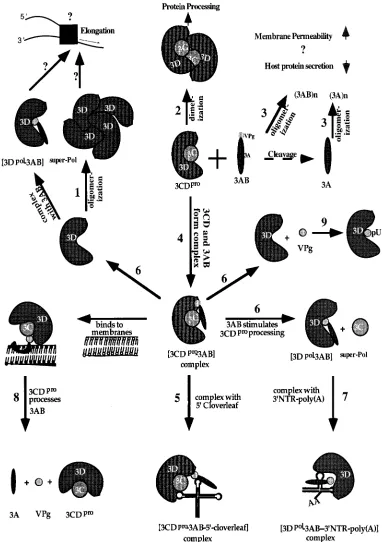
[image:7.612.51.288.69.315.2]FIG. 5. Working model for interactions and diverse functions of the P3 polypeptides. (1) 3Dpolis depicted as a tetramer although its precise status in solution is
not known. 3Dpololigomers are more active than monomers in RNA chain elongation, and the same may be true for the 3AB/3Dpolcomplex. (2) The function of
3CDpro/3CDprodimers is not known, but they may be required for autocleavage and/or viral protein processing. (3) 3AB and its cleavage product 3A alter host
membrane permeability and inhibit protein secretion. Whether these proteins must assemble to dimers or multimers (through interactions of their hydrophobic domain) to produce these effects is unknown. (4) 3AB and 3CDprocan form a complex in solution that (5) can bind with high specificity to the 59cloverleaf, a process required
for the initiation of positive-strand RNA synthesis. (6) When 3AB and 3CDproare bound to each other, 3AB can stimulate the autocleavage of 3CDprointo 3C and
3Dpol. 3Dpoland 3AB may remain in a tight complex with increased RNA polymerization activity. (7) A 3AB/3Dpolcomplex is likely to recognize the 39NTR of the
genome for the initiation of negative-strand RNA synthesis. (8) The half-life of 3AB is regulated by 3CDpro, since membrane-bound 3AB can be cleaved by 3CDpro
into 3A and 3B (VPg), two cleavage products whose functions are distinct from those of the 3AB precursor. (9) VPg, in turn, is uridylylated by 3Dpolto VPgpU(pU),
the primer for 3Dpol. For references, see Table 1. 39NTR, 39nontranslated region.
on November 9, 2019 by guest
http://jvi.asm.org/
a strong defect in poliovirus RNA replication (4, 18, 19). It is possible, therefore, that oligomerization is an important deter-minant of 3AB in RNA replication.
The interaction of 3AB with the viral polymerase 3Dpolmay
play a central role in the enzyme’s template recognition and polymerase activity (27, 33, 44, 46, 56). Particularly intriguing is
3AB’s ability to bind to 3Dpoland stimulate 3Dpolpolymerase
activity at low enzyme concentrations (33, 44, 46, 56). 3AB and
3Dpolmay form a complex that we have described as [3AB/
3D]super-pol(44). We have previously shown that certain
clus-tered-charge-to-alanine mutations in 3AB diminish this stim-ulating activity (56). We now correlate the lack of [3AB/ 3D]super-polformation with the apparent loss of binding of 3AB
to 3Dpol (Fig. 3A). The 3AB mutations abrogating enzyme
stimulation as well as 3AB/3Dpol binding map to the same
basic amino acids in the 3B moiety (Fig. 3B). Thus, specific residues in the 3B moiety of 3AB form contact points between this protein and the polymerase, an interaction that we spec-ulate is true also for the complex formation between VPg and
3Dpol. It should be noted, however, that the M20 mutation in
the hydrophobic domain of 3AB and the M1 mutation in the
3A moiety reduced binding to 3Dpolin the two-hybrid system
(Fig. 3). This may explain why both 3A and 3B portions of 3AB
are required to yield a maximally functioning [3AB/3D]super-pol
complex (44). At least one residue in 3Dpol, Val391, has been
identified as being required for binding of 3Dpolto 3AB in the
two-hybrid system and in biochemical assays (27).
The interaction in the two-hybrid system between 3AB and
3CDpro was relatively weak (Fig. 2A, bar 38). On the other
hand, we have reported that 3AB/3CDpro coprecipitated in
immune reactions, an observation suggesting interaction in solution (37). Indeed, 3AB has been reported to stimulate
proteolysis of 3CDpro, and 3AB/3CDpro exhibits a strong and
very specific affinity to the 59-terminal cloverleaf of the viral genome (25, 56, 57). When tested by far-Western blotting, a
strong interaction between 3AB and 3CDpro was observed
(Fig. 4B). This result underscores the contention that weak signals in the two-hybrid assay are not necessarily indicative of the strength of specific protein-protein interaction.
The complexity of the different interactions of the P3 polypeptides of poliovirus is summarized in Fig. 5. It is appar-ent that the polypeptide chains produced during the cascade of proteolytic cleavages engage in a variety of very different bio-chemical reactions. Several of these reactions have been de-duced by genetic analyses only (55). In addition to the viral polypeptides, there may also be cellular proteins that specifi-cally interact with P3 cleavage products of the poliovirus polyprotein (1, 6, 42). The recently described poly(C) binding protein 2 may be such a host factor (6, 42).
ACKNOWLEDGMENTS
We are indebted to Stanley Fields, Roger Brent, and Bert S. Semler for generously providing materials crucial for this study. We also thank Stanley Fields, Xuemei Cao, Meijia Yang, and Aniko V. Paul for valuable discussions and advice.
W.X. was a member of the graduate training program in Biochem-istry and Cell Biology, A.C. was a member of the training program in Molecular Genetics and Microbiology, SUNY at Stony Brook. D.H. was a member of the training program in Molecular, Cellular and Developmental Biology, University of Colorado at Boulder. This work was supported in part by grants AI-25166 and PN3929 (HSFPO) to K.K. and by grants AI-15122 and AI-32100 to E.W.
REFERENCES
1. Andino, R., G. E. Rieckhof, P. L. Achacoso, and D. Baltimore. 1993. Polio-virus RNA synthesis utilizes an RNP complex formed around the 59-end of viral RNA. EMBO J. 12:3587–3598.
2. Bartel, P. L., C.-T. Chien, R. Sternglanz, and S. Fields. 1993. Using the two-hybrid system to detect protein-protein interactions, p. 153–179. In D. A. Hartley (ed.), Cellular interactions in development: a practical approach. Oxford University Press, Oxford, England.
3. Bartel, P. L., J. A. Roecklein, D. SenGupta, and S. Fields. 1996. A protein linkage map of Escherichia coli bacteriophage T7. Nat. Genet. 12:72–77. 4. Bernstein, H. D., and D. Baltimore. 1988. Poliovirus mutant that contains a
cold-sensitive defect in viral RNA synthesis. J. Virol. 62:2922–2928. 5. Bernstein, H. D., P. Sarnow, and D. Baltimore. 1986. Genetic
complemen-tation among poliovirus mutants derived from an infectious cDNA clone. J. Virol. 60:1040–1049.
6. Blyn, L. B., K. M. Swiderek, O. Richards, D. C. Stahl, B. L. Semler, and E. Ehrenfeld.1996. Poly(rC) binding protein 2 binds to stem-loop IV of the poliovirus RNA 59 noncoding region: identification by automated liquid chromatography-tandem mass spectrometry. Proc. Natl. Acad. Sci. USA 93:11115–11120.
7. Cao, X. M., R. J. Kuhn, and E. Wimmer. 1993. Replication of poliovirus RNA containing two VPg genes leads to a specific deletion event. J. Virol. 5572–5578.
8. Chien, C.-T., P. L. Bartel, R. Sternglanz, and S. Fields. 1991. The two-hybrid system: a method to identify and clone genes for proteins that interact with a protein of interest. Proc. Natl. Acad. Sci. USA 88:9578–9582.
9. Cho, M. W., O. C. Richards, T. M. Dmitrieva, V. Agol, and E. Ehrenfeld. 1993. RNA duplex unwinding activity of poliovirus RNA-dependent RNA polymerase 3Dpol. J. Virol. 67:3010–3018.
10. Clark, M. E., T. Hammerle, E. Wimmer, and A. Dasgupta. 1991. Poliovirus proteinase 3C converts an active form of transcription factor IIIC to an inactive form: a mechanism for inhibition of host cell polymerase III tran-scription by poliovirus. EMBO J. 10:2941–2947.
11. Clark, M. E., P. M. Lieberman, A. J. Berk, and A. Dasgupta. 1993. Direct cleavage of human TATA-binding protein by poliovirus protease 3C in vivo and in vitro. Mol. Cell. Biol. 13:1232–1237.
12. Cuervo, A. M., and J. F. Dice. 1996. A receptor for the selective uptake and degradation of proteins by lysosomes. Science 273:501–503.
13. Datta, U., R. Banerjee, X. Liu, and A. Dasgupta. 1996. A point mutation in the glycosylation site of poliovirus polypeptide 3AB produces a dominant lethal phenotype. Presented at the ASV Meeting, Ontario, Canada. 14. Datta, U., and A. Dasgupta. 1994. Expression and subcellular localization of
poliovirus Vpg-precursor protein 3AB in eukaryotic cells: evidence for gly-cosylation in vitro. J. Virol. 68:4468–4477.
15. Doedens, J. R., and K. Kirkegaard. 1995. Inhibition of cellular protein secretion by poliovirus proteins 2B and 3A. EMBO J. 14:894–907. 16. Fields, S., and O.-K. Song. 1989. A novel genetic system to detect
protein-protein interactions. Nature 340:245–246.
17. Flanegan, J. B., and D. Baltimore. 1977. Poliovirus-specific primer-depen-dent RNA polymerase able to copy poly(A). Proc. Natl. Acad. Sci. USA 74:3677–3680.
18. Giachetti, C., S.-S. Hwang, and B. L. Semler. 1992. cis-acting lesions targeted to the hydrophobic domain of a poliovirus membrane protein involved in RNA replication. J. Virol. 66:6045–6057.
19. Giachetti, C., and B. L. Semler. 1991. Role of a viral membrane polypeptide in strand-specific initiation of poliovirus RNA synthesis. J. Virol. 65:2647– 2654.
20. Hammerle, T., C. U. Hellen, and E. Wimmer. 1991. Site-directed mutagen-esis of the putative catalytic triad of poliovirus 3C proteinase. J. Biol. Chem. 266:5412–5416.
21. Hammerle, T., A. Molla, and E. Wimmer. 1992. Mutational analysis of the proposed FG loop of poliovirus proteinase 3C identifies amino acids that are necessary for 3CD cleavage and might be determinants of a function distinct from proteolytic activity. J. Virol. 66:6028–6034.
22. Hansen, J. L., A. M. Long, and S. C. Schultz. 1997. Structure of the RNA-dependent RNA polymerase of poliovirus. Structure 5:1109–1122. 23. Harris, K. S., C. U. T. Hellen, and E. Wimmer. 1990. Proteolytic processing
in the replication of picornaviruses. Semin. Virol. 1:323–333.
24. Harris, K. S., S. R. Reddigari, M. J. H. Nicklin, T. Ha¨mmerle, and E. Wimmer.1992. Purification and characterization of poliovirus polypeptide 3CD, a proteinase and a precursor for RNA polymerase. J. Virol. 66:7481– 7489.
25. Harris, K. S., W. Xiang, L. Alexander, A. V. Paul, W. S. Lane, and E. Wimmer.1994. Interaction of the polioviral polypeptide 3CDprowith the 59 and 39termini of the poliovirus genome: identification of viral and cellular cofactors necessary for efficient binding. J. Biol. Chem. 269:27004–27014. 26. Heinz, B. A., and L. M. Vance. 1995. The antiviral compound enviroxime
targets the 3A coding region of rhinovirus and poliovirus. J. Virol. 69:4189– 4197.
27. Hope, D. A., S. E. Diamond, and K. Kirkegaard. 1997. Genetic dissection of interaction between poliovirus 3D polymerase and viral protein 3AB. J. Virol. 71:9490–9498.
28. Joachims, M., K. S. Harris, and D. Etchison. 1995. Poliovirus protease 3C mediates cleavage of microtubule-associated protein 4. Virology 211:451– 461.
29. Kitamura, N., B. L. Semler, P. G. Rothberg, G. R. Larsen, C. J. Adler, A. J.
on November 9, 2019 by guest
http://jvi.asm.org/
Dorner, E. A. Emini, R. Hanecak, J. Lee, S. van der Werf, C. W. Anderson, and E. Wimmer.1981. Primary structure, gene organization and polypeptide expression of poliovirus RNA. Nature 291:547–553.
30. Kuhn, R. J., H. Tada, W. M. Ypma, J. J. Dunn, B. L. Semler, and E. Wimmer. 1988. Construction of a “mutagenesis cartridge” for poliovirus genome-linked viral protein: isolation and characterization of viable and nonviable mutants. Proc. Natl. Acad. Sci. USA 85:519–523.
31. Kuhn, R. J., H. Tada, M. F. Ypma-Wong, B. L. Semler, and E. Wimmer. 1988. Mutational analysis of the genome-linked protein VPg of poliovirus. J. Virol. 62:4207–4215.
32. Lama, J., and L. Carrasco. 1995. Mutations in the hydrophobic domain of poliovirus protein 3AB abrogate its permeabilizing activity. FEBS Lett. 367: 5–11.
33. Lama, J., A. V. Paul, K. S. Harris, and E. Wimmer. 1994. Properties of purified recombinant poliovirus protein 3AB as substrate for viral protein-ases and as co-factor for viral polymerase 3Dpol. J. Biol. Chem. 269:66–70. 34. Lawson, M. A., and B. L. Semler. 1991. Poliovirus thiol proteinase 3C can utilize a serine nucleophile within the putative catalytic triad. Proc. Natl. Acad. Sci. USA 88:9919–9923.
35. Lawson, M. A., and B. L. Semler. 1992. Alternate poliovirus nonstructural protein processing cascades generated by primary sites of 3C proteinase cleavage. Virology 191:309–320.
36. McBride, A. E., A. Schlegel, and K. Kirkegaard. 1996. Human protein Sam68 relocalization and interaction with poliovirus RNA polymerase in infected cells. Proc. Natl. Acad. Sci. USA 93:2296–2301.
37. Molla, A., K. S. Harris, A. V. Paul, S. H. Shin, J. Mugavero, and E. Wimmer. 1994. Stimulation of 3Cpro-related proteolysis by the genome-linked protein, VPg, and its precursor, 3AB. J. Biol. Chem. 269:27015–27020.
38. Neufeld, K. L., J. M. Galarza, O. C. Richards, D. F. Summers, and E. Ehrenfeld.1994. Identification of terminal adenylyl transferase activity of the poliovirus polymerase 3Dpol. J. Virol. 68:5811–5818.
39. Neufeld, K. L., O. C. Richards, and E. Ehrenfeld. 1991. Purification, char-acterization, and comparison of poliovirus RNA polymerase from native and recombinant sources. J. Biol. Chem. 266:24212–24219.
40. Nicklin, M. J. H., K. S. Harris, P. V. Pallai, and E. Wimmer. 1988. Poliovirus proteinase 3C: large-scale expression, purification, and specific cleavage ac-tivity on natural and synthetic substrates in vitro. J. Virol. 62:4586–4593. 41. Nomoto, A., B. Morgan-Detjen, R. Pozzatti, and E. Wimmer. 1977. The
location of the polio genome protein in viral RNAs and its implication for RNA synthesis. Nature 268:208–213.
42. Parsley, T. B., J. S. Towner, L. B. Blyn, E. Ehrenfeld, and B. L. Semler. 1997. Poly (rC) binding protein 2 forms a ternary complex with the 59-terminal sequences of poliovirus RNA and the viral 3CD proteinase. RNA 3:1124– 1134.
43. Pata, J. D., S. C. Schultz, and K. Kirkegaard. 1995. Functional
oligomer-ization of poliovirus RNA-dependent RNA polymerase. RNA 1:466–477. 44. Paul, A., X. Cao, K. S. Harris, J. Lama, and E. Wimmer. 1994. Studies with
poliovirus polymerase 3Dpol: stimulation of poly(U) synthesis in vitro by purified poliovirus protein 3AB. J. Biol. Chem. 269:29173–29181. 45. Paul, A. V., J. V. Boom, D. Filipov, and E. Wimmer. 1998. Protein-primed
RNA synthesis by purified poliovirus RNA polymerase. Nature, 393:280– 284.
46. Plotch, S. J., and O. Palant. 1995. Poliovirus protein 3AB forms a complex with and stimulates the activity of the viral RNA polymerase, 3Dpol. J. Virol. 69:7169–7179.
47. Reuer, Q., R. J. Kuhn, and E. Wimmer. 1990. Characterization of poliovirus clones containing lethal and nonlethal mutations in the genome-linked pro-tein VPg. J. Virol. 64:2967–2975.
48. Rueckert, R. R. 1996. Picornaviridae and their replication, p. 507–548. In B. Fields (ed.), Virology. Raven Press, New York, N.Y.
49. Schultz, S. C., J. L. Hansen, and S. Hobson. 1996. Three-dimensional struc-ture of the RNA-dependent RNA polymerase of poliovirus. Presented at the Keystone Symposia on Viral Genome Replication, Tamarron, Colo. 50. Semler, B., C. Anderson, R. Hanecak, L. Dorner, and E. Wimmer. 1982. A
membrane-associated precursor to poliovirus VPg identified by immunopre-cipitation with antibodies directed against a synthetic heptapeptide. Cell 28:405–412.
51. Takeda, N., R. J. Kuhn, C. F. Yang, T. Takegami, and E. Wimmer. 1986. Initiation of poliovirus plus-strand RNA synthesis in a membrane complex of infected HeLa cells. J. Virol. 60:43–53.
52. Takegami, T., R. J. Kuhn, C. W. Anderson, and E. Wimmer. 1983. Mem-brane-dependent uridylylation of the genome-linked protein VPg of polio-virus. Proc. Natl. Acad. Sci. USA 80:7447–7451.
53. Takegami, T., B. L. Semler, C. W. Anderson, and E. Wimmer. 1983. Mem-brane fractions active in poliovirus RNA replication contain VPg precursor polypeptides. Virology 128:33–47.
54. Towner, J. S., T. V. Ho, and B. Semler. 1996. Determinants of membrane association for poliovirus protein 3AB. J. Biol. Chem. 271:26810–26818. 55. Wimmer, E., C. U. T. Hellen, and X. M. Cao. 1993. Genetics of poliovirus.
Annu. Rev. Genet. 27:353–436.
56. Xiang, W., A. Cuconati, A. Paul, X. Cao, and E. Wimmer. 1995. Molecular dissection of the multifunctional poliovirus RNA binding protein 3AB. RNA 1:892–904.
57. Xiang, W., K. S. Harris, L. Alexander, and E. Wimmer. 1995. Interaction between the 59-terminal cloverleaf and 3AB/3CDproof poliovirus is essential for RNA replication. J. Virol. 69:3658–3667.
57a.Yin, J., and E. Wimmer. Unpublished data.
58. Zervos, A. S., J. Gyuris, and R. Brent. 1993. Mxi1, a protein that specifically interacts with Max to bind Myc-Max recognition sites. Cell 72:223–232.