COMPARING THE EFFICACY OF VISUAL
INSPECTION WITH ACETIC ACID AND LUGOL’S
IODINE AS A SCREENING TOOL FOR DETECTING
CERVICAL LESIONS IN ASYMTOMATIC WOMEN OF
REPRODUCTIVE AGE GROUP WITH COLPOSCOPY
AS GOLD STANDARD
Dissertation submitted to
THE TAMILNADU Dr.M.G.R. MEDICAL UNIVERSITY
in partial fulfillment for the award of the Degree of
M.D. OBSTETRICS AND GYNAECOLOGY
BRANCH II
CERTIFICATE
This is to certify that the dissertation titled “
COMPARING
THE EFFICACY OF VISUAL INSPECTION WITH ACETIC
ACID AND LUGOL’S IODINE AS A SCREENING TOOL FOR
DETECTING CERVICAL LESIONS IN ASYMTOMATIC
WOMEN OF REPRODUCTIVE AGE GROUP WITH
COLPOSCOPY AS GOLD STANDARD”
is the bonafide work done
by
Dr. M. KAVITHA
between September 2008 to August 2009 during
her M.D.,O.G., course at ISO -KGH, MMC Chennai.
DEAN DIRECTOR
ACKNOWLEDGEMENT
I would like to thank
Prof. Dr.J.MOHANASUNDARAM M.D,PhD.,DNB, Dean, Madras Medical College for having permitted me to do
this dissertation work.
It is my pleasure to express my thanks to
Prof. Dr. M. MOHANAMBAL MD, DGO, Director, Institute of Social
Obstetrics and Govt. Kasturba Gandhi hospital, for her valuable guidance,
interest and encouragement in this study.
I take this opportunity to express my deep sense of gratitude and
humble regards to my beloved teacher and guide, Dr.Rathnakumar.S for
his timely guidance, suggestion and constant inspiration enabled me to
complete this dissertation.
My sincere thanks to Prof.Ramani Rajendran M.D. D.G.O., for her
guidance throughout the study.
My sincere thanks to Prof.K. Rama M.D., (Patho) for her guidance
throughout the study.
I thank all my professors, assistant professors & paramedical staff
I thank Mr.Padmanaban, statistician, who helped me for statistical
analysis.
I thank my family & friends for their inspiration & support given to
INTRODUCTION
Cancer Cervix:
Cancer cervix is the most common genital cancer in developing countries
and second most common genital cancer in developed countries.
Carcinoma cervix has long pre-invasive stage of over 10-15 years.
Availability of effective screening programmes and effective treatment along with
long pre-invasive stage, make road for reducing morbidity and mortality due to
cervical cancer by early detection of pre-invasive lesions. Cervical cancer
screening programmes to detect and treat cervical neoplasia have dramatic impact
on incidence of invasive cancer.
Incidence:
Worldwide, cervical cancer is the third most common
malignancy and second most common cancer after breast cancer in women.
Highest incidence of disease is seen in Caribbean and African countries.
Incidence in Africa : 55/100,000 women
New Delhi : 20.5/100,000 women
An estimated 4,70,000 new cases of cervical cancer are diagnosed each year
world wide and 80% of them occur in developing countries. A quarter of global
burden is experienced in India, where about 1, 26,000 new cases and 71,000
deaths attributable to cervical cancer are estimated to occur each year. Cervical
cancer constitute 15-55% of all female cancer and value of age standardized
incidence ranges from 17.2 to 55 per 1 lakh women in different region in India
with 5 year survival rate of less than 40% as most are detected at advanced
stage1.
Routine screening for cervical cancer with pap smear for all
women who are or have been sexually active was done previously. In India, lack
of experts in cytopathological studies and long period to get cytopathology report
makes distinct need for alternate strategy. Now visual inspection of cervix with
acetic acid and lugol’s iodine is being recommended especially in low resource
settings as a screening modality for cervical lesions.
The primary goal of cervical screening is to prevent cervical cancer which is
achieved by early detection; eradication and follow-up of pre-invasive cervical
lesions. The ability to detect preinvasive cervical lesions coupled with easy
Anatomy of cervix:
Cervix is the lower fibro muscular portion of uterus ,measures 3-4 cm in length
and 2.5 cm in diameter. However, it varies in size and shape depending on age,
parity and menstrual status of the women.
Ectocervix is the most readily visible portion and the endocervix is
largely invisible and lies proximal to the external os.
Epithelium of cervix:
Endocervix is lined by tall columnar epithelium in single layer with basal
solid nucleus2. On visual inspection ,it appears red in color as the underlying
vasculature is readily visible through single layer of glandular epithelium. This
epithelium is thrown into multiple longitudinal folds protruding into the lumen of
the canal, giving rise to papillary projections. It invaginates into the substance of
cervical stroma, resulting in formation of endocervical crypts, giving columnar
epithelium a grainy appearance. Beneath this layer are cubical or reserve cells
from which new surface cells are believed to develop and undergo squamous
metaplasia.
Ectocervix lined by stratified squamous epithelium which is composed of
4 layers.
1. Superficial cell layer
3. Parabasal layer
4. Basal layer
The important zones in colposcopy are:
Squamo
-
columnar junction:
Junction between stratified squamous epithelium and columnar
epithelium
Transformation zone:
Area between new squamocolumnar junction and old or original
NATURAL HISTORY OF DISEASE3:
Understanding the natural history of various degrees of CIN is the cornerstone
for the appropriate clinical management.
In addition to the degree of dysplasia,it is likely that the course of a specific
lesion is also influenced by number of other factors such as patient’s
age,inciting HPV type,immune competence and smoking
ISO&GOVT.KGH
Normal
Cervix HPV
Infection
Pre-cancer Cancer
Infection Progression Invasion
Regression Clearance
Natural History of HPV & Cervical Cancer
Persistence
Comparison of the terminology for cervical preinvasie squamous
disease
4.
Bethesda System
WNL Benign cellular
changes
ASCUS Lsil Hsil Carcinoma
Dysplasia/CIN System
Normal Inflammati on
Atypia
Mild dysplasia CIN I
Koilocytosis
Moderate dysplasia CIN2
Severe Dysplasia
CIS Cancer CIN 3
Old Pap System
Epidemiological model for cervical carcinogenesis
Level of Mild Moderate Severe CIS Normal
Dysplasia
Normal CIN I CIN II CIN III CIS LSIL HSIL
Frequency
Of spont. 40% 20% 1% 0 regression
Factors affecting natural history Malnutrition
Vit A,C, β carotene Smoking
Pathogenesis of CIN and invasive cancer:
Sexual activity
Hpv exposure
Cervical TZ
Squamous Epithelium columnar epithelium
Squamous intraepithelial lesion Glandular intra epithelial lesion
Low grade High grade Adenocarcinoma insitu
Low risk High risk High risk HPV-16,18 HPV 6,11,42,44 HPV 16,18,33,35
Approximate value of spontaneous regression or persistence and progression of CIN5.
COURSE OF CIN
CIN I CIN II CIN III
Regression to normal
60% 40%- 50% 33%
Persistence 30% 40% 55%
Progression to cancer
1% 5% >12%
(Ref): OSTOR AG, Int j Gynaecol.pathology 1993.
Visual screening approaches:
Visual screening is a process of identifying cervical lesions with or
without aided eye. Recently several researches has been undertaken to explain the
accuracy and acceptability of visual inspection methods as a means of detecting
pre-cancerous cervical lesions. Early methods included downstaging i.e., direct
inspection of cervix by unaided eye. The outcome was not encouraging. So a
promising approach VIA i.e., inspection of cervix after applying acetic acid
which stains the abnormal areas white and VILI i.e., inspection of cervix after
applying lugol’s iodine which does not stain the abnormal areas or stains it yellow
Terminology used for visual screening methods7:
Term used for method magnification enhancement 1. Schiller’s test
Lugol’s iodine test no iodine Visual inspection with
Lugol’s iodine
2. Down staging no no
3. Direct visual inspection no 3-5% acetic acid Acetic acid washes
Acetic acid visualization Acetic acid screening test
Visual inspection with acetic acid Acetic acid test
4. Aided visual inspection 2.5-4 × 3-5% acetic acid Gynoscopy
Avioscopy
DVI with magnification Visual inspection with acetic Acid & magnification
Visual inspection with acetic acid: Advantages:
Promising tool in low resource settings
Simple low tech approach
Minimally reliant on infrastructure for adequate performance
Cost for launching and sustaining is less than other methods
Sensitivity of VIA in detecting high grade lesions is near equal to
cytology though specificity is somewhat lower.
Pathophysiological basis of VIA:
On application of acetic acid,
Normal squamous epithelium appears pink and columnar epithelium
appears as bright red due to reflection of light stroma that is highly vascular.
Whitening effect of acetic acid depends upon the amount of cellular proteins
present in the epithelium. Areas with increased nuclear activity and DNA content
exhibit the most dramatic white color change.
Acetowhitening due to inflammation and healing is usually distributed widely
and restricted to transformation zone and may quickly disappear .Acetowhitening
of CIN takes up promptly and reverses very slowly.
Pathophysiological basis of VILI:
Normal glycogen containing squamous epithelium stains mahogany brown or
black after application of iodine.
Columnar epithelium does not take up iodine and remains unstained.
Areas of CIN and invasive cancer do not take up iodine and appear as thick
mustard yellow or saffron colored areas
Colposcopy:
In 1925, in Germany Hans Hinselmann introduced colposcopy.
Plays singular role in early diagnosis of cancer cervix
Colposcope is typically defined as a stereoscopic binocular field microscope with
a long focal length and powerful light source.
Modern colposcope permits magnification between 2X and 40X although most
routine colposcopic work can be accomplished at 10X to 15X magnification.
Indications for colposcopy:
Suspicious looking cervix
Invasive carcinoma on cytology
CIN 2 or CIN 3 on cytology
HPV infection
Aceto positivity on VIA
Positive lesion on VILI
Advantages of colposcopy:
Both diagnostic and therapeutic procedures can be done.
- to locate the lesions
- selection of biopsy site
- in selection of treatment of early invasive cancer and CIN
-reduction in unnecessary biopsy
REVIEW OF LITERATURE:
Pap smear has been recognized widely as the most effective cancer
screening test in history of medicine. Pap smear introduced by George
papanicoloau into clinical practice circa 1940. It was widely believed that use of
this test has been responsible for drastic reduction in the incidence and mortality
of cervical cancer in United States, Canada and much of Western Europe in the
past 50 years.
The first documented incident of deficiencies in gynecologic cytology
laboratories was reported by United States air force. Allegations that claimed
inaccuracies on Pap smear diagnosis performed by contract laboratory between
1972 -1977.8
Limitations of conventional pap smear:9
1. Failure to capture the entire specimen obtained from the patient.
2. Inadequate fixation of the sample.
3. Random distribution of abnormal cells in the sample.
4. Obscuring elements such as blood, inflammation or thick areas of
overlapping epithelial cells.
5. Technical variability in the quality of the smear
These obstacles made a way to successful development of computer-assisted
Liquid-based, thin layer cytology:
Liquid-based, thin layer cytology was developed to overcome the
technical limitations of the conventional Pap smear. Limitations like failure to
capture the entire specimen obtained from the patient and inadequate fixation are
overcome by collection of cells directly into liquid fixative.
Limitation of random distribution of cells overcome by mechanical
mixing of the cells creates a homogenous sample in which abnormal cells if
present are evenly distributed throughout the sample assuring the sample
homogenicity.10
Obscuring elements and technical variability in smear preparation
addressed in different fashion by the procedures currently available.i.e Thin- prep,
autocyte pap. Both these techniques result in consistent thin layer preparation of
epithelial cells that are depleted of extraneous elements.
Computer assisted screening devices:
Computer assisted devices designed to screen liquid based, thin layer slides
circumvent many of the technical problems faced in conventional pap smear. In
1999, Takahashi et al found that an interactive computer analysis system, the
98% sensitivity rate compared to a sensitivity rate of 89% by manual screening
alone12.
When used in conjunction with thin layer slides, computer assisted
screening devices offer tremendous promise for the future, particularly at a time
when the number of cytotechnologists is decreasing and demand for screening
increasing.
To date no study has subjected these technologies to the rigor of the current
gold standard colposcopy.
In 1998, Papillo et al, found a statistically significant increase in
specificity of diagnosis of SIL of the thin prep 81% over the conventional Pap
smear 72%.13
In 1999, Diaz Rosario et al, found equivalent specificity as determined
by biopsy proven dysplasia between thin prep 74% and conventional pap 79%14.
Molecular testing of residual material in vial:
An unexpected benefit of liquid based thin layer cytology was
discovered upon the realization that abundant cellular material remained in the
vial after production of the slide. With the advancement of molecular testing,
infectious organism. To date, successful out of vial testing has been shown for
HPV, Chlamydia gonorrhea and HSV.
Recently results of 2 large clinical trials showed that detection of high
oncogenic risk HPV types using hybrid capture ll assay effectively separates
patients with a cytological diagnosis of ASCUS group into a group of high
likelihood of having high grade CIN 2 or CIN 3 and a group that has no increased
risk of having high grade CIN15.
This triage strategy has been shown to reduce unnecessary colposcopic
examination in 45% to 60% of women with ASCUS reduce the morbidity,
anxiety and cost associated with that procedure. As these procedures require lab
facility and trained man power these things are not followed in low resource
settings.16
Screening with combined modalities:
In 2002, Vassilokos et al, found that combination of high risk HPV
detection and automated screening of liquid based thin layer pap smears could
separate women with cervical lesions that were ASCUS or higher grade lesions
into a human review group from a group with negative cytological findings for an
in an effort to achieve more sensitive yet cost – effective cervical cancer
screening programmes and probably represents the future of cervical screening17.
Down staging:
Screening for cervical cancer by visual inspection was widely advocated by
WHO in 1980s as a way to provide screening services in low resource settings in
which cytology was not available.18
Large study with 44,970 women was conducted which detected HSIL and
cancer with 62% sensitivity and 89% specificity for invasive cancers.19
Bharva et al and Sujatha et al used down staging as screening method in
3600 women and detected HSIL with sensitivity of 93% and specificity of 37%
Because of the poor performance of down staging in these studies most
authorities have concluded that downstaging offers little merit as a cancer
Cervicography:
When cervicography was first introduced several small studies were
conducted that suggested that it was superior to cervical cytology for detection of
CIN II, III and cervical cancer. More recently, two large well designed screening
studies critically evaluated the performance of cervicography and compared it
with HPV DNA testing and cervical cytology as a screening test. The sensitivity
of cervicography when performed under routine conditions was found to be poor.
Cervicography correctly identified only 52% of all the biopsy confirmed HSIL
and invasive cervical cancers. The specificity of cervicography was reasonable,
however. Only 5% of women were referred for colposcopy on the basis of an
abnormal cervigram .20
Direct visual inspection methods:
These include inspection of cervix after applying acetic acid and lugol’s
iodine. Many studies confirm that DVI is more sensitive but less specific.DVI
identified 88% of biopsy confirmed SIL, whereas cytology and cervicography
identified only 63%.so the Italian study by cecchini et al concluded that DVI
more sensitive but less specific.
In 1999, six large well controlled studies of DVI have been published from
Study by sankaranarayanan et al in 1999, showed that DVI has sensitivity ratio of
1.54(p<0.001) but the specificity was lower than cytology. (DVI specificity 68%
while cytology has 89%)
In 1999, chirenge et al in Zimbabwe studied DVI on 2148 patients and
found that DVI is sensitive in 77% and specific in 64% in detection of HSIL and
cancer.
Studies by Denny et al in 2002, in South Africa on 2698 women revealed
that VIA are 69.8% sensitive and 79.3% specific in detecting HSIL.21
Basu et al in 2003, studied VIA on 5881 women in India. The study had
sentivity of 55.7% and specificity of 82% in detecting CIN II and above lesions.
Sankaranarayanan et al in 2003, studied 4444 women with VIA to detect
CIN II and above lesions. This study showed that VIA is sensitive in detecting
82.6% and the specificity was 86.5%. All patients were subjected to VIA, VILI &
PAP SMEAR CYTOLOGY and positive cases were subjected to biopsy. Based
on the result VIA & VILI were considered to be suitable alternative screening test
to cytology for detecting cervical neoplasia in low resource settings.22
A study was conducted by SANKARANARAYANAN et al in
collaboration with Calcutta cervical cancer early detection group on visual
inspection with acetic acid and cytology in early detection of cervical neoplasia.
Study was conducted in 5881 women between 30 – 64 years of age who were
Study conducted at Fatima Jinnah College, Department of obstetrics and
gynecology, Lahore involving 501 women for comparison of visual inspection of
cervix and Pap smear for cervical cancer screening. Of these, 156 subjects were
positive with VIA (28.96%) ,while Pap smear was positive in 78 cases (14.4%).
The accuracy of VIA was 77.5% compared to 52.8% for Pap smear. They
concluded that VIA was more sensitive and highly specific.
Cervical cancer project at university of Zimbabwe / JHPIEGO conducted
a study in 1090 women to compare VIA with cytology .They concluded that VIA
was highly sensitive and could be valuable in detection of precancerous lesions of
the cervix but emphasis to increase the specificity of VIA was made.23
The study by Division of Cancer Epidemiology and Genetics, National
cancer Institute, Maryland, USA (Jeronimo J, Morales O) involving 1921
asymptomatic women compared the efficacy of VIA AND PAP SMEAR as a
screening modality for detecting cancer cervix. They concluded that VIA is good
screening method not only in low resource settings but also in well equipped
health centers.
Study conducted by Department of Obstetrics and Gynecology, John
Hopkins Medical center, Baltimore, MD (Biumenthal PD, Gaffikin L, Chirenje
involving the use of VIA followed by HPV could yield fewer false positive cases
than the use of VIA alone.
Department of Obstetrics and Gynecology, Cleveland Clinic Foundation
conducted a study with 1997 women of age 35-45 years to estimate the sensitivity
and specificity of VIA and to use it as a primary screening for intraepithelial
neoplasia. Visual inspection yielded normal results in 1445 women low grade
intraepithelial neoplasia in 525 (36%), high grade in 21(1%), cancer cervix in
6(0.3%). The sensitivity was 65% for smaller lesions and 89% for larger lesions.
They concluded that VIA can be used as a screening modality in developing
countries.
Cost effectiveness of cervical cancer screening strategies:
Goldie et al, in 2001 estimated the clinical benefits and cost
effectiveness of cytology, VIA and HPV testing in South Africa. According to
this model, using VIA and treating women with positive results of screening
during the same visit was effective. This single visit strategy was estimated to be
cost effective and reduce cervical cancer incidence by 26% with a cost of US $ 14
per YLS. The least effective strategy was cytologic examination, which would
reduce cervical cancer incidence by 19%, with a cost of US$ 81 per YLS.
Mandelblatt et al, in 2002, estimated the cost effectiveness of screening
using VIA to screen women every five years reduced cervical cancer incidence by
31% at a cost of US$263 per YLS and HPV DNA testing reduces cancer
incidence by 20% at a cost of 672 to 3477 US $ per YLS. The model suggested
that cytologic screening would be least cost effective in reducing cancer incidence
Geneva Foundation for Medical Education and Research
25On-going IARC collaborative studies on VIA for cervical cancer screening
Program design
Interventions evaluated
Location of the study
Number of
participants End points of the program Randomised, controlled intervention study VIA, cervical cytology, HPV testing Osmanabad district, India 160,000 women 30-59 years
Cervical cancer incidence/ mortality; Cost-effectiveness; establishment of a service and training platform for cervical cancer prevention. Randomised, controlled intervention study VIA Dindigul District, India 73,000 women aged 30-59 years
Detection rates of CIN2-3Cervical cancer incidence/mortality; Cost-effectiveness; establishment of a service and training platform for cervical cancer prevention. Cross-sectional
study
VIA, VILI Burkina Faso,
Republic of Congo, Guinea, Hyderabad, India, Laos, Mali, Niger, Mauritania, Tanzania 5000 women aged 30-59 in each location
Test characteristics; Acceptability, efficacy, complications of cryotherapy; establishment of a service and training platform for cervical cancer prevention.
Cross-sectional study
VIA, cytology Nigeria 2000 women
aged 30-64 years
Test characteristics; Acceptability, efficacy, complications of cryotherapy; establishment of a service and training platform for cervical cancer prevention. Cross-sectional study VIA, VILI, cervical cytology Trivandrum and Jaipur India 6000 women 30-59 years in each location
Test characteristics; Acceptability, efficacy, complications of cryotherapy; establishment of a service and training platform for cervical cancer prevention. Cross-sectional study VIA, VIAM, VILI, cervical cytology, HPV testing
Calcutta, India 12,000
women aged 30-64 years
Test characteristics; Acceptability, efficacy, complications of cryotherapy; establishment of a service and training platform for cervical cancer prevention. Cross sectional study Cervical cytology, HPV testing, VIA, VIAM, VILI
Bombay, India 5000 women aged 30-59 years
Cervical screening recommendations 1988 and 2002 – 2003:
1988consensus guidelines 2002 ACS guidelines 2003 ACOG guidelines 2003 USPSTF guidelinesWhen to start screening
Age 18 or with onset of sexual intercourse
Age 21 or about 3 yrs after onset of vaginal
intercourse
Age 21 or about 3 yrs after onset of vaginal
intercourse
Age 21 or about 3 yrs after onset of vaginal
intercourse Screening
Interval
Annually until 3 consecutive
satisfactory
negatives then internal may be extended at discretion of provider
Annually until age 30,
Biennially if liquid based cytology used
Annually until age 30 using either
conventional or liquid based cytology
Every 3 yrs
Age 30 or
older after 3 consecutive satisfactory negatives may screen every 2-3 yrs
Age 30 or older after 3 consecutive satisfactory negatives and no history of CIN 2 or 3 may screen every 2-3 yrs When to stop
screening
No upper limit Age 70 in well screened, low risk women
Evidence inconclusive to set upper age
Age 65 in well screened, low risk women Post hysterectomy No recommendations Screening not recommended after hysterectomy for benign indications if cervix
removed, if no prior CIN-2or3 Screening not recommended after hysterectomy for benign indications if cervix
removed, if no prior CIN-2or3
Existing methods of screening for cervical lesions
7:
1. Cytological evaluation:
*Pap smear
*Liquid based cytology
*Computerized devices like auto pap,
*Autonet, auto screen.
2. Visual inspection approaches:
*Visual inspection with acetic acid and lugol’s iodine.
*Cervicography
*Speculoscopy
*Down staging
3. Aided visual inspection
*gynoscopy
*avioscopy
3. COLPOSCOPY
* Colposcopy
*Colpomicroscopy
*Videocolpomicroscopy
*Computerized digital imaging colposcopy
4. NOVAL APPROACHES
* AgNor: new molecular marker that stands for silver stained nucleolar region.
Alternate screening methods:
A. carcinogenic HPV testing.
b.cellular markers like mRNA expression of E6/E7 transcript, p-16 markers
of disease progression.
Existing screening outcome
26Method sensitivity specificity a. pap smear
CIN I 68.1% 94.6% CIN II 83.3% 90.3% b. VIM
CIN I 60.5% 80% CIN II 73.3% 77.6% c. cervicography
AIM OF STUDY:
¾ To identify the incidence of cervical lesions in sexually active
asymptomatic women using VIM.
¾ To compare the efficacy of VIM with colposcopy.
¾ To evaluate the feasibility of VIM as a mass screening for cervical lesions
TYPE OF STUDY:
A prospective observational study on asymptomatic women of reproductive age
group.
DURATION OF STUDY
:
August 2008 to September 2009
MATERIAL AND METHODS
:
This is a hospital based prospective study conducted at Institute of social
obstetrics and Government Kasturba Gandhi Hospital for Women and children,
Triplicane, Chennai-5 from August 2008 to September 2009.
This study comprises study subject of 734 women who were attending
general and gynecology OPD. All 734 patients were subjected to visual
inspection and magnification (VIA/VILI) and bimanual pelvic examination. All
these patients were subjected to colposcopy and biopsy was done in all patients.
INCLUSION CRITERIA
¾ Sexually active women between 20-40 yrs
¾ Non-pregnant women
¾ Both nulliparous and multiparous
EXCLUSION CRITERIA
¾ Women with symptoms like vaginal discharge, pain etc
¾ Pregnant women
¾ Severe ill health
¾ Postpartum until 12 wks
¾ Overt growth in cervix
¾ Previous treatment for cancerous lesions
¾ Allergy to acetic acid and iodine
¾ Those who had undergone hysterectomy
¾ Women on hormonal therapy
¾ Women below 20yrs and above 40yrs of age
¾ Women those who are not sexually active
¾ Women with h/o surgery on cervix
IARC CRITERIA FOR INTERPRETATION OF VIA/VILI:
VIA POSITIVE:
Well defined, sharp, distinct, dense acetowhite areas with or without raised
margins abutting the squamo columnar junction in transformation zone.
Condyloma and leukoplakia occurring closer to the squamocolumnar
junction turning intensely white after application of acetic acid.
VIA NEGATIVE:
¾ No acetowhite lesion on cervix.
¾ Polyp protruding through the cervix with bluish white acetowhite areas.
¾ Nabothian cysts.
¾ Faint line or ill-defined acetowhitening at squamo columnar junction
¾ Shiny, pinkish white, cloudy white, bluish white faint patchy or
indefinite margins, blending with rest of the cervix.
¾ Angular, irregular dilating, acetowhite lesion resembling geographical
area far away from the transformation zone (satellite lesion).
¾ Ill defined patch, pale acetowhite in inflamed unhealthy, ulcerated
cervix with bleeding and mucopurulent discharge.
¾ Red spots on cervix against pinkish white background after applying
acetic acid.
¾ Streak like acetowhitening in the columnar epithelium.
¾ Dot like areas in endocervix, which are due to grape like columnar
VILI POSITIVE:
Dense, thick, bright mustard yellow or saffron yellow iodine non uptake areas
abutting the squamocolumnar junction in transformation zone.
VILI NEGATIVE:
• Normal cervix when squamous epithelium turns mahogany brown or black
and columnar epithelium does not change colour, no yellow areas seen.
• In ectropion, when an extension area of columnar epithelium with regular
margins on ectocervix remaining without colour change.
• Patchy, indistinct, illdefined, colourless or partially brown areas are seen in
the cervix.
• Non or partial iodine uptake, pale areas comparable to preexisting
nabothian follicle or polyps are seen.
• Stripping or leopard skin appearance associated with T.vaginalis infection.
• When pepper like non iodine uptake areas seen in the squamous epithelium
far away from squamocolumnar junction.
• When satellite, thin, yellow, non iodine uptake areas with angular or
digitating margins resembling geographical areas are seen far away from
Reporting visual inspection findings:
27Normal:
Smooth pink
Clear mucoid secretion
External os: central hole round in nulliparous and slit like in multiparous
Atrophic in postmenopausal women
Abnormal findings:
Infection -redness and congestion in general .
a. Chlamydia - mucopurulent activity
b. Gonorrhea - prominent vascularity,
ectopy, hypertrophy.
C.Trichomoniasis - strawberry cervix.
LSIL:
Flat smooth surface, indistinct borders like feathered or geographic
pattern, faint aceto white change.
HSIL:
Sharply demarcated areas, straight contour, distinct aceto white reaction,
most prompt, persistent, prominent aceto whitening, coarse vascular pattern,
INVASIVE CANCER
Malignant change:
Squamous carcinoma- irregular surface, sharply defined borders, coarse
punctuations and mosaicism, atypical vessels with hallmark of
neovascularisation.
Adenocarcinoma- stark acetowhiteness of fused, irregular heaps of glandular
villi seen in transformation zone.
CRITERIA FOR SCREENING
¾ Should be accurate and reproducible
¾ Test should be acceptable to people and reasonably inexpensive
¾ Adequate follow up of positives should be ensured
¾ Involves minimal or no expenditure to individuals and health services
¾ Undesired harm due to screening should be avoided
MATERIALS NEEDED
¾ Examination table with leg rest
¾ Good light source and ring lens magnifying system
¾ Digital video colposcope with magnification 25X
¾ Sterile bivalve speculum
¾ Normal saline,3%acetic acid,lugol’s iodine solution,10%formalin,
Monsel’s solution
¾ A steel/plastic container with 0.5%chlorine in which used gloves are
immersed
¾ A plastic bucket with polythene bag to dispose contaminated swabs and
waste items
METHODOLOGY OF STUDY
¾ Patient in lithotomy position
¾ Introduce bivalve self retaining speculum
¾ Characteristic of discharge noted if present
¾ Inspection of unstained cervix
¾ Inspection after application of acetic acid
¾ Inspection after application of Lugol’s iodine
¾ Inspection of fornices and vaginal wall
¾ Findings recorded and patient was advised to come after 2 to 3 weeks
¾ Patients undergone VIA/VILI were subjected to colposcopy
¾ Cervix, vagina and vulva inspected for lesions
¾ Colposcopic directed biopsy with punch biopsy done in all patients
¾ Specimen sent for histopathological examination and results obtained.
RESULTS AND ANALYSIS
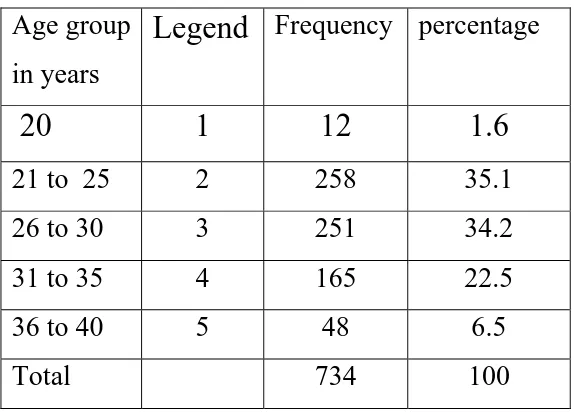
Table 1:
Frequency of age group in this study
(n=734)
Age group in years
Legend
Frequency percentage20
1
12
1.6
21 to 25 2 258 35.1 26 to 30 3 251 34.2 31 to 35 4 165 22.5 36 to 40 5 48 6.5
Total 734 100
In this study, age group taken was between 21-35 years which
CHART : 1
AGE GROUP
5.00
4.00
3.00
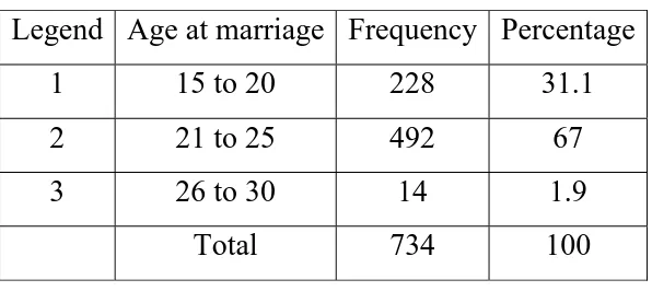
Table 2:
AGE AT MARRIAGE
(n=734)
Legend Age at marriage Frequency Percentage 1 15 to 20 228 31.1 2 21 to 25 492 67 3 26 to 30 14 1.9
Total 734 100
CHART : 2
AGE AT MARRIAGE GROUP
AGE AT MARRIAGE GROUP
3.00 2.00
1.00
Percent
80
70
60
50
40
30
20
10
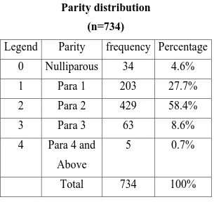
Table 3:
Parity distribution
(n=734)
Legend Parity frequency Percentage
0 Nulliparous
34
4.6%
1 Para
1 203 27.7%
2 Para
2 429 58.4%
3 Para
3 63 8.6%
4
Para 4 and
Above
5 0.7%
Total 734
100%
CHART : 3
PARITY
PARITY
4.00 3.00
2.00 1.00
.00
Percent
70
60
50
40
30
20
10
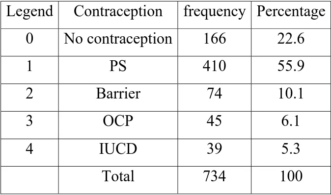
Table 4:
Contraception practiced in this study group:
(n=734
)
Legend Contraception frequency Percentage
0 No
contraception
166
22.6
1 PS 410
55.9
2 Barrier 74
10.1
3 OCP 45
6.1
4 IUCD 39 5.3
Total 734
100
Our study showed that most of our Para 2 women undergo permanent
method of sterilization. Hence with the loss of fear of pregnancy their
sexual activity also increases putting them at higher risk for cervical
CHART : 4
CONTRACEPTION
CONTRACEPTION
4.00 3.00
2.00 1.00
.00 Percent
60
50
40
30
20
10
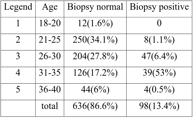
Table 5:
Comparison of Biopsy results and age group
(n=734)
Legend Age Biopsy
normal Biopsy
positive
1 18-20
12(1.6%)
0
2 21-25
250(34.1%) 8(1.1%)
3 26-30
204(27.8%) 47(6.4%)
4 31-35
126(17.2%) 39(53%)
5 36-40
44(6%)
4(0.5%)
total
636(86.6%) 98(13.4%)
Symmetrical measure:
The appropriate significance is established (0.000).
This study showed that age distribution in positive lesion was between
CHART : 5
AGE GROUP
5.00 4.00
3.00 2.00
1.00
Count
300
200
100
0
BIOPSY
.00
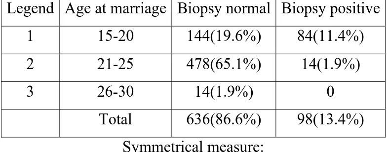
Table 6:
Comparison of Age at marriage with lesion:
(n=734)
Legend Age at marriage Biopsy normal Biopsy positive
1 15-20
144(19.6%)
84(11.4%)
2 21-25
478(65.1%)
14(1.9%)
3 26-30 14(1.9%) 0
Total
636(86.6%)
98(13.4%)
Symmetrical measure:
The appropriate significance is established(0.000).
In this study, there is higher incidence of positive biopsy when these
women marry at early age and expose themselves to longer period of
CHART : 6
AGE AT MARRIAGE GROUP
3.00 2.00
1.00
Count
600
500
400
300
200
100
0
BIOPSY
.00
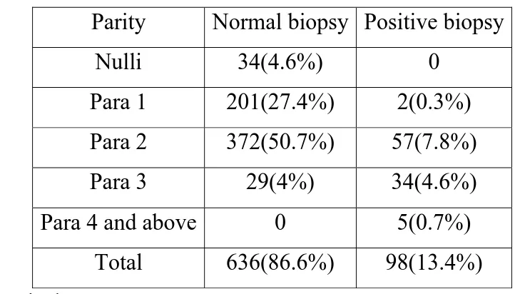
Table 7:
Comparison of Parity with biopsy results:
(n=734)
Parity Normal
biopsy Positive
biopsy
Nulli 34(4.6%) 0
Para 1
201(27.4%)
2(0.3%)
Para 2
372(50.7%)
57(7.8%)
Para 3
29(4%)
34(4.6%)
Para 4 and above
0
5(0.7%)
Total 636(86.6%)
98(13.4%)
Symmetrical measure:
The appropriate significance is established(0.000).
In this study among positive lesions ie, 13.4%, 13.1% belong to Para 2
CHART : 7
PARITY
4.00 3.00
2.00 1.00
.00
Count
400
300
200
100
0
BIOPSY
.00
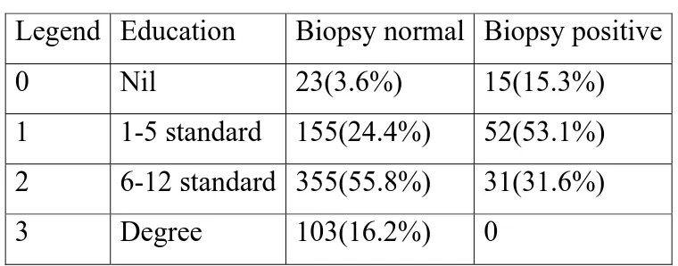
Table 8:
Education compared with positive lesions in colposcopy guided
biopsy:
(n=734)
Legend Education
Biopsy normal Biopsy positive
0 Nil
23(3.6%) 15(15.3%)
1 1-5
standard
155(24.4%)
52(53.1%)
2 6-12
standard 355(55.8%)
31(31.6%)
3 Degree 103(16.2%)
0
Symmetrical measure:
The appropriate significance is established(0.000).
In this study positive lesions are significantly associated with low level
of literacy. Those who undergone graduation has nil lesions indicating
CHART : 8
EDUCATION
3.00 2.00
1.00 .00
Count
400
300
200
100
0
BIOPSY
.00
1.00
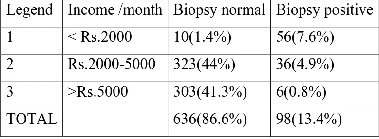
Table 9:
Comparing Socioeconomic status with lesion:
(n=734)
Legend Income /month Biopsy normal Biopsy positive
1 <
Rs.2000
10(1.4%)
56(7.6%)
2 Rs.2000-5000
323(44%)
36(4.9%)
3 >Rs.5000
303(41.3%)
6(0.8%)
TOTAL
636(86.6%)
98(13.4%)
Symmetrical measure:
The appropriate significance is established(0.000).
In this study out of 13.4% positive lesions 7.6% belong to very low
income group of < Rs.2000/month and 4.9% belong to income of Rs.
2000- 5000. Only 0.8% of income more than Rs.5000 /month showed
positivity.
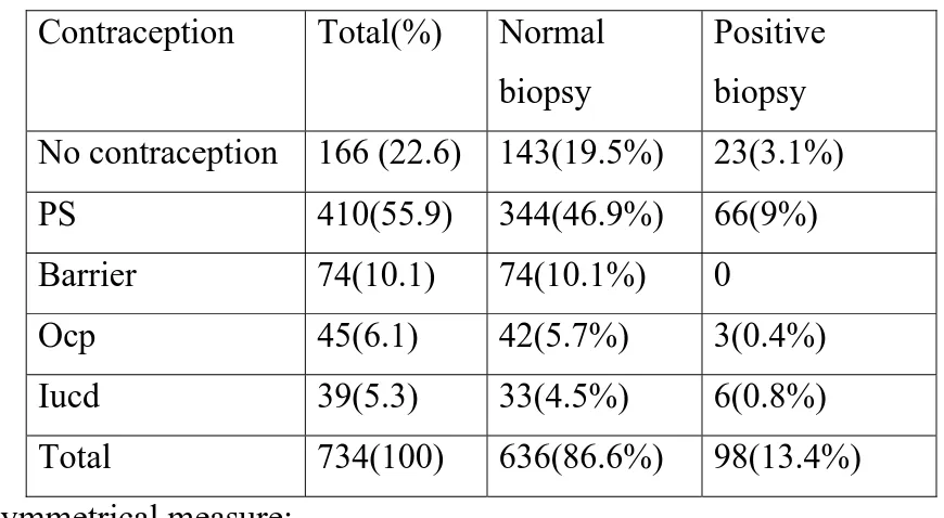
Table 10:
Influence of Contraception method adopted on cervical lesion :
(n=734)
Contraception Total(%)
Normal
biopsy
Positive
biopsy
No contraception 166 (22.6) 143(19.5%)
23(3.1%)
PS 410(55.9)
344(46.9%)
66(9%)
Barrier 74(10.1)
74(10.1%)
0
Ocp 45(6.1)
42(5.7%)
3(0.4%)
Iucd 39(5.3)
33(4.5%)
6(0.8%)
Total 734(100)
636(86.6%)
98(13.4%)
Symmetrical measure:
The appropriate significance is not established(0.124).
In our study positive lesions are more in women practicing
permanent sterilization as they were out of fear of pregnancy and prone
to have increased sexual activity making them a high risk group.
women practicing barrier methods(condoms) are at nil risk in this
CHART : 10
CONTRCEPTION
4.00 3.00
2.00 1.00
.00
Count
400
300
200
100
0
BIOPSY
.00
[image:68.612.116.522.191.413.2]
Table 11:
Result of VIA/VILI in this study:
(n=734)
LEGEND VIA/VILI FREQUENCY PERCENTAGE
0 NO
LESION
64487.7
1 POSITIVE
90
12.3
TOTAL
734 100
Table 12:
COLPOSCOPY result in this study:
(n=734)
Legend COLPOSCOPY FREQUENCY PERCENTAGE
0 NO
LESION
637
86.8
1 POSITIVE 97
13.2
CHART : 11
CHART : 12
COLPOSCOPY
1.00
VIA / VILI
1.00
Table 13:
Comparision of VIA/VILI result with BIOPSY report:
(n=734)
VIA/VILI Biopsy normal Biopsy positive
Normal 636(86.6%) 8(1.1%)
Positive 0
90(12.3%)
Total 636(86.6%)
98(13.4%)
Symmetrical measure:
The exact significance is established(0.000).
Table 14:
Comparison of Colposcopy with biopsy:
(n=734)
colposcopy Biopsy normal Biopsy positive
Normal 636(86.6%) 1(0.1%)
Positive 0
97(13.2%)
Total 636(86.6%)
98(13.4%)
Symmetrical measure:
[image:70.612.166.465.463.615.2]CHART : 13
VIA / VILI
1.00 .00
Count
700
600
500
400
300
200
100
0
BIOPSY
.00
1.00
CHART : 14
COLPOSCOPY
1.00 .00
Count
700
600
500
400
300
200
100
0
BIOPSY
.00
Table 15:
Comparison of VIA/VILI WITH COLPOSCOPY:
(n=734)
VIA/VILI Colpo normal Colpo positive
Normal 636
8
Positive 1
89
Total 637
97
Symmetrical measure:
The exact significance is established(0.000).
Both in VIA/VILI and Colposcopy, it was noted that all the positive
women were found to be positive when biopsy confirmation was
sought. However, there were minor differences with both the methods
missing on negative women. Hence though the methods were near
complete in their positive predictive value, they were lacking when
CHART : 15
VIA / VILI
1.00 .00
Count
700
600
500
400
300
200
100
0
COLPOSCOPY
.00
Table 16:
COMPARISION OF BIOPSY TYPES:
(n=734)
Biopsy positive
Frequency Percentage
Normal 636
86.6%
Chronic non specific
Cervicitis
32 4.4%
LSIL 57
7.8%
HSIL 9
1.2%
Total 734
100%
Most of the positive biopsies were LSIL lesions and the women were
treated accordingly. 1.2% of HSIL women were counseled for further
CHART : 16
BIOPSY TYPE
3.00
2.00
1.00
STUDY RESULTS
VIA/VILI WITH
BIOPSY
COLPO WITH BIOPSY
VIA/VILI WITH COLPO
SENSITIVITY 91.84% 98.98% 91.75%
SPECIFICITY 100% 100% 99.84%
PPV 100% 100% 98.89%
NPV 98.76% 99.84% 98.76%
DISCUSSION
This prospective observational study analyses the efficacy of visual inspection
methods (VIA/VILI) with colposcopy and cervical biopsy and to choose VIA and
VILI as an easily interpretable low cost but effective method for detecting
cervical lesions.
In our study, age group taken was between 21-35 years which constitute about
91.8% of patients studied. Similarly, study conducted in AIIMS, New Delhi in
2003 to evaluate and compare test performance of visual inspection of cervix by a
doctor and paramedical worker, included study group of patients with 64%
belonging to 30-39 years28.
In a study conducted in Mumbai, regarding concurrent evaluation of
visual, cytological HPV testing as screening methods for detection of cervical
neoplasia had study group of age between 30-39 in majority and showed that
younger women had higher rate of positive result in visual tests.
In our study, age at marriage was less than 20 in 31.1% which is
similar to study conducted in obs & gyn department in institute of medical
Similarly, study conducted in AIIMS, New Delhi in March 2003 for
detecting cervical lesions on women with mean age at first sexual intercourse was
19+/- 3.3 years.
In our study most of women were Para 1 and Para 2 which is consistent
with practice of our women for small family norm.
Our study showed that most of our Para 2 women undergo permanent
method of sterilization. Hence with the loss of fear of pregnancy their sexual
activity also increases, putting them at higher risk for cervical lesions
In this study, positive lesions are significantly associated with low level
of literacy. Those who undergone graduations have nil lesions indicating that
literacy have positive influence in reducing cervical lesions.
Similarly, study on Health literacy, cervical cancer risk factor and
distress in low income Africo- American women seeking colposcopy concluded
that low level of health literacy is associated with increased level of distress
among women at high risk for developing cervical cancer29.
In this study ,out of 13.4% positive lesions 7.6% belong to very
low income group of < Rs.2000/month and 4.9% belong to income of Rs. 2000-
5000. Only 0.8% of the study group with income more than Rs.5000 /month
showed positivity.
Similarly, study conducted in Institute of medical sciences, Lahore in
about 24-80% belonged to very low socio economic status of income between Rs.
3000- 5000/month.
This study showed that age distribution in positive lesion was
between 26-35 years (47% in 26- 30 years, 53% in 31-35 years). A study
conducted at Institute of medical sciences, Lahore in 2007 showed that majority
of women with CIN (60%) were between 35-45 years which also showed that
there is higher incidence of positive biopsy when this women marry at early age
and expose themselves to longer period of sexual activity.
In this study, among positive lesions ie, 13.4%, 13.1% belong to Para
2 and above and no nulliparous women was positive.
In a study conducted by IARC,a multicentric case control study
on role of parity and HPV in cervical cancer, found that there was direct
association between number of full term pregnancy and squamous cell carcinoma
risk30. The odds ratio for seven full term pregnancy or more was 3.8(95% CI
2.7-5.5) compared with nulliparous women and 2.3(CI1.6-3.2) compared with women
who had one or two full term pregnancy.
In our study, positive lesions are more in women practicing
permanent sterilization as they were out of fear of pregnancy and prone to have
In a paper published on cancer of cervix and its prevention : still a
public health concern which highlighted the risk factors showed that recent
research is showing that long term user of OCP are at high risk of cervical cancer
and regular user of barrier methods of contraception have low risk for cervical
lesions.
In our study, both in VIA/VILI and Colposcopy, it was noted that
all the positive women were found to be positive when biopsy confirmation was
sought. However, there were minor differences with both the methods missing on
negative women. Hence though the methods were near complete in their positive
predictive value, they were lacking when negative predictive value was
considered.
In this study, most of the positive biopsies were LSIL lesions and the
women were treated accordingly. 1.2% of HSIL women were counselled for
further definitive management.
Results of our study was comparable with study conducted in
Institute of medical sciences ,Lahore showed LSIL in 4.1% and HSIL in 1.8%
Variables in the Equation
33.029 4 .000
-5.175 10205.923 .000 1 1.000 .006
9.738 2.105 21.403 1 .000 16949.295
8.211 1.498 30.062 1 .000 3682.657
5.640 1.056 28.500 1 .000 281.417
8.011 2 .018
16.204 9804.510 .000 1 .999 1.1E+07
14.848 9804.510 .000 1 .999 2807355
3.985 4 .408
-36.924 17174.738 .000 1 .998 .000
-20.997 16022.507 .000 1 .999 .000
-20.198 16022.507 .000 1 .999 .000
-19.404 16022.507 .000 1 .999 .000
.774 .132 34.255 1 .000 2.168
-11.711 18784.217 .000 1 1.000 .000
VAR00004 VAR00004(1) VAR00004(2) VAR00004(3) VAR00004(4) VAR00007 VAR00007(1) VAR00007(2) VAR00008 VAR00008(1) VAR00008(2) VAR00008(3) VAR00008(4) VAR00016 Constant Step
1a
B S.E. Wald df Sig. Exp(B)
Variable(s) entered on step 1: VAR00004, VAR00007, VAR00008, VAR00016. a.
BINARY LOGISTIC REGRESSION MODEL WAS USED TO IDENTIFY
THE ASSOCIATION OF RISK FACTORS WITH RESPECT TO BIOPSY
ABNORMALITY.
THE VARIABLES AGE(4),AGE AT MARRIAGE(GROUP)(7),
DURATION OF SEXUAL EXPOSURE (16) ARE SIGNIFICANTLY
SUMMARY
Of 734 cases, 90 cases were found to be VIA/VILI positive and 644
cases were VIA/VILI negative.
Of 734 cases studied, colposcopy was positive in 97 (13.2%). Among 97
cases who were colposcopy positive, VIA/ VILI was positive in 90 cases.
Colposcopy guided Biopsy was done in all 734 cases. Of that, biopsy was
positive in 98 cases. Among 98 cases who were biopsy positive, 97 cases were
positive for colposcopy.
The sensitivity of VIA/VILI in detecting preinvasive lesions was
91.84% and specificity was 100% when compared with colposcopy which has
sensitivity 98.98% and specificity 100%.
The positive predictive value of VIA/VILI was 100% and its negative
predictive value was 98.76%. Similarly the positive and negative predictive value
in detecting cervical lesions with colposcopy in asymptomatic women was found
to be 100% and 99.84% respectively.
The diagnostic accuracy of VIA is 98.91% and for colposcopy ,it is
99.86% .
CONCLUSION
• There is an enormous increase in the incidence of cancer cervix and in
India, it is increasing in geometric proportion.
• This can be controlled only with the introduction of mass screening
programme in a coordinated way.
• Till recently, all our screening programmes were Pap smear based and with
the inherent difficulties in performing and interpreting Pap smear results in
our set up, it was not surprising that these programmes could not give the
expected results.
• Hence, the emphasis was shifted to visual inspection methods with acetic
acid and Lugol’s iodine.
• This method could sustain due to its simplicity and ease of performing in
mass programmes.
• The other advantage with this method is that the results are available
immediately thereby precluding with the need for the women to visit the
health centers on more than one occasion.
• Hence for resource restricted settings, VIA /VILI is a real boon for mass
screening.
• This study further emphasizes the need for programmed screening of all
women who are sexually active whatever may be their literacy and
BIBILIOGRAPHY
1.Bulletein of the WHO ISSN 0042 – 9680.
2.Colposcopy and treatment of cervical intraepithelial neoplasia. A beginner’s
manual published by international agency for research on cancer, Lyon ,2003.
3.M.schiffman national cancer institute.
4.Practical Gynecologic oncology third edition ,chapter 8, page 273.
5.Ostor AG, Int J gynaecol . pathology 1993.
6.Dr.M.Henry U.S Naval hospital, Bethesada M. D.
7. T.C. Wright, Jr et al / obstet. Gynec clin N Am 29 (2002) 701- 734.
8.New papanicolaou test suggested for Air force dependents U.S MED 1978 ;
8 – 15.
9.Felix Jc, Lonky Nm, Tamura k, Yu K – J, Naidu Y , Lai C – R , et al. abberant
expression of E- Cadherin in cervical intraepithelial neoplasia correlates with a
false negative papanicolaou smear. Am J Obstet Gynaecol 2002; 186: 1308 – 14.
10.Hutchinson ML, Zahniser DJ , Sherman ME , Herrero R, Affaro . M , Bratti
MC, et al. utility of liquid based cytology for cervical carcinoma screening:
11.Takahashi M , Kimura M , Akagi, Naitoh m. Auto-cyte screen interactive
automated primary cytology screening system : a preliminary evaluation . Acta
cytol 1998 ; 42 : 185 – 8.
12.Bishop JW , Bigner SH , Colgan TJ, Husain M, Howell LP, Mc Intosh KM, et
al.multicenter masked evaluation of Auto – cyte prep thin layers with matched
conventional smears , including initial biopsy results , Acta cytol 1998 ; 42: 189 –
97.
13.Papillo JL, Zarka MA .St John TL. Evaluation of the thin prep pap test in
clinical practice: a seven month , 16, 314 - case experience in Northern Vermont.
Acta cytol 1998; 42 : 203 – 8.
14.Diaz – Rozario LA, Kabawat SE. Performance of a fluid – based, thin – layer
papanicolaou smear method in the clinical setting of an independent laboratory
and an out patient screening population in New England. Arch pathol Lab med
1999 ; 123 : 817 – 21.
15. Manos MM, Kinney WK , Hurley LB, Sherman ME , Shieh – Ngai J .
Kurman RJ , et al. Identifying women with cervical neoplasia : using papilloma
virus DNA testing for equivocal papanicolaou results . JAMA 1999; 281 : 1605 –
10.
16.Solomon D , Schiffman M , Tarone R . Comparison of three management
strategies for patients with atypical squamous cells of undetermined significance :
17.Vassilakos P , Petignat p, Boulvain m , campana A . Primary screening for
cervical cancer precursors by the combined use of liquid – based cytology ,
computer assisted cytology and HPV testing. Br J cancer 2002 ; 86 : 382 – 8.
18.World Health Organisation . control of cancer of cervix uteri. Bulletien World
Health Organ . 1986 64 : 607 – 18.
19.Singh V, Seghal a , Luthra UK . screening for cervical cancer by direct
inspection BMJ 1992 , 304 : 534 – 5.
20. Scheinder DL, Burke L, Wright Tc, et al . Can cervicography can be
improved. An evaluation with arbitrated cervicography interpretations Am J
Obstet Gyn 2002 ; 187 : 15 – 23 .
21. Denny l, Kuhn , Pollack A, Wright Jr TC. Direct visual inspection for cervical
cancer screening : an analogue of factors influencing test performances . cancer
2002 ; 94 : 1699 – 707.
22.Shankaranarayanan R, Wesley R, Somanathan T et al. Test charecteristics of
visual inspection with acetic acid and lugol’s iodine(VILI) in cervical cancer
screening in Kerala, India. Int J Cancer 2003; 106 : 404- 408.
23.Zimbabwe project . Visual inspection with acetic acid for cervical cancer
screening . Test Qualities in a primary care setting. University of Zimbabwe /
25.IARC/ORCI/INCTR. http: screening IARC fr.
26. current diagnosis and treatment in obstetrics and treatment : 2007.
27.Tamil Nadu Heath Systems Project . Pilot project for screening cervical
cancer. A practical manual on colposcopy and cryotherapy. Health and family
welfare Dept. Govt. of Tamil Nadu.
28. Indian Journal Of Cancer . Jan – Mar 2004 . vol 41, Issue -1 , page 32.
29.Ethinicity and disease .vol 12, autumn 2002 ; 541.Lisa K.Sharp , Jill M.
Zusawski, MD ; Phillip Y . Roland. Health literacy, cervical cancer risk factor
and distress in low income African – American women seeking colposcopy .
ANNEXURE – I PROFORMA
COMPARING THE EFFICACY OF VISUAL INSPECTION
WITH ACETIC ACID AND LUGOL’S IODINE AS A
SCREENING TOOL FOR DETECTING CERVICAL LESIONS
IN ASYMPTOMATIC WOMEN OF REPRODUCTIVE AGE
GROUP WITH COLPOSCOPY AS GOLD STANDARD.
INSTITUTE OF SOCIAL OBSTETRICS
CHENNAI 5.
1.Serial Number ………… [ ] [ ] [ ]
2.Date of recruitment ……… [ ][ ]-[ ][ ]-[ ][ ][ ][ ]
3.Name………
4.Address……… ………
5.Age……… [ ][ ] S.E.S:[ ] .
6.Education……… [ ] (1-Nil; 2-Primary; 3-Middle; 4-High school; 5-College; 9-Not known)
7.Age at menarche(99-Not known)……… [ ][ ]
8.When did you have your last menstruation?...[ ]. (1-less than 12 months ago ; 2-more than 12 months ago)
9.Marital status……… [ ] (1-Married;2-Widow;3-Separated;8-Other;9-Not known)
13.Findings of VIA………[ ] (1-Negative;2-Positive;3-Positive,Invasive)
14. .Findings of VILI………[ ] (1-Negative; 2-Positive;3-Positive,Invasive)
15.Colposcopy……… [ ]
(1-Not done;2-Satisfactory,entireSCJ seen;3-Unsatisfactory,SCJpartially seen; 4-Unsatisfactory,SCJ not seen;5-Invasive cancer)
16.Colposcopic diagnosis(Date________ )………[ ] (1-Not done;2-Normal;3-Squamous metaplasia;4-Leukoplakia;
5-Condyloma/wart 6-Propbable low grade lesion:Atypia/CIN-1; 7- Propbable high grade lesion CIN-2,3;8-invasive cancer;9-other explain---)
A B C 17.Colposcopy findings
(A-to mark the findings of initial examination;B&C-for review
examinations after treatment;AW-acetowhite area;M-mosaic;P-punctation; AV-atypical vessels;G-growth;I-iodine negative area)
18.Biopsy taken?(1-Yes;2-No)………...[ ]
19.Histopathology of biopsy………[ ][ ] (00-not done;01-inflamation/chronic cervicitis;02-squamous metaplasia; 03-HPV infection;04-atypia;05-CIN-I;06-CIN-II;07-CIN-III;08-early invasive carcinoma 09-invasive squamous cell carcinoma;
10-invasive adeno carcinoma;99-other
(Explain __________________________________)
MASTER CHART- ANNEXURE II
COLPO NO. NAME AGE AGE GROUP S.E.S EDUCA
AGE AT
MARRIAGE-GROUP PARITY CONTRA VAI/ VILI COLPOS BIOPSY
6 anitha 20 1 2 1 1 0 0 0 0 0
2 asha 20 1 2 2 1 0 0 0 0 0
341 farida 20 1 2 2 1 1 0 0 0 0
351 farida 20 1 2 2 1 0 0 0 0 0
3 fathima 20 1 2 2 1 0 0 0 0 0
353 kondamma 20 1 2 1 1 1 0 0 0 0
5 priya 20 1 3 3 1 0 0 0 0 0
4 seetha 20 1 3 3 1 0 0 0 0 0
352 sriradha 20 1 3 3 1 1 0 0 0 0
1 usha 20 1 3 3 1 0 0 0 0 0
9 fousiya 21 2 2 1 1 1 3 0 0 0
8 harini 21 2 2 1 1 1 2 0 0 0
442 jeeva 21 2 2 2 1 0 0 0 0 0
441 jenifier 21 2 2 1 1 0 0 0 0 0
7 lakshmi 21 2 2 2 1 1 2 0 0 0
10 sampathku 21 2 3 3 1 1 0 0 0 0
443 leela 22 2 2 2 1 2 0 0 0 0
11 logeswari 22 2 2 2 1 1 3 0 0 0
13 sinduja 22 2 3 3 1 0 0 0 0 0
12 thiruseivi 22 2 3 3 1 1 0 0 0 0
18 ammu 23 2 2 2 1 1 2 0 0 0
355 asha 23 2 2 2 1 0 0 0 0 0
126 ayesa 23 2 2 1 2 1 3 0 0 0
132 anitha 23 2 2 1 2 1 3 0 0 0
19 aseena 23 2 2 3 1 0 0 0 0 0
116 afdab 23 1 2 2 2 1 2 0 0 0
465 bakiyam 23 2 2 2 2 2 1 0 0 0
15 barathy 23 2 2 2 1 2 1 0 0 0