EVALUATION OF PERIPHERAL NEUROPATHY
IN TYPE 2 DIABETES MELLITUS AND ITS
CORRELATION WITH OTHER
MICROVASCULAR COMPLICATIONS
Dissertation submitted in partial fulfillment of regulation for
the award of M.D. Degree in General Medicine (Branch I)
The Tamilnadu
Dr. M.G.R. MEDICAL UNIVERSITY
Chennai
CERTIFICATE
Certified that this is the bonafide dissertation done by
DR NEETHA BALARAM and submitted in partial fulfillment of the
requirements for the Degree of M.D., General Medicine, Branch I of The
Tamilnadu Dr. M.G.R. Medical University, Chennai.
Date : Professor DR. S. Veerakesari
Head Of Department
Department of General medicine Coimbatore Medical College
Date : ProfessorDr. K.Govindarajan
Guide & Professor
Department of Neurology Coimbatore Medical College
Date : Dr.R Vimala,MD
The Dean,
DECLARATION
I solemnly declare that the dissertation titled “EVALUATION
OF PERIPHERAL NEUROPATHY IN TYPE 2 DIABETES MELLITUS AND ITS CORRELATION WITH OTHER MICROVASCULAR COMPLICATIONS” was done by me from September 2009 to September 2010 under the guidance and supervision
of ProfessorDr.K..Govindarajan M.D., DM.
This dissertation is submitted to the Tamilnadu Dr. MGR Medical
University towards the partial fulfillment of the requirement for the award
of MD Degree in General Medicine (Branch I).
Dr NEETHA BALARAM
Place : Coimbatore
ACKNOWLEDGEMENT
I wish to express my sincere thanks to our respected Dean
Dr. R.Vimala and Medical Superintendent Dr. Mathivanan A, M.S.
for having allowed me to conduct this study in our hospital.
I am extremely grateful to Dr. Veerakesari MD Professor and
HOD of Medicine, CMCH, Coimbatore for his encouragement, and help
in preparing this dissertation. I express my heartfelt thanks and deep
gratitude to the professor of the Department of Neurology, also my guide,
Prof. Dr.K.G.Govindarajan. MD, DM for his able guidance,
supervision, invaluable suggestions and kind help, rendered throughout
the course of my study, and in the preparation of this dissertation.
I sincerely thank all Professors Dr. D. Nedumaran MD, DM,
Dr. S Usha MD, Dr. Raveendran M. MD, and all Asst.
Professors Dr.Kumar Natarajan MD, Dr.V.Neelakandan MD,
Dr.Manohari R MD, for their guidance and kind help. I express my
grateful thanks to Dr. Sacrates MD, DM for his valuable help in
completing this study.
No amount of words can measure up to the deep sense of gratitude
and thankfulness that I feel towards My family whose cherished
blessings and countless sacrifices are behind whatever success I have
achieved in my life. Last but not least I express my gratitude to all
CONTENTS
S.NO. TITLE PAGE NO.
1. INTRODUCTION 1
2. AIMS & OBJECTIVES 5
3. REVIEW OF LITERATURE 6
4. MATERIALS & METHODS 41
5. RESULTS AND ANLAYSIS 48
6. DISCUSSION 69 7. CONCLUSIONS 74
8. BIBLIOGRAPHY
9. ANNEXURES
10. 1) PROFORMA
11. 2) MASTERCHART
12. 3) CONSENT FORM
13. 4) ABBREVIATIONS
LIST OF TABLES
S.NO. TITLE PAGE NO.
1 AGE AND SEX DISTRIBUTION 48
2 DISTRIBUTION OF DURATION OF DIABETES
MELLITUS
49
3 HBA1C AND DURATION ON DIABETES 50
4 BODY MASS INDEX 51
5 HYPERTENSION AND DURATION OF
DIABETES
52
6 DISTRIBUTION OF PATIENTS ACCORDING
TO DNE SCORES
52
7 TOTAL SERUM CHOLESTEROL 54
8 DIABETIC RETINOPATHY 54
9 MICROALBUMINURIA 55
10 NERVE CONDUCTION STUDY 56
11 COMPARATIVE ANALYSIS OF CLINICAL
EXAMINATION SCORE(DNE) AND NERVE CONDUCTION STUDY
56
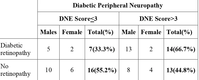
12 DIABETIC PERIPHERAL NEUROPATHY AND
RETINOPATHY
58
13 DIABETIC PERIPHERAL NEUROPATHY AND
MICROALBUMINURIA
59
14 DIABETIC PERIPHERAL NEUROPATHY AND
SEX
60
15 DISTRIBUTION OF DIABETIC PERIPHERAL
NEUROPATHY IN DIFFERENT AGE GROUPS
61
16 DIABETIC PERIPHERAL NEUROPATHY AND
DURATION OF DIABETES
17 DIABETIC PERIPHERAL NEUROPATHY AND HBA1C LEVELS
64
18 DIABETIC PERIPHERAL NEUROPATHY AND
BODY MASS INDEX
65
19 DIABETIC PERIPHERAL NEUROPATHY AND
SYSTEMIC HYPERTENSION
66
20 DIABETIC PERIPHERAL NEUROPATHY AND
TOTAL SERUM CHOLESTEROL
LIST OF CHARTS
S.NO. TITLE PAGE NO.
1. AGE AND SEX DISTRIBUTION 49
2 DISTRIBUTION OF DURATION OF DIABETES
MELLITUS
50
3 HBA1C AND DURATION ON DIABETES 51
4 DISTRIBUTION OF PATIENTS ACCORDING
TO DNE SCORES
53
5 DISTRIBUTION OF DIABETIC RETINOPATHY 55
6 DISTRIBUTION OF MICROALBUMINURIA 55
7 COMPARATIVE ANALYSIS OF CLINICAL
EXAMINATION SCORE (DNE) AND NERVE CONDUCTION STUDY
57
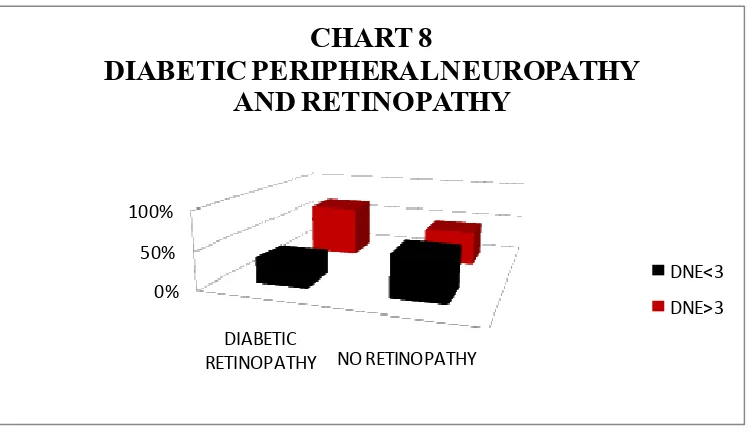
8 DIABETIC PERIPHERAL NEUROPATHY AND
RETINOPATHY
59
9 DIABETIC PERIPHERAL NEUROPATHY AND
MICROALBUMINURIA
60
10 DISTRIBUTION OF DIABETIC PERIPHERAL
NEUROPATHY IN DIFFERENT AGE GROUPS
62
11 DIABETIC PERIPHERAL NEUROPATHY AND
DURATION OF DIABETES
63
12 DIABETIC PERIPHERAL NEUROPATHY AND
HBA1C LEVELS
64
13 DIABETIC PERIPHERAL NEUROPATHY AND
BODY MASS INDEX
66
14 DIABETIC PERIPHERAL NEUROPATHY AND
SYSTEMIC HYPERTENSION
67
15 DIABETIC PERIPHERAL NEUROPATHY AND
TOTAL SERUM CHOLESTEROL
INTRODUCTION
Diabetes Mellitus is a common and a serious disease with chronic
complications and constitutes a substantial burden for both patient and
health care system. In 2010, the global prevalence of diabetes was
estimated at 285 million, corresponding to 6.4% of world’s adult
population. This figure is predicted to reach 438 million by 2030 as a
consequence of longer life expectancy, sedentary life style and changing
dietary patterns. According to recent estimates, presently India has 32
million diabetic subjects, and this is projected to increase to 100million
i.e. rise by 250% by the year 2035. In the CUPS (Chennai urban
population) study by Mohan et al, 12% of individuals above age of 20
years in Chennai were found to be diabetic in the year 1997. In the more
recent CURES study, conducted on 26,001 individuals showed that 16% now have diabetes in Chennai. 1,2,3
Vascular complications both micro and macrovascular predominate
the features of Indian diabetes due to delayed diagnosis. Various
microvascular complications in diabetes mellitus includes
• Diabetic Retinopathy
• Diabetic Nephropathy
Presence of microvascular complications at the time of diagnosis of
diabetes mellitus are showing increasing trend in India. Early detection of
microvascular complications and its treatment at this time by intensive
therapy can prevent progression of these complications and hence
morbidity and mortality among patients.
Diabetic Neuropathy is one of the most common troublesome
complications of diabetes mellitus. The prevalence of neuropathy is
related to age, duration of diabetes and the quality of metabolic control,
by the time a diabetic patient has severe neuropathy, retinopathy and
albuminuria are also usually present. Diabetic neuropathy is clinically
present in 30-50% of all diabetes patients. Diabetic foot accounts for one
of the largest inpatients admissions in India.4,5
The primary pathological role of hyperglycemia in diabetic
complications is well established. With the increasing knowledge that
maintenance of euglycemia greatly reduces, if not prevents the risk of
diabetic complications and at times helps even in regression of such
complications, monitoring the control of diabetes is essential for the
successful management of the diabetes6.
Keontg and Gabbay and their co-workers have suggested
measurement of glycosylated hemoglobin (HbA1C) as an indicator of
glycosylation of HbA at the amino-terminal valine of Beta chain. When
properly assayed HbA1C level in a blood sample gives an estimate of
diabetic control for preceding 3-4 month period (i.e. life span of RBC)7,8.
DCCT study proved that a glycated Hemoglobin (HbA1C) reduction
from 9 to 7% for a mean follow up of 6.5 years was able both to reduce
the onset of diabetic neuropathy (from 9.6% 2.8%) and to slow its
progression. However, euglycemia is only able to halt the progression,
rather than reverse it, once the nerve damage is established.9,10
The relative effect of cardiovascular risk factors specifically
associated with diabetes (e.g., hypertension, dyslipidemia, and increased
weight) or not associated with diabetes (smoking)on the development of
neuropathy are incompletely elucidated. The Diabetes Control and
Complications Trial (DCCT) reported a 60 percent reduction in
neuropathy in the intensively treated groups after five years but the
cumulative incidence of neuropathy (15 to 21 percent) and abnormal
nerve conduction (40 to 52 percent)remained substantial. Such findings
suggest that neuropathy can develop, despite intensive control of the
glucose level.Thus, risk factors besides hyperglycemia are probably
involvedin the evolution of neuropathy.9,10 Identifying them, particularly
if they are modifiable, might lead to new risk-reduction strategies. The
aimed to define and assess the relative importance of other, potentially
modifiable factors that increase the risk of distalsymmetric neuropathy in
patients with type 1 diabetes mellitus.
The present study aims to evaluate peripheral neuropathy in type
2 diabetes patients by clinical examination and electrophysiologicaly by
nerve conduction studies and to correlate it with the other
microvascular complications and various risk factors such as age, sex,
duration of diabetes, HbA1C, body mass index, systemic hypertension
and hypercholesterolemia. The importance of this study lies in the fact
that the morbidity and mortality in diabetes is attributed to a large
extend by its complications. Early detection and treatment of these
complications therefore plays a crucial role. Other than glycemic control,
there are no treatments for diabetic neuropathy. Thus, identifying
potentially modifiable risk factors for neuropathy is crucial. Detecting
these risk factors helps in correcting these at an early stage. Correlation
between the various microvascular complications helps in better
AIMS AND OBJECTIVES
To evaluate peripheral neuropathy in type 2 diabetes patients by
clinical examination and electrophysiologicaly by nerve
conduction study and thus do a comparative analysis.
To study the correlation of diabetic peripheral neuropathy with
other microvascular complications by assessing microalbuminuria
and retinopathy.
To study the correlation of diabetic peripheral neuropathy with
age, sex, duration of diabetes, body mass index, systemic
hypertension and total serum cholesterol.
REVIEW OF LITERATURE
“To know diabetes is to know medicine”
Historic Review
Knowledge of diabetes dates back to Centuries before Christ. The
Egyptian PAPYRUS EBCRS (Ca.1500 B.C) described an illness
associated with the passage of much urine. First clear clinical description
was given by CELSUS in the first century (30BC to 50 A.D).
The word “DIABETES” coined by ARETAEUS, of
CAPPADOCIA probably derived from Greek work Indicating “Running
Through as a Siphon”. “because the fluid does not remain in the body but
uses the man’s body as a ladder whereby to leave it”. The disease is better
understood in India. CHARAKA (2nd century AD) in his “Charaka
Samhita” has mentioned the sweetness of urine in addition to the
symptom of polyuria. The ancient Indian surgeon Sushruta (500 AD)
described the disease as “Madhu Meha” (Honey Urine) with symptoms of
polyphagia, polydypsia and polyuria. Avicenna (980–1037), an Arab
Physician gave the first description of diabetic gangrene.
FREDRICK-BANTING AND CHARLES BEST discovered insulin in 1922.
JOHN ROLLO IN 1978 recorded the involvement of Nervous
system in diabetes. MARACHAL DE CALVI (1864) first suggested that
PAVY (1885) gave a detailed description of the clinical
manifestation of diabetic neuropathy. R.W. RUNNDLES wrote that
entire nervous system may be involved including central, autonomic and
peripheral systems, but more commonly peripheral system. JORDON
(1936) AND RUDDLES (1945) gave the first clear description of
autonomic neuropathy.
IN 1936, Jordon tried to classify the neurological manifestation into 3
groups.
1. Hyperglycemic symptoms-Reversed upon treatment.
2. Circulatory degenerative type
3. Neurotic type
THOMAS AND LESCELLES (1966) described an increased
incidence of segmental demyclination in diabetic Neuropathy. BISHOP
(1968) postulated that a disordered lipid metabolism in the Schwann cells
results in demyelination. In monitoring the control of diabetes,
estimation of urine sugar and blood sugar have drawback as it requires
patient compliance and frequent measurements.
Consequently GABBAY et al, in 1976 suggested measurement of
Diabetes Mellitus
Diabetes mellitus is a group of metabolic diseases characterized by
hyperglycemia, resulting from defects in insulin secretion, insulin action
or both. The metabolic dysregulation associated with diabetes mellitus
causes secondary pathophysiological changes in multiple organ systems.
With an increasing incidence world wide, diabetes mellitus will be the
leading cause of morbidity and mortality in the future.
Etiologic classification of diabetes mellitus17
1. Type 1 Diabetes (β-cell destruction, usually leading to absolute insulin deficiency).
A. Immune mediated
B. Idiopathic
2. Type 2 diabetes (may range from predominantly insulin resistance with relative insulin deficiency to a predominantly insulin secretory
defect with insulin resistance).
3. Other specific types of diabetes
A. Genetic defects of β-cell function characterized by mutation in:
• Hepatocyte Nuclear Transcription Factor (HNF) 4α (MODY 1)
• Glucokinase (MODY 2)
• Insulin promoter factor (IPF) 1 (MODY 4)
• HNF-1β (MODY 5)
• Neuro D1 (MODY 6)
• Mitochondrial DNA
• Proinsulin or insulin conversion
Genetic defects in insulin action
1. Type A insulin resistance
2. Leprechaunism
3. Rabson Mendenhall syndrome
4. Lipodystrophy syndromes
B. Diseases of the exocrine pancreas–pancreatitis, pancreatectomy,
neoplasia, cystic fibrosis, hemochromatosis, fibrocalculous
pancreatopathy.
C. Endocrinopathies–acromegaly, Cushing’s syndrome, glucagonoma,
pheochromocytoma, hyperthyroidism, somatostatinoma,
aldosteronoma.
D. Drug or chemical induced–Vacor, pentamidine, nicotinic acid,
glucocorticoids, thyroid hormone, diazoxide, beta – adrenergic
agonists, thiazides, phenytoin, α-interferon, protease inhibitors,
E. Infections – congenital rubella, cytomegalovirus, coxsackie.
F. Uncommon forms of immune mediated diabetes–“stiff-man”
syndrome, anti-insulin receptor antibodies.
G. Other genetic syndromes sometimes associated with diabetes.
Down’s syndrome, Klinefelter’s syndrome, Turner’s syndrome,
wolfram’s syndrome, Friedreich’s ataxia, Huntington’s chorea,
Laurence Moon Biedl Syndrome, myotonic dystrophy, porphyria,
Prader – Willi syndrome.
4. Gestational Diabetes
Diagnostic criteria for diabetes mellitus17
The criteria for diagnosis of diabetes are shown in the table:
DIAGNOSTIC CRITERIA FOR DIABETES MELLITUS
• Symptoms of diabetes plus random blood glucose concentration
≥11.1 mmol/L (200 mg/dL)a or
• Fastng plasma glucose ≥ 7.0 mol/L (126 mg/dL)b or
• Two-hour plasma glucose ≥ 11.1 mmol/L (200 mg/dL) during an
Three ways to diagnose diabetes are possible and each, in the
absence of unequivocal hyperglycemia, must be confirmed, on a
subsequent day by any one of the 3 methods, given in the table. The use
of the hemoglobin A1C (HbA1C) for the diagnosis of diabetes is not
recommended at this time.
COMPLICATIONS OF DIABETES
Diabetes has both acute and long-term complications. They are :
Acute
• Diabetic ketoacidosis
• Hyperglycemic hyperosmolar state
• Hypoglycemia
Long term
• Retinopathy
• Neuropathy
• Nephropathy
• Ischemic heart disease
• Cerebrovascular disease
• Peripheral vascular disease
Others
• Infections
Tuberculosis
Candidiasis – Oral / Vulvovaginal
Mucormycosis
Necrotising fasciitis
Periodontitis
Microvascular complications in Diabetes Mellitus
All forms of diabetes, both inherited and acquired, are
characterized by hyperglycemia, a relative or absolute lack of insulin, and
the development of diabetes specific microvascular pathology in the
retina, renal glomerulus, and peripheral nerve.
Pathogenesis
Diabetes mellitus causes both microvascular and macrovascular
complications. It has been showed in studies that microvascular
complications are mainly because of hyperglycemia, whereas insulin
resistance is the major determinant in macrovascular disease.
Atherosclerosis is the pathological entity in macrovascular disease.
Microvascular complications are due to following mechanisms18
Most cells are able to reduce the transport of glucose inside the cell
when they are exposed to hyperglycemia, so that their internal glucose
concentration stays constant. In contrast, the cells damaged by
Thus, diabetes selectively damages cells, like endothelial cells and
mesangial cells.
i. Myoinositol Metabolism
Glucose acts a virtually the sole source of energy in peripheral
nerves as well as in the brain. It enters nerve cells through insulin –
independent pathways and is used in the production of ATP. Although,
ATP production does not appear impaired, experimental diabetic nerves
do demonstrate a reduction in ATP utilization, thought to be secondary to
decrease Na/K ATPase activity. Decreased Na/K ATPase activity has
been shown to correlate to decreased myoinositol concentrations within
peripheral nerves in diabetic animals.
Myoinositol is a normal dietary hexose having structural similarity
to glucose. It is an important constituent of phospholipid and cell
membrane, is 90 to 100 times more concentrated in peripheral nerves than
Hyperglycemia results in competitive inhibition of the sodium-
dependent transport system responsible for myoinositol uptake. This
decreased uptake is hypothesized to contribute to the decreased
concentration of myo-inositol found in the peripheral nerve, as well as
decreased Na/K ATPase activity. Na/K ATPase impairment not only
results in decreased nerve cell membrane potential and, therefore
decreased nerve conduction, but also decreased sodium dependent
myo-inositol uptake creating a worsening cycle. Myo-myo-inositol replacement
Increased flux through the polyol pathway
The polyol pathway focuses on the enzyme aldose reductase.
Aldose reductase normally has the function of reducing toxic aldehydes
in the cell to inactive alcohols, but when the glucose concentration in the
cell becomes too high, aldose reductase also reduces that glucose to
sorbitol, which is later oxidized to fructose. In the process of reducing
high intracellular glucose to sorbitol, the aldose reductase consumes the
cofactor NADPH. NADPH is also the essential cofactor for regenerating
a critical intracellular antioxidant, reduced glutathione. By reducing the
amount of reduced glutathione, the polyol pathway increases
susceptibility to intracellular oxidative stress.
Intracellular production of advanced glycation endproducts (AGE)- It is harmful by three mechanisms
The first mechanism, is the modification of intracellular proteins
including, most importantly, proteins involved in the regulation of gene
transcription
NADPH NADPH
Glucose Aldose
Reductase
NAD+ NADP
Sorbitol Dehydogenase Sorbitol
Water accumulation Change in ionic gradients
Axonal constriction Schwann cell malfunction
Neuronal degeneration
1. Glycosylated nerve protein – alteration in myelin macrophage
interaction – segmental demyelination.35
2. Glycosylated protein in RBC membrane – decreased RBC
deformability and Gly. Serum protein – Hyperviscosity -- Tissue
hypoxia – Nerve dysfunction.
3. Glycosylated Hemoglobin – greater affinity for oxygen – Tissue
hypoxia – nerve dysfunction (Ditizel and Strandl, 1975).
Second mechanism, is that these AGE precursors can diffuse out of
the cell and modify extracellular matrix molecules nearby, which changes
signaling between the matrix and the cell and causing cellular
dysfunction. . Advanced glycation end products have been shown to have
an effect on matrix metalloproteinases, which might damage nerve fibers
The third mechanism is that these AGE precursors diffuse out of the cell
and modify circulating proteins in the blood such as albumin. These
modified circulating proteins can then bind to AGE receptors and activate
them, thereby causing the production of inflammatory cytokines and
growth factors, which in turn cause vascular pathology
Protein kinase activation (PKC activation)
Hyperglycemia inside the cell increases the synthesis of a molecule
isoforms of protein kinase. When PKC is activated by intracellular
hyperglycemia, it has a variety of effects on gene expression. In each
case, the things that are good for normal function are decreased and the
things that are bad are increased. For example, the vasodilator producing
endothelial nitric oxide (NO) synthase (eNOS) is decreased, while the
vasoconstrictor endothelin-1 is increased.
Increased hexosamine pathway activity
Next mechanism is increased flux through the hexosamine
pathway. When glucose is high inside a cell, most of that glucose is
metabolized through glycolysis,going first to glucose-6 phosphate, then
fructose-6 phosphate, and then on through the rest of the glycolytic
pathway. However, some of that fructose-6-phosphate gets diverted into
signaling pathway in which an enzyme called GFAT (glutamine
fructose-6 phosphate amidotransferase) converts the fructose-fructose-6 phosphate to
glucosamine-6 phosphate and finally to UDP (uridine diphosphate) N
-acetyl glucosamine. The N-acetyl glucosamine gets put onto serine and
threonine residues of transcription factors, just like the more familiar
process of phosphorylation, and over modification by this glucosamine
often results in pathologic changes in gene expression. For example,
increased modification of the transcription factor Sp1 results in increased
expression of transforming growth factor ß1 and plasminogen activator
Hyperglycemia increases superoxide production by the mitochondria and causes oxidative stress on the cells
In diabetic cells with high glucose inside, there is more glucose
being oxidized in the TCA cycle, which, in effect, pushes more electron
donors (NADH and FADH2) into the electron transport chain. As a result
of this, the voltage gradient across the mitochondrial membrane increases
until a critical threshold is reached. At this point, electron transfer inside
complex III is blocked, causing the electrons to back up to coenzyme Q,
which donates the electrons one at a time to molecular oxygen, thereby
generating superoxide.
Mitochondrial overproduction of superoxide activates the four
major pathways of hyperglycemic damage by inhibiting GAPDH. This is
the key pathway, which activates all other pathways thus unifying the
mechanism.
Macrovascular Pathogenesis
For microvascular disease end points, there is a nearly 10-fold
increase in risk as HbA1C increases from 5.5 to 9.5%. In contrast, over
the same HbA1C range, macrovascular risk increases only about twofold.
Hence hyperglycemia is not the major determinant of diabetic
Insulin resistance is the main pathophysiologic abnormality found
in these patients. Insulin resistance causes mitochondrial overproduction
of ROS in macrovascular endothelial cells by increasing FFA flux and
oxidation. And, as hyperglycemia, this FFA-induced increase in ROS
activates the same damaging pathways: AGEs, PKC and hexosamine
pathway.
Diabetic neuropathy General Consideration
Diabetic neuropathy is one of most common long-term,
complications of diabetes mellitus and is clinically present in 30-50% of
all diabetic patients. The clinical and electro-physiological evidence of
diabetic peripheral neuropathy is estimated to be about 70% in both
type-I and type type-Itype-I diabetes mellitus.
Distal symmetrical polyneuropathy (DSPN) is a symmetrical,
length-dependent sensorimotor polyneuropathy attributable to metabolic
and microvessel alterations as a result of chronic hyperglycemia exposure
(diabetes) and cardiovascular risk covariates. The various risk factors
were studied, which predispose to the early development of diabetic
neuropathy. The most important-factor is the level of hyperglycemia. The
fasting blood sugar is major determinant of neuropathy independent of
hemoglobin associated with increased incidence of distal symmetrical
neuropathy (DSN) and DAN.Orskoy. L et al found that neuropathy only
rarely developed in patients with Glycosylated hemoglobin below 7%.19.
Classification20,21
Since mixed syndromes are frequent in diabetic neuropathy it is
very difficult to classify the diabetic neuropathy as observed by many
workers.
1. Bruyn and Garland Classification
I. Symmetrical, predominantly sensory, and distal polyneuropathy
A. Diabetic pseudotabes
B. Hyperalgesic type
II. Asymmetrical, predominantly motor, and often proximal neuropathy
A. Mononeuropathy
B. Multiple neuropathy
C. Autonomic visceral neuropathy
D. Radiculopathy
2. Thomas’ Classification
I. Symmetrical polyneuropathies
A. Sensory or sensorimotor polyneuropathy
B. Acute or subacute motor neuropathy
II. Focal and multifocal neuropathies
A. Cranial neuropathy
B. Trunk and limb mononeuropathy
C. Proximal motor neuropathy
3. Boluton and Ward Classification
I. Mononeuropathy
A. Cranial
B. Truncal
C. Multiple
II. Polyneuropathy
A. Acute sensory neuropathy
B. Chronic sensori motor
C. Autonomic
D. Proximal
E. Truncal motor
4. Classification by topography Somatic Neuropathies
I. Distal Symmetric Diabetic Neuropathy
A. Predominantly sensory
• Larger fibre (Propricoeptive, vibrational, and muscle reflex
sensory function).
• Mixed large and small fibre
B. Predominantly motor
• With sensory neuropathy
• With hypoglycemia
II. Proximal Symmetric Diabetic Neuropathy
III. Asymmetric Diabetic Neuropathy
A. Predominantly sensory : Intercostal radiculopathy , Truncal
radiculopathy
B. Predominantly Motor : Cranial neuropathy, Peripheral
neuropathy{Median (Carpal’s tunnel syndrome), ulnar,
popliteal}
C. Proxmial neuropathy
IV. Autonomic Neuropathies
I. Cardiovascular
Exercise intolerance, Cardiac denervation syndrome.
II. Gastrointestinal
Gastric emptying abnormalities, Constipation, Diabetic
diarrhea, Incontinence
III. Genito-urinary
Bladder dysfunction, Sexual dysfunction
IV. Counter – Regulatory
V. Sudo Motor
EXAMINATION SCORES FOR DIABETIC NEUROPATHY22
Frequently used and accepted examination scores for diabetic
neuropathy are the Neuropathy Disability Score (NDS) , the Neuropathy
Impairment Score in the Lower Limbs (NISLL) , various modified NDS
scores, the Neuropathy Deficit Score, the Michigan Neuropathy
Screening Instrument (MNSI), and the Clinical Examination Score of
Valk (CE-V).
The NDS was designed for neuropathy in general. Although the
score is well founded and complete, it is difficult to perform in clinical
practice on patients with diabetic foot problems. Precise descriptions of
how the tests should be performed and how items should be scored are
lacking. The NISLL is a modification of the NDS specific for distal PNP,
maximum of 88 points . The NIS-LL and the Neuropathy Deficit Score
has not been validated.Feldman et al. developed a combination of 2
scoring systems: the MNSI (symptom and examination score) and the
Michigan Diabetic Neuropathy Score (neurological examination and
nerve conduction studies). These scores do not have a separate
examination score as advised by consensus reports. The CE-V can be
used to examine sensory functions, tendon reflexes, and muscle strength
in the lower extremities . The scoring systems of Feldman et al. and Valk
et al.have been validated and are easy to perform in clinical practice.
None of the aforementioned scores is known to be hierarchical.
The Symptom Score (DNS), and Examination Score (DNE), which
were designed by Meijer, are simple, reproducible, fast and easy to
perform and were modified from the widely used Neuropathy Symptom
Score and Neuropathy Disability Score of Dyck.
Diabetic neuropathy symptom score
All subjects were questioned regarding the presence or otherwise
of symptoms, either positive or negative suggesting the presence of
neuropathy. The questionnaire was the Diabetic Neuropathy Symptom
DNS Score adopted from the Neuropathy Symptom Score (NSS) of
Diabetic neuropathy symptom Score: The questions should be
answered ‘yes’ (positive: 1 point) if a symptom occurred more times a
week during the last 2 weeks or ‘no’ (negative: No point) if it did not.
1. Symptoms of unsteadiness in walking?
2. Do you have a burning, aching pain or tenderness of your legs or
feet?
3. Do you have pricking sensations at your legs and feet?
4. Do you have places of numbness on your legs or feet?
Maximum score: 4 points; 0 points- PNP absent; 1-4 points - PNP present
THE DIABETIC NEUROPATHY EXAMINATION (DNE) SCORE
The NDS, as the most complete and accepted score, was used for
item selection to develop the DNE. The new instrument is the DNE,
which is a scoring system with 8 items. It was validated in diabetic
patients with a wide spectrum of complications. The DNE is hierarchical,
sensitive, fast, and easy to perform in clinical practice (application takes 5
Diabetic Neuropathy Examination Muscle strength
1. Quadriceps femoris: extension of the knee
2. Tibialis anterior : dorsiflexion of the foot
Reflex
3. Triceps surae
Sensation: index finger
4. Sensitivity to pinpricks
Sensation: big toe
5. Sensitivity to pinpricks
6. Sensitivity to touch
7. Vibration perception
8. Sensitivity to joint position
Only the right leg and foot are tested.
Scoring from 0 to 2:
0 = Normal
1 = Mild/moderate deficit
• Muscle strength: Medical Research Council scale 3–4
• Reflex: decreased but present
2 = Severely disturbed/absent
• Muscle strength: Medical Research Council scale 0–2
• Reflex: absent
• Sensation: absent
Maximum score: 16 points
A score of > 3 indicates presence of polyneuropathy. Electrodiagnostic studies.24
No single reference standard defines distal symmetric
polyneuropathy. The most accurate diagnosis of distal symmetric
polyneuropathy comprises a combination of clinical symptoms, signs, and
electrodiagnostic findings. Electrodiagnostic findings should be included
as part of the case definition since they provide a higher level of
specificity for the diagnosis.
Electrodiagnostic studies are sensitive, specific, and validated
measures of the presence of polyneuropathy. Electrodiagnostic
evaluations commonly include both nerve conduction studies (NCSs) and
needle EMG. In the diagnosis of polyneuropathy, NCSs are the most
informative part of the electrodiagnostic evaluation. NCSs are
noninvasive, standardized, and provide a sensitive measure of the
functional status of sensory and motor nerve fibers. NCSs are also widely
The inclusion of NCSs in the assessment of polyneuropathy adds a higher
level of specificity to the diagnosis.For these reasons, NCSs are included
as an integral part of the case definition of polyneuropathy.
The protocol for performing NCSs was determined by a structured
consensus process. There are many previous recommendations regarding
NCS criteria for the diagnosis of polyneuropathy, but no formal
consensus exists. The recommendations that follow are based on
electrophysiologic principles that combine both the highest sensitivity
and specificity as well as the highest efficiency for the diagnosis of distal
symmetric polyneuropathy.
Technique of nerve conduction study25
NCS involve the application of a depolarising square wave
electrical pulses to the skin over a peripheral nerve producing: (1) a
propagated nerve action potential (NAP) recorded at a distant point over
the same nerve: and (2) a compound muscle action potential (CMAP)
arising from the activation of muscle fibres in a target muscle supplied by
the nerve. In both cases these may be recorded with surface or needle
electrodes.
Motor nerve conduction studies : Motor studies are performed by electrical stimulation of a nerve and recording the compound muscle
action potential (CMAP) from surface electrodes overlying a muscle
the individual muscle fibre action potentials. The recording electrodes are
performed using adhesive conductive pads placed onto the skin overlying
the target muscle. The active electrode is placed over the muscle belly
and the reference over an electrically inactive site (usually the muscle
tendon). A ground electrode is also placed somewhere between the
stimulating and recording electrodes providing a zero voltage reference
point. A supramaximal stimulus is applied to the nerve at a distal and a
proximal site. Fastest motor nerve conduction velocity can be calculated
as follows: FMNCV (m/s) = distance between stimulation site 1 and site
2 (mm)/[latency site 2 – latency site 1 (ms)].
Sensory conduction studies : The sensory nerve action potential (SNAP) is obtained by electrically stimulating sensory fibres and recording the
nerve action potential at a point further along that nerve. Once again the
stimulus must be supramaximal. Recording the SNAP orthodromically
refers to distal nerve stimulation and recording more proximally (the
direction in which physiological sensory conduction occurs). Antidromic
testing is the reverse. Different laboratories prefer antidromic or
orthodromic methods for testing different nerves.. The sensory latency
and the peak to peak amplitude of the SNAP are measured. The velocity
correlates directly with the sensory latency and therefore either the result
Nerve Conduction Study
F waves : F waves (F for foot where they were first described) are a type of late motor response. When a motor nerve axon is electrically
stimulated at any point an action potential is propagated in both directions
away from the initial stimulation site. The distally propagated impulse
gives rise to the CMAP. However, an impulse also conducts proximally
to the anterior horn cell, depolarising the axon hillock and causing the
axon to backfire. This leads to a small additional muscle depolarisation
(F wave) at a longer latency. F wave abnormalities can be a sensitive
indicator of peripheral nerve pathology, particularly if sited proximally.
Recommended protocol for nerve conduction studies as advised by American association of electrodiagnostic medicine
A simplified NCS protocol may be used for the purpose of defining
the presence of distal symmetric polyneuropathy. However, the
abbreviated protocol is not sufficient to determine the subtype or severity
of the polyneuropathy. For these purposes as well as for clinical trials in
which electrodiagnostic measures will be tracked serially, the more
comprehensive set of NCSs is recommended.
The simplified NCS protocol is as follows:
1. Sural sensory and peroneal motor NCSs are performed in one lower
extremity. Taken together, these NCSs are the most sensitive for
normal, there is no evidence of typical distal symmetric
polyneuropathy. In such a situation, no further NCSs are necessary.
2. If sural sensory or peroneal motor NCSs are abnormal, the
performance of additional NCSs is recommended. This should
include NCS of at least the ulnar sensory, median sensory, and ulnar
motor nerves in one upper extremity. A contralateral sural sensory
and one tibial motor NCS may also be performed according to the
discretion of the examiner. Caution is warranted when interpreting
median and ulnar studies since there is a possibility of abnormality
due to compression of these nerves at the wrist or ulnar neuropathy at
the elbow.
3. If a response is absent for any of the nerves studied (sensory or
motor), a NCS of the contralateral nerve should be performed.
4. If a peroneal motor response is absent, an ipsilateral tibial motor NCS
should be performed.
The minimum case definition criterion for electrodiagnostic
confirmation of distal symmetric polyneuropathy is an abnormality
(≥99th or ≤1st percentile) of any attribute of nerve conduction in two
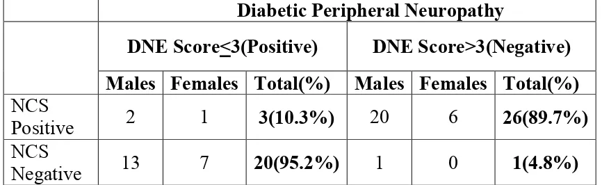
Nerve conduction studies (NCS) are the most objective noninvasive
measures of nerve function. They represent a valuable tool of evaluation
of neuropathy in large clinical and epidemiological studies. In clinical
practice, however, NCS should not be considered a substitute for careful
clinical examination, because NCS have many pitfalls and their results
must be interpreted in the context of clinical data. In the case small-fiber
polyneuropathies, the main drawback of NCS is that small myelinated
and unmyelinated nerve fibers, which are affected early in the disease
course of diabetic neuropathy, do not contribute to the sensory action
potential detected by routine NCS. The sensory action potential is altered
only after involvement of larger myelinated fibers, which is often a late
event in patients with diabetes. Electrophysiological data must, therefore,
always be evaluated in a clinical context.
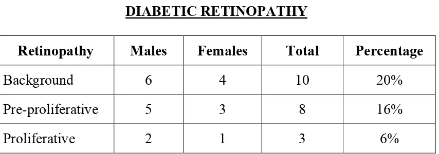
Diabetic retinopathy
Diabetic retinopathy is a well-characterized, sight-threatening,
chronic microvascular complication that eventually afflicts virtually all
patients with diabetes mellitus. Diabetic retinopathy is characterized by
gradually progressive alterations in the retinal microvasculature, leading
to areas of retinal non-perfusion, increased vasopermeability, and
Epidemiology of diabetic retinopathy
All patients with type 1 diabetes and more than 60% of patients
with type 2 diabetes develop some degree of retinopathy after 20 years. In
patients with type 2 diabetes, approximately 20% have retinopathy at the
time of diabetes diagnosis and most have some degree of retinopathy over
subsequent decades about 4% of patients younger than 30 years of age at
diagnosis and nearly 2% of patients older than 30 years of age at
diagnosis were legally blind.26 Approximately 25% of patients with type
1 diabetes have retinopathy after 5 years, with this figure increasing to
60% and 80% after 10 and 15 years, respectively.27
Pathophysiology
The earliest histologic effects of diabetes mellitus in the eye
include loss of retinal vascular pericytes (supporting cells for retinal
endothelial cells), thickening of vascular endothelium basement
membrane, and alterations in retinal blood flow. With increasing loss of
retinal pericytes, the retinal vessel wall develops outpouchings
(microaneurysms) and becomes fragile. With time, increasing sclerosis
and endothelial cell loss lead to narrowing of the retinal vessels, which
decreases vascular perfusion and may ultimately lead to obliteration of
the capillaries and small vessels the resulting retinal ischemia is a potent
development of new vessel growth and retinal vascular permeability.
Proliferating new vessels in diabetic retinopathy have a tendency to
bleed, which results in preretinal and vitreous hemorrhages and later
macular edema.
Risk factors
1. Duration of diabetes is closely associated with onset and severity
of diabetic Retinopathy.26
2. Lack of glycemic control.
3. Renal disease, as manifested by microalbuminuria and
Proteinuria.28
4. Hypertension.29
5. Elevated serum lipid levels are associated with extravasated lipid
in the retina (hard exudates) and visual loss.30
Clinical findings31
Clinical findings associated with early and progressing diabetic
retinopathy include hemorrhages or microaneurysms (H/Ma),
cotton-wool spots (CWSs), hard exudates, intraretinal microvascular
abnormalities (IRMAs), and venous caliber abnormalities (VCABs), such
as venous loops, venous tortuosity, and venous beading.
The intraretinal hemorrhages can be “flame-shaped” or “dot/blot”
tissue itself or shunt vessels through areas of poor vascular perfusion. It is
common for IRMAs to be adjacent to CWSs, which are caused by micro
infarcts in the nerve fiber layer. VCABs are a sign of severe retinal
hypoxia. In some cases of extensive vascular loss, however, the retina
may actually appear free of non-proliferative lesions. Such areas are
termed “featureless retina” and are a sign of severe retinal hypoxia.
Symptoms of diabetic retinopathy
1) Patient complaints of blurred vision, usually central vision and
metamorphosia as a result of maculopathy with foveal involvement.
2) Black spots, floaters or sudden visual loss may be experienced by
patients with vitreous hemorrhage, depending on quantum of bleed.
Kanski classification of diabetic retinopathy31 1. Background diabetic retinopathy
a. Haemorrhages (Dot and Blot, Flame shaped)
b. Microaneurisms (Located in inner nuclear layer)
c. Hard exudates (Lipoprotien and lipid filled macrophages.
Located within the outer plexiform layer).
d. Retinal edema (Located between outer plexiform and inner
Background Retinopathy : Retinal microaneurysms at the posterior pole; FA shows scattered hyperfluorescent spots in the posterior fundus
Pre-proliferative diabetic retinopathy : Cotton-wool spots, IRMA and venous changes
Severe disc new vessels Mild new vessels elsewhere
2. Pre proliferative
a. Vascular changes(beading , looping)
b. Dark blot haemorrhages
c. Cottonwool spots
d. Intraretinal microvascular abnormalities
e. Shunt vessels
3. Proliferative
a. Neo vascularisation
b. Fibrous proliferation
c. Vitreous detachment and haemorrhages
4. Maculopathy
a. Focal
b. Diffuse
c. Ischaemic
d. Clinically Significant Macular Oedema (CSMO)
Classification of diabetic maculopathy Intraretinal
• Macular edema
• Macular hard exudates
Comprehensive eye examination
Most of the blindness associated with advanced stages of
retinopathy can be averted with appropriate and timely diagnosis and
therapy.
Diabetic nephropathy Pathogenisis30
Like other microvascular complications pathogenesis of Diabetic
nephropathy is related to chronic hyperglycemia. The mechanisms by
which chronic hyperglycemia leads to ESRD are :
Yes Diabetes
Has patient had eye exam since diagnose of diabetes? Diabetes type Age >10 years Ophthaimic follow-up per patient's diabetic retinopathy level
Refer to eye care provider for
comprehensive eye exam Is patient pregnant? Is diabetes duration >5 years Initial opthaimic exam not yet
required
No Type 1 No
Type 2
No No
a) Soluble factors (Growth factors, Angiotensin II, Endothelin,
AGEs)
b) Hemodynamic alteration in renal microcirculation.
c) Structural changes in Glomerulus.
Because only 20-40% of patients with diabetes develop Diabetic
nephropathy, additional susceptible factors remain unidentified.
Epidemiology
Both type 1 and type 2 diabetes cause renal disease. Compared to
type 1, a slightly smaller and imperfectly defined proportion of type 2
patients progress to ESRD, but they represent more than 90% of those
receiving renal replacement therapy with the diagnosis of diabetes. The
distribution of renal disease due to type 2 diabetes is uneven among racial
groups. American Indians, African Americans, and Mexican Americans
have a greater incidence than non-Hispanic whites. Genetic
predisposition, environmental factors, delayed diagnosis of type 2
diabetes, and sub adequate medical care in minority groups contribute in
undefined amounts to such disparity.
Clinical features
Symptoms suggestive of renal impairment :
Oliguria, Anuria, Puffiness of face, Distension of abdomen, Pedal
Nephropathy in type 1 diabetes33
The course of diabetic nephropathy can be followed by two main
variables:
Proteinuria and GFR.
There are five distinct stages:
1. Stage 1: Glomerular hyper filtration and renal enlargement.
2. Stage 2: Early glomerular lesions or silent stage with normal albumin
excretion. Early glomerular lesions, consisting of glomerular
basement membrane thickening and mesangial matrix expansion,
characterize the second stage. Those structural changes appear 18 to
36 months after onset of type 1 diabetes and may become prominent
after 3.5 to 5 years. During this stage of morphologic changes,
microalbuminuria, seen only after exercise or during episodes of very
poor metabolic control.
3. Stage 3: Incipient diabetic nephropathy or micro albuminuric stage
Microalbuminuria, defined as 30-300mg per day in a 24 hour
collection or 30-300µg/mg of creatinine in spot collection, represents
the first laboratory evidence of diabetic renal disease. It is increased
by hypertension, strenuous exercise, fever, poor glycemic control, and
congestive heart failure. Therefore, a diagnosis of incipient diabetic
nephropathy is made only when Microalbuminuria is detected in at
4. Stage 4: Clinical or Overt diabetic nephropathy: proteinuria and falling
glomerular filtration rate:
Albuminuria greater than 300 µg/mg of creatinine relentless decline of
renal function, and hypertension define the fourth stage of diabetic
nephropathy. This stage, though variable, usually occurs 15 to 20
years after the onset of type 1 diabetes and after 5 or more years of
diagnosed type 2 diabetes. The amount of urinary protein can be as
little as 500 mg, but it can reach massive proportions, such as 20 to 40
g/24 hours.
5. Stage 5: End-stage renal disease.
Nephropathy in type 2 diabetes32, 33
Although renal structural changes and severity of target organ
damage are similar in both types of diabetes, delayed diagnosis has
complicated the construction of the natural history of diabetic renal
disease in type 2 diabetes. Hypetension more commonly accompanies in
MATERIALS AND METHODS
DESIGN
Cross sectional study
STUDY POPULATION
50 patients with type 2 diabetes attending diabetology OP in
Coimbatore medical college hospital from September 2009 to September
2010 were selected for this study after getting informed written consent.
INCLUSION CRITERIA
Patients were included for study ifthey had
o Onset of type 2 diabetes > 35 years of age.
o Duration of diabetes > 5 yrs.
EXCLUSION CRITERIA
Patients were excluded from study if they had
o Gross albuminuria.
o Non diabetic neuropathies.
o Non diabetic retinopathy and other chronic ophthalmological
illness.
Detailed history, complete clinical examination including body mass
index, blood pressure and neurological examination with special
nerve conduction studies for sensory and motor neuropathy, HbA1C,
serum total cholesterol, ophthalmolgical examination for diabetic
retinopathy and urine analysis for microalbuminuria was done for all
patients.
TECHNIQUES
Assessment and Definition of Diabetic Peripheral neuropathy
In this study the diabetic neuropathy examination (DNE) score is
used for assessment of distal symmetrical polyneuropathy.
DIABETIC NEUROPATHY EXAMINATION(DNE) SCORE Muscle strength
1. Quadriceps femoris: extension of the knee
2. Tibialis anterior: dorsiflexion of the foot
Reflex
3. Triceps surae
Sensation: index finger
4. Sensitivity to pinpricks
Sensation: big toe
5. Sensitivity to pinpricks
6. Sensitivity to touch
7. Vibration perception
Only the right leg and foot are tested.
Scoring from 0 to 2:
0 = Normal
1 = Mild/moderate deficit
• Muscle strength: Medical Research
Council scale 3–4
• Reflex: decreased but present
• Sensation: decreased but present 2 = Severely disturbed/absent
• Muscle strength: Medical Research Council scale 0–2
• Reflex: absent
• Sensation: absent
Maximum score: 16 points
A score of > 3 indicates presence of polyneuropathy.
Vibration Sensation: Vibration sensation is performed with the great toe unsupported. Vibration sensation will be tested bilaterally using a 128 Hz
tuning fork placed over the dorsum of the great toe on the boney
prominence of the DIP joint. Patients, whose eyes are closed, will be
asked to indicate when they can no longer sense the vibration from the
vibration from the hand-held tuning fork for 5 seconds longer on his
distal forefinger than a normal subject can at the great toe (e.g.
examiner’s DIP joint of the first finger versus patient’s toe). If the
examiner feels vibration for 10 or more seconds on his or her finger, then
vibration is considered decreased. A trial should be given when the tuning
fork is not vibrating to be certain that the patient is responding to
vibration and not pressure or some other clue. Vibration is scored as 1)
present if the examiner senses the vibration on his or her finger for < 10
seconds, 2) reduced if sensed for ≥10 or 3) absent (no vibration
detection.)
Muscle Stretch Reflexes : The ankle reflexes will be examined using an appropriate reflex hammer. The ankle reflexes should be elicited in the
sitting position with the foot dependent and the patient relaxed. For the
reflex, the foot should be passively positioned and the foot dorsiflexed
slightly to obtain optimal stretch of the muscle. The Achilles tendon
should be percussed directly. If the reflex is obtained, it is graded as
present. If the reflex is absent, the patient is asked to perform the
Jendrassic maneuver (i.e., hooking the fingers together and pulling).
Reflexes elicited with the Jendrassic maneuver alone are designated
“present with reinforcement.” If the reflex is absent, even in the face of
1 10-g Semmes-Weinstein monofilament
Monofilament Testing : For this examination, it is important that the patient’s foot be supported (i.e., allow the sole of the foot to rest on a flat,
warm surface). The filament should initially be prestressed (4-6
perpendicular applications to the dorsum of the examiner’s first finger).
The filament is then applied to the dorsum of the great toe midway
between the nail fold and the DIP joint. Do not hold the toe directly. The
filament is applied perpendicularly and briefly, (<1 second) with an even
pressure. When the filament bends, the force of 10 grams has been
applied. The patient, whose eyes are closed, is asked to respond yes if
he/she feels the filament. Eight correct responses out of 10 applications is
considered normal: one to seven correct responses indicates reduced
sensation and no correct answers translates into absent sensation.
Electrophysiological studies will be performed in all cases. The
parameters recorded included distal latencies, amplitudes of compound
motor action potentials (CMAP), duration of CMAP, F wave latencies
and conduction velocities in motor nerves. In sensory nerves, latencies
and amplitudes of the sensory nerve action potentials and their
conduction velocities were documented.
The presence or absence of neuropathy in these subjects was defined
5. Sural sensory and peroneal motor NCSs are performed in one lower
extremity. Taken together, these NCSs are the most sensitive for
detecting a distal symmetric polyneuropathy. If both studies are
normal, there is no evidence of typical distal symmetric
polyneuropathy. In such a situation, no further NCSs are necessary.
6. If sural sensory or peroneal motor NCSs are abnormal, the
performance of additional NCSs is recommended. This should
include NCS of at least the ulnar sensory, median sensory, and ulnar
motor nerves in one upper extremity. A contralateral sural sensory
and one tibial motor NCS may also be performed according to the
discretion of the examiner. Caution is warranted when interpreting
median and ulnar studies since there is a possibility of abnormality
due to compression of these nerves at the wrist or ulnar neuropathy at
the elbow.
7. If a response is absent for any of the nerves studied (sensory or
motor), a NCS of the contralateral nerve should be performed.
8. If a peroneal motor response is absent, an ipsilateral tibial motor NCS
should be performed.
The minimum case definition criterion for electrodiagnostic
confirmation of distal symmetric polyneuropathy is an abnormality
(≥99th or ≤1st percentile) of any attribute of nerve conduction in two
Assessment and Definition of Diabetic microvascular Complications Direct ophthalmoscopic examination of fundus
Fundus examination was done and results will be classified as
normal, background, pre-proliferative and proliferative retinopathy. It
was confirmed by a senior ophthalmologist.
Microalbuminuria detection was done by Albumin Creatinine Ratio estimation. Urinary albumin measured by rate Nephelometry and Urinary
Creatinine measured by modified Jaffe’s method. Albumin Creatinine
Ratio of 30-300 µg/mg of creatinine was defined as microalbuminuria;a
ratio greater than 300 µg/mg of creatinine was defined as
macroalbuminuria.
Assessment and Definition of various risk factors
The specific risk factors studied were age, sex, duration of
diabetes, HbA1C, BMI, systemic hypertension and total serum
cholesterol. In the present study the definition of systemic hypertension
was taken as subjects with either a history of systemic hypertension on
treatment or whose blood pressure measurement shows >140/90 mm
Hg in two different occasions .
Total serum cholesterol >200mg/dl was taken as positive. BMI was
calculated as
(
)
2Weight in Kg
Height in mt and the normal BMI was 18.5-24.9. HbA1C
measured by bidirectionally interfaced fully automated turbidometry by
RESULTS AND ANALYSIS
AGE
The mean age of the study subjects was 51.66 years; standard
deviation (SD) of 11.03. Most of the patients belonged to the age group
35-44 yrs
Sex
Out of 50 cases, 36 were males and 14 were females. The ratio of
[image:64.595.102.534.386.588.2]males to females was 2.6:1
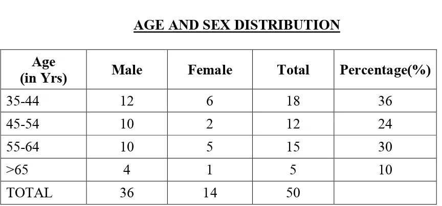
TABLE 1
AGE AND SEX DISTRIBUTION Age
(in Yrs) Male Female Total Percentage(%)
35-44 12 6 18 36
45-54 10 2 12 24
55-64 10 5 15 30
>65 4 1 5 10
TOTAL 36 14 50
18 patients (36%) in the study were in the age group of 35 – 44
years. Of these 12 were males and 6 were females. 12 patients (24%)
were in the age group of 45 – 54 years. Of these 10 were males and 2
were females. 15 patients (30%) in the study were in the 55 – 64 age
in the age group of>65 years, of these 4 were males and 1 was female.
[image:65.595.112.521.181.505.2]Most patients were between the age group of 35-44 years.
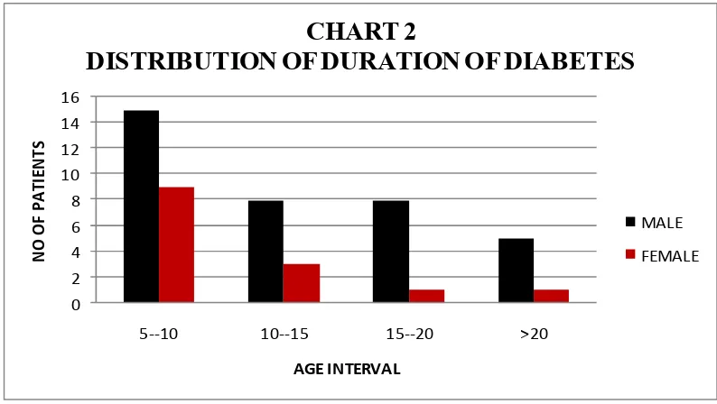
TABLE 2
DISTRIBUTION OF DURATION OF DIABETES MELLITUS Duration of
Diabetes (Yrs) Male Female Total (%)
5-10 15 9 24(48%)
10-15 8 3 11(22%)
15-20 8 1 9(18%)
>20 5 1 6(12%)
TOTAL 36 14 50
0 2 4 6 8 10 12 14
35 - 44 45 - 54 55 - 64 >65
N o o f p a ti e n ts Age intervals CHART I
AGE AND SEX DISTRIBUTION
Male
In the study population, more number of patients (24 i.e 48%) were
having diabetes with duration 5-10 years, followed by 11 patients (22%)
with the duration of 10 to 15 years, next 9 patients(18%) between 15 to
20 years and only 6 patients(12%) were having duration of diabetes >20
[image:66.595.116.513.74.298.2]yrs.
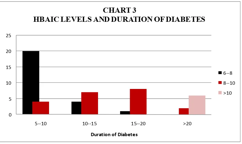
TABLE 3
HbA1C AND DURATION ON DIABETES
Duration of Diabetes (Years) HbA1C 6-8% (No. of patients) 8-10% (No. of patients) >10% (No. of patients)
Total Average HbA1C
5-10 20 4 0 24 7.44
10-15 4 7 0 11 8.27
15-20 1 8 0 9 9.22
>20 0 2 4 6 11.13
TOTAL 25 21 4 50
0 2 4 6 8 10 12 14 16
5--10 10--15 15--20 >20
N O O F P A T IE N T S AGE INTERVAL CHART 2
DISTRIBUTION OF DURATION OF DIABETES
MALE
Among the 4 patients who had glycosylated hemoglobin >10% all
had duration of diabetes >20 years. The average glycosylated hemoglobin
(HbA1C) was 7.44,8.27,9.22 and 11.33 for duration of diabetes 5-10 yrs,
10-15 , 15-20 , >20years respectively. This shows that those patients with
[image:67.595.113.513.73.311.2]duration of diabetes more than 10 years have poorer control.
TABLE 4
BODY MASS INDEX
BMI(Kg/M2) Males Females Total Percentage (%)
18.5-24.9 30 10 40 80%
25-29.9 4 4 8 16%
30-34.9 2 0 2 4%
>35 0 0 0 0
0 5 10 15 20 25
5--10 10--15 15--20 >20
Duration of Diabetes
CHART 3
HBAIC LEVELS AND DURATION OF DIABETES
6--8
8--10
TABLE 5
HYPERTENSION AND DURATION OF DIABETES Duration of
Diabetes (Years)
Number of Hypertensive Patients
Male Female Total (%)
6-10 6 1 7 33%
11-15 4 1 5 24%
16-20 4 1 5 24%
>20 3 1 4 19%
TOTAL 17 4 21
All patients with history of hypertension on antihypertensives
and/or whose blood pressure while clinical examination is > 140/90 mm
Hg on two separate occasions is included in Hypertensive group. Out of
total of 50 patients included in study 21 patients were found to be
hypertensive. Out of it 17 were males and 4 were females. Out of these 21
patients 33%,24%,24% and 19% were having duration of diabetes
6-10,11-15,16-20,>20 years respectively.
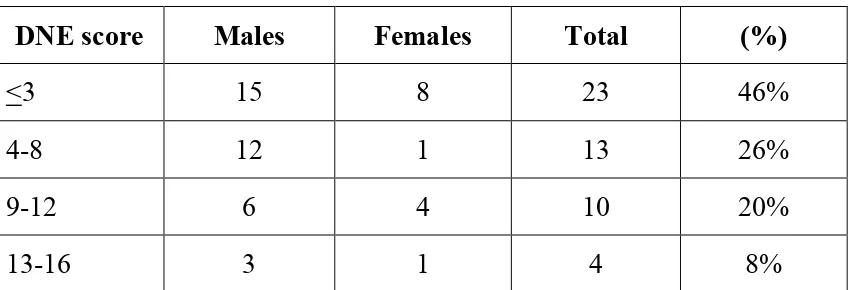
TABLE 6
DISTRIBUTION OF PATIENTS ACCORDING TO DNE SCORES DNE score Males Females Total (%)
<3 15 8 23 46%
4-8 12 1 13 26%
9-12 6 4 10 20%
According to definition those patients with DNE score > 3 is having
diabetic peripheral neuropathy (DSPN).
DNE score Male Female Total (%)
<3 15 8 23 46%
>3 21 6 27 54%
So, in this study out of total 50 patients, 27 (54%)patients were
found to have peripheral neuropathy. Among them 21 were males and 6
were females. Out of total 36 males in the study 21 patients(58%) had
DNE scores >3 and out of total 14 females in the study 6 patients (43%)
had DNE scores >3.
23
27
0 5 10 15 20 25 30
DNE <3 DNE>3
CHART 4
DISTRIBUTION OF PATIENTS ACCORDING TO DNE SCORES
MALE
FEMALE
TABLE 7
TOTAL SERUM CHOLESTEROL
Total Cholesterol