Copyrightq1995, American Society for Microbiology
Negative Regulation of the Adeno-Associated Virus (AAV) P
5Promoter Involves both the P
5Rep Binding Site and the
Consensus ATP-Binding Motif of the AAV Rep68 Protein
SIRKKA R. M. KYO¨ STIO¨ , RAMANI S. WONDERLING,ANDROLAND A. OWENS*
Laboratory of Molecular and Cellular Biology, National Institute of Diabetes and Digestive and Kidney Diseases, Bethesda, Maryland 20892
Received 7 April 1995/Accepted 28 July 1995
Transcript levels from the P5promoter of adeno-associated virus type 2 (AAV) are negatively regulated by
the AAV Rep78 and Rep68 proteins in the absence of helper virus. We have identified a Rep-responsive negative ciselement of the P5promoter between the P5TATA box and transcription start site by using 5*and 3*deletions
of the P5promoter fused to the chloramphenicol acetyltransferase gene. This element contains four imperfect
GAGC repeats similar to the Rep recognition sequences (RRSs) in the AAV inverted terminal repeats and in the AAV preferred integration locus in chromosome 19. Band shift analyses showed that human 293 cell nuclear extracts containing Rep68 or Rep68/K340H, a putative nucleoside triphosphate (NTP)-binding-site mutant of Rep68, formed Rep-specific complexes with this P5RRS DNA. Within the P5RRS, mutation of a
cytosine at position 273 in the AAV sequence to guanine abolished Rep68 binding to the DNA. A mutation in the P5RRS within a full-length AAV genome, which abolished Rep binding, resulted in a 40 to 50% reduction
in the ability of wild-type Rep68 to inhibit the accumulation of P5 transcripts in vivo. In contrast, the
Rep68/K340H mutant was unable to down-regulate this mutated promoter. These results indicate that there are at least two mechanisms involved in the negative regulation of P5transcript levels by Rep68; one involves
Rep68 binding to the P5RRS, and another requires the region of Rep68 containing the consensus NTP-binding
motif. Furthermore, our studies of AAV genomes containing mutated RRS- and/or YY1-binding elements suggest that transcription factor YY1 binding to the transcription start site of P5 interferes with Rep68
repression of the P5promoter.
Adeno-associated virus type 2 (AAV) is a nonpathogenic human parvovirus that requires coinfection with adenovirus or herpesvirus as a helper virus for efficient replication (5). In the absence of the helper virus, the AAV DNA integrates into the host genome and is maintained as a latent provirus. The linear single-stranded viral genome of 4.7 kb contains two open read-ing frames (ORFs) flanked by inverted terminal repeats (ITRs) at either end (6, 50). The first ORF encodes the nonstructural (NS) Rep proteins; the larger Rep proteins, Rep78 and Rep68, are made from transcripts initiating from the P5 promoter, while the smaller Rep proteins, Rep52 and Rep40, are ex-pressed from the downstream P19promoter (6, 37, 51, 55). The second ORF of AAV encodes the viral capsid proteins (VP-1, VP-2, and VP-3) expressed from the P40promoter (50, 53).
The AAV Rep proteins have multiple functions and are involved in both the replication and gene regulation of the virus. Several of the known properties of the Rep proteins, such as the binding to the ITR DNA, the ATP-dependent site-specific endonuclease, and the DNA helicase activities, are important for its replication function (21–23, 49). The Rep proteins also contain a consensus nucleoside triphosphate (NTP)-binding motif which is involved in the endonuclease, helicase, and ATPase activities of Rep78 and Rep68 (10, 12, 34, 42, 57). This motif has also been shown to be necessary for the cell cytotoxicity by the analogous NS proteins of the
au-tonomous parvoviruses B19 and minute virus of mice (MVM) (28, 38). The Rep functions important for gene regulation have been less well characterized and may vary depending on the presence or absence of the helper virus. In the absence of a helper virus, the larger Rep proteins, Rep78 and Rep68, effi-ciently inhibit the production of P5and P19transcripts, while Rep52 and Rep40 have a lesser effect on these transcript levels (25). A mutant Rep78 with an alteration of a lysine to a histidine in the putative NTP-binding site (K340H) retains the ability to decrease P5 mRNA levels but lacks the ability to down-regulate P19mRNA levels (25). The presence of helper virus results in the activation of AAV promoters by Rep pro-tein(s) (2, 26), suggesting interaction with or modification of Rep proteins by host or helper virus proteins. Mutation of two amino acids (T341I and N342Y) in the putative NTP-binding site of Rep abolished the ability of the AAV Rep proteins to activate the AAV P19 and P40 promoters in the presence of helper virus (34). Furthermore, the ability to activate promot-ers by an analogous NS protein of MVM was also dependent on the conserved lysine in the NTP-binding site, since its change to serine abolished MVM P38activation (24).
The complexity of the AAV life cycle is reflected in the regulation of the viral promoters by proteins produced by the helper virus and the host cell, as well as by AAV. Conse-quently, several known and putative cis elements for both viral and host trans activators and repressors have been localized in the AAV P5promoter. These include cis elements such as the binding sites for CREB/ATF (17, 29) and major late transcrip-tion factor (MLTF; USF) (8) in the region upstream of the P5 TATA box. The P5promoter also contains two binding sites for the host cell transcription factor YY1 (46, 47). One is located upstream from the TATA box at260 (in relation to the
tran-* Corresponding author. Mailing address: Laboratory of Molecular and Cellular Biology, National Institute of Diabetes and Digestive and Kidney Diseases, Bldg. 8, Rm. 309, National Institutes of Health, 8 Center Drive MSC 0840, Bethesda, MD 20892-0840. Phone: (301) 496-3359. Fax: (301) 402-0053. Electronic mail address: rolando@ bdg8.niddk.nih.gov.
6787
on November 9, 2019 by guest
http://jvi.asm.org/
scription start site), where YY1 binding results in repression of P5expression in the absence of helper virus (47). In the pres-ence of E1a protein produced by the coinfecting adenovirus, this site mediates transcription activation of the P5promoter by a direct E1a-YY1 interaction (47). Binding of YY1 to the second binding site located at the transcription start site (11) enhances and directs the accurate positioning of P5transcript initiation (46). Recently, a Rep68-binding site has been located just upstream of the11 YY1-binding site, although its function in AAV gene regulation was not characterized (35).
In this study, the objective was to analyze the mechanism of the repression of the P5promoter by Rep68. The Rep-respon-sive negative cis element within the P5promoter was localized between the P5TATA box and transcription initiation site and matches the recently reported Rep-binding sequence (35). We also report a mutational analysis of this binding site and its functional significance in the context of the full-length AAV genome during conditions nonpermissive for AAV replication.
MATERIALS AND METHODS
Cells and viruses.Adenovirus-transformed human embryonic kidney 293-31 cells (293 cells) (18) were maintained as monolayer cultures and grown in Eagle’s minimal essential medium (Quality Biological, Inc., Gaithersburg, Md.) supple-mented with 10% fetal calf serum (Inovar, Gaithersburg, Md.), 2 mML -glu-tamine, and 100 U each of penicillin, neomycin, and streptomycin (Life Tech-nologies, Gaithersburg, Md.) per ml.
Plasmids.The AAV plasmids used in this study were constructed by using standard techniques (44). Plasmid pHIVrepamD(encoding the protein desig-nated Repam) is similar to pHIVrepam described previously (1) except that the right-hand ITR has been deleted by SnaBI-SphI digestion. The overhangs of the larger fragment were then filled in by T4 DNA polymerase, and the blunt ends were religated. The right-hand ITR was also deleted from plasmid pSK9, which contains the AAV Rep68-coding region expressed from the long terminal repeat (LTR) promoter of human immunodeficiency virus type 1 (HIV-1) (42). pSK9/ NTP is derived from pSK9 and contains the same mutation in the region encod-ing the NTP-bindencod-ing site as pHIVrepNTP (42).
The plasmids containing the 39deletion series of P5-chloramphenicol
acetyl-transferase gene (cat) fusions (Fig. 1A) were derived from plasmid pAV2, which contains the entire AAV genome (27). pAV2 was cleaved with PvuII and SphI and religated in order to delete the NruI site in the pBR322-derived region. A 2.4-kb fragment downstream from the P40promoter was removed by cleavage
with HindIII and KpnI and replaced with the HindIII-KpnI fragment of plasmid pTS1 (52), which contained the cat gene. The resulting plasmid was cleaved with
NruI at AAV nucleotide 658 and then digested with BAL 31 exonuclease.
Aliquots taken at various times during the BAL 31 treatment were cut with
HindIII, and the termini were filled in with the Klenow fragment of DNA
polymerase and blunt-end ligated. Of the plasmids generated, two in which a
HindIII site had been reconstituted, plasmids pYT45 (17) and pYT36, were
identified. The SnaBI-NdeI fragments of pYT45 and pYT36, which included the right-hand ITR, were deleted to produce pRO45 (17) and pRO36, respectively. pRO45 and pRO36 contain AAV nucleotides 1 to 263 and 1 to 505, respectively, ligated to the cat gene. pRO1472 was made by inserting an annealed synthetic oligonucleotide pair (AAV nucleotides 266 to 321) flanked by HindIII-compat-ible ends into HindIII-cleaved pRO45 (17). Two tandem repeats of AAV nucle-otides 266 to 287 with HindIII-compatible ends were inserted into the HindIII site of pRO45 to make pRO1511. An insertion of a single copy of the same oligonucleotide pair, but in the reverse orientation, resulted in pRO1517. Be-cause of the presence of the HindIII site, pRO1472 and pRO1511 have nucle-otides 264 and 265 changed from CC to TT. An oligonucleotide pair containing AAV nucleotides 287 to 321 with HindIII-compatible ends was inserted into the
HindIII-cleaved pRO45 in the correct orientation to make pRO1535 or in the
reverse orientation to make pRO1538.
pNTC244 and pNTC3 (see Fig. 5A) contain a wild-type AAV genome and an AAV genome with an amber mutation in the rep gene, respectively, cloned into pTZ19U (Bio-Rad, Melville, N.Y.), and they have been described before (9). Mutations between the TATA box and the transcription initiation site in pNTC3 and pNTC244 were created by PCR. A 59primer (59 GGCCCGAGATATC GATCAGGGTCTCCATTTTG 39) containing the native AvaI site and a novel
EcoRV site (mRRS-3 [RRS5Rep recognition sequence]) and a 39primer located at AAV positions 1062 to 1085, downstream from a BamHI site, were used to synthesize a 0.8-kb fragment. This fragment was cleaved with AvaI and
BamHI, gel purified, and then cloned into pNTC3 that had been partially cleaved
with AvaI and BamHI. A double mutation containing both the EcoRV site (mRRS-3) and an altered11 YY1 site that abolishes YY1 binding (47) was constructed similarly except that the 59primer (59GGCCCGAGATATCGAT CAGGGTCTAAATTTTGAAGC 39) additionally contained mutations at
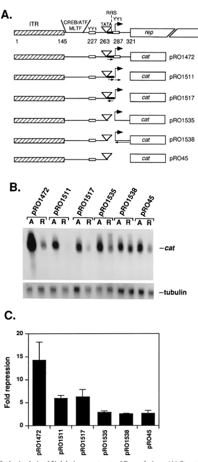
nucle-FIG. 1. Analysis of 39deletion constructs of P5-cat fusions. (A) Structures of
the 39deletion constructs of P5-cat fusions. Numbers indicate positions in the
AAV nucleotide sequence. ITR (hatched box), binding sites for CREB, ATF, MLTF, YY1 (at260 and11), and Rep68 (RRS), TATA box, transcription start site (bent arrow), and the beginning of the rep ORF (position 321) are shown. The short arrows indicate the number of copies and the orientation of the region between the TATA box and transcription start site. The long arrow indicates the reversal of the region between the transcription start site and the beginning of the rep ORF. (B) Northern analysis of RNA from cells transfected with the P5-cat
constructs. The P5-cat constructs (10mg) indicated at the top were cotransfected
with plasmids (5mg) producing either Repam (lanes A) or Rep68 (lanes R) into human 293 cells. The cytoplasmic RNA was isolated after 48 h and analyzed by Northern blotting using a32
P-labeled cat fragment. Hybridization for tubulin gene transcripts was performed to indicate the intactness of the RNA. The RNA blot shown represents one of the three independent experiments performed. (C) Quantitation of P5repression by Rep68, using 39deletion constructs. Three
independent transfection experiments were performed and analyzed by Northern blotting. The cat transcripts were cut and counted, and the values were normal-ized for transfection efficiency by values obtained from DNA slot blots (P5-cat
constructs). The level of repression is expressed as fold repression by comparing the unrepressed (in the presence of Repam) and repressed (in the presence of Rep68) levels. Error bars indicate standard deviations between experiments.
on November 9, 2019 by guest
http://jvi.asm.org/
[image:2.612.333.535.73.543.2]otides 285 and 286 (cytosines to adenines) of the AAV nucleotide sequence. A derivative of the pNTC3 construct with the same altered YY1-binding site but a wild-type P5RRS was also created by using a 59 primer (59GGCCCGAGT
GAGCACGCAGGGTCTAAATTTTG 39) with this mutation.
For electrophoretic mobility shift analyses, several double-stranded oligonu-cleotides containing flanking BamHI overhangs (59GATC 39) were cloned into
BamHI-digested pBlueScriptII SK1(pBluescript; Stratagene, La Jolla, Calif.). These include the P5RRS (AAV nucleotides 259 to 285), various mutant forms
of the P5RRS (with AAV cytosines 265, 273, and/or 277 changed to guanines)
(see Fig. 4), P19(AAV nucleotides 847 to 874), the HIV-1 LTR (40) (130 to154
relative to the mRNA start site), and a series of oligonucleotide pairs (AAV nucleotides 259 to 298) containing wild-type or mutant forms of the P5RRS and
the11 YY1-binding site (see Fig. 5A). The YY1-binding-site mutations were designed such that they abolish YY1 binding to the DNA (47). For competition assays, double-stranded oligonucleotides flanked by BamHI overhangs and con-taining only the wild-type (59AGGGTCTCCATTTTGAAGCGG 39) or mutated (59AGGGTCTAAATTTTGAAGCGG 39)11 YY1-binding site were cloned into pBluescript.
The presence of the mutations or cloned double-stranded oligonucleotides in all constructs was confirmed by DNA sequencing (45) using a Sequenase DNA sequencing kit (United States Biochemical Corp., Cleveland, Ohio). All plasmids used for transfections were grown in Escherichia coli DH5 (Life Technologies, Inc.), prepared by alkaline lysis and purified on cesium chloride gradients (44).
DNA transfections.Transfections for RNA analyses were performed by the calcium phosphate coprecipitation method (19). For transfections, 6 to 10mg of the template plasmids and 3 to 5mg of rep plasmids were used as indicated in the figure legends. All transfections were adjusted with vector DNA (pBR322) to contain a total of 15mg of DNA. Human 293 cells were plated on 100-mm-diameter dishes at a density of 33106cells per plate 24 h prior to transfection.
All cells were harvested 48 h after transfection and used for RNA isolation.
RNA isolation and Northern (RNA) analysis.Cytoplasmic RNA was isolated and analyzed as described previously (25). The 0.7-kb HindIII-ScaI fragment of pRO5 and tubulin DNA (Oncor, Inc., Gaithersburg, Md.) were used for the detection of cat and tubulin gene transcripts, respectively. For the detection of
rep transcripts, a 1.6-kb HindII fragment of pNTC3 was used. The fragments
were labeled by the random-priming method (16), using [a-32
P]dCTP (specific activity, 3,000 Ci/mmol; DuPont NEN, Boston, Mass.) and a random-primer labeling kit (Boehringer Mannheim, Indianapolis, Ind.). The transcript levels were quantitated by cutting and counting the radioactive signals in a liquid scintillation counter. These values were normalized by plasmid uptake into the nuclei, as measured by DNA slot blot analysis (25).
Primer extensions.Ten micrograms of cytoplasmic RNA was mixed with 40 pmol of32
P-labeled primer (59GCCATTGGGATATATCAACGGTGGT 39) complementary to the cat gene (105cpm) in primer extension buffer (50 mM
Tris-HCl [pH 8.3], 50 mM KCl, 10 mM MgCl2, 10 mM dithiothreitol, 1 mM each
dNTP, 0.5 mM spermidine). The primer was annealed by heating the tubes at 708C for 10 min followed by slow cooling to room temperature. The volume was doubled to 20ml by adding primer extension buffer, sodium pyrophosphate to a final concentration of 2.8 mM, and 2 U of avian myeloblastosis virus reverse transcriptase (Promega, Madison, Wis.). The reaction mixtures were incubated at 428C for 1 h, after which 20ml of loading dye was added. Samples were heated at 908C for 10 min and loaded on an 8% polyacrylamide sequencing gel. A
32
P-end-labeled primer or [35
S]dATP (DuPont NEN) was used for the labeling of sequencing reactions that served as DNA size markers.
Gel mobility shift assays.Annealed oligonucleotide pairs or oligonucleotide pairs cloned into pBluescript were used in band shift analyses. The plasmids containing the double-stranded oligonucleotides were cut with XbaI and HindIII to release a 70- to 90-bp fragment containing the oligonucleotide pair flanked with DNA sequences of the multiple cloning site of pBluescript. Both the an-nealed oligonucleotide pairs and the restriction fragments were labeled with Klenow polymerase and [a-32
P]dCTP (3,000 Ci/mmol; DuPont NEN). The la-beled fragments were then purified on a 6% polyacrylamide gel. Nuclear extracts (600 mM NaCl) of 293 cells transfected with pHIVrepam, pSK9, or pSK9/NTP were prepared as described previously (42). The protein concentrations were equalized with extraction buffer, and the samples were diluted 10-fold in buffer A (25 mM Tris-HCl [pH 7.5], 0.1 mM EDTA, 1 mM dithiothreitol, 0.1 mM phenylmethylsulfonyl fluoride, 20% [vol/vol] glycerol) to reduce the NaCl con-centration. Labeled DNA (0.02 or 0.002 pmol) was incubated with 0.44mg of nuclear extract protein for 20 min at 48C in a 20-ml volume containing 5.5mg of acetylated bovine serum albumin (BSA; New England Biolabs, Beverly, Mass.), 50 mM NaCl, 25 mM N-2-hydroxyethylpiperazine-N9-2-ethanesulfonic acid (HEPES)-KOH (pH 7.5), 10 mM MgCl2, 1 mM dithiothreitol, 2% (vol/vol)
glycerol, 0.01% (vol/vol) Nonidet P-40, and 1mg of poly(dI-dC) (Boehringer Mannheim). A rabbit antibody against the S18K oligopeptide (Rep78 amino acids 516 to 533) was added in a volume of 1ml where indicated (37). In some experiments, various amounts of ATP were added to the mixture. The samples were electrophoresed in a 4% polyacrylamide gel as described previously (21) except that the gel was run at 48C.
RESULTS
Localization of a negative Rep-responsiveciselement in the AAV P5 promoter.The AAV Rep proteins have been
previ-ously shown to inhibit transcript accumulation from the AAV P5promoter in 293 cells in the absence of the helper virus (25). To localize a negative Rep-responsive element in the AAV P5 promoter, several 39and 59deletions of the P5promoter and upstream regions were constructed and fused to the cat gene (Fig. 1A and data not shown). The constructs were tested in human embryonic kidney cells (293 cells) which have been immortalized with the adenovirus E1a and E1b genes (18). Expression of these adenovirus proteins enhances the levels of AAV P5 and P19 transcripts but is not sufficient for AAV replication in the absence of the helper virus (8, 27). The effect of Rep proteins on cat transcript levels from the P5-cat fusions was measured by Northern analysis after cotransfection of the fusion plasmids with plasmids producing either wild-type Rep68 (pSK9) or a truncated Repam protein (pHIVrepam) as a negative control. The cat transcript levels were normalized for transfection efficiency by measuring the template (P5-cat fusions) uptake in DNA slot blots. CAT activities were not measured because the Rep proteins may negatively regulate translation of the cat mRNA (54). Similarly, expression of a control reporter gene was not used because of the effects of Rep proteins on heterologous promoters.
We first analyzed the effects of Rep68 on the 39 deletion constructs of the P5promoter fused to the cat gene (Fig. 1B and C). Plasmids pRO1472, pRO1511, and pRO1517 all con-tained the previously identified Rep68-binding site between the P5TATA box and the transcription start site (35) and were all clearly down-regulated approximately 6- to 14-fold, depend-ing mainly on the level of unrepressed transcription. Further-more, the orientation of the region between the P5TATA box and the transcription start site did not appear to be critical, since in construct pRO1517, this region was reversed. Further deletions from the 39 direction (pRO1535, pRO1538, and pRO45), which deleted the Rep68-binding site, however, abol-ished efficient down-regulation (to threefold or less) by Rep68 without changing the unrepressed level of expression com-pared with pRO1511 and pRO1517 (Fig. 1B). These results suggested that the region between the TATA box and tran-scription start site containing the Rep68-binding site was re-quired for efficient Rep68 repression. However, the remaining repression activity by Rep68 suggested that other factors be-sides this cis region may also be involved.
We also analyzed the effects of Rep68 on the 59 deletion constructs, all of which contained the known Rep68-binding site between the TATA box and transcription start site (35). The Northern analysis indicated that all of the 59 deletion constructs (deletions up to AAV base 217) tested were down-regulated to some degree by the Rep68 protein (data not shown). However, the differences in fold repression were strongly influenced by the differences in the unrepressed levels of expression, and thus comparison of repression efficiencies of the various constructs was difficult. The results showed that the AAV ITR and several upstream cis elements such as binding sites for CREB, ATF, or MLTF (USF) were not required for the negative effect by Rep68. Furthermore, a construct con-taining a deletion to just upstream of the260 YY1-binding site (AAV nucleotide 217) and a mutation in the260 YY1-binding site (47) had reduced mRNA levels but was still repressed by Rep.
Since several of the 39 deletion constructs contained an altered DNA sequence around the transcription start site, due to the deletions and reversals, the initiation sites of cat
on November 9, 2019 by guest
http://jvi.asm.org/
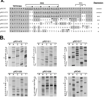
scripts from these constructs were analyzed by primer exten-sion using a primer complementary to the cat gene (Fig. 2). Previous results have located the start site of the P5mRNA at nucleotide 287 of the AAV sequence (32). Our analyses showed that in constructs (pRO36, pRO1472, and pRO1511) containing the native AAV transcription initiation site, which is normally within a known YY1-binding site (46), single major transcripts were initiated accurately at the correct AAV start site 26 bp downstream from the end of the P5TATA box at 261. The alteration in the YY1-binding site in pRO1517 re-sulted in the appearance of two equally strong start sites lo-cated one nucleotide from each other. The effect of muta-tions at the 11 YY1-binding site resulting in multiple start sites has been reported before (46). Interestingly, the lack of the normal initiator sequence in the other constructs (pRO1535, pRO1538, and pRO45) did not appear to decrease the levels of the cat mRNA relative to pRO1511 (Fig. 1B) or result in multiple strong start sites (Fig. 2B). With pRO1535 and pRO1538, transcription was initiated consistently 23 bp from the TATA box, using either purines or pyrimidines as starting nucleotides. Similarly, with the pRO45 construct,
which lacks all of the native AAV sequence downstream from the P5TATA box, the transcript initiated 23 bp downstream of the TATA box (13 bp upstream of the ATG of the cat gene). These results indicated that the P5 repression by Rep68 re-quired the Rep68-binding region between the TATA box and the mRNA initiation site (AAV nucleotides 265 to 287). The alterations in the YY1-binding site at the transcription start site also correlated with the reduced repression by Rep68, suggesting that Rep-mediated repression might be affected by host transcription factor YY1 binding to the transcription start site (Fig. 2A). Therefore, we next analyzed the properties and the significance of Rep68 binding to the P5promoter as well as the role of YY1 binding to the P5transcript start site.
Wild-type and NTP-mutant Rep68 proteins bind to the re-gion between the P5TATA box and the transcription initiation
[image:4.612.120.486.75.415.2]site.The region between the P5TATA box and transcription initiation site that was important for the P5negative regulation by Rep68 contained four imperfect GAGC repeats. These repeat sequences are similar to those of the GAGC repeats observed previously in the RRSs identified in the AAV ITRs and within the preferred AAV integration locus on human
FIG. 2. Mapping of transcription start sites in 39deletion constructs of P5-cat fusions and their locations with respect to the P5TATA box. (A) Summary of primer
extension results with P5-cat constructs. The DNA sequence in the P5promoter between the TATA box and transcription start site are shown (AAV positions 255 to
292). Some constructs contain additional changes due to deletions and reversals of sequences (see Fig. 1A). Regions identical to the wild-type AAV sequence are shaded. The wild-type AAV start site at 287 is shown as a bent arrow; the vertical arrows indicate the major transcript start sites determined by three primer extension assays. The horizontal arrows show the positions of the native or serendipitous GAGC boxes. The YY1-binding site in the transcription initiation site is also indicated. On the right side are shown the relative levels of repression by Rep68 on each construct. (B) Primer extension analysis of P5-cat constructs. All nucleotides detected
as start sites for transcripts are underlined; the major start sites are indicated by asterisks. Cytoplasmic RNA from transfections containing the P5-cat constructs (10
mg) indicated and pHIVrepam (5mg) was annealed to a primer complementary to the cat gene. The primer extension products (lane P) were run on an 8% polyacrylamide sequencing gel, and the start site was localized by using standard sequencing reactions (lanes A, C, G, and T) as DNA markers.
on November 9, 2019 by guest
http://jvi.asm.org/
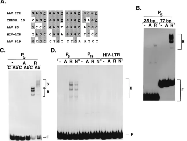
chromosome 19, which have been shown to be bound by Rep proteins (Fig. 3A) (12, 56). Furthermore, McCarty et al. (35) have recently demonstrated that the P5RRS can also be bound by purified Rep68 produced in Sf9 insect cells. Since our ex-periments were performed in human 293 cells, we analyzed the abilities of Rep proteins in 293 cell nuclear extracts to bind to this P5sequence. The band shift reaction mixtures contained equal amounts of nuclear proteins, and results of Western blot (immunoblot) analyses indicated that the concentrations of Rep68 and Rep68/K340H were within twofold of each other within their respective extracts (not shown).
The gel mobility shift analyses with Rep68-containing ex-tract and an annealed oligonucleotide pair containing the P5 RRS showed a Rep68-specific shifted band which was not seen with Repam-containing extract (Fig. 3B). This 35-bp double-stranded oligonucleotide produced a single Rep68-specific shifted band and thus did not produce the characteristic mul-tiple shifted band pattern observed previously for Rep68 bind-ing to the RRS sequences within the ITR or chromosome 19 (42, 56). However, when the annealed oligonucleotide pair was cloned into pBluescript and isolated within a larger fragment (77 bp), the formation of the Rep68-specific DNA-protein complexes was enhanced and the typical multiple band pattern was detected (Fig. 3B). The effect of the length of the fragment in stabilizing Rep binding has also been observed with the linear ITR DNA by using a purified maltose-binding protein–
Rep68 fusion protein (13). Consequently, all of the oligonu-cleotide pairs analyzed in band shift analyses were cloned into pBluescript and tested as larger fragments. The retarded com-plexes contained Rep protein(s), as the shifted bands were supershifted by an anti-Rep antibody (Fig. 3C). The specificity of the anti-Rep antibody has been shown before (42). Further-more, the Rep68-dependent shift was specific for the P5RRS (region between the TATA box and the transcription initiation site), since a region from the P19promoter between the TATA box and the mRNA start site or a Rep-responsive region from the HIV-1 LTR (40) produced no Rep68-specific shifted bands (Fig. 3D). Nuclear extracts containing Rep68/K340H produced Rep-specific shifted bands with P5 DNA similar in intensity and pattern to the bands produced by wild-type Rep68. This result indicated that the NTP mutant of Rep68 was compara-ble to wild-type Rep68 in its DNA-binding properties analyzed in vitro.
[image:5.612.145.469.75.319.2]Previous methylation interference assays of Rep binding to the AAV integration site in human chromosome 19 or to the AAV ITR indicated the importance of four guanines comple-mentary to the underlined cytosines shown in Fig. 3A (42, 56). One of these residues is not present in the P5RRS, while the remaining three are all conserved in the RRS of P5, the AAV ITR, and chromosome 19. To analyze the significance of these three cytosines (and/or their complementary guanines) in the P5RRS, we tested several altered P5RRS sequences flanked
FIG. 3. Rep68 forms specific protein-DNA complexes with the P5RRS. (A) Sequence comparison of the P5negative cis element with the RRSs located in the AAV
ITRs and the AAV integration site in human chromosome (CHROM.) 19. Sequence identity with the AAV ITR sequence is indicated by shading. Underlined cytosines in the human chromosome 19 sequence and in the AAV ITR are complementary to guanines which have been previously shown to be important for Rep binding (42, 56). For comparison, also shown are the region between the TATA box and the transcription start site from the AAV P19promoter and the Rep-responsive negative
element from the HIV-1 LTR. (B) Effect of length of the P5RRS-containing DNA on the Rep-specific binding. An oligonucleotide pair containing the P5RRS was 32
P end labeled by fill-in and resulted in a 35-bp fragment used for the band shift assay (35 bp). The oligonucleotide pair was also cloned into the multiple cloning site of pBluescript. The P5RRS together with a part of the multiple cloning site of pBluescript was then excised as a 77-bp fragment, labeled with
32
P, and used for the band shift assay (77 bp). The DNA was incubated with no extract (2) or nuclear extracts prepared from 293 cells transfected with a plasmid producing Repam (A) or Rep68 (R). The samples were electrophoresed in a 4% polyacrylamide gel run at 48C. The Rep-specific shifted (B) and unshifted (F) fragments are indicated. (C) The Rep68-specific P5DNA-protein complexes are supershifted with an anti-Rep antibody. The incubation mixtures contained either BSA as a control (C) or
anti-Rep/S18K antibody (Ab) together with the nuclear extracts and the 77-bp P5RRS DNA. The gel mobility shift analysis was performed as described above. The
supershifted fragments are indicated by S. (D) Rep68- and Rep68/K340H-containing extracts result in a P5-specific mobility shift. For comparison, the DNA from the
HIV-1 LTR or from the region between the TATA box and the transcription start site of P19are not shifted by the Rep68-containing extracts. 32
P-labeled fragments containing the cloned oligonucleotides were incubated with no extract (2) or nuclear extracts prepared from 293 cells transfected with a plasmid producing Repam (A), Rep68 (R), or Rep68/K340H (N).
on November 9, 2019 by guest
http://jvi.asm.org/
by a portion of the pBluescript multiple cloning site sequence for Rep68 binding, using 293 nuclear extracts (Fig. 4). The band shift analyses were performed with equal picomole amounts of DNA fragments. A DNA fragment (mRRS-2) that had all three cytosines changed did not show any detectable Rep-specific binding (Fig. 4), indicating the importance of these cytosines and/or their complementary guanines in the P5 RRS for Rep68 binding. We then changed these cytosines either as a single mutation or as double mutations. The results indicated that a mutation of the cytosine (AAV nucleotide 273) in the third GAGC repeat abolished the Rep-specific shift both as a single mutation (oSK103/104) and as part of a dou-ble mutation (oSK95/96 and oSK101/102). Mutation of the other two cytosines alone had a smaller effect (oSK93/94 and oSK105/106).
We also tested Rep68 binding to another mutant P5RRS (mRRS-3). This sequence had changes in the second, third, and fourth GAGC boxes which created an EcoRV site but had all three conserved cytosines intact (Fig. 4). Interestingly, the mutations in mRRS-3 also completely abolished Rep68-spe-cific binding in vitro. These results indicate that although the RRS is somewhat degenerate, multiple sequence elements are required for Rep68 binding. It is also possible that in vivo, the Rep68-binding properties are affected by other components such as DNA conformation and the presence of other proteins on the P5promoter.
Mutations both in the P5 Rep-binding region within the
context of the entire AAV genome and in the consensus NTP-binding motif of Rep68 eliminate the negative regulation by Rep68.The correlation between Rep68 binding to the P5RRS and negative regulation of P5transcript levels was tested by mutating the P5 RRS in a plasmid (pNTC3) containing the entire AAV genome (Fig. 5A). This construct produces unre-pressed levels of P5and P19transcripts as a result of an amber mutation in the rep gene (9, 25). The P5mRRS-3 mutation was used to abolish Rep68-specific binding, since the creation of a novel EcoRV site facilitated the screening of the constructs.
Plasmids containing mutations in the11 YY1-binding site (47) alone or in combination with the P5mRRS-3 mutation were also made to test the possible effect of the YY1-binding site at
11 on Rep68-mediated repression. This possibility was
sug-FIG. 4. Mutational analysis of the P5RRS. Several 31-base oligonucleotides
containing the indicated changes in the P5RRS were synthesized (only the core
16-mers containing the imperfect GAGC repeats are shown). After being an-nealed with their complementary oligonucleotides (oSK series), they were cloned into pBluescript and used to isolate a 77-bp fragment. For band shift analyses, the labeled fragments were incubated with 293 nuclear extracts containing either Repam or Rep68, and the samples were electrophoresed in a 4% polyacrylamide gel run at 48C. The radioactive bands were cut and counted in a scintillation counter. The background with the Repam extract was subtracted, and the amount of Rep68 binding was calculated as a percentage of Rep68 binding to the wild-type P5RRS. The sequences with below 10% binding did not show any
[image:6.612.336.531.70.432.2]detectable Rep68-specific band shifts even after long exposures (4 days), except for construct oSK97/98 (p), which after a long exposure had very faint shifted bands.
FIG. 5. Effects of P5RRS mutations in the context of a full-length AAV
genome on the negative regulation of P5by wild-type Rep68 and Rep68/K340H.
(A) Construction of mutated P5promoter sequences into the pNTC3
back-ground. Mutations (underlined) that abolished Rep68 binding (mRRS-3) and/or YY1 binding (mYY1) were created by PCR using 59primers containing the sequences shown and were introduced into pNTC3 as described in Materials and Methods. (B) Band shift analyses with DNA containing the P5RRS- and
YY1-binding sites. Oligonucleotides containing either wild-type or mutated P5RRS
and11 YY1-binding sites (AAV nucleotides 259 to 298) with BamHI overhangs were synthesized, annealed, and cloned into pBluescript. A 90-bp fragment was isolated, labeled, and then incubated with no extract (2) or extract containing Repam (A), Rep68 (R), or Rep68/K340H (N) in band shift analyses. The sam-ples were electrophoresed in a 4% polyacrylamide gel run at 48C. The Rep-specific and YY1-Rep-specific shifted bands are indicated on the right. F, free DNA. (C) Northern analysis of RNA from 293 cells transfected with different AAV genomes. The cells were transfected with 6mg of pNTC3 (wild type [WT]) or pNTC3 derivatives containing a mutated P5 promoter sequence (mRRS-3,
mRRS-3/mYY1, or mYY1) plus 3mg of plasmids encoding Repam (A), Rep68 (R), or Rep68/K340H (N). The RNA blots were hybridized with a32
P-labeled AAV fragment. The tubulin gene transcripts were analyzed to show the intact-ness of the RNA. To quantitate the mRNA levels, several independent trans-fection experiments were performed (n57 for pNTC3 and pNTC3/mRRS-3, n
53 for pNTC3/mRRS-3/mYY1, and n52 for pNTC3/mYY1). The values at the bottom indicate levels of repression expressed as fold repression by comparing the unrepressed (in the presence of Repam) and repressed (in the presence of Rep68 or Rep68/K340H) P5transcript levels. These values were calculated by
cutting and counting the P5transcripts and normalizing the values for
transfec-tion efficiency by counts obtained from DNA slot blots (pNTC3 and its deriva-tives; data not shown).
on November 9, 2019 by guest
http://jvi.asm.org/
[image:6.612.75.276.74.212.2]gested by the partially overlapping regions of Rep68- and YY1-binding sites as determined by DNase I protection assays (35, 47). Furthermore, our findings with the assays of 39 -de-leted P5-cat constructs showed that the constructs not effi-ciently repressed by Rep68 also lacked the 11 YY1-binding site (Fig. 1 and 2).
The effects of mutations in the Rep68- and YY1-binding sites were first analyzed by gel mobility shift assays (Fig. 5B). The cloned oligonucleotide pairs with combinations of wild-type or mutant Rep- and YY1-binding sites (Fig. 5A) were analyzed for Rep68- and YY1-specific retarded complexes, using 293 nuclear extracts containing Rep68, Rep68/K340H, or Repam. A double mutant (mRRS-3/mYY1) abolished all de-tectable Rep68- and YY1-related DNA-protein complexes as expected. Three protein-DNA complexes which were depen-dent on the presence of a wild-type YY1-binding site were identified. The amount of each of these complexes was not affected by the presence of wild-type or NTP-mutant Rep68 in the nuclear extract. The YY1 specificity of these three com-plexes was tested by a competition experiment with various amounts of unlabeled DNA containing either wild-type or mu-tant YY1-binding sites. The wild-type YY1-binding site com-peted with the formation of all three DNA-protein complexes, while the mutant binding site did not (data not shown). La-beled DNA fragments containing the wild-type P5 RRS showed the typical Rep68-specific, multiple shift pattern both in the presence and in the absence of a wild-type YY1-binding site (Fig. 5B). When both the wild-type Rep68- and YY1-binding sites were present on the same DNA fragment, only the Rep68- and YY1-specific bands were detected, and no novel bands suggestive of simultaneous binding to the same DNA molecules were observed. Thus, YY1 and Rep binding on the same DNA may be mutually exclusive. However, since not all of the labeled DNA was shifted, it is possible that by random chance Rep68 and YY1 did not interact with the same DNA molecules. We tried band shift experiments with lesser amounts of template DNA, but this abolished both YY1- and Rep68-specific binding (data not shown).
The significance of the Rep68-binding site in the full-length AAV genome (pNTC3 and derivatives) for Rep68-mediated repression was tested in vivo in 293 cells (Fig. 5C). Rep68 decreased P5transcript levels from the wild-type P5promoter approximately 14-fold as shown before (25). The NTP mutant also reduced the P5mRNA levels but was less efficient than the wild-type Rep68. The mutation in the Rep68-binding site (pNTC3/mRRS-3) reduced the ability of Rep68 to decrease transcript levels by about 40 to 50% compared with its ability to repress the wild-type P5promoter. In contrast, the Rep68/ K340H mutant was virtually unable to inhibit the P5transcript levels from the altered P5 promoter. Thus, a wild-type NTP-binding motif of Rep68 was necessary for P5repression in the absence of stable Rep68 binding to the P5 RRS. When the mRRS-3 mutation was cloned into a wild-type AAV genome (Rep1; pNTC244), similar results were obtained; the ability of additional Rep68 to inhibit P5 mRNA levels was decreased, and there was no inhibition by the Rep68/K340H mutant in
trans (data not shown). Although not the sole determinant,
these results indicate the importance of Rep68 binding to the P5 RRS in repression. This binding site was particularly im-portant for the ability of the NTP mutant to repress. The band shift analyses indicated that neither Rep68 nor Rep68/K340H in the 293 nuclear extracts bound the mRRS-3 sequences in vitro (Fig. 5B and data not shown). However, we do not know whether this mutation abolishes Rep68 binding in vivo, where DNA structure and other DNA-binding proteins may modify Rep68-binding properties.
The effect of YY1 binding at11 on Rep68-mediated repres-sion was studied next (Fig. 5C). The mutations in the 11 YY1-binding sites in the constructs (pNTC3/mRRS-3/mYY1 and pNTC3/mYY1) reduced P5 mRNA levels approximately to 50 to 70% of that from pNTC3 in the presence of Repam. The pNTC3/mYY1 construct, which contained the wild-type P5 RRS, was very efficiently down-regulated by the wild-type Rep68. There was an approximately two- to threefold increase in the level of P5repression by Rep68 in the absence of YY1 binding, suggesting that the YY1 binding interfered with Rep68 repression. The results with the double mutant (pNTC3/ mRRS-3/mYY1) containing both the mutated Rep68- and YY1-binding sites show the importance of normalizing the data. The Northern analyses indicated that Rep68, but not the NTP mutant, was able to repress comparably to the wild-type P5 promoter. However, normalizing the P5 transcript levels with the template amount showed that Rep68 repression of the double mutant was comparable to its repression of the pNTC3/ mRRS-3 construct. This finding implied that the YY1-binding-site mutation had no effect in the presence of mutated Rep68-binding site.
Rep68/K340H interferes with the ability of wild-type Rep68 to negatively regulate P5mRNA levels.To further analyze the
properties of Rep68/K340H in P5negative regulation, we per-formed a mixing experiment with wild-type Rep68 and the NTP mutant in vivo (Fig. 6). The Rep68- and Rep68/K340H-producing plasmids were cotransfected in different ratios (4:1, 1:1, and 1:4) with template plasmid pNTC3 or pNTC3/mRRS-3. On the wild-type P5promoter (pNTC3), mixing Rep68/K340H with Rep68 reduced the ability of wild-type Rep68 to decrease P5mRNA levels (from 15- to 9-fold). In the case of the mutant P5 promoter (pNTC3/mRRS-3), the NTP mutant was clearly dominant over the wild-type Rep68 at all tested ratios, result-ing in a complete lack of down-regulation of P5 transcript levels. Additionally, Rep68/K340H was unable to down-regu-late the P19transcript levels on either the wild-type or mRRS-3 template and was dominant over wild-type Rep68 at the P19 promoter. These results suggested that the Rep proteins reg-ulated P5and P19mRNA levels as multimeric complexes and that the presence of the NTP mutant in the Rep complex rendered the complex functionally defective.
ATP increases Rep68 binding to the P5RRS.The absence of
[image:7.612.338.536.69.186.2]negative regulation by Rep68/K340H on the mutated P5RRS
FIG. 6. The NTP mutant of Rep68 interferes with the ability of the wild-type Rep68 to negatively regulate the P5mRNA levels. The Northern blot shows the
analysis of the RNA from 293 cells transfected with 6mg of pNTC3 or pNTC3/ mRRS-3 plus different amounts of plasmids encoding Repam (3mg), Rep68 (0.8 or 3mg), and Rep68/K340H (0.8 or 3mg) as indicated at the top. Amounts of total transfected DNA were equalized by the addition of vector plasmid pBR322. The Northern blot and the quantitations (two independent transfections) were performed as described for Fig. 5C. The samples were compared with the control, pNTC3 (wild-type [WT] P5promoter) plus pHIVrepam, which
repre-sents no repression (51).
on November 9, 2019 by guest
http://jvi.asm.org/
(mRRS-3) indicated the importance of the NTP-binding motif of Rep68 in negative regulation. To investigate whether the presence of ATP modified the binding characteristics of Rep68, gel shift analyses with the P5RRS were performed with various concentrations of ATP. The results in Fig. 7, as well as results of other experiments in which the radioactive bands were cut out and counted (data not shown), showed that the addition of ATP increased the formation of Rep68-specific DNA-protein complexes about two- to fourfold and peaked between 1.6 and 3.2 mM ATP. The presence of ATP prefer-entially enhanced the formation of the more slowly migrating complexes. These results suggested that in the presence of ATP, some properties of Rep68 were changed. In contrast, the presence of increasing concentrations of ATP did not repro-ducibly or significantly enhance (never greater than 30%) re-tarded complexes formed by the Rep68/K340H-containing nu-clear extract, indicating that the effect of ATP was mediated by the putative NTP-binding motif of Rep68. We also performed a mixing experiment with Rep68 and Rep68/K340H in the presence of 3.2 mM ATP. These results showed an additive formation of protein-DNA complexes by the wild-type and mutant Rep68, and we did not see any indication of a domi-nant-negative effect on Rep68 binding by Rep68/K340H in the presence of ATP (data not shown).
We also performed gel shift analysis in the presence of 3.2 mM ATP, using the HIV-1 LTR RRS-like motif and the mRRS-3 sequence (data not shown). No Rep-specific com-plexes were observed in the presence of ATP with these se-quences, as with earlier experiments in the absence of ATP (Fig. 3D).
DISCUSSION
The Rep78 and Rep68 proteins of AAV have pleiotropic effects on gene expression from both AAV and heterologous promoters. In this study, we have investigated how Rep pro-teins mediate their negative effect on expression of the AAV P5 promoter. The results indicate that there are at least two mechanisms that contribute to the reduction of P5transcript levels. One mechanism involves the binding of Rep proteins to the AAV P5promoter, specifically between the TATA box and the transcription initiation site. The other mechanism may be independent of stable Rep binding to DNA and requires the consensus NTP-binding motif of the Rep proteins.
Our deletion analyses indicated that the P5promoter region between the TATA box and the transcription initiation site is important for the negative regulation by Rep68. This region is similar to the RRSs identified previously in the AAV ITR and in the AAV integration site in human chromosome 19, which contain three perfect GAGC repeats and are bound by Rep proteins (12, 13, 22, 23, 42, 56). Although the P5RRS contains only one perfect GAGC box, it is bound by Rep68, as shown by the gel shift analyses. McCarty et al. (35) have also demon-strated Rep68 binding to P5by gel shifts with purified Rep68 from recombinant baculovirus-infected Sf9 cells. Their DNase I protection assays localized Rep68 binding to an approxi-mately 26-bp region, which coincides with the location indi-cated in our studies. Similarly loindi-cated negative regulatory ele-ments have been detected in several herpesvirus promoters, such as the binding sites for the cytomegalovirus IE2 protein and the herpes simplex virus ICP4 protein (11, 30, 39). This location suggests a mechanism of repression in which Rep binding overlaps or interferes with the assembly of the tran-scription initiation complex. The negative regulation by Rep68 may be affected by the binding of the ubiquitous eukaryotic transcription factor YY1 to the P5transcription start site. The lack of a YY1-binding site at11 in the P5promoter resulted in enhanced Rep68-mediated repression in the presence of the Rep68-binding site. The down-regulation of P5transcript levels by Rep68 in the absence of a YY1-binding site indicates that YY1 binding at the transcription start site is not necessary for regulation by Rep68, and thus Rep proteins probably affect additional components in the process of transcription complex assembly. It is tempting to speculate that Rep68 bound to the P5RRS simultaneously interacts with some of the basal tran-scription factors, thereby possibly interfering with the recruit-ment of RNA polymerase II or other factors. Several viral transcription factors have been previously reported to interfere with transcription initiation by interacting with TATA-binding protein and/or TFIIB (7, 48, 58).
[image:8.612.77.272.71.204.2]The data presented here demonstrate an interesting ability of the wild-type Rep68 to inhibit P5transcript levels, even in the presence of a mutated P5RRS, which did not allow Rep68 binding in vitro. Whether the remaining Rep68-mediated re-pression was due to weak Rep68 binding to the mutated P5 RRS which was undetectable in our gel shift assays, was due to Rep68 binding to somewhere else in the AAV genome, or was entirely independent of Rep68 binding to DNA is unclear. This property was dependent on the wild-type consensus NTP-bind-ing motif, since Rep68/K340H was unable to repress the mu-tated P5promoter, in contrast to wild-type Rep68. Rep-medi-ated negative regulation of promoters which show weak or no detectable Rep binding to the DNA has been observed previ-ously (25, 35, 40). The AAV P19promoter region is only weakly bound by Rep68 (35), and we have previously shown that in contrast to wild-type Rep68, the NTP mutants of Rep68 or Rep78 are unable to repress P19transcript levels (Fig. 5C and 6) (25). Mixing with the NTP mutant also interferes with the ability of the wild-type Rep68 to inhibit P19mRNA levels (Fig. 6). Similarly, expression of CAT activity from the HIV-1 LTR is negatively regulated by the wild-type Rep proteins in the absence of detectable Rep binding to the Rep-responsive, neg-ative element (40). The wild-type Rep proteins can also inhibit HIV-1 replication, whereas the Rep proteins with the K340H mutation cannot (43). The putative NTP-binding motif of Rep68 may be crucial for protein-protein interactions that normally stabilize Rep68 binding to the P5 promoter region. An altered protein structure of the NTP mutant or its putative inability to bind NTP could contribute to a lack of protein-protein interaction. ATP has been reported to enhance DNA
FIG. 7. Effect of ATP on Rep68-dependent P5RRS binding. Band shift
analysis was performed with the cloned P5RRS (77-bp) fragment, using 293
nuclear extract containing Repam (A), Rep68 (R), or Rep68/K340H (N). Dif-ferent concentrations of ATP were included in the incubations as indicated at the top. The gel mobility shift analysis was performed as described in Materials and Methods. The positions of bound (B) and free (F) DNA are indicated at the right.
on November 9, 2019 by guest
http://jvi.asm.org/
binding of several sequence-specific DNA-binding proteins, many of which, such as the yeast origin-binding complex, sim-ian virus large T antigen, the NS1 protein of MVM, polyoma-virus large T antigen, bovine papillomapolyoma-virus E1, and herpes simplex virus UL9, are involved in the recognition of origins of replication (3, 4, 14, 15, 20, 31, 33). Similar to the increased DNA binding of the MVM NS1 protein to the MVM origin of replication in the presence of ATP, our results with Rep68 showed that the addition of ATP could increase the formation of protein-DNA complexes mediated by the wild-type protein but not by an NTP-binding-site mutant (14). It is possible that Rep undergoes a change in conformation upon binding and possible hydrolysis of ATP. The possibility that ATP causes a rearrangement of Rep multimers is supported by the increased appearance of more slowly migrating DNA-protein complexes in the presence of ATP, similar to the observations with the polyomavirus large T antigen (31). Lastly, some of the enzy-matic activities of Rep proteins which are dependent on the putative NTP-binding regions such as ATPase or DNA-RNA helicase activities may also be involved in AAV gene regula-tion (57).
Multiple Rep68 molecules may be involved in negative reg-ulation of the P5promoter. This possibility was suggested by the mixing experiments in vivo that demonstrated the negative dominance of Rep68/K340H over the wild-type Rep68 protein on the mutant P5promoter. The negative dominance of Rep proteins with the K340H mutation has previously been de-tected with the DNA helicase, DNA-RNA helicase, and ter-minal resolution site endonuclease activities of Rep proteins in vitro (41, 42, 57) as well as AAV replication in vivo (10). The action of Rep proteins as multimeric complexes is also sug-gested by the multiple shifted band patterns observed in the gel shift assays with the P5RRS. The multiple shifts are similar to those previously detected for Rep binding to the AAV ITR and to the preferred AAV integration site in chromosome 19 both with purified Rep proteins and with Rep-containing nu-clear extracts (12, 13, 36, 42, 56).
Knowledge of the role of individual bases in RRS binding or the number of perfect GAGC repeats required for Rep bind-ing is important in evaluatbind-ing the Rep effects on AAV and heterologous gene expression. A sequence comparison shows that the third GAGC box is the most conserved of the four boxes among the RRSs (Fig. 3A). While this third GAGC box was also reported to be important in Rep binding to the AAV ITR, its alteration did not obliterate Rep68 binding if the first two GAGC boxes were left intact (36). The P5RRS contains only one perfect GAGC box, which is surrounded by imperfect boxes. We analyzed the sequence requirements for Rep68-specific binding to the P5RRS by changing nucleotides that have been previously shown to be important for Rep binding to the AAV integration site in chromosome 19 and the AAV ITR by methylation interference assays (42, 56). Our studies indi-cated that the alteration of a complementary cytosine in the third box of the P5 RRS had the strongest effect on Rep68 binding. The importance of the flanking sequence is apparent in the comparison between the P5RRS and the HIV-1 LTR RRS-like region (40). Both of these elements contain only a single perfect GAGC box and are surrounded by approxi-mately equal numbers (8 of 12 and 7 of 12) of residues in common with the AAV ITR (Fig. 3A), but the HIV-1 LTR sequence showed no detectable Rep binding (Fig. 3D) (40). Thus, a single GAGC box alone is not sufficient for stable Rep binding. While we did not test the region downstream from the P5 TATA box in pRO1535, pRO1538, or pRO45 for Rep68 binding, a sequence analysis of these constructs also revealed a single, serendipitous GAGC box located in the vicinity of the
transcription initiation site (Fig. 2A). Similarly, the single GAGC boxes in these constructs were not enough for efficient repression by Rep68 (Fig. 1). Further studies will be necessary to evaluate what flanking sequences are required for Rep68 binding.
In summary, our studies indicate mechanisms of negative regulation by Rep proteins that require either DNA binding in the vicinity of the transcription initiation site or the wild-type consensus NTP-binding motif of Rep68. The mechanism by which Rep68 inhibits P5 transcription in the absence of the specific DNA-binding site may be similar to the mechanism by which it affects some heterologous promoters and may involve Rep interactions with basal transcription factors. Furthermore, in the advent of the development of AAV vectors for human gene therapy, it is necessary to evaluate the effects of Rep proteins on cellular gene expression.
ACKNOWLEDGMENTS
We thank Jacov Tal for constructing pYT36 and pYT45. We also thank Nancy Nossal, Deborah Hinton, and John Brady for critically reviewing the manuscript.
REFERENCES
1. Antoni, B. A., A. B. Rabson, I. L. Miller, J. P. Trempe, N. Chejanovsky, and
B. J. Carter.1991. Adeno-associated virus Rep protein inhibits human im-munodeficiency virus type 1 production in human cells. J. Virol. 65:396–404. 2. Beaton, A., P. Palumbo, and K. I. Berns. 1989. Expression from the adeno-associated virus p5 and p19 promoters is negatively regulated in trans by the Rep protein. J. Virol. 63:4450–4454.
3. Bell, S. P., and B. Stillman. 1992. ATP-dependent recognition of eukaryotic origins of DNA replication by a multiprotein complex. Nature (London)
357:128–134.
4. Borowiec, J. A., and J. Hurwitz. 1988. ATP stimulates the binding of simian virus 40 (SV40) large tumor antigen to the SV40 origin of replication. Proc. Natl. Acad. Sci. USA 85:64–68.
5. Carter, B. J. 1990. Adeno-associated virus helper functions, p. 255–282. In P. Tijssen (ed.), Handbook of parvoviruses, vol. 1. CRC Press, Inc., Boca Raton, Fla.
6. Carter, B. J., J. P. Trempe, and E. Mendelson. 1990. Adeno-associated virus gene expression and regulation, p. 227–254. In P. Tijssen (ed.), Handbook of parvoviruses, vol. 1. CRC Press, Inc., Boca Raton, Fla.
7. Caswell, R., C. Hagemeier, C. J. Chiou, G. Hayward, T. Kouzarides, and J.
Sinclair.1993. The human cytomegalovirus 86K immediate early (IE) 2 protein requires the basic region of the TATA-box binding protein (TBP) for binding, and interacts with TBP and transcription factor TFIIB via regions of IE2 required for transcriptional regulation. J. Gen. Virol. 74:2691–2698. 8. Chang, L. S., Y. Shi, and T. Shenk. 1989. Adeno-associated virus P5
pro-moter contains an adenovirus E1A-inducible element and a binding site for the major late transcription factor. J. Virol. 63:3479–3488.
9. Chejanovsky, N., and B. J. Carter. 1989. Replication of a human parvovirus nonsense mutant in mammalian cells containing an inducible amber sup-pressor. Virology 171:239–247.
10. Chejanovsky, N., and B. J. Carter. 1990. Mutation of a consensus purine nucleotide binding site in the adeno-associated virus rep gene generates a dominant negative phenotype for DNA replication. J. Virol. 64:1764–1770. 11. Cherrington, J. M., E. L. Khoury, and E. S. Mocarski. 1991. Human cyto-megalovirus ie2 negatively regulatesagene expression via a short target sequence near the transcription start site. J. Virol. 65:887–896.
12. Chiorini, J. A., M. D. Weitzman, R. A. Owens, E. Urcelay, B. Safer, and R. M.
Kotin.1994. Biologically active Rep proteins of adeno-associated virus type 2 produced as fusion proteins in Escherichia coli. J. Virol. 68:797–804. 13. Chiorini, J. A., S. M. Wiener, R. A. Owens, S. R. M. Kyo¨stio¨, R. M. Kotin,
and B. Safer.1994. Sequence requirements for stable binding and function of Rep68 on the adeno-associated virus type 2 inverted terminal repeats. J. Virol. 68:7448–7457.
14. Cotmore, S. F., J. Christensen, J. P. F. Nu¨esch, and P. Tattersall.1995. The NS1 polypeptide of the murine parvovirus minute virus of mice binds to DNA sequences containing the motif [ACCA]2-3. J. Virol. 69:1652–1660.
15. Deb, S. P., and P. Tegtmeyer. 1987. ATP enhances the binding of simian virus 40 large T antigen to the origin of replication. J. Virol. 61:3649–3654. 16. Feinberg, A. P., and B. Vogelstein. 1983. A technique for radiolabeling DNA
restriction endonuclease fragments to high specific activity. Anal. Biochem.
132:6–13.
17. Flotte, T. R., R. Solow, R. A. Owens, S. Afione, P. L. Zeitlin, and B. J. Carter. 1992. Gene expression from adeno-associated virus vectors in airway epithe-lial cells. Am. J. Respir. Cell Mol. Biol. 7:349–356.
on November 9, 2019 by guest
http://jvi.asm.org/
18. Graham, F. L., J. Smiley, W. C. Russell, and R. Nairn. 1977. Characteristics of a human cell line transformed by DNA from human adenovirus type 5. J. Gen. Virol. 36:59–72.
19. Graham, F. L., and A. J. van der Eb. 1973. A new technique for the assay of infectivity of human adenovirus 5 DNA. Virology 52:456–467.
20. Gustafsson, C. M., O. Hammarsten, M. Falkenberg, and P. Elias. 1994. Herpes simplex virus DNA replication: a spacer sequence directs the ATP-dependent formation of a nucleoprotein complex at oriS. Proc. Natl. Acad.
Sci. USA 91:4629–4633.
21. Im, D. S., and N. Muzyczka. 1989. Factors that bind to adeno-associated virus terminal repeats. J. Virol. 63:3095–3104.
22. Im, D. S., and N. Muzyczka. 1990. The AAV origin binding protein Rep68 is an ATP-dependent site-specific endonuclease with DNA helicase activity. Cell 61:447–457.
23. Im, D. S., and N. Muzyczka. 1992. Partial purification of adeno-associated virus Rep78, Rep52, and Rep40 and their biochemical characterization. J. Virol. 66:1119–1128.
24. Jindal, H. K., C. B. Yong, G. M. Wilson, P. Tam, and C. R. Astell. 1994. Mutations in the NTP-binding motif of minute virus of mice (MVM) NS-1 protein uncouple ATPase and DNA helicase functions. J. Biol. Chem. 269: 3283–3289.
25. Kyo¨stio¨, S. R. M., R. A. Owens, M. D. Weitzman, B. A. Antoni, N. Che-janovsky, and B. J. Carter.1994. Analysis of adeno-associated virus (AAV) wild-type and mutant Rep proteins for their abilities to negatively regulate AAV p5 and p19 mRNA levels. J. Virol. 68:2947–2957.
26. Labow, M. A., P. L. Hermonat, and K. I. Berns. 1986. Positive and negative autoregulation of the adeno-associated virus type 2 genome. J. Virol. 60: 251–258.
27. Laughlin, C. A., J. D. Tratschin, H. Coon, and B. J. Carter. 1983. Cloning of infectious adeno-associated virus genomes in bacterial plasmids. Gene 23: 65–73.
28. Legendre, D., and J. Rommelaere. 1992. Terminal regions of the NS-1 protein of the parvovirus minute virus of mice are involved in cytotoxicity and promoter trans inhibition. J. Virol. 66:5705–5713.
29. Leza, M. A., and P. Hearing. 1988. Cellular transcription factor binds to adenovirus early region promoters and to a cyclic AMP response element. J. Virol. 62:3003–3013.
30. Liu, B., T. W. Hermiston, and M. F. Stinski. 1991. A cis-acting element in the major immediate-early (IE) promoter of human cytomegalovirus is required for negative regulation by IE2. J. Virol. 65:897–903.
31. Lorimer, H. E., E. H. Wang, and C. Prives. 1991. The DNA-binding prop-erties of polyomavirus large T antigen are altered by ATP and other nucle-otides. J. Virol. 65:687–699.
32. Lusby, E. W., and K. I. Berns. 1982. Mapping of the 59termini of two adeno-associated virus 2 RNAs in the left half of the genome. J. Virol.
41:518–526.
33. Lusky, M., J. Hurwitz, and Y. S. Seo. 1993. Cooperative assembly of the bovine papilloma virus E1 and E2 proteins on the replication origin requires an intact E2 binding site. J. Biol. Chem. 268:15795–15803.
34. McCarty, D. M., T. H. Ni, and N. Muzyczka. 1992. Analysis of mutations in adeno-associated virus Rep protein in vivo and in vitro. J. Virol. 66:4050– 4057.
35. McCarty, D. M., D. J. Pereira, I. Zolotukhin, X. Zhou, J. H. Ryan, and N.
Muzyczka.1994. Identification of linear DNA sequences that specifically bind the adeno-associated virus Rep protein. J. Virol. 68:4988–4997. 36. McCarty, D. M., J. H. Ryan, S. Zolotukhin, X. Zhou, and N. Muzyczka. 1994.
Interaction of the adeno-associated virus Rep protein with a sequence within the A palindrome of the viral terminal repeat. J. Virol. 68:4998–5006. 37. Mendelson, E., J. P. Trempe, and B. J. Carter. 1986. Identification of the
trans-acting Rep proteins of adeno-associated virus by antibodies to a
syn-thetic oligopeptide. J. Virol. 60:823–832.
38. Momoeda, M., S. Wong, M. Kawase, N. S. Young, and S. Kajigaya. 1994. A putative nucleoside triphosphate-binding domain in the nonstructural pro-tein of B19 parvovirus is required for cytotoxicity. J. Virol. 68:8443–8446.
39. Muller, M. T. 1987. Binding of the herpes simplex virus immediate-early gene product ICP4 to its own transcription start site. J. Virol. 61:858–865. 40. Oelze, I., K. Rittner, and G. Sczakiel. 1994. Adeno-associated virus type 2 rep
gene-mediated inhibition of basal gene expression of human immunodefi-ciency virus type 1 involves its negative regulatory functions. J. Virol. 68: 1229–1233.
41. Owens, R. A., and S. R. M. Kyo¨stio¨.1994. Mutant adeno-associated virus Rep proteins which are dominant-negative for helicase activity, abstr. W33-3, p. 225. In Abstracts of the 13th Annual Meeting of the American Society for Virology.
42. Owens, R. A., M. D. Weitzman, S. R. M. Kyo¨stio¨, and B. J. Carter.1993. Identification of a DNA-binding domain in the amino terminus of adeno-associated virus Rep proteins. J. Virol. 67:997–1005.
43. Rittner, K., R. Heilbronn, J. A. Kleinschmidt, and G. Sczakiel. 1992. Adeno-associated virus type 2-mediated inhibition of human immunodeficiency virus type 1 (HIV-1) replication: involvement of p78rep/p68rep and the HIV-1 long terminal repeat. J. Gen. Virol. 73:2977–2981.
44. Sambrook, J., E. F. Fritsch, and T. Maniatis. 1989. Molecular cloning: a laboratory manual, 2nd ed. Cold Spring Harbor Laboratory Press, Cold Spring Harbor, N.Y.
45. Sanger, F., S. Nicklen, and A. R. Coulson. 1977. DNA sequencing with chain-terminating inhibitors. Proc. Natl. Acad. Sci. USA 74:5463–5467. 46. Seto, E., Y. Shi, and T. Shenk. 1991. YY1 is an initiator sequence-binding
protein that directs and activates transcription in vitro. Nature (London)
354:241–245.
47. Shi, Y., E. Seto, L. S. Chang, and T. Shenk. 1991. Transcriptional repression by YY1, a human GLI-Kru¨ppel-related protein, and relief of repression by adenovirus E1A protein. Cell 67:377–388.
48. Smith, C. A., P. Bates, R. Rivera-Gonzalez, B. Gu, and N. A. DeLuca. 1993. ICP4, the major transcriptional regulatory protein of herpes simplex virus type 1, forms a tripartite complex with TATA-binding protein and TFIIB. J. Virol. 67:4676–4687.
49. Snyder, R. O., D. S. Im, and N. Muzyczka. 1990. Evidence for covalent attachment of the adeno-associated virus (AAV) Rep protein to the ends of the AAV genome. J. Virol. 64:6204–6213.
50. Srivastava, A., E. W. Lusby, and K. I. Berns. 1983. Nucleotide sequence and organization of the adeno-associated virus 2 genome. J. Virol. 45:555–564. 51. Tratschin, J. D., I. L. Miller, and B. J. Carter. 1984. Genetic analysis of adeno-associated virus: properties of deletion mutants constructed in vitro and evidence for an adeno-associated virus replication function. J. Virol.
51:611–619.
52. Tratschin, J. D., J. Tal, and B. J. Carter. 1986. Negative and positive regulation in trans of gene expression from adeno-associated virus vectors in mammalian cells by a viral rep gene product. Mol. Cell. Biol. 6:2884–2894. 53. Trempe, J. P., and B. J. Carter. 1988. Alternate mRNA splicing is required for synthesis of adeno-associated virus VP1 capsid protein. J. Virol. 62:3356– 3363.
54. Trempe, J. P., and B. J. Carter. 1988. Regulation of adeno-associated virus gene expression in 293 cells: control of mRNA abundance and translation. J. Virol. 62:68–74.
55. Trempe, J. P., E. Mendelson, and B. J. Carter. 1987. Characterization of adeno-associated virus rep proteins in human cells by antibodies raised against rep expressed in Escherichia coli. Virology 161:18–28.
56. Weitzman, M. D., S. R. M. Kyo¨stio¨, R. M. Kotin, and R. A. Owens.1994. Adeno-associated virus (AAV) Rep proteins mediate complex formation between AAV DNA and its integration site in human DNA. Proc. Natl. Acad. Sci. USA 91:5808–5812.
57. Wonderling, R. S., S. R. M. Kyo¨stio¨, and R. A. Owens.1995. A maltose-binding protein/adeno-associated virus Rep68 fusion protein has DNA-RNA helicase and ATPase activities. J. Virol. 69:3542–3548.
58. Wu, J., R. Jupp, R. M. Stenberg, J. A. Nelson, and P. Ghazal. 1993. Site-specific inhibition of RNA polymerase II preinitiation complex assembly by human cytomegalovirus IE86 protein. J. Virol. 67:7547–7555.