Predictors of Recurrence of Syncope in
Patients with Unexplained Syncope
Undergoing Head Up Tilt Testing:
A Study Using Clinical, Hemodynamic and
Echocardiographic Variables
A DISSERTATION SUBMITTED IN PARTIAL FULFILLMENT OF THE REQUIREMENTS FOR DM (BRANCH ii,
CARDIOLOGY) EXAMINATION, OF THE TAMILNADU DR.M.G.R MEDICAL UNIVERSITY TO BE HELD ON
JULY/AUGUST 2011
BONAFIDE CERTIFICATE
This is to certify that the work presented in this dissertation titled “Predictors
of Recurrence of Syncope in Patients with Unexplained Syncope Undergoing Head
Up Tilt Testing: A Study Using Clinical, Hemodynamic and Echocardiographic
Variables” done towards fulfillment of the requirements of the Tamil Nadu Dr.
M.G.R. Medical University, Chennai, for the DM (Branch II) (Cardiology)
examination to be conducted in July/August 2011, is a bonafide work of the
candidate Dr. Anoop Mathew, post graduate student at the Department of
Cardiology, Christian Medical College, Vellore, performed under my guidance and
supervision. This dissertation has not submitted, fully or in part to any other Board
or University.
Dr. Bobby John
M.D., D.M., Ph.D.
Professor, Department of Cardiac Electrophysiology,
Christian Medical College Hospital, Vellore 632004.
BONAFIDE CERTIFICATE
This is to certify that the work presented in this dissertation titled “Predictors
of Recurrence of Syncope in Patients with Unexplained Syncope Undergoing Head
Up Tilt Testing: A Study Using Clinical, Hemodynamic and Echocardiographic
Variables” done towards fulfillment of the requirements of the Tamil Nadu Dr.
M.G.R. Medical University, Chennai, for the DM (Branch II) (Cardiology)
examination to be conducted in July/August 2011, is a bonafide work of the
candidate Dr. Anoop Mathew, Post graduate student at the Department of
Cardiology, Christian Medical College, Vellore. This dissertation has not
submitted, fully or in part to any other Board or University.
Dr. Sunil Chandy
M.D., D.M.
Professor, Head, Department of Cardiology,
I, Dr Anoop Mathew hereby declare that this dissertation entitled “Predictors of
Recurrence of Syncope in Patients with Unexplained Syncope Undergoing Head
Up Tilt Testing: A Study Using Clinical, Hemodynamic and Echocardiographic
Variables” has been prepared by me under the direct supervision of Dr Bobby
John, Professor, Department of Cardiology, Christian medical College, Vellore.
This is being submitted to the Tamil Nadu Dr. M.G.R. Medical University,
Chennai, in partial fulfillment of regulations for the DM (Branch II) (Cardiology)
examination to be conducted in July/August 2011.
This dissertation has not been submitted by me either in part or in full on any
previous occasion to any University or Institution for the award of any degree or
diploma
Place: Vellore
Dr Anoop Mathew
Postgraduate Student
Department of Cardiology
This dissertation would not have been possible without the support and encouragement of many
people.
I am indebted to Dr Bobby John, Professor, Department of Cardiology, Christian Medical
College and Hospital (CMCH), Vellore for his valuable inputs and constant guidance throughout
the study.
I thank Dr V Jacob Jose, Professor, Department of Cardiology, CMCH, Vellore, for help in
conception of the research concept and for support throughout the study.
My special thanks to Dr Sunil Chandy, Profesor and Head, Department of Cardiology, CMCH,
Vellore for the constant encouragement and support.
I thank Dr George Joseph, Professor, Department of Cardiology, CMCH, Vellore, for helping me
recruit patients from his unit.
I thank Dr David Chase, Dr John Roshan and Dr John Jose for their valuable inputs.
I thank Dr Basu G for help with the statistics.
I thank Dr Sherin George for the constant support.
I am grateful to all the patients who gave consent to take part in this study, without whose
1.
Abstract 2
2.
Introduction 3
3.
Aims and Objectives 6
4.
Review of Literature 7
5.
Materials and Methods 40
6.
Results 45
7.
Discussion 63
8.
Limitations 70
9.
Conclusions 72
10.
Bibliography 73
Appendix:
Study Proforma
Informed Consent Form
Glossary of Master Chart
Master Chart
Predictors of Recurrence of Syncope in
Patients with Unexplained Syncope
Undergoing Head Up Tilt Testing:
ABSTRACT
Background: Recurrent syncope is a common clinical problem. Head up tilt (HUT) testing
reproduces reflex syncope in controlled laboratory settings. Echocardiographic monitoring of
parameters including change in fractional shortening (FS) have been used to identify false
positive responses on HUT testing. We assessed predictors of recurrent syncope in patients with
unexplained recurrent syncope undergoing HUT testing.
Methods: This study is a prospective follow up of a cohort of patients undergoing HUT for
unexplained recurrent syncope, with additional monitoring of echocardiographic left ventricular
(LV) dimensions/ FS during HUT. The study was performed from Jan 2010 to Jan 2011.
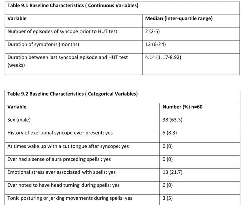
Results: Sixty patients underwent HUT testing. Mean age was 46 ± 15 years and median
duration of symptoms was 12 months (IQR 6 to 24 months). Thirty five (58.3%) patients had
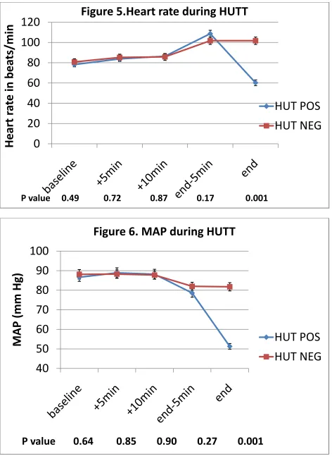
positive HUT response. Mean time to syncope was 31.5± 6.9 minutes. At the end of the tilt
phase, FS in the HUT positive group increased significantly from baseline (32.4±0.68% to
37.5±0.64 %, p< 0.001), while FS did not change significantly in the HUT negative group. Ten
(16.7%) patients had recurrent syncope on follow up. During HUT test, achieving a maximum
heart rate of ≥ 108 beats per minute was predictive of recurrent syncope [OR 8.62 (1.002-73.84),
p=0.049]
Conclusions: In patients with a positive response on HUT testing, there is a significant increase
in LV FS during tilt as compared to those with a negative response. Patients who have recurrence
of syncope on follow up tend to have higher peak heart rate attained during HUT. Hence peak
heart rate attained during HUT testing can be used to identify patients at high risk of recurrence
INTRODUCTION
Recurrent syncope is a commonly encountered clinical problem and remains a
majordiagnostic as well as therapeutic challenge in any clinical setting. Even though only a
small percentage of patients experiencing syncope in the community present themselves to a
hospital, syncope still accounts for 1% of presentations in European acute medical care settings
(1)(2) (Figure 1). In order to prognosticate or risk-stratify patients presenting with syncope, two
major aspects ought to be considered: first the risk of mortality and major adverse cardiac events
and second the risk of recurrent syncope and consequent injury/ trauma. Syncope especially
when recurrent has a pervasive detrimental influence on the quality of life of patients. Syncopal
burden has inverse correlation with quality of life (3). Recurrent syncopal episodes are
associated with orthopedic fractures and soft tissue injury in 12% of patients (4). While a
detailed preliminary evaluation including an exhaustive clinical history, physical examination
and a resting ECG will diagnose the underlying cause of syncope in 23 - 50% of subjects
presenting with syncope, the remaining patients will require further risk stratification and
additional diagnostic testing (5).
Reflex (neurally mediated) syncope, also known as neurocardiogenic syncope continues
to be the leading cause of syncope in any clinical setting. In contemporary clinical practice, tilt
testing remains the singular investigation that is available to the clinician, in order to demonstrate
the propensity for reflex syncope in patients presenting with otherwise unexplained syncope.
Head up tilt (HUT) testing reproduces reflex syncope in controlled laboratory settings and this
has been shown to correlate with the patients original symptoms. Even in patients estimated to
have a high risk of major adverse cardiac events and in those with clinical markers indicative of
an underlying cardiovascular etiology has been ruled out by focused and comprehensive
investigations. Since 1986 when HUT testing was originally introduced, the tilt protocol has
evolved through multiple modifications. Studies have looked at the effect of modifying the
duration of the pre-tilt supine phase, duration of tilt phase, angle to which the table is tilted and
different drugs for pharmacological challenge, in order to arrive at an optimal protocol (6). HUT
testing has become widely adopted in clinical practice, especially in the diagnostic evaluation of
reflex syncope. However the absence of a “gold standard” for diagnosing syncope has made the
sensitivity of HUT test questionable.
Apparently healthy subjects without any indication of cardiovascular disease may have a
positive response to tilt testing (7) (8). The false positivity rate of HUT test is influenced both by
patient characteristics including age and protocol characteristics including duration, angle of tilt
and use of provocative drugs (9) (10). Studies comparing patho-physiological changes during
positive tilt testing in healthy volunteers and patients presenting with reflex syncope have shown
that the hemodynamic as well as humoral mechanisms leading to syncope are qualitatively and
quantitatively different in these two groups. In patients presenting with reflex syncope, studies
show that there is accelerated peripheral venous pooling as indicated by a more marked and
exaggerated decline in left ventricular end diastolic dimension (LVEDD) as compared to HUT
positive healthy volunteers. These patients were also demonstrated to have higher left ventricular
(LV) contractility as indicated by values of fractional shortening (FS) associated with elevated
levels of epinephrine (11).
Hence we postulated that, echocardiographic parameters such as FS and decrease in
LVEDD may help in identifying a false positive group of patients in those undergoing HUT
may help in assessing the risk of recurrence of syncope, thereby helping to direct therapy and
interventions in order to limit recurrences and prevent physical injury and morbidity from
AIMS AND OBJECTIVES
Aim:
1. The aim of the study was to characterize the clinical, hemodynamic and
echocardiographic variables including LV contractility during tilt in patients with
unexplained recurrent syncope undergoing HUT test and to correlate the same with
clinical outcomes
Objectives:
1. To determine the LV contractility during tilt in patients with unexplained recurrent syncope
undergoing tilt testing by measuring the LV end diastolic and systolic dimensions and FS,
using echocardiography during tilt
2. To determine if change in FS during HUT test will predict recurrence of syncope during
follow up
3. To determine other significant predictors of recurrent syncope in the study population
REVIEW OF LITERATURE
Syncope is a common presenting complaint in any clinical setting. Clinically syncope can
be defined as a transient loss of consciousness caused by cerebral hypoperfusion, the hallmarks
of which include abrupt onset, brief duration, and spontaneous complete recovery without
residual neurological deficits. The term pre-syncope is commonly used in clinical parlance to
depict a spell that is similar to the prodrome of syncope but which is not followed by loss of
consciousness. Questions have been raised as to whether the patho-physiology of pre-syncope is
same as that of syncope. In the 17th century John Hunter, a surgeon, documented the
vasodepressor manifestations of syncope in a patient undergoing phlebotomy. In the 18th century,
researchers including Foster identified vagally mediated cardio-inhibition as the causative
mechanism underlying syncope. Lewis coined the term ´vasovagal syncope´ (12).
The prognosis of patients with syncope is not homogenous. Syncope can be benign. Less
commonly syncope can be a harbinger of sudden cardiac death. Even though the cause is benign,
recurrent syncopal episodes result in substantial morbidity. Syncope can cause injury. Syncope
causes significant anxiety among subjects and their relatives, resulting in substantial functional
limitation comparable to the level of debilitation seen in many chronic illnesses. Syncope poses a
huge burden on the society in terms of medical, social, and economic impact. In the United
States alone more than one million patients are worked up for syncope every year. Different
registries have shown that that upto 5% of emergency department consultations and upto 6% of
hospitalizations are for evaluation of syncope in Western populations (13). In a recent European
series the median duration of hospitalization was 5.5 days (2). The most commonly encountered
form of syncope is vasovagal syncope (common faint). It is also known as neurocardiogenic
neurocar managem for preve only subo as episod Figure A by globa which le mediated syncope. diogenic syn
ment of recur
enting recurr
optimal. Dru
dic nature of
1.Syncope eve
An etiologic c
al cerebral hy
eads to sync
d bradycardia
Secondly, c
ncope rema
rrent of vaso
rence of sync
ug trials for
f neurocardio
ents/ visits per
classification
ypoperfusion
cope, as sho
a which is al
cardiovascul
G
9.3
Ge
18.1
‐
3
ins elusive.
ovagal synco
cope, the cho
this purpose
ogenic synco
1000 patient-y
n system for
n. There ar
own in tabl
lso known a
lar causes a
E
0.7
p
enera
per
1000
eneral
p
39.7per
1
There are o
ope. Even tho
oice of thera
e have been m
ope and by th
years [data from
r syncope ha
re three mai
le 1. The co
s cardio-inh
also contribu
ED
per
1000
l
pract
0
patient
popula
1000
pati
only limited
ough multip
apy is mostly
mostly hamp
he heterogen
m (14)] ED Em
as been prop
n causes of
ommonest u
ibitory type
ute to the eti
tice
years
tion
ent
years
d therapeutic
ple drugs hav
y empirical a
pered by the
neity of the s
mergency depar
osed (15). S
transient lo
underlying c
of reflex (ne
iology, inclu
s
c options fo
ve been evalu
and its effica
e sporadic as
study popula
rtment
Syncope is ca
w cardiac o
cause is a r
and structural cardiac diseases. Yet another cause is inadequate venous return, which in turn may
[image:15.612.69.549.143.603.2]be due to dehydration or venous stagnation in the lower limbs.
Table 1. Etiological Classification of Syncope (15)
Reflex Syncope
- Vasovagal or neurocardiogenic
- Situational (e.g. cough, micturition)
- Carotid Sinus Hypersensitivity
- Syncope without obvious triggers or non-typical presentations
Syncope resulting from Orthostatic Hypotension
- Medications causing Orthostatic Hypotension
- Primary Autonomic Failure
- Secondary Autonomic Failure
- Volume Depletion
Cardiovascular Syncope
- Arrhythmias: bradycardia and tachy-arrhythmias
- Structural Cardiovascular Diseases: Valvular heart diseases, aortic dissection, acute myocardial
infarction, hypertrophic cardiomyopathies, cardiac tumors, cardiac tamponade, coronary artery
anomalies, pulmonary hypertension and pulmonary embolism
Pathophysiology of Neurocardiogenic Syncope
Blood pressure is regulated at the central nervous system (CNS) level by a
impulses relayed from the aortic and carotid sinus baroreceptors continually regulate the arterial
blood pressure levels. These neural impulses are carried in the vagus nerve and glossopharyngeal
nerve respectively and they relay to the CNS. These neural impulses are processed in the the
nucleus tractus solitarius. This will result in inhibition of efferent sympathetic outflow and
augmentation of the efferent vagal outflow (12). A fall in the arterial blood pressure leads to an
augmentation of the sympathetic outflow and suppression of vagal output. Thus this neural reflex
system modulates the balance between the sympathetic and parasympathetic system thereby
regulating the blood pressure levels.
In addition to the above mentioned reflex system there are cardiopulmonary
baroreceptors which are located in the ventricular walls and in the vasculature. These
baroreceptors also regulate the sympathetic outflow (16). Ventricular stretch mediated by
increased filling pressures is the main stimulus for these receptors. Based on the underlying
pathogenesis, a classification system for neurocardiogenic syncope has been proposed.
Neurocardiogenic syncope is classified as ‘vasodepressor type’ if hypotension occurs due to a
loss of the vasoconstrictor sympathetic tone and ‘Cardio-inhibitory’ when bradycardia or
asystole occurs. Neurocardiogenic syncope is classified as mixed if both these mechanisms
contribute.
On assumption of erect posture a number of compensatory physiologic changes involving
the sympathetic and parasympathetic reflex systems come into effect. When a patient assumes
upright posture these is pooling of more than half a liter of blood in the lower extremities. Thus
reduced venous return will decrease the filling pressure in the ventricle. Hence the mean arterial
pressure as sensed by the aortic arch and carotid sinus drops (12). This would trigger a
impulses relayed to the cardiac and vascular structures. This cascade of events will ultimately
result in peripheral vasoconstriction, increased inotropic and chronotropic response. As a result
the mean arterial blood pressure is maintained and the subject does not faint.
While much research has gone into delineating the patho-physiology of neurally
mediated syncope, not all aspects have been explained adequately thereby rendering the
diagnosis and treatment of this common condition difficult. Hence therapies targeting different
aspects of the purported patho-physiologic mechanism of syncope have not yielded optimal
results. The most widely implicated patho-physiological mechanism causing neurally mediated
syncope is the Bezold–Jarisch reflex (16,17).Bezold–Jarisch reflex is a neurocardiogenic reflex
that is triggered by venous pooling in the lower limbs especially in dehydrated subjects. This
results in decrease in ventricular volume and cardiac filling pressures. Compensatory
baroreceptors reflex comes into play and resultant sympathetic discharge mediates increase in
contractility of the left ventricle. Consequently mechanoreceptors that are activated by changes
in wall tension located in the left ventricle wall are activated inappropriately. The C- fiber
mediated afferent signals are relayed through the vagus nerve to the CNS. That mediates a reflex
decrease in the sympathetic outflow to the vasculature and the cardiac structures, and inverse
changes occur in parasympathetic activity. Marked vasodilatation occurs. This is accompanied
by bradycardia and hypotension. These changes are the hallmarks of the loss of consciousness
that characterize neurally mediated syncope. These changes are promptly reversed once the
subject assumes supine posture. Thus we may conclude that neurally mediated syncope mainly
takes place in predisposed patients due to excess lower limb venous pooling that leads to a
sudden drop in venous return leading to inappropriate activation of LV mechanoreceptors
Mechanoreceptors are present at multiple other sites including the urinary bladder,
rectum, and respiratory organs. It has been postulated that abrupt activation of these receptors
groups during micturition, defecation or coughing transmits afferent signals to the CNS thereby
triggering a reflex mediated syncope. This is considered to be the maladaptive mechanism
underlying situational syncope.
In humans Bezold–Jarisch reflex, has been implicated in the pathogenesis of reflex
syncope largely based on indirect evidence. In patients with reflex syncope, during tilt, a surge in
catecholamine levels has been demonstrated occurring just prior to loss of consciousness (18).
However, exaggerated activation of the sympathetic system prior to syncope has not been
demonstrated across studies in a consistent manner. In human studies, recording of neural
impulses in peroneal nerve fibers using microelectrodes has demonstrated an increase in
sympathetic neuronal traffic in the presyncopal phase, followed by a sudden withdrawal of
sympathetic neuronal activity just prior to onset of syncope. Echocardiographic studies have
demonstrated decrease in the LVEDD and end diastolic volume in syncopal subjects during tilt
testing (19,20).
However neurally mediated syncope in humans, cannot be entirely attributed to the
Bezold–Jarisch reflex mechanism. This point has been illustrated by the fact that a pure
vasodepressor response without a drop in heart rate has been demonstrated during tilt induced
neurocardiogenic syncope in post heart transplant patients (12). Neural reflex mechanism is
effectively ruled out in this case as cardiac afferent and efferent pathways are denervated in a
transplanted heart. This partly explains the lack of bradycardia in these patients. Hence multiple
triggers beyond those described in the Bezold–Jarisch reflex are likely to be operative in neurally
VASIS classification) also known as pure vasodepressor response occurring in cardiac transplant
recipients and not due to the surgical cardiac denervation alone (21)(22,23). EEG monitoring
during HUT has shown increased activity in the left cerebral hemisphere in subjects developing
syncopal response on tilt test. This lateralization occurs prior to the onset of loss of
consciousness and accompanies the drop in heart rate and blood pressure and onset of symptoms.
Hence the left cerebral hemisphere may be a part of the neural circuit that mediates vasovagal
syncope (24).
Studies have indicated that multiple neurotransmitters and neuro-modulators influence
the pathogenesis of neurally mediated syncope. These neurotransmitters include serotonin (25),
adenosine, and opioids (26). Emotional stress may precipitate reflex syncope. The neural reflex
mechanism underlying vasovagal syncope may be modulated by impulses from the
hypothalamus and forebrain. Prior to vasodepressor syncope a surge in the levels of beta
endorphins have been demonstrated (27). Opioid µ receptor antagonists have been demonstrated
to augment the cardiac baroreflex mediated stimulation of sympathetic pathway (28). The µ
receptor antagonist naloxone has not been successful in aborting syncope during HUT (29).
Not only central neurotransmitters but also peripheral neuronal triggering mechanisms
other than those typified by the Bezold–Jarisch reflex may play a role in the pathogenesis of
reflex syncope. Intravenously administered adenosine has been shown to trigger sympathetic
afferent activity (30). This fact underlies the use of adenosine as a pharmacological challenge
during tilt testing. However adenosine induced tilt test response has shown no correlation with
the recurrence rate of syncope or with the mechanism of spontaneous syncope as demonstrated
by studies using implantable loop recorders (31). Pharmacological challenge with isoproterenol
during tilt testing (32). Thus a decrease in ventricular volume leading to triggering of LV
mechanoreceptors may not an imperative step in the pathogenesis of reflex syncope. Also the
hemodynamic responses preceding syncope are distinctly different in those with passive tilt
induced syncopal response as compared to those with a positive response to tilt testing with
isoproterenol pharmacological challenge (33). Just prior to the onset of loss of consciousness,
patients in the isoproterenol challenged tilt test group had a higher heart rate and cardiac output
as compared to patients with passive tilt induced syncopal response. This heterogeneity in the
hemodynamic response in these two groups of patients suggests differences in the syncopal
triggering mechanisms in these groups.
The sudden fall in peripheral vascular resistance that is one of the final steps in the
pathophysiology of reflex syncope, has been investigated by many groups. One postulate is that
the peripheral vasodilatation is a passive mechanism mediated by a withdrawal of sympathetic
activity. However multiple studies have demonstrated the presence of a sympathetic mediated
active vasodilatation in humans. This active skeletal muscle vasodilatation may be initiated by
changes in sympathetic tone mediated by cholinergic vasodilator nerves or by release of nitric
oxide (NO). Patients with positive response to HUT test have been shown to have excessive
metabolism of NO (34). Also the presence of NO as a peripheral neurotransmitter has been
demonstrated in skeletal muscles.
Thus it can be concluded that the Bezold–Jarisch reflex is one of the many causative
History and Physical Examination
Reflex syncope can be precipitated by a number of triggers including prolonged standing,
strenuous exertion on a hot day, emotional stress, sight of blood and excessive physical pain.
Patient may complaint of prodromal symptoms including lethargy, giddiness, sweating, dimness
of vision, heaviness of head and nausea. Transient clinical signs that may be demonstrated at this
stage include facial pallor, dilatation of pupils and flushing. This prodromal phase may be very
brief or may last up to a few minutes prior to the actual occurrence of loss of consciousness.
Some patients successfully abort the syncopal episode by recognizing this prodromal phase and
assuming a supine position or by employing certain “physical counter pressure maneuvers”.
However, a minority of patients especially elderly subjects do not have a prodrome preceding the
syncopal episode hence they are at risk of physical injury resulting from abrupt loss of postural
tone (35).
Syncope is characterized by short duration of loss of consciousness, which may last for
up to five minutes. Elderly patients have a number of atypical features including lack of
prodromal symptoms, longer duration of syncope and post spell confusion which may last up to
ten minutes or rarely longer. Syncope on occasions can have atypical features including jerky
seizure-like movements. Some researchers refer to this as “convulsive syncope” (36). When the
patient regains consciousness, he/ she may experience lethargy or fatigue. Patient may appear
pale and at times can have profuse sweating after the spell. Post spell confusion is in favor of
seizure disorder. Recovery from syncope is characteristically abrupt, and without any residual
neurological deficits. A simple questionnaire has been used to differentiate syncope from
seizures with a high degree of sensitivity as well as specificity (37). Questions enumerating
A focused and exhaustive clinical evaluation should also include an electrocardiogram,
and a detailed drug history to identify the use of any proarrhythmic drugs including the use of
Class IA and IC anti-arrhythmic medications. If available, bystanders who witnessed the
syncopal spell should be interviewed to identify etiological markers. Jerky limb movements and
tonic posturing may be associated with both cardiac and neurological causes of syncope.
Absence of a prodrome is a clinical marker of cardiac arrhythmia. Rarely absence of prodromal
symptoms may be indicative of dysautonomia (35). Recurrent syncope presents sporadically. It is
not clear why syncopal spells occur in clusters, interspaced by relatively long asymptomatic
periods. Hence compliance with drugs and other therapy becomes an issue. Family history of
sudden cardiac death is important to elicit. History of exertional syncope points to structural
heart disease especially those with fixed cardiac output.
A detailed clinical examination including postural blood pressure response should be
documented in every patient. This includes a cardiovascular and neurologic examination in all
patients. The clinical examination may provide clues to the etiological diagnosis including the
presence of LV dysfunction, pulmonary arterial hypertension, valvular heart disease,
hypertrophic cardiomyopathy and other structural cardiac diseases. Carotid bruits point to
underlying carotid artery stenosis. In the majority of patients, a detailed clinical history and
physical examination has high efficacy in identifying the etiology of syncope, though the
underlying pathology of syncope remains unidentified in about 40% of patients (38).
Investigating Syncope
In cases where there is an underlying conduction disturbance or arrhythmia, ECG
atrioventricular conduction blocks, presence of an accessory pathway, channelopathies including
Long QT Syndrome and Brugada syndrome, causing syncope, may be diagnosed on the ECG.
The ECG may suggest a diagnosis of arrhythmogenic right ventricular dysplasia (ARVD).
Frequent ventricular ectopics or a short run of nonsustained ventricular tachycardia especially in
patients with underlying structural cardiac disease increases the likelihood of an underlying
arrhythmic cause of syncope. However since the underlying arrhythmic episode is usually
sporadic the diagnostic yield of a 12 lead surface ECG for this purpose may be low.
A variety of options for ambulatory ECG monitoring is available to the clinician. The
“gold standard” for diagnosing cardiac arrhythmia as the cause of syncope is ECG
documentation of the arrhythmia coinciding with the patients’ symptoms. The singular factor
that determines the device selection and duration of ambulatory monitoring is frequency of
symptoms. Holter monitoring is widely available and is suitable for spells that recur on a daily
basis. When the spells recur on a monthly basis an event monitor may be prefered. An
implantable loop recorder is suitable for those with very infrequent symptoms. Implantable
recorders can be used for up to 14 months and permits the clinician to correlate the patient’s
symptoms with the underlying cardiac rhythm. Implantable loop recorders have a high diagnostic
yield. These devices are able to diagnose up to 90% of patients presenting with unexplained
recurrent syncope (39). The use of implantable loop recorders has revolutionized the
management of syncope by providing insights into the pathophysiology of syncope in an
individual patient and it is shown to be cost-effective (40).
Cardiac imaging including echocardiogram is indicated only in select patients with
syncope. Echocardiogram is a indicated if underlying structural heart disease is suspected
have not yielded a diagnosis. Echocardiogram is an appropriate imaging technique to diagnose
many underlying causes of syncope including valvular lesions, hypertrophic obstructive
cardiomyopathy (HCM), pulmonary embolism, LV dysfunction and ARVD. When patients
known to have ischemic heart disease or those at high risk for ischemic heart disease, present
with unexplained syncope, further evaluation is indicated. When there is history of exertional
syncope, exercise stress testing is indicated. During exercise stress testing, a hypotensive
response or a failure of blood pressure to increase with exercise is of particular significance. This
may point towards a diagnosis of HCM, high risk ischemic heart disease and autonomic failure.
Exercise stress testing may also suggest the presence of catecholaminergic polymorphic
ventricular tachycardia. A diagnosis of neurocardiogenic syncope or reflex syncope can be made
in a patient presenting with a typical history in the absence of other plausible explanations for the
syncopal episode (42). Further evaluation is indicated if the clinical features are atypical.
Syncope in the Patient with an Apparently Normal Initial Evaluation
After a detailed clinical evaluation and focused comprehensive investigations, if no
cardiac disease is detected, it is likely that the syncopal episodes are not associated with
increased mortality. In such a scenario the main objective of further evaluation is to identify the
risk of physical injury and occupational risks from recurrence of syncope. For this purposes the
AHA/ACCF has defined a “malignant episode of syncope” as a syncopal spell that occurs
without any prodromal warning symptoms resulting in physical injury to self or causing property
damage (41). Hence occupational groups like drivers and pilots presenting with syncope may
require further detailed evaluation for medico-legal purposes. However deciding on further
many of these investigations are low. Even after the above mentioned investigations do not yield
a diagnosis one should still actively consider alternative diagnoses like reflex syncope, carotid
sinus hypersensitivity especially in elderly patients, arrhythmias including atrial and ventricular
tachycardia.
The Tilt Table Test
Ever since it was introduced into clinical practice in 1986 by Kenny, et al, tilt testing has
become the investigation of choice for evaluating patients presenting with unexplained recurrent
syncope (7). HUT testing is primarily used for arriving at a diagnosis of reflex syncope, in a
patient with a compatible history. Even though HUT testing has become widely acceptable as a
feasible test for diagnosing reflex syncope, the clinical implications of a positive test response is
still uncertain. The sensitivity, positive yield, and reproducibility of HUT testing reported in
literature, vary widely (43), (44). The estimated sensitivity and specificity of HUT testing
depends on patient factors as well as multiple factors in the tilt protocol (45). The sensitivity of
HUT test is reported to vary from 26% to 80%, however these estimates may be inaccurate as
there is no established “gold standard” for diagnosing neurocardiogenic syncope. The estimated
specificity is about 90%; however it is well known that many healthy volunteers have a positive
response on HUT (46). Even when patients have a positive response on tilt testing, the
hemodynamic changes and underlying pathophysiological mechanism may not be the same as
those occurring during a spontaneous syncopal spell (as recorded by implantable loop monitors)
(47).
Table 2 illustrates the positive and negative results of HUT testing in adults. Table 3
modify future responses to HUT testing. The reproducibility of HUT test (when a second tilt test
is repeated after a variable time period ranging from hours to weeks) is 80% to 95% for an
initially negative test response. However the reproducibility is much lower for an initially
positive HUT response (30% to 90%). This has implications for the use of HUT testing in
assessing the response to therapy. Indications for HUT testing are enumerated in Table 4. In
subjects presenting with unexplained recurrent syncope, if the initial work up is negative and the
clinically estimated likelihood of the diagnosis being reflex syncope is high, little incremental
diagnostic or prognostic information is obtained by performing a HUT test. The negative
predictive value of tilt table test is low in an individual patient with a high pretest probability for
reflex syncope. In patients presenting with unexplained syncope and physical injury resulting
from syncope and for those in high risk occupations, further investigations are warranted when
the tilt response is negative. A positive HUT test response does not predict the risk for recurrence
[image:26.612.69.549.498.698.2]of syncope in these patients.
Table 2. Passive Head Up Tilt Testing in Subjects with Unexplained Syncope
Study Total
patients
Positive
result on
HUT
Controls Controls
with
positive
HUT
Tilt angle Tilt
duration
n n (%) N n (%) degrees minutes
Kerry et
al(7)
Fitzpatrick
et al(48)
71 53 (75) 27 2 (7) 60 60
Strasberg et
al(49)
40 15 (38) 10 0 60 60
Raviele et
al(50)
30 15 (50) 8 0 60 60
Abi Samra
et al(51)
151 63 (42) 15 0 60 20
Almquist et
al (52)
15 4 (27) 18 0 80 10
Grubb et
al(53)
25 6 (24) 6 0 80 30
Pongiglione
et al(54)
20 4 (20) 0 - 90 15
Shen et
al(55)
Figure 2. Head Up Tilt table testing © N Engl J Med 2005;352:1004-10.
Table 3. Reproducibility of the Initial Results of Tilt-Table Testing
Study (Reference)
Protocol for
Tilt-Table Test
Time between
Tests
Positive
Reproducibility
Negative
Reproducibility
de Buitleir et al (56)
80° tilt, 10-min duration
5 min 8/14 (57) 16/17 (94)
Brooks et al (57) 70° tilt, 25-min duration
1 d 11/30 (37) 45/56 (80)
Raviele et al (50) 60° tilt, 60-min duration
3 d 10/14 (71) -
Fitzpatrick et al(48)
60° tilt, 60-min duration
- 24/31 (77) -
Blanc et al(58) 60° tilt, 60-min duration
7 d 8/13 (62) -
Fish et al(59) Isoproterenol used
30 min 14–18/21 (67–
86)
-
Chen et al (60) Isoproterenol
used 30 min 12/15 (80) 8/8 (100)
Grubb et al (61) Isoproterenol used
5 d 13/14 (93) 6/7 (86)
Sheldon et al
(62) Isoproterenol used 2 wk 23/26 (88) 17/20 (85)
G. Foglia-Manzillo (63)
60º for up to 45 min. 400 µg sublingual spray nitroglycerin
Table 4. Indications for Head Up Tilt Testing (64)(15)
Definite indications
Recurrent syncope in the absence of underlying cardiac disease or in the presence of organic
cardiac disease, after cardiac causes of syncope has have been ruled out using appropriate
investigations (Class I B)
A single episode of unexplained syncope occurring in a high risk setting (occupational
implications or trauma resulting from syncope) (Class I B)
HUT testing is used for demonstrating the susceptibility to neurocardiogenic syncope to the
patient (Class I C)
Possible indications
Differentiation between reflex and orthostatic hypotension syncope (Class IIa C)
Differentiation between syncope with jerking movements and seizure disorders (Class IIb C)
Evaluation of repeated inexplicable falls (Class IIb C)
Evaluation of patients with frequent syncope and psychiatric disease (Class IIb C)
Evaluation of recurrent syncope in patients with neurological disorders like dysautonomia or
peripheral neuropathy
Not indicated in
HUT testing is not recommended for evaluation of response to treatment (Class III B)
Isoproterenol HUT testing is contraindicated in patients with ischemic heart disease (Class III C)
Head-up tilt testing using sublingual nitroglycerin (NTG) challenge
HUT testing augmented by sublingual nitroglycerin was advocated by an Italian group
(65) in 1995 by addition to the standard Westminster protocol (48)(7) of a NTG provocation
phase. The nitroglycerin-head-up tilt is more sensitive than the non-medicated passive tilt test
(65), and it is simpler and is increasingly used across the world. Thus, the test consisted of the 45
minute passive phase directly followed, if negative, by nitroglycerin administration with the test
continuing for further 20 min. This protocol, although twice as sensitive as the passive tilt alone,
is time-consuming and not well accepted by many cardiologists. From the available evidence,
shortening the passive phase from 45 minutes to 20 minutes results in a significant reduction in
the positive response rate. On the other hand, this was balanced by an increase in the positive
responses during the provocation phase (Tables 5, 6). Thus, the final positivity rate of the test
remained unchanged (6). A possible explanation is that nitroglycerin ensures the positive
responses of the late passive phase of the test, besides including an additional number of
drug-induced positive responses. In fact, assuming similar conditions of tilt angle, drug and its dose,
the number of positive responses during the provocation phase of the test is always greater after a
shortened rather than after a conventional passive phase (Tables 5, 6). Furthermore, the reduction
responses’. Finally, it was found that the specificity of the test was not affected by the duration
of the passive phase (Table 7).
Stabilization phase
If invasive maneuvers are avoided before the test, 5 minutes are sufficient for the patient
to achieve a stable physical condition.
Passive phase Tilt angle
As previously shown (66), the best tilt angle is 60º
Passive phase duration
For all the above-mentioned reasons that the optimal duration of the passive phase is
considered to be 20 minutes.
Drug Challenge/ provocation phase
Multiple studies have evaluated testing protocols (table 6) using sublingual crushed
nitroglycerin tablets at a dosage of 300 µg dosage. However significant individual variation in
pharmacokinetics resulted, including differences in the mucosal absorption rate depending on the
amount of saliva available. This was an important consideration in older patients. In many
subsequent studies (table 5), a protocol using 400 µg nitroglycerine oral spray has been reported.
This protocol has many advantages including a higher dose of nitroglycerine used, better
pharmacokinetics, ease of administration and reduced time to syncope. This protocol imparts
better sensitivity to the tilt testing while maintaining comparable levels of specificity. Using
This interval was reduced to 5 ± 4 minutes using nitroglycerin in the spray form (67) ((68). This
finding underlies a recommendation to shorten the duration of test by cutting down on the
duration of the provocation phase from 20 to 15 minutes. This reduction in the duration of the
provocation phase does not seem to affect the sensitivity of the test (6).
Interruption of tilt
The tilt phase is terminated and the patient made supine when the following end points
occur: First, at the end of the drug challenge stage if the patient remains asymptomatic (test
negative). Second, if there is loss of consciousness (test positive). The test is deemed positive
whenever loss of consciousness (that is, the reproduction of the patients’ presenting symptom of
syncope) occurs in association with hypotension and/ or bradycardia, with sudden (<5 minutes)
onset. This positive HUT response is considered to be analogous to the prototypical vasovagal
syncope. Recommendations suggest that the tilt phase should be terminated just when syncope is
initiated with loss of postural tone, rather than at the onset of hypotension. The response can be
classified according to the VASIS recommendations. However for classification purposes the
hemodynamic responses until the onset of syncope has to be taken into account. The subsequent
bradycardia should not be used for classifying the response. The tilt may also be terminated if
there is a tardy onset, prolonged (>5 minutes) hypotension. This is an indication of orthostatic
intolerance and is associated with minor symptoms usually. The clinical significance of this
particular response is still debated. This response at times can be documented in healthy subjects
also. However in patients with orthostatic intolerance without syncope, autonomic nervous
Diagnostic Criteria for Tilt Testing (15)
In patients presenting with unexplained syncope, but without organic cardiac disease the
induction of hypotension and/ or bradycardia with reproduction of syncope is diagnostic
of reflex syncope
In patients without organic cardiac disease the induction of progressive hypotension is
diagnostic of orthostatic hypotension
In patients without organic cardiac disease, the induction of reflex hypotension and/ or
bradycardia without reproduction of syncope may be diagnostic of neurocardiogenic
syncope
In patients with structural heart disease, first tachy/bradyarrhythmias and alternative
cardiovascular causes of syncope should be reasonably ruled out before interpreting the
positive tilt test result as diagnostic
If the subject has loss of consciousness without accompanying hypotension and/ or
[image:34.612.72.548.512.708.2]bradycardia, a diagnosis of psychogenic pseudosyncope may be considered
Table 5. Results of tilt table testing potentiated by nitroglycerin sublingual spray 400 µg
Study, year Patient nos
Passive phase Duration (min)
Passive phase positivity (%)
NTG phase positivity (%)
Total Positive response
Exaggerated responses
Natale, et al 1998(69)
33 20 4(12) 22(67) 26(78)
Del Rosso, et al. 1998(70)
202 20 22(11) 119(59) 141 (70) 8 (4)
et al. 1999(71) Total passive phase 20 min
304 20 33 (11) 177(58) 210(69) 23 (8)
Bartoletti, 1999(67)
84 45 15(18) 28(33) 43 (51) 18(21)
Fogila Manzillo, et al. 1999(63)
48 45 9 (19) 25 (52) 34 (71) 2 (4)
Del Rosso, et al. 2000(68)
31 45 3 (10) 21 (68) 24(77) 2(6)
Total passive phase 45 min
[image:35.612.69.549.68.418.2] [image:35.612.68.547.74.419.2] [image:35.612.67.552.476.714.2]163 45 27 (17) 74 (45) 101 (62) 22(13)
Table 6. Results of head-up tilt + use of nitroglycerin sublingual tablets 300 µg
Study, year Patient number
Duration of passive phase
Passive phase positivity (%)
NTG phase positivity (%)
Total positive responses
Exaggerated responses
Raviele, et al. 2000(72)
71 20 9 (13) 26 (36) 35(51) 3(4)
Raviele, et al. 1995(65)
235 45 59 (25) 60 (26) 119(51) 33 (14)
Kurbaan, et al. 1999(73)
102 45 35 (34) 38 (37) 73 (72) -
Total (only 55, 65)
Table 7. Response to sublingual nitroglycerin challenge tilt table test in healthy controls
Study, year Number of patients Age (Mean SD) HUT protocol Positive response (%)
Raviele, et al. 1995 (65)
35 54± 19 60º×45 + 20 min
NTG 0.3 mg
2(6)
Aerts, et al. 1997 (74)
20 27± 4 70º×45 + 15 min
ISDN 5 mg
6 (30)
Natale, et al. 1998 (69)
16 67± 9 70º× 20 + 15 min
NTG 0.4 mg
2 (12)
Del Rosso, et al. 1998(70)
34 45±17 60º×20 + 25 min
NTG spray 0.4 mg 2(6)
Ammirati, et al. 1998(75)
23 36 ± 12 60º×30 + 15 min
ISDN 1.25 mg
0 (0)
Bartoletti, et al. 1999(67)
25 49 ± 17 60º×45 + 20 min
NTG spray 0.4 mg 1(4)
Del rosso, et al. 2000(68)
47 52± 20 60º×20 + 20 min
NTG spray 0.4 mg 2(4)
Raviele, et al. 2000(72)
30 44± 10 60º×20 + 20 min
NTG 0.3 mg
3 (10)
Total 230 18(8)
Classification of vasovagal syncope by the Vasovagal Syncope International Study
(VASIS) in 1992 provided insight into the different types of vasovagal responses observed
during tilt-induced syncope (76). This classification system has been extended to tilt testing with
pharmacological challenge (70) (73). Based on the hemodynamic changes preceding syncope the
positive response to HUT testing can be categorized into different subgroups. This classification
system may be of use in making the choice of permanent pacing versus drug therapy for the
[image:37.612.65.548.176.508.2]treatment of recurrent reflex syncope.
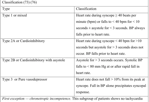
Table 8: Classification of reflex syncope induced by HUT table testing- the modified VASIS Classification (73) (76)
Type Classification
Type 1 or mixed Heart rate during syncope ≥ 40 beats per
minute (bpm) or falls to < 40 bpm for < 10 seconds ± asystole for < 3 seconds. BP always falls prior to heart rate.
Type 2A or Cardioinhibitory Heart rate during syncope < 40 bpm for >10 seconds but asystole for > 3 seconds does not occur. BP falls prior to heart rate.
Type 2B or Cardioinhibitory with asystole Asystole for > 3 seconds occurs. Systolic BP falls to < 80 mm Hg at or after rapid fall in heart rate.
Type 3 or Pure vasodepressor Heart rate does not fall > 10% from its peak at syncope. Fall in BP alone precipitates syncopal response.
First exception — chronotropic incompetence. This subgroup of patients shows no tachycardia
response during the tilt (i.e. maximum heart rate during tilt < 10% from the pre-tilt rate). However there is no hypotensive response.
Second exception — excessive heart rate increase. These patients show an excessive heart rate
rise both at the onset of the upright posture and during the entire duration of tilt, till prior to onset of syncope (i.e. heart rate greater than 130 bpm).
Subsequently a classification system based on interpretation of the hemodynamic patterns
during the pre-syncopal phase of the HUT test with and without drug challenge using sublingual
pre-syncopal period, subjects had a rapid and full compensatory reflex adaptation to the upright
posture. This lead to a stabilization of the blood pressure levels until abrupt onset of the reflex
syncope took place at the end of tilt. Second type is the dysautonomic (vasovagal) syncope
pattern in which steady-state adaptation to upright posture does not take place. This
mal-adaptation leads to a steady and progressive fall in the blood pressure levels till the occurrence of
a typical vasovagal reaction. Third is the orthostatic intolerance pattern in which there was a
progressive fall in the blood pressure levels, similar to that of the dysautonomic group, but this
hypotensive response was not followed by vasovagal syncope.
Complications and contraindications of tilt table testing
Tilt table testing is generally considered to be safe. There has been no report of mortality
during tilt testing. Few cases of ventricular tachycardia with the use of isoproterenol in patients
with ischemic heart disease or sick sinus syndrome have been reported in literature (77).
However, no life threatening adverse effects from the use of nitroglycerine have been reported.
Minor adverse reactions are common with both these drugs. These include palpitations with the
use of isoproterenol and headache with the use of nitroglycerine. Rarely arrhythmias including
atrial fibrillation can be triggered during or following a positive HUT test. This usually is
transient (78). Despite the low risk involved in tilt testing, resuscitation equipment including
defibrillators should be readily available in the HUT room. Caution should be exercised in
False Positive HUT Test and Use of Echocardiography during HUT test
Echocardiographic studies have provided insight into the pathogenesis of syncope
during tilt testing. The loss of consciousness due to reflex syncope has been by convention
attributed to the activation of LV mechanoreceptors, which in turn trigger the Bezold-Jarisch
reflex. Peripheral venous pooling, sympathetic nervous system activation, as well as
hypercontractility of a relatively empty left ventricle are the other potential triggers involved in
this reflex pathway that mediates reflex syncope. Indirect evidence for the role of LV
mechanoreceptors in pathogenesis of reflex syncope in humans is largely derived from studies
demonstrating increased LV FS and decreased LV volumes in subjects with syncope and positive
tilt testing (19). Studies using echocardiography during tilt testing show an accelerated rate of
reduction of end-diastolic volume index as well as significant decrease in stroke volume index
and ejection fraction in subjects presenting with reflex syncope as compared to healthy
volunteers. This phenomenon is due to redistribution of blood to the peripheral venous system
during tilt and an early parasympathetic effect on LV contractility (79).
The incidence of false positive responses has been demonstrated to be a problem inherent
in HUT test. This has led many investigators to opine that tilt testing should only be used to
confirm a clinically based diagnosis (10).
Even though tilt induced syncopal response can occur in healthy volunteers (false
positive), the hemodynamic as well as humoral changes that accompany this response are
qualitatively distinct from those of HUT positive patients who complaint of syncopal spells
during normal daily activities (true positive). These distinct hemodynamic and humoral
alterations are enumerated below (11).
This was about twice as long in HUT positive healthy volunteers as compared to
HUT positive patients with neurocardiogenic syncope.
b) Time to drop in blood pressure:
The true positives had a drop in blood pressure as early as two minutes into the
test accompanied, at the same time, by tachycardia, while in HUT positive healthy
controls (false positive cases), these alterations were absent till about two minutes
before the onset of symptoms. The normal controls did not demonstrate either of
these characteristics.
c) LV end-diastolic dimension:
The pattern of peripheral venous pooling in the three groups (true positives, false
positives and true negatives) was indirectly estimated by analyzing changes in
LVEDD. During tilt, both HUT positive groups ( true positives and false
positives) exhibited a progressive decline in LVEDD, significantly different from
pre-tilt LVEDD value, while the HUT negative subjects (true negatives)
demonstrated an initial decline that promptly stabilized (11)(19). There is more
rapid and exaggerated peripheral pooling of intravascular volume in patients
presenting with neurocardiogenic syncope. The hypotensive response and the
abrupt reduction in peripheral venous return during HUT are indicative of an
abnormality in vascular control. Studies have demonstrated impaired
vasoconstrictor response in patients with neurally mediated syncope during HUT
(80) and during dynamic leg exercise (81)(82).
In patients with true positive reflex syncope, it has been demonstrated that FS
assessed by echocardiography, significantly increases throughout the HUT test to
become, at 2 min before the end of HUT, statistically different from the false
positive and true negative groups (11).
e) Epinephrine levels:
During HUT, patients with reflex syncope had a sixfold increase in the
epinephrine levels as compared to the baseline value, accompanying the
bradycardia and hypotension. This increase in epinephrine level was significantly
more than the level of increase seen in patients with false positive HUT results
(11).
Monitoring of select echocardiographic parameters like LVEDD and FS to enhance the
specificity and sensitivity of HUT testing for the diagnosis of neurally mediated syncope has
been proposed (11). However this awaits controlled trials with larger numbers of patients. The
population of HUT positive patients is heterogenous when classified in terms of LV contractility
change during HUT test. It has been shown that LV hyper-contractility preceding syncope exists
in about 50% of HUT positive patients (83). In the remaining patients without increase in LV
contractility prior to the syncope, it could be that a mechanism other than Bezold Jarisch reflex
may be underlying the syncope. Or else it could be that this group of patients may be false
positive cases, behaving like the HUT positive normal volunteers in whom LV contractility does
not increase before syncope.
No correlation has been made between monitoring parameters like LVEDD and FS
been questioned in some earlier studies (84). The use of echocardiography during HUT testing
has been largely limited to research settings.
We may conclude that the use of echocardiographic monitoring of select parameters
during HUT provides valuable insight into the pathogenesis of neurally mediated syncope and
these findings may have major clinical and prognostic implications.
Electrophysiological study in Unexplained Syncope
In patients presenting with syncope of unknown cause, recent studies show that positive
yield with electrophysiology study (EPS) occur mainly in the subgroup of patients with structural
cardiac disease (85). The ability of EPS to arrive at an etiological diagnosis for syncope is
determined by the pre-test probability of the disease as well as the protocol used for the study.
The advent of efficacious non-invasive electrocardiographic monitoring including prolonged
monitoring has increased the diagnostic yield and in turn has diminished the prominence of EPS
as a diagnostic test. In those presenting with syncope and bi-fascicular block, the high degree AV
block may be transient. Hence it may not be picked up on routine 24 hour ambulatory Holter
monitoring and, hence prolonged period of monitoring may be required to document it by ECG
(86). In those subjects presenting with syncope associated with bi-fascicular block, an EPS has
high sensitivity for diagnosing intermittent high degree AV block. However the negative
predictive value of EPS is limited while evaluating intermittent AV block as the etiology of loss
of consciousness.
EPS may help in risk stratifying syncope. The induction of a sustained ventricular
tachycardia and presence of severe LV systolic dysfunction can predict a life-threatening
more favorable outcome. It can be concluded that EPS with programmed electrical stimulation is
a test with high diagnostic and prognosticating value in those presenting with unexplained
recurrent syncope in the presence of coronary artery disease and markedly depressed cardiac
function. However the diagnostic efficacy of EPS is questionable in patients with dilated
cardiomyopathy (4).
The current ESC guidelines suggest the following: In patients presenting with syncope in
the presence of concomitant ischemic heart disease, EPS is warranted when the initial work up
indicates an underlying arrhythmia (15). In patients presenting with syncope and underlying
bundle branch block, EPS is to be performed when noninvasive investigations have not yielded
an etiological diagnosis. In patients with syncope preceded by abrupt onset short duration
palpitations, EPS can be considered when noninvasive tests have not yielded the correct
diagnosis. In patients with high risk occupations and in patients with a diagnosis of Brugada
syndrome, ARVD or HCM presenting with syncope, EPS may be performed in a select subgroup
of cases (15).
Therapeutic Options in Reflex Syncope:
Counter-pressure maneuvers
With the paucity of data on the effectiveness of pharmacological data,
non-pharmacological therapy, including physical counter-pressure maneuvers are emerging as the
preferred treatment of reflex syncope. Recent studies have shown that isometric physical
counter-pressure maneuvers involving the legs including leg crossing, or of the arms including
hand grip and arm tensing, may successfully counter the hypotensive phase of an impending
the subject to abort or delay the frank syncopal spell in many cases, thereby preventing injury
(87) (88).
Tilt training exercises
Tilt training involves the patient assuming an enforced period of upright posture, at a
prescribed angle. The duration of tilt is progressively increased till the prescribed time period is
reached. In highly motivated young patients with recurrent syncope and/ or presyncope triggered
by orthostatic challenge, tilt training may reduce or prevent the recurrence of syncope (89). The
main drawback of tilt training is the poor long term compliance as this requires high patient
motivation. Many studies including randomized controlled trials have shown that tilt training
may not be efficacious in preventing recurrence of syncope in patients undergoing repeat tilt
table testing (90).
Drug therapy
Since Bezold- Jarisch reflex is known to be the major underlying pathophysiological
mechanism of reflex syncope, the use of beta blockers may decrease the ventricular
mechanoreceptor activation, by virtue of their negative inotropic effect in neurally mediated
syncope. However the outcomes of multiple randomized control trials have not lent credence to
this plausible hypothesis. The use of beta blockers may not be optimal in subtypes of reflex
syncope in which mechanisms other than increased sympathetic activation are operational. Beta
blockers may aggravate the bradycardia in carotid hypersensitivity syndrome. Majority of
randomized trials with long term follow up have failed to prove the effectiveness of beta
Alpha receptor agonists including etilefrine and midodrine have been used in the
management of neurally mediated syncope. Peripheral blood pooling due to failure to achieve
adequate vasoconstriction of the peripheral blood vessels underlie many cases of neurally
mediated syncope. Etilefrine, an alpha agonist vasoconstrictor, at a dose of 25 mg twice daily
failed to reduce the frequency or time to recurrent syncope in a randomized clinical trial as
compared to placebo (93). The major drawback with midodrine is the mulpile and frequent
dosing. This limits long-term compliance in most patients (94). Alpha agonists may cause
urinary retention, hence caution to be exercised in elderly male patients. Based on available data
it may be concluded that long term drug treatment with alpha-agonists alone may not be of
benefit in neurally mediated syncope. Also long-term becomes a problem in patients with
sporadic symptoms.
Paroxetine has been shown to be efficacious in the management of frequent recurrent
syncopal episodes (95). Tough fludrocortisone is widely used there are no randomized trials in
adults which show fludrocortisone to be efficacious.
Dual Chamber pacing with rate drop algorithm
Studies have yielded conflicting results on the efficacy of cardiac pacing in patients with
recurrent syncope (96) (97). A recent meta-analysis has shown a non-significant 17% reduction
in the recurrence of syncope by pooling data from multiple double-blind trials. There was an
84% reduction in those trials where the control group did not have a pacemaker implantation,
probably contributed to by a placebo effect (98). Different explanations for the suboptimal
results with pacing have been offered. The most plausible one is that cardiac pacing may affect
the cardio-inhibitory component of the vasovagal reflex; however pacing has no impact on the
MATERIALS AND METHODS
Study design:
This study is a prospective follow up of a cohort of patients undergoing tilt table testing
for unexplained recurrent syncope in the absence of structural heart disease, with additional
monitoring of echocardiographic LV dimensions/ FS during tilt table testing. The study was
performed during a 13 month period from Jan 2010 to Jan 2011.
Setting:
The study was conducted at the department of Cardiology, Christian Medical College Hospital,
Vellore. Patients were recruited from the Cardiology outpatient department as well as the
Electrophysiology outpatient department. The patients were followed up till the end of the study
period. Consecutive patients referred for HUT test were enrolled provided they met the inclusion
and exclusion criteria. Oral questionnaire was administered by one of the investigators prior to
the tilt test in all the cases. Patients were followed up during review consults. If the patient was
not able to return for a review consult, then telephonic interview and follow up was performed.
Subjects:
Inclusion Criteria:
1. At least two syncopal episodes, with minimum one episode in the last one year that
remained unexplained in spite of a detailed history, comprehensive physical examination,
12-lead ECG and echocardiography. 24 hour ambulatory Holter recording and
electrophysiology study were performed if indicated. Neurology consultation was
2. Normal heart structure and function by echocardiographic criteria.
Exclusion Criteria:
1. Technically inadequate echocardiographic images/ window
2. History of usage of drugs known to cause orthostatic hypotension at the time of tilt
testing. For those patients on beta blockers, beta blockers needed to be interrupted two
days prior to tilt table testing.
Tilt Table Testing Protocol:
After obtaining documentary informed consent from all participants in the study, the tilt
table testing was performed in a quite dedicated room. The patient was fasting for 4 hours prior
to the test. All medications that could interfere with tilt table testing including diuretics,
vasodilators and beta blockers were withheld for at least 48 hours prior to the test. The HUT
table testing was performed by using an electrically controlled tilt table with a foot board for
weight bearing (Ausmedic supplies, Australia) and three safety straps across the bed to hold the
patient in case of loss of consciousness. The heart rate was continuously monitored using a 3-
lead ECG monitor. Radial intra-arterial blood pressure monitoring was performed during the test.
Electrocardiographic and BP data were continuously displayed on a monitor (Hewlett Packard).
We used a modification of the “Italian Protocol” with a longer stabilization phase and
longer provocative phase.
Stabilization phase: The tilt was performed only after an initial observation period in the supine
Passive phase: 20 minutes of passive tilt at 60 degree tilt
Provocation phase: If there were no hypotension and/or bradycardia in the passive phase,
sublingual spray of nitroglycerin 400 µg was administered at 60 degree tilt and patient was
monitored for further 20 minutes
The test was interrupted, and the patient was brought down to the supine position in the
following situations:
(1) Completion of the schedule in the absence of symptoms (test negative).
(2) Syncope (test positive).
(3) Progressive, prolonged (>5 minutes) orthostatic hypotension associated with minor
symptoms (exaggerated response)
The test was considered positive whenever syncope (that is, the reproduction of the
patient’s original symptoms) occurred in association with hypotension, bradycardia or both, with
rapid (<5 minutes) onset. For the categorization of the type of response the patterns of
hemodynamic changes until that moment (but not bradycardia occurring after the onset of
syncope (71)) were considered.
The patients were observed for a minimum period of 20 minutes after the test and longer
if they continued to be symptomatic. All positive responses were classified according to the
modified VASIS classification (21). An “exaggerated response” to nitroglycerine spray was
interpreted as a negative response. This response was considered to be due to the
pharmacological effects of nitrates. This response was identified by the prog