COMPARISON OF THE REFRACTIVE OUTCOME
FOLLOWING CORNEAL SECTION
PHACOEMULSIFICATION VERSUS BLUMENTHAL
TECHNIQUE OF MANUAL SMALL INCISION CATARACT
SURGERY AT 6 WEEKS
...
BONAFIDE CERTIFICATE
This is to certify that this dissertation entitled “‘COMPARISON OF THE REFRACTIVE OUTCOME FOLLOWING CORNEAL SECTION PHACOEMULSIFICATION VERSUS BLUMENTHAL TECHNIQUE OF MANUAL SMALL INCISION CATARACT SURGERY AT 6 WEEKS’ done towards fulfilment of the requirements of the Tamilnadu Dr. MGR Medical University, Chennai, for the M.S. Branch III (Ophthalmology) examination to be conducted in March 2013 is a bonafide work of Dr. Rashmi Mittal, postgraduate student in the department of ophthalmology, Christian Medical College, Vellore.
Dr Thomas Kuriakose Professor,
...
CERTIFICATE
This is to certify that the subject matter of this thesis entitled, ‘COMPARISON OF THE
REFRACTIVE OUTCOME FOLLOWING CORNEAL SECTION
PHACOEMULSIFICATION VERSUS BLUMENTHAL TECHNIQUE OF MANUAL SMALL INCISION CATARACT SURGERY AT 6 WEEKS’ is the result of original work undertaken by Dr Rashmi Mittal under our supervision and submitted to the faculty of the Tamilnadu Dr. MGR Medical University, Chennai in partial fulfilment of the requirements of the M.S. Branch III (Ophthalmology) final examination to be conducted in March 2013.
Dr Sarada David
Professor,
Department of Ophthalmology, Christian Medical College, Vellore.
Dr Jayanti Peter
Assistant Professor,
……….
ACKNOWLEDGEMENTS
As I start to write the acknowledgements, I feel so happy, relieved and also a bit proud. At the same time I realize that this thesis would have never been possible without the help of numerous friends and colleagues. At the end of my thesis, it is a pleasant task to express my thanks to all of them.
I begin by paying my respects to The Almighty God for bringing me to this level of mental and physical capability. Secondly, I thank my family for their constant support and encouragement.
I owe my deepest gratitude to my guide Prof. Dr Sarada David for her encouragement, guidance and support from the initial to the final level.
I would like to thank Dr Jayanti Peter who offered her continuous advice and encouragement throughout the course of this thesis.
My very sincere thanks to Dr. Smita Jasper for helping me with the data analysis. But for her help I would have been lost in my vast data. Thank-you very much Maam.
My special thanks go to a colleague and best friend Dr Smita Dikshit for being my moral support. It was our mutual encouragement that kept us going through the tough times. I will always cherish the memory of us working till late night in Schell library.
My colleagues, Ruthika and Shishir, thank you for helping me with patient recruitment. I thank Mr. Deendayalan, our librarian, for helping with the translations of the information sheets and consent forms to Tamil, assessing visual function scores for the patients, explaining the methodology to patients right till the end. Thank you very much Sir.
I also grateful to our optometrists Mr. Vishwanath, Ms Jayanti, Mr John and Ms. Nithya, for their invaluable help.
I am also thankful to our biostatistician Mr. Prasanna for helping me with the complex statistics involved in the thesis.
...
TABLE OF CONTENTS
INTRODUCTION...07
AIMS AND OBJECTIVES...10
REVIEW OF LITERATURE...12
MATERIALS AND METHODS...35
RESULTS...41
DISCUSSION...78
CONCLUSIONS...86
LIMITATONS...88
BIBLIOGRAPHY... ...89
INTRODUCTION
Cataract is one of the leading causes of blindness in India and cataract surgery is the most common and most successful surgery performed by all ophthalmologists which enables millions of people to restore or improve their vision (1)(2). Although cataract surgery has being performed since ancient times, the last half century has seen remarkable refinements of the procedure in order to achieve good and early visual rehabilitation.
The procedure of cataract extraction has witnessed enormous evolution over time. Intracapsular cataract extraction was a commonly performed surgery in olden times. Surgical aphakia was corrected with spectacles. But with the advent of intraocular lenses in 1940s extracapsular cataract extraction with rigid intraocular lens implantation became the surgical technique of choice. In late 1960 Charles Kelman came up with the idea of using ultrasonic sound waves to emulsify the lens material and then removing it through a very small incision, a technique referred to as phacoemulsification(3). Today “phacoemulsification” with foldable intraocular lens implantation is state of the art technique of cataract extraction.
commonly performed in our institution. Phacoemulsification has several advantages like smaller incision size, less corneal complications, less post-operative discomfort, which leads to quicker visual recovery (4). But it is machine dependent, has a longer learning curve, not suitable for hard cataracts and also a costly affair. On the other hand MSICS is a safe, cost effective, faster technique which can be used for all types of cataracts (5).
Therefore, for a developing country like ours where the prevalence of cataract is still very high and where most people can’t afford phacoemulsification, MSICS offers the advantages of sutureless cataract surgery as a low cost alternative to Phacoemulsification with the added advantages of having wider applicability and an easier learning curve (5).
AIM AND OBJECTIVES
Aim:
To compare the refractive outcome following corneal section (CS) Phacoemulsification versus Blumenthal technique of manual small incision cataract surgery (MSICS) at 6 weeks.
Objectives:
To compare unaided visual acuity following CS-Phacoemulsification versus Blumenthal technique of manual small incision cataract surgery.
To compare surgically induced astigmatism (SIA) following CS-Phacoemulsification versus Blumenthal technique of manual small incision cataract surgery.
To compare endothelial cell loss after cataract surgery – CS Phacoemulsification versus MSICS (Blumenthal technique).
To compare the effect of surgical technique on the central corneal thickness – CS Phacoemulsification vs. MSICS (Blumenthal technique).
REVIEW OF LITERATURE
Epidemiology:
Senile cataract is irreversible opacification of the lens or its capsule due to denaturation of the lens protein. It is usually a bilateral disease but can be asymmetric. It causes gradual, painless, progressive diminution of vision and is a potentially blinding condition (3).
With increasing longetivity of life in the modern era the prevalence of senile cataract associated visual impairment is also increasing and this can have a huge impact on the country’s economy(7) (8). Moreover, if left untreated, cataract can lead to various complications like lens induced glaucoma and uveitis which can be a source of great nuisance to the patients and their families(9).
most developed countries (10)(3). It accounts for 48% of global blindness which represents 18 million people currently. It is also estimated that the current estimate of 20 million cataract blind will double by the year 2020 (3).
The burden of cataract blindness is highest in developing countries. In India alone an estimated 9 million people are cataract blind and additional 1.8–3.8 million go blind from cataract every year.(1)(11). But only 0.5 million cataract surgeries were performed in 1981-1982 and the numbers had increased to approximately 4.8 million in 2006.(11) 90% had intraocular lens implantation (11). Thus we see that India still needs to increase the cataract surgical rate to combat cataract blindness
Both refractive errors and cataract are treatable cause of blindness and much can be done to cure them. Other ocular pathologies like age related macular degeneration, glaucoma, trachoma and diabetic retinopathy contribute very little to the global burden of blindness and also they do not have any cost-effective treatment. Thus cataract and refractive errors are the major target diseases for all the blindness control programs all over the world.
Cataract surgical techniques:
performed by all ophthalmologists. The technique of cataract extraction has witnessed major advancements over time in order to improve the visual outcome.
A variety of surgical techniques are available for cataract extraction and then replacing it with an intraocular lens. These methods include phacoemulsification, extracapsular cataract extraction and intracapsular cataract extraction. Each method has many variations which undergoes further modifications depending on the surgeon preference.
In 1903, before the advent of intraocular lenses, Colonel Henry Smith developed the technique of intracapsular cataract extraction (ICCE) to deal with the high volume cataract surgery in India. In this technique the whole lens along with the capsule was removed through a large 12mm corneal incision which then required suturing. This resulted in unpredictably large surgically induced astigmatism and thus delayed visual rehabilitation. It was also associated with high rates of posterior segment complications like retinal detachment because of the pressure placed on the vitreous body during the procedure (14). The patients were left aphakic most of the times and the post operative residual refractive error were corrected with thick aphakic spectacles which suffered from poor image quality due to image distortion and magnification. But still this procedure was very popular in the developing countries in the past because it was quick and cost-effective. It did not require any sophisticated instrumentation and had a very short learning curve.
an intact membrane between the aqueous and vitreous greatly reduces the incidence of post-operative complications like cystoid macular edema, retinal detachment, endophthalmodonesis and post operative glaucoma. ECCE is now the surgical technique of choice as it offers many advantages over ICCE. Since the many techniques of ECCE has been introduced and includes conventional ECCE, manual small incision cataract surgery, and phacoemulsification (3).
Conventional ECCE involves manual expression of the entire lens through a 10- 12mm corneal incision followed by implantation of intraocular lens in the capsular bag. The large corneal incision requires suturing which results in unwanted astigmatic errors. But the visual outcome in terms of image quality and clarity is much better after ECCE as compared to ICCE. Conventional ECCE is a cost effective procedure and can be performed for all grades of cataract.
In developing countries phacoemulsification is still not a popular method of cataract surgery in view of the cost of the procedure. As an alternative to phacoemulsification, another surgical technique has been developed that combines the advantages of phacoemulsification but without the accompanying costs. It is commonly referred as manual small incision cataract surgery (MSICS) (5). In manual SICS the cataractous lens is removed through a 5 to 6mm scleral incision. The scleral tunnel wound of MSICS has two incisions – external scleral incision and internal corneal incision. The scleral incision is either in shape of a frown or a straight scratch 5-6 mm in length, 1.5mm behind the limbus with or without, back cuts with side pockets. The internal incision is in the cornea and therefore does not lend itself to stretching. So it has to be large enough to allow expulsion of the entire nucleus from the eye and implantation of an IOL into the eye. 8-10mm internal wound, parallel to the limbus, usually suffices. So finally there is a relatively small external incision with a geometrical shape that lends itself to stretching and a tunnel that flares to a larger internal incision. This ensures the self sealing nature of a relatively large wound, thus avoiding any sutures and thereby minimizing post-operative astigmatic shift(5).
So we can see that the procedure of cataract surgery has taken major leaps over time which is still continuing (3). With so many options available one has to decide which cataract surgery technique to opt for. In less developed or developing countries with high prevalence of cataract blindness, the single most important factor that guides this decision is the cost effectiveness of the procedure.
performed in India and phacoemulsification accounts for only about 10%. Phacoemulsification is slowly becoming the option of choice in urban settings(11).
Monitoring cataract surgical outcome:
The primary aim of cataract surgery is to visually rehabilitate the patient so as to improve their vision related quality of life and visual function and also to help them attain functional independence rapidly and this would indirectly help them contribute towards the country’s economy (17). Therefore just expanding the cataract surgical services and increasing the cataract surgical rates is not enough. The focus should be more on postoperative visual outcome.
The visual outcome following any technique of cataract surgery can be assessed using various parameters like post operative visual acuity, contrast sensitivity, subjective improvement in vision as assessed by visual function score etc. Of this visual acuity assessment using Snellen’s chart can be considered the best and quick method for objective assessment of visual outcome (17). Both unaided and best corrected visual acuity should be evaluated
Post-operative acuity With available Correction With best Correction
Good 6/6 – 6/18 >80% > 90%
Borderline <6/18 – 6/60 <15% <5%
Poor <6/60 <5% <5%
Causes of poor visual outcome can be classified in four groups:
Selection: due to pre-existing concurrent eye disease
Surgery: due to surgery or immediate pre-or post-operative complications Spectacles: due to inadequate optical correction
Sequels: due to late post-operative complications (posterior capsule opacification, retinal detachment, etc.)
If the WHO guidelines are not met then the visual outcomes after cataract surgery should be attempted to be improved by any measures that will:
Improve case selection and avoid surgery in patients who will not benefit Improve the quality of surgery and avoid surgical complications
Role of postoperative residual refractive error in patient rehabilitation:
With evolution of cataract surgery patient’s tolerance for post operative residual refractive error is also decreasing. Expecting spectacle independence after cataract surgery is very common (18). Need to wear glasses post operatively for clear visual acuity is often a source for patient dissatisfaction and
complaint.
The residual refractive error after surgery has a spherical and an astigmatic component. With the
advent of modern biometry techniques and latest generation formulas to calculate the intra-ocular lens power the spherical defects can be minimized. Thus control of the astigmatic component is becoming increasingly important to the refractive outcomes after cataract surgery (19).
The amount of residual astigmatic error after cataract surgery is dependent on two factors:–
1) The preexisting astigmatism, intrinsic to the patient 2) The surgically induced astigmatism.
For best post operative visual outcome the surgically induced astigmatism should be minimal, and if possible favorable, to counteract the preexisting astigmatism.
Surgically induced astigmatism:
The change in astigmatism that follows any ocular surgery is known as surgically induced astigmatism (SIA). Change in corneal curvature is a well documented finding after cataract surgery and this induces a change in astigmatism which reflects the SIA. SIA is one of the major obstacles in achieving good visual rehabilitation because it necessitates spectacle wear for clear vision which is not desirable by most of the patients in view of the cost as well as the inconvenience. Though no one can see clearly at all distances (far, intermediate and near) without glasses after cataract, most patients desire optimal vision at least for distance and for this the amount of blur caused by the induced astigmatism must be minimal.
in keratometry reading after surgery. There are a number of ways to calculate SIA as described below.
Calculation of surgically induced astigmatism
The amount of SIA, can be calculated by comparing pre- and postoperative keratometry values with vector or polar analysis (21) (22). Using standard keratometry as a sole guide to astigmatism planning can be at times misleading because it fails to identify any irregular astigmatism which can limit optimum surgical results. In such cases corneal topography would be the preferable (23).
Refractive data are usually consists of sphere, cylinder, and axis. This conventional format, which characterizes a single refraction, is not suitable for statistical analysis. The spherical component can be analysed without difficulty but the problem resides with the cylindrical component. The cylinder is denoted by a magnitude expressed in diopters and a direction reported in degrees. For statistical analysis of such directional data these values must be converted to vectors or as polar values (21) (24) .
Polar analysis - This technique was specifically developed for analysis of the astigmatic component of refractive surgery. The refractive data is converted to polar values which characterizes regular astigmatism completely(24).
Online calculators – most of these use the method of SIA calculation described by Holladay et al(22).
Factors affecting surgically induced astigmatism:
SIA depends upon various factors like type of incision, size and location of the incision, placement of any suture, suture material used and technique of suturing , amount of scleral cauterization, use of steroids post operatively and also on the pre-existing astigmatism (25)(26)(27)(28)(29)(30)(31)(32)(33). Each of these factors play an important role in determining the final post operative residual astigmatism.
Incision characteristics are “the” most important factor in determining the amount of surgically induced astigmatism. In a cataract surgery an incision has to be described in terms of its position, location, distance from the limbus, size and distance from the limbus.
Incisions made in cataract surgery can be corneal, limbal or scleral. Studies comparing SIA associated with each of these have shown that it is highest with corneal incisions, intermediate with limbal and minimum with scleral incisions.
Temporal scleral wounds are purported to cause less astigmatism than superior wounds as they are farthest away from centre of the cornea and therefore least likely to affect the corneal curvature in the visual asix (36). Temporal incisions also have a counterbalancing effect on the natural ATR shift that occurs with age. However, most surgeons are familiar with the superior location, and the larger wound size and conjunctival dissection in SICS make the temporal site less appropriate for wound placement.
An important concept in understanding incision design in SICS is that of the incisional funnel. This is an area bounded by a pair of curvilinear lines whose shape is based upon the relationship between astigmatism and two characteristics of the incision – length and distance from limbus. Incisions made within this funnel are astigmatically stable.
Short linear incisions made close to the limbus and longer incisions farther away are equally stable. The frown incision or the Chevrolet ‘v’ incision incorporate a larger incision into this funnel and hence are more desirable. Though moving farther away from the limbus makes an incision more stable, it increases the surgical difficulty by limiting access and maneuverability (37). Clear corneal tunnels have significant demerits – difficulty in obtaining square geometry due to limited length of tunnel, difficulty in anterior chamber manipulation , and less security due to long healing time and lack of fibrosis (37) .
It has been shown that a 3mm clean corneal incision correlated with least surgically induced astigmatism as compared to 2.5mm or 3.5mm incisions (26). There is no significant difference between SIA induced by 2.5mm and 3.5mm clear corneal incisions (27). Studies have also shown that clear corneal temporal incisions are associated with much less SIA as compared to nasal incisions (28).
Presence of a large preoperative astigmatism, a low postoperative IOP, and high age, all results in more against-the-rule surgically induced astigmatism (30). It has also been shown that the postoperative astigmatic shift is same with both mersilene and nylon suture(31) (32). Use of scleral cautery at or 2mm within the limbus induces significant astigmatic change during cataract surgery (33). Therefore excessive, unnecessary cautery should be avoided intraoperatively.
Methods to control post operative residual astigmatism
The post op astigmatism can be minimized by intelligent preoperative planning, appropriate intraoperative interventions and post operative correction of any residual astigmatism. To be able to manipulate the astigmatic component one should be familiar with the SIA with that technique. Keeping a consistent technique allows the surgeon to have a reasonable estimation of SIA and thus plan to compensate for SIA and other astigmatic components. Some intraoperative manipulations that can help minimize astigmatism are as follows:
Peripheral corneal Relaxing incisions (PCRI) – 90% deep diametrically opposite paired incisions are made in the peripheral cornea. Nomograms exist to specify the length of the incisions. The phacoemulsification incision is placed through one of the PCRIs. This technique is useful to treat 1-1.5D of astigmatism when a monofocal intraocular lens is planned. Beyond this the risks associated outweigh the benefit of the procedure due to the increased length of incision required. In such situations toric IOLs are a better option(20).
Compression sutures – wound compression steepens the corneal curvature in that meridian (42) and this can be utilized to control astigmatism intraoperatively.
Toric IOLs – These IOLs not only have a spherical power but also have an astigmatic component which cancels out the corneal astigmatism. But such a combination of two toric surfaces leads to unwanted image distortion post operatively. Upto 3 D of astigmatism can be corrected by toric IOLs. Present generation toric lenses have good rotational stability. It has many other advantages like it does not require any sophisticated instruments; it is a reversible procedure and can be easily performed by surgeons who are not so comfortable with corneal surgeries(20).
Studies comparing surgically induced astigmatism after different techniques of cataract extraction:
New innovations in the technique of cataract surgery reflect an attempt to improve outcome of surgery by trying to reduce the SIA. Original intra and extra capsular cataract extraction techniques (ICCE and ECCE) which had large incisions and required sutures to close the wound induced unwanted change in the corneal curvature and hence were associated with high SIA. Thus cataract surgery wounds were modified to make them sutureless and with this there was a drastic decrease in SIA. The incision made in suture less cataract surgery is a three step wound involving the sclera and the cornea so that it is self sealing and also large enough to allow placement of a rigid intraocular lens. This technique of cataract extraction is known as manual small incision cataract surgery (MSICS). But the latest and gold standard of all cataract surgery techniques today is Phacoemulsification (PE) wherein an even smaller beveled wound is made in the cornea which is self sealing and adequate to insert a foldable IOL.
It has already been showed that there is significantly higher SIA after ECCE as compare to MSICS (43). But there have been only few studies to compare the SIA after phacoemulsification versus manual small incision cataract surgery.
surgically induced astigmatism. Also there was no significant difference in the complication rates between the two groups(44)
Another RCT from Nepal comparing visual outcomes of ECCE with MSICS in 100 eyes shows that uncorrected VA of 6/12 and better was achieved in 34% of the MSICS group and only14% in the ECCE group at six to eight weeks postoperatively. Astigmatism of ≥ 2D was 35.4% and 72.9% participants from MSICS and ECCE groups respectively at eight weeks. Thus they concluded that a better and rapid visual rehabilitation can be achieved with MSICS as compared to conventional ECCE (45)
Another randomized control trial from India , where they studied 186 eyes with visually significant cataract, showed the mean SIA to be 1.77D for the ECCE group, 1.17D for the SICS group and 0.77D for the PE group. SIA was significantly higher in the ECCE group but the magnitude of the difference between the SICS and the PE group was not statistically significant. But in this study phacoemulsification was performed through a 5.5mm scleral incision rather than a 2.8mm clear corneal incision which is the gold standard (6).
One other study from Nepal showed that both phacoemulsification and MSICS technique of cataract surgery achieve excellent visual outcomes with low complication rates and the averaged keratometric astigmatism was 0.7D and 0.88D in phacoemulsification and MSICS group respectively. But in this study vector analysis of the astigmatic change was not performed and also all the surgeries were performed through a temporal approach unlike most other places where a superior approach is favored (47)
Endothelial cell loss after cataract surgery:
(48)(49)(50)(51)(52). Low endothelial cell density can lead to corneal oedema and, in extreme cases, pseudophakic bullous keratopathy which can negatively affect the visual outcome (48). But unless the endothelial count falls below a certain threshold corneal decompensation is unlikely (6)
The amount of endothelial cell loss depends on a number of factors which includes the preoperative endothelial cell count, associated medical comorbodities like diabetes, and the surgical technique used for cataract extraction. Loss varies with different techniques of cataract surgery. Many studies have been done in the past to compare the endothelial cell loss after different techniques of cataract surgery.
MSICS and phacoemulsification are associated with comparable endothelial loss (46) (6) (53). Gogate et al studied 400 patients and reported surgically induced endothelial cell loss of 474.2 cells/mm2 after phacoemulsification and 456.1 cells/mm2 after manual small incision cataract surgery at 6 weeks. The difference between the two groups was not statistically significant (p = 0.98) (46).
George et al studied 53 eyes with MSICS 60 eyes with phacoemulsification and have reported a mean percentage reduction in endothelial cell count of 4.21% after SICS and 5.41% after phacoemulsification (p = 0.85) (6).
Another recent study from Kerela also showed that MSICS is associated with lesser endothelial loss as compared to phacoemulsification, but the difference is not statistically or clinically significant (53).
Authors say that endothelial cell loss during surgery is significantly affected by the diabetic status of the patient (53) (54). In one study, in the diabetic group there was a 9.26 ± 9.55% drop in endothelial cell density as compared to only 7.67 ± 9.2 & drop in the control group. One other study also showed a statistically significant more surgically induced endothelial loss in diabetics than non-diabetics (p=0.05) (53).
Central corneal thickness after cataract surgery:
is no correlation between moderate endothelial cell damage and change in central corneal thickness in long term (56).
Role of visual function assessment in cataract surgery:
In a patient with cataract the need for surgery is based on the visual disability caused by the cataract. Various techniques available for assessing visual disability include Snellen’s visual acuity, near visual acuity, contrast sensitivity assessment, glare disability measurement and patients’ perceived visual disability as quantified by a questionnaire (57). Visual acuity assessment alone may underestimate the value of cataract surgery as it ignores the overall postoperative functional improvement and patient satisfaction. But it still remains the most commonly used physiological measure for evaluating patients for cataract surgery because of its simplicity and ease of measurement (58). Vision of 6/12 or less is taken as an indication for cataract surgery in most centres (57) (59). It has been shown that presenting visual acuity correlates poorly with the subjective visual disability(57) (60). However binocular contrast sensitivity correlates well with the patients perceived visual disability (60). Thus contrast sensitivity measurement can provide useful information on need for cataract surgery in patients with reasonably good visual acuity. Near visual acuity has also been found to be a better predictor of patients’ trouble with vision as compared to the Snellen’s visual acuity (61). But use of near vision has rarely been used to predict cataract surgery outcome.
postoperative visual acuity but still highly unsatisfied with the visual outcome. Therefore patient perceived outcomes needs to be recognized as the most important tool for assessing visual outcome after cataract surgery.
Reporting of visual function is still not popular among ophthalmologists, though it is slowly gaining momentum. Significant progress has been made in the past few years to assess the patients’ visual function. Several questionnaires have been designed that correlates well with the visual disability and also predict well the outcome of surgery (62).
Thus a reliable, easy-to-use and valid measurement of patients visual function would be the most accurate method to recognize patients for whom cataract surgery is needed and the improvements obtained after the surgery (57).
MATERIAL AND METHODS
Study design
This is a cross sectional observational study conducted in the department of ophthalmology (Schell eye hospital), Christian Medical College, Vellore from March 2012 - December 2012.
Subjects
Inclusion criteria:
Visually significant cataract
Grade of cataract - Nuclear sclerosis grade III or less Preoperative refraction should be possible
No other co-existing ocular disease contributing to the low vision Patients willing to participate in the study
Exclusion criteria:
Pre-existing corneal scar or a pterygium (can alter the surgically induced astigmatism)
Axial length of the eyeball <20mm or >25mm Pre-existing high astigmatism (>3 Dioptre)
Patients fulfilling the inclusion and exclusion criterion and willing to participate in the study by signing consent were included in the study.
Method
196 patients planned cataract surgery who met our inclusion and exclusion criteria and who are willing to undergo the examination were invited to participate in the study.
Preoperative work-up - Informed consent in the local language was taken. Then the participants underwent objective and subjective refraction, automated keratometry, pachymetry and specular microscopy by trained optometrists and a detailed slit lamp examination by a trained ophthalmologist as a part of routine preoperative work-up. The participants were also asked to fill a visual function score sheet in order to assess their visual disability.
Surgical intervention - Participants underwent either corneal section Phacoemulsification with intraocular lens implantation or Blumenthal technique of manual small incision cataract surgery with intraocular lens implantation, as planed in the outpatient department. No randomization was attempted.
- Foldable IOL will be implanted in the capsular bag. Blumenthal technique of MSICS
- 5mm long scleral wound with 2-2.5mm back-cuts and 8-10mm internal wound will be made 1-2mm from the superior limbus.
- Rigid PMMA IOL will be implanted.
The intra ocular lens (IOL) power will be calculated using SRK II formula and an IOL with power aiming for near emmetropia was implanted in the eye during the surgery. All surgeries were performed by experienced surgeons who have performed at least 500 cataract surgeries till date.
Follow up - Participants were followed up on day one, 1st week and at 6 weeks after surgery. Objective and subjective refraction, automated keratometry, pachymetry and specular microscopy by repeated at 6 weeks by the same optometrists and a detailed slit lamp examination was done by an ophthalmologist. The participants were once again asked to fill the visual function score sheet in assess their current visual function.
Institutional review board
The study protocol was approved by the institutional review board which constituted members outside the instution as per the ICMR guidelines required for any study conducted in the institution.
Outcomes measured
Primary outcome:
- Surgically induced astigmatism - This will be calculated from pre and post operative keratometric value at 6 weeks as described by Holladay et al.
Secondary outcomes:
- To compare the unaided postoperative visual acuity, endothelial cell loss, central corneal thickness following CS-Phacoemulsification and Blumenthal technique of MSICS at 6 weeks postoperatively.
- Patient satisfaction in terms of improvement in visual function after the cataract surgery – visual function was assessed by the VF-7 Scale (modified according to our social setting) pre- and post-operatively. The following 7 parameters were studied:
1. Reading a newspaper or book
4. Reading traffic/street/store signs
5. Doing fine handiwork, sewing, knitting, or carpentry 6. Cooking
7. Watching TV
The patients’ difficulty, because of vision, with each of the above mentioned activities is given a numerical value
- No difficulty = 0
- A little = 75
- A moderate deal = 50
- A great deal = 25
- Unable to do activity at all because of vision = 0
- The score is left blank where the activity is not applicable to the patient The composite score (0 to 100) is the average of all valid values
Sample size
Target sample size - 192 eyes
96 in Corneal section Phacoemulsification group and 96 in Blumenthal technique of manual small incision cataract group
Sample size calculation:
Two Means - Hypothesis testing for two means (equal variances)
Standard deviation in group I = .95
Mean difference = .33
Effect size = 0.4125
Alpha Error (%) = 5
Power (%) = 80
Sided = 2
Required sample size per group = 96
At end of the study there were 99 eyes in the corneal section phacoemulsification group, but 79 eyes the Blumenthal technique of manual small incision cataract surgery group. This was because of the poor follow up n the MSICS group. More patients could not be recruited due to the time constraints.
Statistical Methods
Analysis was done using SPSS version 16
To find the associate between qualitative variables, the chi-square test was used.
To compare the risk factors quantitatively between the two groups the student’s t-test was
RESULTS AND ANALYSIS
[image:41.595.70.530.195.671.2]A total of 178 eyes were recruited for the study of which 99 eyes underwent corneal section phacoemulsification and 79 eyes Blumenthal technique of MSICS.
Table 1: Demographic profile
Characteristics Phacoemulsification (N=99)
Manual small incision cataract surgery
(N=79)
Age (in years) 59.9 ± 9.7 62.7 ± 9.7
Gender
Male 37 (37.4%) 35 (44.3%)
Female 62 (62.6%) 44 (55.7%)
Operated eye
Right 60 (60.6%) 53 (67.1%)
Left 39 (39.4%) 26 (32.9%)
Grade of nucleus
Nuclear sclerosis grade 1 16(16.2%) 6 (7.6%) Nuclear sclerosis grade 2 60 ((60.6%) 35 (44.3%) Nuclear sclerosis grade 3 17 (17.2%) 37 (46.8%) Only posterior subcapsular
Cataract
6 (6.0%) 1 (1.3)
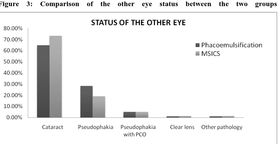
Other eye status
Cataract 64 (64.7%) 58 (73.4%)
Pseudophakia 28 (28.3%) 15(19.0%)
Pseudophakia with posterior capsule opacification
5 (5.0%) 4(5.0%)
Clear lens 1 (1.0%) 1 (1.3%)
Other pathology 1 (1.0%) 1(1.3%)
Other eye vision
6/6 to 6/18 50 (50.5%) 26 (33.3%)
6/24 to 6/60 41 (41.4%) 44 (56.4%)
5/60 to 3/60 5 (5.1%) 7(9.0%)
Less than 3/60 3(3.0%) 1 (1.3%)
DEMOGRAPHICS
Figure 1 - Age and gender distribution
The age distribution in the two groups was analyzed using the independent t-test and the mean age difference between those who underwent phacoemulsification and MSICS was not statistically significant (p = 0.06).
Gender distribution in the two groups was analyzed using the chi-square test which showed that here was no statistically significant difference in the number of men and women who underwent phacoemulsification and manual small incision cataract surgery (p = 0.361).
Gender Female Male A ge ( in y ea rs ) 90 80 70 60 50 40 5
Manual Small Incision Cataract Surgery Phacoemulsification
Thus the demographic profile of the eyes in the two study groups was comparable to each other.
Figure 2 – Comparison of the distribution of eye operated between the two groups
Distribution of eyes in the two groups was analyzed using chi square tests. The p value was 0.372 which suggested that there was no significant difference between the numbers of right or left eyes that were operated in the two groups.
Figure 3: Comparison of the other eye status between the two groups 60.60%
39.40% 67.10%
32.90%
0.00% 10.00% 20.00% 30.00% 40.00% 50.00% 60.00% 70.00% 80.00%
Right eye left eye
DISTRIBUTION OF THE EYE OPERATED
[image:43.595.75.526.510.744.2]Figure 4: Comparison of the other eye visual acuity for distance between the two groups
‘p’ value – 0.117
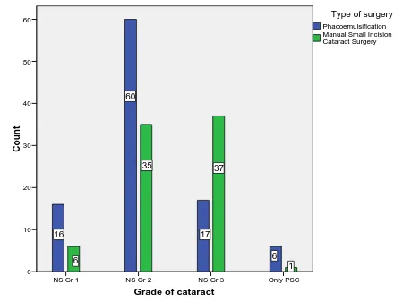
Table 2: Comparison of grades of cataract in the operated eye between the two groups
Grades of nucleus
Phacoemulsification N = 99
MSICS N = 79
n % n %
Nuclear sclerosis grade 1 16 16.16 6 7.6
Nuclear sclerosis grade 2 60 60.60 35 44.30
Nuclear sclerosis grade 3 17 17.17 37 46.83
Posterior subcapsular cataract 6 6.06 1 1.27
99 100 79 100
Figure 6: distribution of grades of cataract in the operated eye between the two groups
Grade of cataract
Only PSC NS Gr 3
NS Gr 2 NS Gr 1
Co unt 60 50 40 30 20 10 0 1 37 35 6 6 17 60 16
Manual Small Incision Cataract Surgery Phacoemulsification
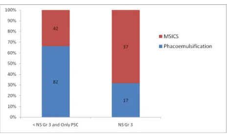
Eyes in the two study groups were subcategorized based on the cataract grade into hard cataracts and soft cataracts. Hard cataracts included nuclear sclerosis of grade 3 or more and the soft ones included nuclear sclerosis of grade 2 or less and/or posterior subcapsular cataracts.
Figure 7: Hard and soft cataracts in the two groups
INTRAOCULAR LENS IMPLANTED
[image:47.595.150.445.241.456.2]All eyes in the MSICS group had a rigid intraocular lens implanted, whereas foldable IOL was put in all eyes that underwent corneal section phacoemulsification. 2 foldable IOLs were placed in the ciliary sulcus due to posterior capsule rent during phacoemulsification.
Table 3: Different types of foldable IOL implanted after phacoemulsification
Type of lens Phacoemulsification N = 99
N %
Auroflex 82 82.8
Acrysof 5 5.1
Acrysof IQ 10 10.1
AMO 2 2
Total 99 100
[image:47.595.76.522.506.759.2]VISUAL PROFILE
Our study shows that there was a definite improvement in visual status after cataract surgery. Though the pre-existing spherical error decreased with cataract surgery, an increase in the mean subjective cylinder was noted post-operatively. But this can be explained based on the fact that the surgical process by itself induces a certain amount of astigmatism (surgically induced astigmatism) which may add on or negate the pre-existing astigmatism. There was a concurrent increase in the keratometric astigmatism postoperatively signifying that some part of the surgically induced astigmatism may be because of the changes in the corneal curvature during and after surgery.
Table 4: Pre and postoperative patient visual data – “Phacoemulsification group”
Parameter Preoperative Postoperative
BCVA
Median 6/18 6/6
Range 6/9 or less 6/6 to 6/12
Sphere (D)
Mean ± S.D -1.49 ± 1.96 -0.26 ± 0.56
Range -7.00 to +2.00 -3.50 to +1.00
Keratometric astigmatism (D)
Mean ± S.D 0.86 ± 0.53 1.07 ± 0.64
Table 5: Preoperative and post operative patient data – “MSICS group”
Parameter Preoperative Postoperative
BCVA
Median 6/36 6/9
Range 6/9 or less 6/6 to 6/36
Sphere (D)
Mean ± S.D -1.68 ± 2.13 -0.30 ± 0.54
Range -8.00 to +0.50 -2.50 to +0.75
Keratometric astigmatism (D)
Mean ± S.D 0.79 ± 0.57 1.56 ± 0.82
Range -0.50 to + 2.75 +0.05 to 4.50
Mean minimum K (D) ± S.D 43.87 ± 1.47 43.46 ± 1.59
Mean maximum K (D) ± S.D 44.66 ± 1.61 45.03 ± 1.65
Figure 9: Gain in visual acuity after cataract surgery
* For one patient post operative unaided visual acuity was not recorded.
The above bar graph shows that there is definite improvement in both unaided and best corrected visual acuity after cataract surgery as one would expect. 46.3% of the patients achieved an unaided visual acuity of better than 6/18. But 98.3% of patients attained a BCVA better than 6/18 which is well within the guidelines laid down by WHO to monitor the cataract surgical outcomes.
Figure 10: Comparison of visual acuity at 6 weeks in eyes that underwent MSICS
Table 6: Statistical analysis comparing the visual outcome in the two study groups Better than 6/18 6/18 to 6/60 Worse than 6/60 ‘p’ value Preoperative Unaided vision Phaco N=99
7 59 33
0.157 MSICS
N=79
1 50 28
BCVA
Phaco N=99
35 49 15
0.006 MSICS
N=79
11 52 16
Postoperative
Unaided vision
Phaco N=99
60 39 0
0.007 MSICS
N=78
26 52 0
BCVA
Phaco N=99
99 0 0
0.27 MSICS
N=79
76 3 0
Table 7: Comparison of post op BCVA for near at 6 weeks between the two groups
Near vision Phacoemulsification N = 99
MSICS N = 79
n % n %
J1 96 97 72 91.1
J2 3 3 5 6.3
J3 0 0 1 1.3
J5 0 0 1 1.3
Figure 12: Post operative best corrected visual acuity for near at 6 weeks
Table 8: Comparison of the number of patients who bought spectacles after cataract surgery in the two groups
Post operative unaided VA
Phacoemulsification N=99
MSICS N=78
Bought glasses Bought glass
Yes No Yes No
6/6 6 11 0 2
6/9 11 9 2 9
6/12 10 13 2 11
6/18 4 13 0 21
6/24 7 9 4 15
6/36 2 3 2 7
6/60 1 0 0 3
TOTAL 41(41.41%) 58 10(12.82%) 68
P value 0.48 0.42
In our study 41.41% of patients bought glasses after phacoemulsification whereas only 12.8% patients did the same after MSICS. This difference can be explained based on the differences in the socio-economic status, occupational needs, and status and visual acuity of the fellow eye in the two study groups.
SURGICALLY INDUCED ASTIGMATISM (SIA)
Table 9: Comparison of SIA after Phacoemulsification & MSICS
SIA Phacoemulsification
N= 99
MSICS N = 79
n % n %
<0.5D 14 14.14 11 13.92
0.5 to <1.00D 38 38.38 19 24.05
1 to <2.00D 44 44.45 34 43.04
2 to <3.00D 2 2.02 14 17.72
3 to 3.50D 1 1.01 1 1.27
The distribution of surgically induced astigmatism after the two techniques of cataract surgery was as shown in the table above. Only two of the 99 patients in the corneal section phacoemulsification group had high surgically induced astigmatism of ≥ 2.0D. There was no notable cause for this extreme SIA in these eyes. Following Blumenthal technique of manual small incision cataract surgery 15 eyes had high astigmatism of ≥ 2.0D. In the MSICS group three patients had wound problems during the surgery. Scleral sutures were placed in two cases. At 6 weeks these eyes had SIA of 2.22D and 0.84D
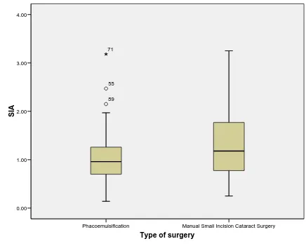
Table 10: Statistical analysis of the SIA following phacoemulsification and MSICS
SIA
Corneal section Phacoemulsification
Blumenthal technique of manual small incision
cataract surgery
P value
Mean 1.01 ± 0.49 D 1.29 ± 0.71 D
0.003
Median 0.96D 1.18D
Minimum 0.14D 0.25D
Figure 13: SIA after different techniques of cataract surgeries
The mean surgically induced astigmatism was calculated and compared between the two surgical groups using independent t-test. The mean SIA in those who underwent Phacoemulsification was 1.01 D ± 0.49 D and in those who underwent manual small incision cataract surgery was 1.29 D ± 0.71 D. There was a mean difference of 0.28 D surgically induced astigmatism between the 2 groups. Phacoemulsification induces 0.28 D astigmatism less than MSICS (p<0.05). This result though statistically significantly different may not be of much clinical significance.
Type of surgery
Manual Small Incision Cataract Surgery Phacoemulsification
SIA
4.00
3.00
2.00
1.00
0.00
71
55
FACTORS AFFECTING SURGICALLY INDUCED ASTIGMATISM
[image:57.595.73.528.396.658.2]Position of intraocular lens
Table 11: Comparison of the position of the IOL after the two surgical techniques
Position of IOL Phacoemulsification N= 99
MSICS N = 79
n % n %
In the bag 94 95 29 36.7
In the sulcus 2 2.0 20 25.3
Partly in the bag 1 1.0 6 7.6
Indeterminate 2 2.0 24 30.4
99 100 79 100
Figure 14: Comparison of the position of the IOL in the two study groups
Effect of the position of intraocular lens in the eye on SIA
Figure 15: Comparison of SIA in eyes with different positions of IOL
In a cataract surgery the ideal placement of the IOL would be in the bag. But sometimes it may not be possible and the lens may be in the ciliary sulcus or partly in the bag and partly in the sulcus. In such situations one would expect induced lenticular astigmatism which can affect the SIA. There we analyzed our data using ANOVA test to compare the SIA with different positions of IOL. There is no statistically significant difference in the IOL position in affecting surgically induced astigmatism (p>0.05).
Effect of age on surgically induced astigmatism
iol position
indeterminate mixed
its itb
SIA
4.00
3.00
2.00
1.00
0.00
71
155 146
138 55
89
Figure 16: effect of age on SIA in the two groups
With increasing age changes in corneal characteristics have been described in the past. Therefore we decided to study whether SIA differs with age in our patients or not. A regression analysis was done and a graph was plotted as shown above. Different values of SIA were seen to be equally distributed in all age groups in both phacoemulsification and MSICS arms. Thus it can be said that age does not affect SIA significantly.
SIA 4.00 3.00 2.00 1.00 0.00 Ag e ( in yea rs) 90 80 70 60 50 40
Fit line for Total Manual Small Incision Cataract Surgery Phacoemulsification Type of surgery
Effect of position of the incision (Supero-temporal or Supero-nasal) on SIA in the
Phacoemulsification group
[image:60.595.62.524.480.719.2]In our study superior scleral section incision was made for all patients who underwent manual small incision cataract surgery. For phacoemulsification clear corneal section incision was made at 10:30 ‘O’ clock position for all patients. This meant that the incision was made superotemporally in the right eye and superonasally in the left eye. Superonasal incisions cause more surgical discomfort for the surgeon and therefore technically slightly more difficult. SIA has been described in literature to vary with the side operated (right or left eye). Therefore we subdivided out phacoemulsification arm into right and left eye and then further analyzed the SIA in the two subgroups.
Table 13: Comparison of SIA between the two eye following phacoemulsification
SIA Right eye
(Supero-temporal insicion) N= 60
Left eye
(Supero-nasal incision) N = 39
n % n %
<0.5D 10 16.67 4 10.26
0.5 to <1.00D 25 41.67 13 33.34
1 to <2.00D 24 40.00 20 51.28
2 to <3.00D 1 01.66 1 02.56
Figure 17: Variation in SIA based on the difference in the site of incision between the two eyes in phacoemulsification
Table 14: Statistical analysis of the difference in astigmatism with change in site of the cataract incision SIA Right eye (Supero-temporal incision) Left eye (supero-nasal incision)
Mean 0.94D 1.11D
Std. error of mean 0.059D 0.087D
Median 0.92D 1.02D
Mode 0.88D 1.14D
Std. deviation 0.45D 0.54D
Minimum 0.14D 0.31D
Maximum 2.47D 3.18D
‘p’ value – 0.09 0.00% 10.00% 20.00% 30.00% 40.00% 50.00% 60.00%
<0.5D 0.5 to <1.00D 1 to <2.00D 2 to <3.00D 16.67% 41.67% 40% 1.66% 10.26% 33.34% 51.28% 2.56%
Variation in SIA based on the eye operated
[image:61.595.61.524.462.727.2]In our study left eye had slightly more surgically induced astigmatism as compared to right eye. Mean SIA in the left eye was 1.11D as compared to only 0.94D in the right eye. Statistical analysis was done using the independent t-test test and the difference of means between the two eyes was found to be0.17. This was statistically not significant.
[image:62.595.71.528.277.578.2]CORRELATION BETWEEN SIA & POST OPERATIVE PRESCRIBED CYLINDER
Figure 18: Comparison of the prescribed cylinder at 6 weeks in the two arms
Figure 19: Comparison of the SIA in the two groups
17.17%
14.14%
33.34% 32.32%
3.03%
6.33% 7.59%
34.18% 36.71% 13.92% 1.27% 0.00% 5.00% 10.00% 15.00% 20.00% 25.00% 30.00% 35.00% 40.00%
None 0.5D 0.75 to 1.00D 1.25 to 2.00D 2.25 to 3.00D 3.25 to 3.50D
POST OPERATIVE REFRACTIVE CYLINDER PRESCRIBED
Phacoemulsification
Table 15: Statistical comparison between the SIA and prescribed cylinder
Phacoemulsification MSICS ‘p’ value
Mean induced astigmatism 1.01D ± 0.49D 1.28D ± 0.70D 0.003
Mean of the magnitude of prescribed cylinder
-0.94 ± 0.63D -1.36 ± 0.77D 0.00
Mean SIA and mean of the prescribed cylinder in the two surgical groups was analyzed using Independent t-test and a significant statistical difference was noted.
14.14%
38.38%
44.45%
2.02% 1%
13.92% 24.05% 43.04% 17.72% 1.27% 0.00% 5.00% 10.00% 15.00% 20.00% 25.00% 30.00% 35.00% 40.00% 45.00% 50.00%
None 0.5D 0.75 to 1.00D 1.25 to 2.00D 2.25 to 3.00D 3.25 to 3.50D SURGICALLY INDUCED ASTIGMATISM
Phacoemulsification
TARGET SPHERICAL ERROR
Table 16: Pre operative target spherical error in the two groups IOL power aim Phacomemulsification
N = 97
MSICS N = 79
P value
n % n %
0.50
More than -2D 0 0 0 0
-1.01 to -2D 2 2 0 0
-0.51 to -1.0D 8 8 6 7.5
-0.01 to -0.5D 64 33 60 76
Emmetropia 1 1 1 1.3
+0.01 to +0.5D 22 22 12 15.2
+0.51 to +1.0D 0 0 0 0
97 100 79 100
MEAN TARGET SPHERICAL ERROR
-0.19 ± 0.25D -0.16 ± 0.21D
[image:64.595.76.523.430.711.2]POST OPERATIVE SPHERICAL ERROR
Table 17: Post operative subjective spherical prescribed at 6 weeks
Spherical correction Phacoemulsification N = 99
MSICS N = 79
n % N %
More than -2D 01 1.0 01 1.3
-1.25 to -2D 03 3.0 03 3.9
-0.75 to -1.0D 16 16.2 11 15.9
-0.25 to -0.5D 15 15.2 21 26.6
Emmetropia 57 57.6 36 45.6
+0.75 to +1.0D 01 1.0 01 1.3
+0.25 to +0.5D 06 6.1 06 7.6
[image:65.595.72.531.389.638.2]99 100 79 100
Figure 21: Comparison of the spherical error prescribed at 6 weeks in the two groups
INTRA OPERATIVE COMPLICATIONS
Table 18: Comparison of intraoperative complications between Phaco and MSICS
Complication Phacomemulsification N = 99
MSICS N = 79
n % n %
None 95 96 73 92.4
Posterior capsule rent 2 2 0 0
Hazy cornea 1 1 1 1.3
Bleeding 0 0 3 3.8
Wound suturing 0 0 2 2.5
others 1 1 0 0
[image:66.595.75.515.389.648.2]99 100 79 100
Figure 22: Intraoperative complications
IMMEDIATE POST OPERATIVE COMPILCATIONS – DAY 1
Table 19: Comparison of immediate postoperative complications b/w Phaco & MSICS
Complication Phacomemulsification N = 99
MSICS N = 79
n % n %
None 74 74.47 64 81
Epithelial defect 2 2 1 1.3
Corneal oedema 18 18.2 1 1.3
Bleeding 0 0 2 2.5
High IOP 4 4 6 7.6
Wound leak 0 0 2 2.5
PCO 1 1 0 0
Exudative membrane 0 0 3 3.8
99 100 79 100
Figure 23: Comparison of the immediate postoperative complications in the two groups
LATE POST OPERATIVE COMPLICATIONS – 6 WEEKS
Table 20: Comparison of late postoperative complications b/w Phaco & MSICS
Complication Phacomemulsification
N = 99
MSICS N = 79
n % N %
None 94 95 68 86.1
Posterior capsule opacification 2 2 6 7.6
Corneal oedema 1 1 0 0
Cystoid macular oedema 1 1 0 0
IOL decentration 1 1 3 3.8
Optic capture 0 0 2 2.5
99 100 79 100
[image:68.595.76.503.514.753.2]SUBJECTIVE VISUAL FUNCTION ASSESSMENT
Improvement in visual function after cataract surgery
(Visual Function Score)
[image:70.595.78.480.303.627.2]Visual function score = Difference between the post and pre operative visual function
Figure 25: comparison of the subjective improvement in visual function in the 2 groups
Visual function score was analyzed using the independent t-test. There was a statistically significant difference in the mean visual function score between phacoemulsification and manual small incision surgery (p<0.001). The subjective improvement in visual function was
Type of surgery
Manual Small Incision Cataract Surgery Phacoemulsification
Vi
sua
l F
unc
ti
on
Sc
ore
100.00
80.00
60.00
40.00
20.00
Postoperative visual function
Visual function score indicates the dramatic improvemant in the visual perception after cataract surgery and this is significantly dependent on the pre-operative visual disability. More the visual disability before surgery, better will be the VF-score.
[image:71.595.101.500.310.631.2]Postoperative visual function is a better indicator of patient satisfaction as it tells about the final subjective visual function status as experienced by the patient
Figure 26: Comparison of the post-operative visual function in the two groups
On comparison of post-operative visual outcome between the 2 groups, phacoemulsification had a slightly better outcome, but this was statistically not significant (p>0.05).
Type of surgery
ENDOTHELIAL CELL LOSS FOLLOWING CATARACT SURGERY
[image:72.595.74.527.351.622.2]Valid endothelial cell count pre and post operatively was available only for 73 eyes in the phacoemulsification group and 44 patients in the MSICS group.
Table 21: Comparison of the endothelial cell loss following surgery in the two gropus
Endothelial cell loss
Phacoemulsification N = 73
MSICS N = 44
n % n %
Less than 500 48 65.75 37 84.1
500 to less than 1000 15 20.55 5 11.36
1000 to less than 1500 5 6.85 2 4.54
More than 1500 5 6.85 0 0
73 100 44 100
Table 22: Mean (SD) pre- and postoperative endothelial cell counts and surgically induced endothelial cell loss
Phacoemulsification N = 73
MSICS N = 44
‘p’ Value
Preoperative mean – cells/mm2 (SD) 2566.63 (374.94) 2522.43 (355.05) 0.530
Postoperative mean– cells/mm2 (SD) 2039.71 (514.64) 2251.59 (335.90) 0.008
Endothelial cell loss – cells/mm2 (SD) 526.91 (474.72) 270.84 (280.34) <0.001
FACTORS AFFECTING THE EXTENT OF ENDOTHELIAL CELL DAMAGE DURINF CATARACT SURGERY
Technique of cataract surgery
Table 23: Comparison of the endothelial cell loss after Phacoemulsification & MSICS
Type of surgery N Mean
(cells/mm2)
Std. Deviation
Std. Error Mean
p-value
Phacoemulsification 73 526.92 474.73 55.56
0.002
MSICS 44 270.84 280.35 42.26
Diabetic status
Table 24: Comparison of endothelial cell loss in diabetics versus non diabetics
Diabetes
Group statistics Independent
samples test
N Mean
(cells/mm2)
Std. Deviation
Std. Error Mean
p-value 0.223
Yes 41 496.76 451.01 70.43
No
76 394.93 416.23 47.74
There was no statistically significant effect of diabetes on the extent of endothelial cell damage during cataract surgery in our study
Grades of cataract
CHANGE IN CENTRAL CORNEAL THICKNESS AFTER CATARACT SURGERY
Table 25: comparison of changes in CCT following cataract surgery in the two groups
Change in central corneal thickness
Phacoemulsification N = 96
MSICS N = 77
N % N %
Gain in CCT Less than 10 microns
28 29.17 23 29.88
10 to less than 20 microns
17 17.71 11 14.28
20 to less than 30 7 7.29 5 6.49
30 to less than 40 1 1.04 1 1.3
More than 40 1 1.04 1 1.3
Loss in CCT 42 43.75 36 46.75
Total 96 100 77 100
For three patients in the phacoemulsification group and two in the MSICS group, central corneal thickness values could not be recorded.
Table 26: Independent samples test
N Mean Standard deviation P value
Phacoemulsification 96 -0.22 18.61
0.904
MSICS 77 +0.10 16.05
CORRELATION BETWEEN ENDOTHELIAL CEL LOSS AND CHANGE IN CENTRAL CORNEAL THICKNESS
Figure 28: Correlation b/w endothelial cell loss and increase in CCT
DISCUSSION
In India cataract is still the leading cause of blindness. Surgery being the only treatment option available, low cost surgical techniques that are quick and easy to master are the demand of time. Manual small incision cataract surgery has emerged in a big way because it avoids the disadvantages of ECCE and at the same time enjoys the advantages of phacoemulsification. It is inexpensive, quick, has a shorter learning curve, can deal with all types of cataract, and relatively safe even in inexperienced hands. With improved instrumentation and technological advancements MSICS is slowly becoming the preferred surgical technique for most low cost, high volume surgical setups. The visual outcome following MSICS is slowly improving though not comparable to phacoemulsification. We did the study to compare the visual outcome following corneal section Phacoemulsification and Blumenthal technique of manual small incision cataract surgery.
In our study the demographic profile of the patient in terms of age and gender of the subjects in the two study groups was similar. This made the data in the two arms comparable.
it is well known that phacoemulsificaion is more difficult in hard cataracts especially mature and hypermature (67). Difficulty in completing the continuous curvilinear capsulorhexis and emulsification of the hard nucleus makes the surgery challenging (68). The higher rates of intraoperative complications (69) may lead to surgeons erring on the side of MSICS rather than phacoemulsification for these cases. But studies have shown that phacoemulsification is quite safe and well tolerated procedure for all kinds of cataract in experienced hands (70) (69).
The post operative visual outcome was analyzed for the two surgical groups. In our study overall 48.6% of the patients achieved an unaided visual acuity of better than 6/18. On spectacle correction this figure improved to 98.31%. On analyzing the phacoemulsification and MSICS group separately, visual outcome following corneal section Phacoemulsification was better than Blumenthal technique of MSICS at 6 weeks. 60.6% of patients achieved uncorrected visual acuity of 6/18 or better after phacoemulsification as compared to only 33.33% in the MSICS group. Best corrected visual acuity of better than 6/18 was attained in 100% after phacoemulsification and 96.2% after MSICS. A study done by George et al, where they studied 62 eyes with phacoemulsification and 62 with MSICS, has shown that BCVA of better than 6/18 was achieved in100% of those who had Phacoemulsification and 98.2% of patients who had MSICS. They did not comment on the unaided visual acuity(6). Another Indian study reported unaided visual acuity of 6/18 or better in 47.9% of patients after MSICS at 6 weeks postoperatively (44). Our results are comparable with these studies.