THE VALUE OF AgNOR STUDY IN RADIOTHERAPY TREATED SQUAMOUS
CELL CARCINOMA OF THE CERVIX - AS A DIAGNOSTIC AND
PROGNOSTIC MARKER
DISSERTATION
SUBMITTED FOR M.D.BRANCH III
(PATHOLOGY)
MARCH – 2007
CERTIFICATE
This is to certify that this dissertation entitled “ THE VALUE OF
AGNOR STUDY IN RADIOTHERAPY TREATED SQUAMOUS CELL CARCINOMA OF THE CERVIX - AS A DIAGNOSTIC AND PROGNOSTIC MARKER ” is the bonafide record work done by Dr.D.SATHIYABAMA submitted as partial fulfillment for the requirements of M.D. Degree Examinations Branch III Pathology, March 2007.
Dr.T.B.UMADEVI M.D.,
THE DEAN
Professor & Head of the Department, Thanjavur Medical College, Department of Pathology, Thanjavur.
ACKNOWLEDGEMENT
I wish to express my sincere and profound gratitude to my Professor Dr.T.B.Umadevi
M.D(Pathology) Professor and Head of the Department of Pathology, Thanjavur Medical College, Thanjavur, for her valuable guidance at every stage, constant encouragement and words of advice which have been the motivating forces in bringing forth this piece of work.
My heartfelt thanks to Dr.C.D.Balakrishnan M.D., Reader of Pathology for his valuable guidance and encouragement throughout my study.
I am also extremely grateful to Dr.Arumugam M.D., Reader of Pathology who has offered many valuable suggestions and encouragement during his period, who’s help has been invaluable to me.
My special thanks to Professor, Assistant Professors and working staffs of Radiotherapy Department , TMCH for their valuable help in carrying out this study.
I thank my Assistant Professors for their valuable suggestions and guidance at every stage in this study.
I would also like to express my sincere thanks to my fellow-Postgraduates, and all the technical staffs of the department for their generous help throughout my study.
CONTENTS
Page No.
1.
INTRODUCTION
1
2. AIM OF THE STUDY
4
3. MATERIALS AND METHODS
5
4. REVIEW OF LITERATURE
7
5. OBSERVATION AND RESULTS
31
6. DISCUSSION
42
7. CONCLUSION
53
APPENDIX
INTRODUCTION
Carcinoma Cervix is the second most common cancer worldwide especially in perimenopausal period. In developing countries like Asia, Africa and South America it is the commonest malignancy largely due to lower socioeconomic status, poor hygiene, illiteracy and paucity of screening programs.(14,15,51)
Pre – Cancerous lesions precedes a majority of cervical carcinoma and may exist in the non invasive stage for longer periods and shed abnormal cells which can be detected by exfoliative cytology using papanicoloau stain (2,14,15). Papanicoloau smears remain a simple screening method for detecting the precancerous lesions of cervix inspite of limitations due to sampling and preparation errors. With effectiveness of Papanicoloau cytological test and the accessibility of the cervix by colposcopic examination and biopsy, the detection rate of precancerous lesions have improved.
In invasive carcinoma of cervix, which are largely detected by biopsies, cancer cell proliferation greatly influences the clinical out come of the patients. The available evidences indicate that evaluation of cell kinetic parameters may help surgical Pathologists and Oncologist to define the biological behaviors of cancer.
calculated as the S-Phase fraction. The main disadvantage is that specimen is destroyed in the process, thus cellular subpopulations within the tumour or admixtures of normal cells cannot be evaluated. Immunohistochemical methods for cell proliferation markers like Ki – 67, PCNA (Proliferating Cell Nuclear Antigen), DNA polymerases can be used, but they are more complicated and expensive than the silver stain used to show Nucleolar Organizing regions.(13,15)
A significant correlation between the interphase AgNOR (Argyrophilic Nucleolar Organizer Regions) quantity and prognosis has been reported in many neoplasms like Carcinoma breast, Lung, Gastrointestinal tract, and hematological malignancies (1,3,4,5,6,7,8,13,30). There was A CLOSE CORRELATION BETWEEN THE PROLIFERATIVE BEHAVIORS AND THE QUANTITATIVE AND QUALITATIVE CHANGE OF AgNOR (SILVER STAINED NUCLEOLAR ORGANIZER REGIONS) WAS FOUND IN OBVIOUSLY MALIGNANT NEOPLASMS.
There are also many studies using AgNOR conducted in preinvasive and invasive squamous epithelial lesions of cervix and AgNOR(20,22,2,3,24,27,28,31,32,34,46,48,51,52,54) proved to be a simple inexpensive and reliable proliferation marker in lesions of cervix.
Radiotherapy, which is the main mode of treatment in Carcinoma Cervix. AgNOR proved to be a sensitive marker to predict the prognosis of the patient treated with radiotherapy. In a rural and semi-urban areas where sophisticated methods are not available, AgNOR COUNTS IN CERVICAL SMEARS PROVE TO BE A SIMPLE AND COST EFFECTIVE METHOD IN THE DIAGNOSIS, PROGNOSIS AND FOLLOW UP OF CARCINOMA CERVIX PATIENTS.(16,21,26)
Regions (NOR) are components of ribosomal DNA located in the short arms of acrocentric chromosomes of 13,14,15,21 and 22 and transcribe to ribosomal RNA. NOR’s vary in size and shape according to nucleolar transcription. They are intimately related to the cell cycle and may be RELATED TO PROLIFERATION AND PLOIDY.(1,7,18)
AIM OF THE STUDY
1) To evaluate the incidence and prevalence of Carcinoma Cervix.
2) To assess the value of AgNOR IN CERVICAL CANCERS AS WELL AS IN CONTROL.
3) To assess the AgNOR SENSITIVITY ACCORDING TO DIFFERENTIATION AND MORPHOLOGICAL CHARACTERIZATION.
4) To assess the PROLIFERATIVE ACTIVITY OF NEOPLASTIC CELLS AND IT’S PROGNOSIS.
MATERIALS AND METHODS
This study includes 50 Carcinoma Cervix patients referred from RAJA MIRASUDAR HOSPITAL which is affiliated to THANJAVUR MEDICAL COLLEGE, THANJAVUR during the two year period from JUNE 2004 to JUNE 2006 and 25 cases served as control.
A detailed clinical history like age, parity, age at menarche, years of married life, years after last delivery, hematological investigations, review of previous cytology and histopathological examination, family status/ socio economic status was done in all cases.
Cervical and Vaginal smears were collected from controls and from each patient prior to radiation therapy, using Wooden Ayres Spatula. By putting the woman on the examination table in dorsal position speculum was introduced into the vagina exposing the cervix and Wooden Ayres Spatula was introduced in to the posterior fornix. The Scraped material obtained was then spread on a slide. The pointed end of the Ayres Spatula was introduced into the cervical OS and rotated 360°, sampling the whole ectocervix. For each patient two slides were taken and they were fixed immediately with isopropyl alcohol.
The Cervical and Vaginal smears collected from controls and from patients prior to radiation therapy were subjected to AgNOR staining.
TECHNIQUE:
Single step AgNOR staining technique was employed for the demonstration of AgNOR’s (APPENDIX – 1). The freshly prepared solution was poured on to the smears which were then left in dark at 37°c for 30 minutes. Slides were dehydrated in 3 changes of acetone, cleared in Xylene and mounted in DPX.
The patients were given 40 – 50 gray of cobalt – 60 teletherapy divided in 20 – 25 fraction with daily fractions of 1.8 to 2.0 gray with five fractions per week.
In ideal patients, who respond to radiotherapy, as the tumor shrinks making the introduction of the uterine and vaginal applicators easy, the rest of the dose is delivered by brachytherapy by referring the patients to other higher centers.
REVIEW
OF LITERATURE
INVASIVE CARCINOMA OF CERVIX:
The cervix is the commonest site for female genital tract cancer; statistics vary considerably from country to country and from race to race. So in African and Asian women living in poor conditions, the incidence and relative mortality rate of the carcinoma cervix is 4-5 times higher than those seen in developed countries.(14,15,51)
AEITOLOGY:
Age: Although invasive cancer of cervix is reported at all ages, it has two peaks, one at about 35 yrs and another at 50-55 years, following which there is reduced incidence.(14,15,35,39)
0 10 20 30 40 50 60
20-24 25-29 30-34 35-39 40-44 45-49 50-54 55-59 60-64 65-69 70-74 75-79
AGE
Religion and Race:
The women of Orthodox Jews and in Muslims, the incidence of cervical cancer is less due to ritual circumcision done in Male Children at the childhood. Certain races it is unusually common, for example Africans they are susceptible even if they live in conditions of squalor.
Social and Economic Factors:
The people living in low Socio economic status are more prone to cervical carcinoma. The possible operating factors are low standards of cleanliness (including penile hygiene), coitus at an early stage, and frequency of sexual intercourse and promiscuity of both partners.
Coitus:
The sexually active women are 4-5 times more likely to develop cancer than sexually inactive women. It is postulated that smegma and spermatozoa are themselves carcinogenic.
Cervical Infection:
Human papilloma virus plays an important role in virtually all cases.
15-20 types of HPV have been associated with cervical carcinoma, type 16, 18, 31 and 45 accounts for 80% of cervical carcinoma. HPV 16 is associated with 50% of cases. (14,15,35,44)
Hormonal Factors:
There is some evidence that Oestrogen - Progesterone Oral Contraceptive pill usage favours CIN changes and also for Adeno carcinoma.
Predisposing Histological Status:
are sometimes confused with cancer include basal oell hyperplasia, squamous cell metaplasia and CIN. Of these, only CIN II-III are likely to significant forerunners of invasive carcinoma.(26,50)
Histological Classification of Cancer Cervix:
Carcinoma cervix nearly always starts at the squamo-columnar epithelial junction and 80-90% cases are squamous cell in type.
In 5-10% of cases it is entirely columnar cell in pattern (AdenoCarcinoma) and the frequency is increasing especially in young women who are smokers or pill-users. The remainder constitutes the mixed types.
WHO Histological Classification Of Tumors Of The Uterine Cervix
Epithelial Tumors
Squamous tumors and precursors
Squamous cell carcinoma, not otherwise specified Keratinizing
Non – Keratinizing Basaloid
Verrucous Warty Papillary
Lymphoepithelioma –like Squamotransitional
Early invasive (micro invasive) squamous cell carcinoma Squamous intraepithelial neoplasia
Cervical intraepithelial neoplasia (CIN) 3 / Squamous cell carcinoma in situ
Fibroepithelial polyp Glandular tumors and precursors
Adenocarcinoma
Mucinous adenocarcinoma Endocervical Intestinal Signet-ring cell Minimal deviation Villoglandular
Endometrioid adenocarcinoma Clear cell adenocarcinoma Serous adenocarcinoma Mesonephric adenocarcinoma Early invasive adenocarcinoma Adenocarcinoma in situ
Glandular dysplasia Benign glandular lesions
Müllerian papilloma Endocervical polyp Other epithelial tumors
Adenosquamous carcinoma
Glassy cell carcinoma variant Adenoid cystic carcinoma
Adenoid basal carcinoma Neuroendocrine tumors
Carcinoid
Atypical carcionoid Small cell carcinoma
Large cell neuroendocrine carcinoma Undifferentiated carcinoma
Mesenchymal tumors and tumor – like conditions
Leiomyosarcoma
Sarcoma botryoides Alveolar soft part sarcoma Angiosarcoma
Malignant peripheral nerve sheath tumor Leiomyoma
Genital rhabdomyoma
Postoperative spindle cell nodule
Mixed epithelial and mesenchymal tumors
Carcinosarcoma (malignant müllerian mixed tumors, metaplastic carcinoma) Adenosarcoma
Wilms tumor Adenofibroma Adenomyoma
Melanocytic tumors
Malignant melanoma Blue naevus
Miscellaneous tumors
Tumors of germ cell type Yolk sac tumor Dermoid cyst
Mature cystic teratoma
Lymphoid and haematopoetic tumors
Secondary tumors
Reagan et al (1957) recognized three groups of squamous cell carcinoma
φ Large cell Keratinizing type
φ Large cell Non – Keratinizing type
φ Small cell Non – Keratinizing type
and other categories have been adopted by World Health Organization (Ritton & Christopherson 1973).
Cervical Intraepithelial Neoplasia:
This is an intraepithelial neoplasia that encompasses a continuum of morphologic changes arising in the basal layer of the stratified squamous epithelium of the transformation zone and extending to the entire thickness of the epithelium. The progression is divided into CIN I,II, and CIN III. Almost 60% of CIN I and 50% of CIN II lesions regress spontaneously. The remainder either persists or progresses through intermediate stages to invasive cancer. The majority of CIN III lesions progress to invasive cancer over 10-20 years. The majority of high-grade lesions are associated with HPV-16, 18, 31 and 33.(14,15,35)
Symptoms and Signs:
In its early stage, invasive carcinoma causes no symptoms and is discovered accidentally. Irregular uterine bleeding, post coital bleeding, foul smelling discharge with an offensive odour are present in most cases.
Diagnosis:
Screening for prevention of cervical cancer
Cervical cytology with Papanicoloau Staining (APPENDIX 2) has been the mainstay of screening. The most effective method of obtaining a specimen is to use a cytobrush for the endocervical canal and scraping the ecto cervix with an Ayres spatula.(25,49)
Cytological screening has been established in most developed countries resulting in marked decrease in incidence of invasive cervical cancer.
The pre invasive-stage can be detected by the cervical smear test, which is simple, safe, in expensive and generally acceptable to women. All sexually active women are at risk and should be screened every year.
Cytological terminology of Cervical Precancer:
Papanicoloau’s Classification was used for many years and his classification I to V
have been adhered to until recently by some cytologists and gynecologists.(2,25,49)
Class I - Negative - Absence of atypical / abnormal cells Class II - Negative - Atypical cells present but without
abnormal features
Class III - Suspicious - Cells with abnormal features suggestive but not conclusive for malignancy
Class IV - Positive - Cells and cell clusters fairly conclusive for malignancy
WHO Classification:
Ritton and Christopherson used conventional histological terminology of mild, moderate and severe dysplasia and carcinoma in situ, as well as atypical metaplasia.
British Society for Clinical Cytology Terminology (BSCC):
BSCC’s first working party on terminaly recommended the term dyskaryosis
Cytology Expected histology
Mild dyskaryosis CIN I (Mild dysplasia) Moderate dyskaryosis CIN II (Moderate dysplasia) Severe dyskaryosis CIN III (Severe dysplasia)
Bethesda System:
The 1988 Bethesda system for reporting cervical / vaginal cytologic diagnosis was published. The workshop agreed that the Papanicoloau classification was no longer appropriate and proposed the three essential components of cervical / vaginal smear report.(2,25,49)
The 1991 Bethesda System for Reporting cervical / vaginal cytologic diagnosis: A) Statement of adequacy of the specimen
a) Satisfactory for evaluation when the specimen has correct
identification with a minimum of pertinent clinical information and the smears contain an adequate number of well preserved and well visualized epithelial cells, including endocervical.
b) Satisfactory for evaluation but limited-by lack of pertinent clinical data or smears have an excess of blood, inflammatory cells, contaminants or are poorly fixed with air drying artifacts etc or contain no adequate endocervical or transformation zone cells.
B) General Categorization:
(Optional, used to facilitate the triage of the specimens) a) Within normal limits
b) Benign cellular changes: see descriptive diagnosis c) Epithelial cell abnormality: See descriptive diagnosis
C) Descriptive Diagnosis:
a) Benign Cellular Diagnosis
1) Infection with organisms morphologically consistent with Trichomonas Vaginalis.
2) Fungal organisms consistent with candida species
3) Predominance of coccobacilli consistent with shift in vaginal flora 4) Bacteria consistent with Actinomyces sp.
5) Cellular changes associated with herpes simplex virus 6) others
b) Reactive changes associated with
1) Inflammation (includes typical repair)
2) Atrophy with inflammation (“atrophic vaginitis”) 3) Radiation and chemotherapy effect
4) Intra uterine contraceptive device 5) Other
c) Epithelial cell abnormalities I) Squamous cell
a) Atypical squamous cells of undetermined Significance qualify (AGUS)
encampassing changes of HPV and CIN I
c) High grade squamous intraepithelial lesion (SIL) encompassing changes of CIN II and CIN III
d) Squamous cell carcinoma d) Glandular cell
1) Endo metrial cells cytologically benign in a post meno pausal Woman
2) Atypical glandular cells of uncertain significance qualify 3) Endocervical adenocarcinoma
4) Endometrial adenocarcinoma 5) Extra uterine adenocarcinoma 6) Adenocarcinoma NOS
The Bethesda System includes a new term Squamous Intraepithelial Lesion (SIL) which is divided into two grades. Low grade SIL to include HPV and CIN I and High grade SIL for cells from CIN II and CIN III.
Comparison of terminologies used for abnormal squamous epithelial cells in cervical cytology:
CIN Grade WHO BSCC Bethesda
Borderline Atypia (ASCUS) I Mild dysplasia Mild dyskaryosis Low grade SIL II Moderate dysplasia Moderate dyskaryosis High grade SIL III Severe dysplasia
Carcinoma in Situ
Severe dyskaryosis High grade SIL
Epidemoid Ca Severe dyskaryosis Squamous Carcinoma ? invasive carcinoma
The Cytology of CIN and invasive squamous cell carcinoma Dyskaryosis
The term dyskaryosis means literally “abnormal nucleus”. The morphological abnormalities seen in the nucleus of epithelial cells in cervical smears include a combination of any number of the following.
1) Disproportionate nuclear enlargement 2) Hyperchromasia
3) Bi and Multinucleation
4) Irregularity in form and outline
5) Abnormal chromatin pattern appearing as coarsening, stippling, formation of clumps or strands and sometimes as condensation beneath the nuclear membrane producing apparent irregularities in its thickness
6) Abnormalities of the number, size and form of nucleoli
1) Cytological findings of Mild Dyskaryosis / CIN I
Disproportionate nuclear enlargement, the nucleus usually occupy less than half the area of the cell
Abnormal nuclear pattern
Irregularity of the nuclear membrane
Multiple abnormal nuclei
Cytoplasm reduced and usually thin
Cell borders are usually angular
2) Cytological findings of Moderate Dyskaryosis / CIN II
φ Disproportionate nuclear enlargement, the nucleus usually occupying between one half and two thirds of the total area of the cell
φ Abnormal chromatin pattern, often more marked than in mild dyskaryosis
φ Irregular nuclear membrane
φ Nuclear hyperchromasia
φ Multiple abnormal nuclei
φ Cytoplasm reduced, may be thick or thin
φ Cell borders may be angular / rounded
3) Cytological findings of Severe Dyskaryosis and CIN III
o Disproportionate nuclear enlargement with the nucleus usually occupying at least two thirds of total area of the cell
o Abnormal chromatin pattern
o Nuclear hyperchromasia
o Irregularity of nuclear membrane
o Multiple abnormal nuclei
o Cytoplasm markedly reduced abnormal maturation of cytoplasm including Keratinization
o Nucleoli prominent when CIN III is wide spread
o Bizarrely shaped cells sometimes including fibre cells
4) Micro invasive and Invasive Squamous Cell Carcinoma Cytological features of invasive carcinoma:
Most cytologic specimens are obtained by direct scraping of the lesion. The most important sampling error that may lead to false negative interpretation is when the clinicians scrapes the bottom of the ulceration rather than its margins which will contain only debris, blood and inflammatory cells, mixed with a few hard to recognize degenerated cancer cells.
Well Differentiated (Keratinizing) Squamous Cell Carcinoma
They make up to 20% of invasive cervical squamous cell carcinoma
Cellular Morphology:
-The cancer cells shed singly and in clusters (55%) sheets (30%), or malignant pearl (2%) or in synticial form (13%)
-The shape of the cell is very pleomorphic which may be polygonal (55%), flat, round (10%), tadpole (10%), spindle (5%), irregular or pearl formation (10%)
-The size of the nuclei ranges from 2-10 times the size of normal squamous cells. The shape is round (30%), oval (20%) or irregular (40%) and varies with the shape of the entire cell (elongated) in spindle cells and round, irregular in polygonal cells
-Background of the smear shows a large number of nonspecific Inflammatory cells and cellular debris (Tumor diathesis)
Moderately differentiated (Nonkeratinizing large cells carcinoma)
70 % of the cervical cancers are of this type
Cellular Morphology:
o The numerous diagnostic cells are large either single (70%) or syncytial formation (30%). Epithelial sheets and pearl formations are absent. The cells vary in shape (round to polygonal)
o The cytoplasm is usually basophilic varies in amount and contain little mature keratin
o The nuclei are round or oval and less irregular, chromatin is coarse and irregularly clumped
o The eosinophilic nucleoli are often larger and more prominent than seen in well differentiated cancer cells
o The background of the smear shows severe tumor diathesis, containing large amounts of degenerate cellular debris red blood cells and protein deposits
Poorly differentiated squamous cell carcinoma
Based on their cellular morphology and ultra structures, some of
these cancers are thought to be Neuroendocrine origin. They are often located toward the endocervical canal and represent 7% of invasive cervical carcinoma.
Cellular Morphology:
The cells vary moderately in size and shape and are usually either round or oval. The delicately vacuolated cytoplasm of these cells is basophilic (80%), scanty and often seen as a narrow rim. Because of the fragility of this cytoplasm large number of “naked” malignant nuclei are usually present in the smear.
The nuclei are angular / oval shaped, with irregular chromatin clumps, multi nucleation is common. In clusters, the cells may overlap, with intercellular or slit like spaces.
- Pseudo cannabalism (birds eye appearance) is common - Severe tumor diathesis is seen
HISTOLOGY:
In Micro invasive carcinoma, usually displays extensive CIN III, from which tongues and islands of neoplastic cells extend through the basement membrane in a spray like pattern of later as confluent invasion.
Well Differentiated Squamous Cell Carcinoma includes the presence of large islands of
infiltrating tumor cells with intercellular bridges, epithelial pearl formation and obvious kernatinization of the cell cyto plasm.
Moderately Differentiated Squamous Cell Carcinoma show some evidence of a squamous
origin, consisting of solid islands and smaller groups of polygonal pleomorphic cells, but kertinization and intercellular bridges are less obvious and pearl formation is not seen.
Poorly Differentiated Squamous Cell Carcinoma invades as sheets, islands and single cells
The Nucleolar Organizer Regions – A Review
Nucleolar Organizer Regions (NOR’s) are DNA loops transcribing toribosomal RNA’s. In the human karyotype, NOR are secondary constrictions of metaphase chromosomes and are located in the short arms of the acrocentric chromosomes 13,14,15,21 and 22. In chromosome preparations, NOR can be clearly visualized using In Situ hybridization or by ammoniacal or formic acid silver nitrate staining. The silver stainability or NOR is due to the presence of a peculiar set of acidic proteins, which are highly argyrophilic.(3,37,38)
Structure and Function of Interphase NOR
The nucleolus is a well defined structural, functional domain of the cell nucleolus in which ribosomal genes are located and ribosomal biogenesis occurs. Mammalian cell nucleoli constantly exhibit apart from intranuclear chromatin, three main components.
1) The Fibrillar Centers, which appear as rounded structures of different size composed of very thin loosely, inter oven fibrils.
2) The Dense Fibrillar Component frequently located at the periphery of the fibrillar centers, which is composed of densely packed fibrils.
3) The Granular component composed of granules surrounding the fibrillar components. The fibrillar centers plus the closely associated dense fibrillar component have been identified as the inter phase counterparts of metaphase NOR.(37,38)
Interphase NOR and Nucleolar Pathology:
The size and shape of the nucleoli is highly variable from cell to cell and in the same type of cell from the resting to proliferative state. Ultrastructural studies have shown that this variability is due to different distribution of the nucleolar components. A solitary large fibrillar center surrounded by a rim of ribonucleoprotein characterizes the very small nucleoli of resting lymphocytes. Whereas numerous small fibrillar centers, each surrounded by a rim of dense fibrillar component frequently intermingled with granules is characteristic of large nucleoli of phytohaemagglutinin stimulated lymphocytes.(37,38)
Until recently, a precise evaluation of nucleolar morphology, and its change during cell activation or transformation was impossible to be performed in routine cytopathology and histology at light microscopial level. Small nucleoli are frequently undetected and large nucleoli, independent of their size and shape are generally defined as “Prominent”.
In 1986, Ploton and co-worker applying the formic acid silver nitrate staining method for AgNOR proteins at the light microscopy level, succeeded in a very precise visualization of interphase NOR. By means of this procedure interphase NOR appear as well defined black dots evaluated by light microscopy, and each dot correspond to one interphase NOR as visualized by the electron microscope.(37,38)
How can Interphase AgNOR be quantified?
The method more frequently employed consisted of counting the number of interphase AgNOR per cell, evaluating the sample directly at the light microscope by carefully focusing throughout the section thickness at very high magnification (100 x oil immersion level) (5,7,8).
Interphase AgNOR and Diagnosis of Malignancy:
Ploton and co-workers have demonstrated that malignant cells have a greater quantity of interphase AgNOR than the corresponding benign or normal cells and therefore evaluation of interphase AgNOR distribution may be useful for the diagnosis of malignancy.(37,38)
According to Crocker, interphase AgNOR quantity can be recommended as a suitable parameter for diagnosis of malignancy only in a few type of cancers. AgNOR values were found to be useful in differentiating Nevo cellular nevi and malignant melanoma, between infiltrating lymphocytes and Oat cell carcinoma deposits in bronchial material, between reactive mesothelial proliferation and mesothelioma, normal cirrhotic and carcinomatous liver, between benign and malignant salivary gland tumors and between low grade and high grade Non Hodgkins Lymphoma.
(6,7,8)
Therefore AgNOR quantify can be considered only as one, among the other well established cyto-histopathological parameters to be used for diagnosis of malignancy.
Nevertheless, it is incontestable that in a broad sense, the quantify of interphase AgNOR is greater in cancer than in normal or hyperplastic cells.
Why do Cancer Cells have a large number of interphase AgNOR?
Considering the central role of interphase AgNOR in rRNA synthesis, changes of interphase NOR distribution in cancer cells might be the consequence of the increased demand for ribosomal biogenesis which characterizes dividing cells.
INTERPHASE AgNOR AND CANCER PROGNOSIS
The data reported above concerning the relationship between interphase AgNOR and cell proliferation clearly indicate that the quantity of interphase AgNOR’s can be considered as a parameter of cell kinetics. The greater the interphase AgNOR amount, the shorter the cell doubling time.(2,9,14)
The cell kinetic parameters may help pathologists and clinical oncologists to define the biological behaviors of cancer lesions, obtain valuable prognostic information and indications about specific treatments of cancer.
Neverthless, a significant correlation between interphase AgNOR quantity and patient survival has been reported in many types of neoplastic diseases.(34,41) In two types of cancer lesions interphase AgNOR evaluation has been proved to be of great importance.
1) Patients with lower number of interphase AgNOR in colorectal cancers still alive after 5 years, than those who presented with higher counts.
Multivariate analysis of survival in both these cancers demonstrated that quantity of AgNOR represented the most important prognostic parameter when compared to histological grade.
Interphase AgNOR quantification appears to be very interesting and promising method for the routine evaluation of cell kinetics for prognostic purposes. It is the only method which permits information to be obtained on the rapidity of cell proliferation in routinely processed samples. Only flow cytometry, by simultaneous analysis of DNA content and incorporation of injected bromo deoxyuridine in vivo, permits measurement of cell doubling time.
Interphase AgNOR staining is not expensive and very rapidly executed on the cytological smear and tissue sections. Moreover AgNOR distribution in cancer tissues can be directly perceived at light microscopic examination without using additional instruments for objective AgNOR quantification.(34,39,41)
RADIATION FOR CERVICAL CANCERS:
All cervical cancers, except stage I and II A are primarily treated with radiation therapy. The chief modalities of present day radiation treatment of cancer cervix are external photon beam treatment and brachytherapy.
External Beam Radiotherapy ( EBRT ) (Teletherapy):
Brachytherapy:
The word “Brachy” in Greek means “short distance”. It implies placing radioactive sources in contact with or very close to target tissue. The implants are classified into High Dose Rate (HDR) and Low Dose Rate (LDR). Dose rates of 0.4 to 2 gy/hour are delivered in LDR brachytherapy so that the treatment time is 24-72 hours. In HDR brachytherapy implant uses dose rates > 12 gy/hour and the total dose can be delivered in few minutes.
Isotopes used in LDR brachytherapy are Cs137 (caesium) and for HDR brachytherapy
OBSERVATION AND RESULTS
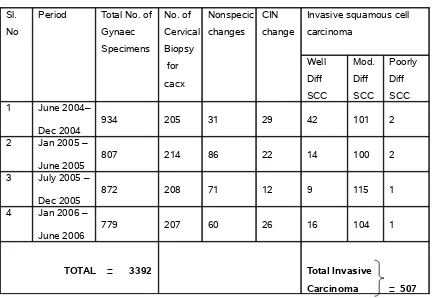
[image:32.612.75.493.401.665.2]The prospective study included 50 exfoliative cytology specimens of clinically evaluated and diagnosed cases of squamous cell Carcinoma of Cervix. This study also includes 25 control specimens for comparison and correlation of efficacy of AgNOR in cervical cancers. The clinical, cytological feature, histopathological data and AgNOR study conducted in cases are listed in the master chart. Table-1 shows the incidence of cervical cancers, referred for cytological evaluation and subsequent confirmation by histopathological examination, during the period from JUNE 2004 TO JUNE 2006.
TABLE-1
Sl.No Period Total No. of Pap
smears Positive cases
1 June 2004 – December 2004 280 10
2 January 2005 – June 2005 1201 15
3 July 2005 – December 2005 396 13
4 January 2006 – June 2006 286 12
Total 50
histopathology evaluation after precytological evaluation (in proportion of cases).
TABLE 1-A
Sl. No
Period Total No. of Gynaec Specimens No. of Cervical Biopsy for cacx Nonspecic changes CIN change
Invasive squamous cell carcinoma Well Diff SCC Mod. Diff SCC Poorly Diff SCC 1 June 2004–
Dec 2004 934 205 31 29 42 101 2
2 Jan 2005 –
June 2005 807 214 86 22 14 100 2
3 July 2005 –
Dec 2005 872 208 71 12 9 115 1
4 Jan 2006 –
June 2006 779 207 60 26 16 104 1
TOTAL = 3392 Total Invasive
Carcinoma = 507
AGE INCIDENCE:
The patients initially diagnosed with Papanicoloau Stain as Cervical Cancer were divided into 6 groups according to age (i.e., 20-30 yrs, 31-40 yrs, 41-50 yrs, 51-60 yrs, 61-70 yrs, and 71-80 yrs). There was increased incidence of cervical cancer observed in the age group of 41-50 years (38%) followed by 51-60 years (30%) and 31-40years (20%).
TABLE-2
Sl.No Age Group No. of Cases Percentage
1 20 – 30 years 2 4
2 31 – 40 years 10 20
3 41 – 50 years 19 38
4 51 – 60 years 15 30
5 61 – 70 years 3 6
6 71 – 80 years 1 2
TOTAL 50 100
Most of the patients referred for Papanicoloau stain were belonging to surrounding villages of low socioeconomic status with poor hygiene and lived in over crowded surrounding. Most of them presented with foul smelling discharge / post coital bleeding, bleeding PV for which initial Papanicoloau cytological examination was undertaken.
In all 50 cases of initially diagnosed cervical cancers with cytology further cervical biopsies were undertaken and histopathological examination was done.
TABLE-3
Sl.No Grade No. of Cases Percentage
1 Well differentiated squamous cell carcinoma
9 18%
2 Moderately differentiated squamous cell carcinoma
37 74%
3 Poorly differentiated squamous cell carcinoma
4 8%
In this study most of the cases are Moderately differentiated (37 cases, 74%), fig-2 followed by Well differentiated squamous cell carcinoma (9 cases, 18%) fig-1. Poorly differentiated carcinoma constitute only 8% of total cases fig-3.
The following Table-3A shows classification of cervical cancers (squamous cell carcinoma) according to morphological subtypes.
TABLE-3A
Sl.No Morphologic Subtype No. of Cases Percentage (%)
1 Non Keratinizing SCC 420 82.8
2 Keratinizing SCC 75 14.8
3 Small Cell SCC 6 1.2
4 Basaloid SCC 1 0.2
5 Verrucous SCC 1 0.2
6 Warty SCC 1 0.2
7 Papillary SCC 2 0.4
8 Lymphoepithelioma Like 1 0.2
The Table-3B shows most of the cases are Non Keratinizing squamous cell carcinoma (35 cases, 70%) followed by Keratinizing squamous cell carcinoma (8 cases, 16%). Small cell, non keratinizing / Poorly differentiated / Neuro endocrine cancer constitute only 10% (5 cases), Papillary squamous cell carcinoma 4% (2 cases) during the prospective study period.
TABLE- 3B
Sl.No Morphologic Subtype No. of Cases Percentage (%)
1 Non Keratinizing SCC 35 70
2 Keratinizing SCC 8 16
3 Small Cell SCC 5 10
4 Verrucous SCC -
-5 Warty SCC -
-6 Papillary SCC 2 4
7 Basolid SCC -
-8 Lymphoepithelioma Like -
-In this study 35 cases of Non Keratinizing squamous cell carcinoma, 8 cases of Keratinizing squamous cell carcinoma, 5 cases of small cell Non Keratinizing squamous cell carcinoma, 2 cases of Papillary variant of squamous cell carcinomas were subjected for further management to radiotherapy.
Evaluation of AgNOR counts in controls and in cancer cervix, AgNOR size variation and distribution was recorded according to the criteria provided by Ashan et all. Students‘t’ test was applied for the statistical analysis of results.
Size variation was graded as follows
0 - More or less uniform size 1+ - Two different sizes
3+ - All grades and sizes heterogenous
Distribution of AgNOR’s in the nucleoli were graded 0 - Limited to nucleoli
1+ - Occasional dispersion outside the nucleoli 2+ - Moderate dispersion outside the nucleoli 3+ - Widely dispersed throughout the nucleus
The following Table-4 shows the comparison of AgNOR counts, size variation, and distribution in control cases (25 cases) Fig-8.
TABLE-4
Sl.No Group AgNOR count / cell AgNOR size Variation
AgNOR distribution Range Mean 0 - 1+ 2+ - 3+ 0 – 1+ 2+ - 3+ 1 Control-25
cases
1 – 3 1.63 22 3 23 2
22 cases exhibit size variation of 0 to 1+ and 3 cases shows 2+ to 3+ size variation. The AgNOR distribution is also 0 to 1+ in 23 cases and 2 cases shows 2+ to 3+. The AgNOR count per cell in all 25 cases ranged 1-3 AgNOR dots/cell, with mean of 1.63.
TABLE - 4A
Sl.No Grade AgNOR count AgNOR size variation AgNOR distribution
Range Mean 0 - 1+ 2+ - 3+ 0 - 1+ 2+ - 3+
1 Well
differentiated SCC
3-8 3.76 2 7 1 8
2 Moderately
differentiated SCC
3-5 3.84 3 34 2 35
3 Poorly
differentiated SCC
3-9 4.12 - 4 - 4
In well differentiated squamous cell carcinoma (9 cases), the AgNOR count/cell ranged from 3-8 cell, with mean of 3.76, AgNOR size variation was 2+ to 3+ (7 cases), and 2 cases show 0 – 1+, AgNOR distribution ranges 2+ - 3+ (8 cases), one case 0 - 1+. Fig-10
In Moderately differentiated squamous cell carcinoma (37 cases), the AgNOR count/cell ranged 3-5/cell with mean of 3.84, AgNOR size variation was 2+ to 3+ in 34 cases and 2 cases show 0 to 1+, AgNOR distribution ranges 2+ to 3+ in 35 cases , two cases 0 to 1+. Fig-14
In Poorly differentiated squamous cell carcinoma (4 cases), the AgNOR count/cell ranged from 3-9/cell with mean of 4.12, AgNOR size variation and distribution ranged 2+ to 3+ in all four cases. Fig-18
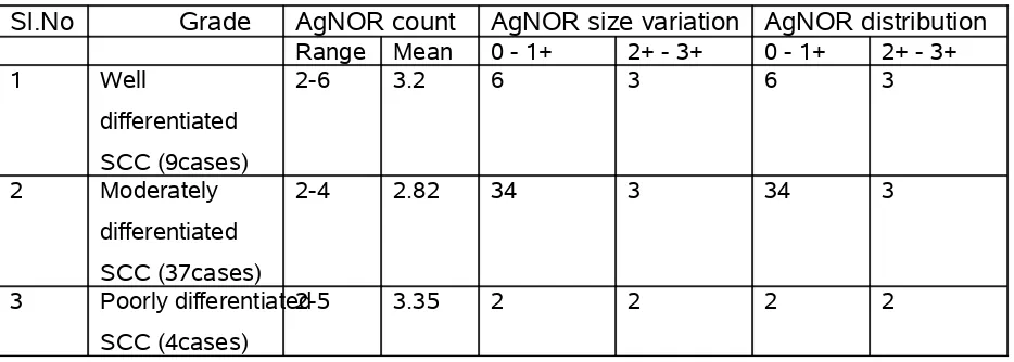
TABLE -4B
Sl.No Grade AgNOR count AgNOR size variation AgNOR distribution
Range Mean 0 - 1+ 2+ - 3+ 0 - 1+ 2+ - 3+
1 Well
differentiated SCC (9cases)
2-6 3.2 6 3 6 3
2 Moderately
differentiated SCC (37cases)
2-4 2.82 34 3 34 3
3 Poorly differentiated SCC (4cases)
2-5 3.35 2 2 2 2
In well differentiated squamous cell carcinoma (9 cases), the AgNOR count/cell ranged from 2-6 cell, with mean of 3.2. AgNOR distribution ranges 0-1+ in 6 cases, 2+ - 3+ in 3 cases, AgNOR size variation ranges from 0 – 1+ in 6 cases and 2+ to 3+ in 3 cases. Fig-11
In Moderately differentiated squamous cell carcinoma, the AgNOR count/cell ranged 2-4/cell with mean of 2.82, AgNOR size variation ranged 0 to 1+ in 34 cases, only 3 cases ranged 2+ to 3+, AgNOR distribution ranges 0 to 1+ in 34 cases and only 3 cases ranged 2+ to 3+. Fig-15
Similarly comparison was done in 50 cases of carcinoma cervix, after 8 weeks of radiation as given in Table -4C.
TABLE -4C
Sl.No Grade AgNOR count AgNOR size variation AgNOR distribution
Range Mean 0 - 1+ 2+ - 3+ 0 - 1+ 2+ - 3+
1 Well
differentiated SCC
(9cases)
2-5 2.67 6 3 6 3
2 Moderately
differentiated SCC
(37cases)
2-3 2.19 34 3 34 3
3 Poorly
differentiated SCC
(4cases)
2-4 2.97 2 2 2 2
In well differentiated squamous cell carcinoma (9 cases), the AgNOR count/cell ranged from 2-5/cell with a mean of 2.67, the AgNOR size ranged 0 to 1+ in 6 cases, 2+ to 3+ in 3 cases. The AgNOR distribution ranged 0 to 1+ in 6 cases and 2+ to 3+ in 3 cases. Fig-12
In Moderately differentiated squamous cell carcinoma (37 cases), the AgNOR count/cell ranged from 2 to 3/cell with mean of 2.19. AgNOR size variation and distribution ranged 0 to 1+ in 34 cases, 3 cases ranged 2+ to 3+. Fig-16
[image:40.612.52.509.151.411.2]DISTRIBUTION OF AgNOR’S IN CARCINOMA CERVIX PRE AND POST RADIATION THERAPY (RT)
TABLE-5
Sl.No Category No. of Cases AgNOR Count/cell
Range Mean
1 Control 25 1-3 1.63
2 Prior to RT 50 3-8 3.98
3 Post RT
a) 4 weeks b) 8 weeks
50 50
2-5 2-4
2.92 2.43
4 Persistence of Malignancy 8 3-6 3.54
1:2 P< 0.001 1:3a P< 0.001 1:3b P< 0.001 1:4 P< 0.001 2:3a P< 0.001 2:3b P< 0.001 2:4 P> 0.05 3a:4 P< 0.001 3b:4 P< 0.001
DISCUSSION
Carcinoma Cervix accounts for 80% of all gynecological cancers including breast cancer being commonest cancer in women in the developing countries. The most common cancer among women in India is Cervical Cancer. The American cancer society estimates that 9,710 women will be diagnosed with and 3700 women will die of cancer of the cervix in 2006, however although globally, 5, 00,000 new cases of carcinoma cervix are recorded of which 1, 00,000 are from India. (15,42)
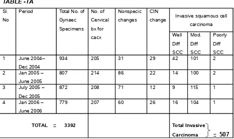
In our prospective study the average incidence of cervical cancer in biopsy specimen is 60.8% which is high when compared with studies conducted by various authors and literature.
[image:42.612.52.518.396.674.2]Present Study Incidence
TABLE -1A
Sl. No
Period Total No. of Gynaec Specimens No. of Cervical bx for cacx Nonspecic changes CIN
change Invasive squamous cell carcinoma Well Diff SCC Mod. Diff SCC Poorly Diff SCC 1 June 2004–
Dec 2004
934 205 31 29 42 101 2
2 Jan 2005 – June 2005
807 214 86 22 14 100 2
3 July 2005 – Dec 2005
872 208 71 12 9 115 1
4 Jan 2006 – June 2006
779 207 60 26 16 104 1
TOTAL = 3392 Total Invasive
79%
21%
Developed Countries Developing Countries
Africa 15%
China 23%
Latin America
15% Other
Asia 47%
Africa China Latin America Other Asia
When considering the Age specific cancer incidence per 1, 00,000 population, the below bar chart shows that our study clearly shows increased ratio 51 / 1,00,000 population well comparable to the incidence data of India/Madras and Indian and Chinese people living in Malaysia and is well compared with various research workers as well as program like SEER (Surveillance Epidemiology and End Results) program.
51
27.7
33.6
12.6
7.7
10.5
7.5
15.4
14.2
38.9
4.5
15.3
9.9
0 10 20 30 40 50 60
In India, carcinoma cervix continues to lead the list of cancers afflicting female genital tract because of poverty, poor socioeconomic status, wide spread ignorance, poor personal hygiene, early marriage and child bearing and high parity status continue to prevail in our country. Religious taboos and social traditions instill a false sense of modesty which inhibits women, particularly from rural backgrounds from seeking medical aid for what they consider a minor gynecological problem.
The average age of women newly diagnosed with cervical cancer is between 50 and 55 years (14,15,35,36,40). This cancer rarely occurs in girls younger than 15. It begins to appear in women in their twenties. Cervical cancer is different from most cancers that tend to occur more often as people get older. Although cervical cancer does affect young women, many older women do not realize that their risk of developing cervical cancer does not go down as they age and that it is important for them to continue having Pap tests.
SEER Incidence:
From 2000-2003, the median age at diagnosis for cancer of the cervix uteri was 48 years of age. Approximately 0.1% were diagnosed under age 20. 15.4% between 20 and 34, 26.4% between 35 and 44, 23.3% between 45 and 54, 14.9% between 55 and 64, 10.4% between 65 and 74, 6.9% between 75 and 84, and 2.6% 85+ years of age.(16,51)
Incidence rates by race were
Race / Ethnicity Women
All Races 8.8 per 1,00,000 women
White 8.5 per 1,00,000 women
Black 11.5 per 1,00,000 women
Asian/Pacific Islands 8.2 per 1,00,000 women American Indian/Alaska Native+ 7.2 per 1,00,000 women
Hispanic 14.2 per 1,00,000 women
In our study the youngest age group at which cervical cancer identified is 28 and 29 years and incidence is common between 41-50 years followed by 51-60 years(42,49). Our study contrast with other studies that the initial age of cervical cancer is 5-10 years earlier than the western population.
[image:46.595.83.512.98.203.2]Among the cervical carcinomas, 80% are in advanced stages, i.e., stage II B, III and IV and only 20% are in early stages i.e., in stage I and II A, while in UK 90% cases are in early stage and only 10% are in advanced stages.
TABLE -6 Patient Distribution According To Stage
Patients Stage
I B II A II B III A III B IV A
Study Burdwan
Medical College
Number
(292) 14 18 98 14 142 8
% 4.76 6.12 33.34 4.76 48.30 2.72
Present Study
Number
(50) 5 8 29 1 2 5
% 10 16 58 2 4 10
of I B and II A which is well correlated. Most of the patients were coming for the first time in an already advanced stage; however the picture is reverse in western population, since females are evaluated consequently from the age of 18 years with Pap smears.
In our study above 74% of the cases are Moderately differentiated squamous cell carcinoma followed by 18% Well differentiated carcinoma. This distribution is well in correlation with various studies that Moderately differentiated infiltrating squamous cell carcinoma being the commonest type all over the world followed by Well differentiated squamous cell carcinoma when considering the morphological subtypes also, Nonkeratinising squamous cell carcinoma predominates (319 cases, 88.12% in total cervical cancers, 35 cases, 70% in the present study).
In this study evaluation of Cervical Cancer with the AgNOR status is done with Pap smears after initial diagnosis with histopathological examination, 4 weeks after radiotherapy, as well as 8 weeks after radiotherapy cytological assessment has become a standard procedure for early detection of squamous cell carcinoma per se, and for assessment of radiation therapy changes and for AgNOR scoring.
TABLE-7
Sl.No Study Group Grade of
SCC
AgNOR Count/Cell
Range Mean
1 Ghazala Mehdi & Kafl Aktar
o Well o Mod o Poor 3.7–3.77 3.75-3.89 3.86-4.2 3.74 3.82 4.03
2 Jyotima Agarwal & J.K Gupta
o Well o Mod o Poor 5.27 5.41 5.37
3 Prathiba & Sarah Kuruvilla o Well
o Poor
4.2 5.3
4
Seema Kashyap & Kusum Kapila o Well o Mod o Poor 2.17-7.52 2.24-4.68 3.01-3.89 3.66 3.04 3.45
5 Miller et al
o Well
o Poor
2.9 4.0
6 Present Study
o Well o Mod o Poor 3.76 3.84 4.12
Our study correlates well with study conducted by Ghalaza Mehdi(18), Prathiba(39) and Miller et al(29) that the count increases when the neoplasm becomes well to poorly differentiated.
differentiated squamous cell carcinoma.
Rowland’s (1988) in a study of AgNOR in Cervical Intraepithelial Neoplasia did not give any significant difference in AgNOR counts in normal squamous epithelium CIN I and CIN II, but there was a significant increase in CIN III group as well as invasive cancer(10,11).
Crocker et al who have done intensive work on NOR’s in various tumors have observed three main types of AgNOR configurations in normal neoplastic cells(6,7,811,12). All three types of NOR patterns were observed in his study
The NOR’s are fully aggregated to form a solitary rounded structure, often seen in resting cell.
Nucleolar pattern in proliferating cells, where NOR’s can be seen within the nucleolus
Dispersion of small NOR’s throughout the nucleoplasm as frequently observed in highly malignant cells.
inflammatory reaction.
These cases are subjected for repeat Pap smears at 3 month intervals, revealed decreased in AgNOR counts to normal ratio at the end of one year.
With the standardization of the Silver staining technique diagnostic pathology has achieved a new milestone. The AgNOR’s have been shown to reflect DNA transcriptional activity. Study of AgNOR’s has been identified as a reliable indicator of cell proliferation and in turn of the malignant potential of a lesion.
[image:50.595.87.514.494.726.2]Malignant tumor cells are characterized by extremely large AgNOR’s which show a random or scattered distribution. They are useful in discriminating between benign and malignant conditions being significantly higher in malignant cells than in normal cells. They also serve as a significant prognostic indicator in malignant lesions.
TABLE -8
Sl.No Grade Prior RT
AgNOR count/cell
4 weeks after RT AgNOR count/cell
8 weeks after RT AgNOR
count/cell
Range Mean Range Mean Range Mean
1 Well differentiated squamous cell carcinoma
3-8 3.76 2-6 3.2 2-5 2.67
2 Moderately
differentiated squamous cell carcinoma
3-5 3.84 2-4 2.82 2-3 2.19
3 Poorly differentiated squamous cell carcinoma
3-9 4.12 2-5 3.35 2-4 2.97
reduced from 3.76 to 3.2 and 2.67 (after 4 weeks and 8 weeks of radiotherapy in Well differentiated squamous cell carcinoma. However in Moderately differentiated squamous cell carcinoma the reduction is so impressive from a mean of 3.84 to 2.82 to 2.19 (after 4 weeks and 8 weeks of Radiotherapy).
Poorly differentiated squamous cell carcinoma also shows a reduction from 4.12 to 3.35 and 2.97. Our study correlates well with Ghazala Mehdi(30) that Moderately differentiated squamous cell carcinoma responds well with Radiotherapy followed by Well differentiated squamous cell carcinoma. Poorly differentiated squamous cell carcinoma even though shows reduction in AgNOR range in the mean, prior and post radiation, there is clear evidence that these cancers responds poorly to external radiotherapy alone, which suggests the use of internal as well as external Radiotherapy in combination in cases of Poorly as well as Well differentiated squamous cell carcinoma.
TABLE-9 Sl. No Grade of Tumor Persistence of malignancy No. of cases Size of Tumor Stage Anaemia Grade Type of Radiotherapy Disrupted Treatment External RT Internal RT 8 wks
> 8 wks
1 Well
Differentiated
Squamous
cell
carcinoma
4 > 7cm III B III B III A IV A +++ ++ +++ +++ √ √ √ √ x x x x √ -√ √ √ 2 Moderately Differentiated Squamous cell carcinoma
2 > 6cm IV A IV A +++ +++ √ √ x x -√ √ -3 Poorly Differentiated Squamous cell carcinoma
2 > 7cm IV A IV A +++ +++ √ √ x x -√ √
CONCLUSION
In the present prospective study comprising of 50 Papanicoloau cervical smears of carcinoma cervix and 25 controls suggest the following conclusions.
1. The incidence of cervical cancers in semi urban areas – Thanjavur is
51/1, 00,000 population.
2. Cervical cancers are common between the age group of 41-50 years which is in
contrast low compared to western population where it is common in 51-60 years.
3. Squamous cell carcinoma of cervix constitutes 80% of the gynecological cancers.
4. Most patients presents initially at an advanced stage (i.e., > II B).
5. Moderately differentiated squamous cell carcinoma is the commonest type observed as in other.
6. AgNOR technique provides an index of cell proliferation.
7. AgNOR count/cell increases gradually from normal to SIL changes to invasive
carcinomas.
8. The AgNOR count is high in Poorly differentiated carcinomas and low in Well differentiated carcinomas.
9. Moderately differentiated squamous cell carcinoma responds well to external
radiotherapy alone.
10. Poorly differentiated squamous cell carcinoma requires both external and internal
radiation for prognosis.
11. Viral induced changes can impart a slight increase in AgNOR count in normal
individuals.
The AgNOR technique which was used extensively in cytogenetics earlier, has now gained importance as an indicator of cell proliferation. In normal cells, the AgNOR are tightly packed in the nucleoli and are indiscernible. In rapidly proliferating neoplastic cells nucleolar disaggregation takes place resulting in dispersion of individual AgNOR’s.
APPENDIX-1
AgNOR STAINING
PREPARATION OF STAINING SOLUTION
Solution A: 2% gelatin in 100 ml of 1% Formic acid
Solution B: 50% aqueous silver nitrate solution in de-ionized water
WORKING SOLUTION
One part of Solution A mixed with two parts of Solution B
STAINING METHOD
The wet fixed cervical smears were exposed to freshly prepared working solution for 30 min at 37°c, left in dark.
Slides were dehydrated in 3 changes of acetone, cleared in Xylene and
APPENDIX-2
PAPANICOLAOU STAINING METHOD
7) Wet fixed smear, rinse in water for 1 min 8) Stain in Harris Haemotoxylin , 5 min 9) Rinse in water, 2 min
10)Differentiate in 0.5% aqueous hydrochloric acid, 10 seconds approx 11) Rinse in water , 2 min
12)“ BLUE “ in Scott’s tap water substitute, 2 min 13)Rinse in water, 2 min
14)Dehydrate, 70 percent alcohol for 2 min 15)Dehydrate, 95 percent alcohol for 2 min 16)Dehydrate, 95 percent alcohol for 2 min 17)Stain in OG 6, 2 min
18)Rinse in 95 percent alcohol, 2 min 19)Rinse in 95 percent alcohol, 2 min 20)Stain in EA 50, 3 min
21)Rinse in 95 percent alcohol, 1 min
From
BIBLIOGRAPHY
φ Arora.B, Sanjay Kumar, Rahul Jain – Morphometric Evaluation of Nucleolar Organizer Regions in Reactive and Neoplastic Lymphnode Lesion – Journal of Indian Medical Association ; January 2006; 104; 1
φ Bibbo Comprehensive cytopathology – 2nd edition , Fadi W.Abdul Karim,
Marluce Bibbo
φ Cabrini RL, Schwint AE, Mendez A, Femopase – A morphometric study of nucleolar organizer regions in human oral normal mucosa, papilloma and squamous cell carcinoma – J Oral Pathol Med ; 1992; 21; 275-279
φ Calore EE, Maeda MY, Cavaliere MJ, Pereira SM, De Melo JR – Study of organizer nucleolar regions by the argyrophil technique in cervical neoplasias – Minerva Ginecol; 1997 March; 49 (3); 59-62
φ Chiu KY, Loke SI, Wang KK – Improved silver technique for showing nucleolar organizer regions in paraffin wax sections – J Clin Pathol ; 1989; 42; 992-994
φ Crocker J Nar P – Nucleolar organizer regions in lymphomas – J Pathol ; 1987; 151; 111-118
φ Crocker J, Boldy DAR, Egan MJ – How should we count AgNORs ? Proposals for a standardized approach – J Pathol ; 1989; 158; 185 -188
φ Crocker J, Skilbeck N – Nucleolar organizer region associated proteins in cutaneous melanotic lesions: a quantitative study – J Clin Pathol ; 1987; 40; 885 – 889
Nucleolar organizer regions in adenocarcinoma in situ and invasive adenocarcinoma – J Clin Pathol ; 1990 Aug; 43(8); 657-60
φ Diadyk EA, Vasilenko IV, Smirnova EA, Raikhlin NT – Argyrophilic Proteins in the nucleolar organizer in the differential diagnosis of cervical dysplasias and cancer – Arkh Patol ; 1993 March-April; 55 (2); 23-7
φ Egan M, Freeth M, Crocker J – Intraepithelia neoplasia, human papilloma virus infection and argyrophilic epithelium – Histopathology ; 1988 Nov; 13 (5); 561-7
φ Egan MJ, Crocker J – Nucleolar organizer regions in pathology – Br J Cancer; 1993; 65; 1-7
φ Gul Naz Akhtar, Naseer Ahmed Chaudry, Muhammad Tayyab – AgNOR Staining in malignant and benign effusions – Pak Journal Med Sci ; Jan-March 2004; 20; 1; 29-32
φ Gynecologic Cancer by David M.Gershensor, William P.Mc.Guire, Gillian Thomas
φ Haines & Taylor , Obstetrical and Gynecological pathology, 5th edition by
Harold Fox, Michael Wells , Vol -1
φ Heber E, Schwint AE, Sartor B, Nishibamas, Sanchez O, Brosto M – AgNORs as an early marker of sensitivity to radiotherapy in gynecologic cancer – Acta Cytol ; 2002; March – April; 46(2) ; 311-6
φ Jyotima Agarwal, JK Gupta – Nucleolar organizer regions in neoplastic and non-neoplastic epithelium of the cervix – Indian J Pathol Microbiol ; 1997; 40(2); 125 -127
-Diagnostic and Prognostic significance of AgNOR counts in radiotherapy treated squamous cell carcinoma of the cervix - Journal of Obstetrics & Gynecological India ; March/April 2005; 55; 2
φ Kashyap S, Kapila K, Kumar N, Kinra G, Rath GK, Verma K – Nucleolar organizer regions and morphologic subtypes of squamous cell carcinoma of cervix – Indian J Pathol Microbiol ; 1998 July; 41(3); 303-8
φ Kaushik R, Sharma V, Gulati A, Sharma BB – AgNOR counts in cervical lesions ; Indian J Pathol Microbiol ; 2003 Apr; 46(2); 201-3
φ Kinoshita Y, Dohi M, Mizutoni N et al - Effects of preoperative radiation and chemotherapy on AgNOR counts in oral squamous cell carcinoma – J Oral Maxillofac Surg ; 1993; 54; 304-7
φ Kodousek R, Dusek J – Demonstration of the nucleolar organizer region by silver staining (AgNOR Method) in research and in histopathological practice – Acta Univ Palacki Olomuc Fac Med ; 1991; 131; 9-37
φ Lakshmi S, Nair SA, Jayashree K, Kannan S, Pillai R – Argyrophilic nucleolar organizer regions in inflammatory premalignant and malignant lesions of the cervix – Cancer Lett; 1993 Jul 30; 71 (1-3); 197-201
φ Leopardi O, Colecchia M, Colavecchio A – Validity of the AgNOR count in cervical pathology – Pathologica ; 1992 May-June; 84 (1091); 287-98
φ Leopold G.Koss , Diagnostic Cytology and its histopathologic bases – 4th
edition
φ M.V.Lelmini, E.Heber, M.E. Itoiz – AgNOR are sensitive markers of radiation lesions in squamous epithelia – J Rent Res ; 2000; 79(3); 850-856
organizer regions in the normal and carcinomatous epithelium of the uterine cervix. A morphometric study – Int J Gynecol Pathol; 1989; 8(3); 237-45
φ Marbaux E, Dewandeleer S, Habbac – NORs in normal and carcinomatous epithelium of uterine cervix. A morphometric study – Int J Gynecol Pathol; 1989; 8; 237-245
φ Miller B, Flex S, Docker M – NOR’s in cancers of uterine cervix – Cancer 1974; 74; 3142-5
φ MK Dube, Anjana Govil – Evaluation of significance of AgNOR counts in differentiating benign from malignant lesions in the breast - Indian J Pathol Microbiol ; 1995; 38; 5-10
φ Murty VV, Mitra AB, Sharma JK, Luthra UK – Nucleolar Organizer Regions in patients with precancerous and cancerous lesions of the uterine cervix. Cancer Genet Cytogenet ; 1985; 18; 275-279
φ Newhold KM, Rollason TP, Luesley DM et al – Nucleolar organizer regions and proliferative index in glandular and squamous carcinoma of the cervix – J Clin Pathol; 1989; 42; 441-2
φ Nuzhat Husain, Manisha Bagchi, Bandana Tiwari – AgNOR expression in CNS Neoplasms – Indian J Pathol Microbial ; 1997; 40(4); 503-509
φ Pahuja S, Chowdhury M, Gupta U – Proliferative activity in squamous intraepithelial and invasive lesions of cervix: analysis by AgNOR staining – 2003 Oct; 46(4) ; 573-5
φ Pathology of the female genital tract Blausteins by Robert J Kurman – 5th
edition
correlate with that of histological sections in CIN – Eur J Histochem ; 1997; 41 (2); 105-110
φ Ploton N, Menager M, Adnet JJ – Simultaneous high resolution localization of AgNOR proteins and nucleoproteins in interphase and mitotic nuclei – Histochem J ; 1984; 16; 1897-906
φ Ploton N, Menager M, Jeannesson P et al – Improvement in staining and visualization of argyrophilic proteins of nucleolar organizer regions at optical level - Histochem J ; 1986; 18; 5-14
φ Prathiba D, Sarah Kuruvilla - Value of AgNORs in premalignant and malignant lesions of the cervix – Indian J Pathol Microbiol ; 1995; 38; 11 -16
φ Principles and Practice of Surgical Pathology and Cytopathology, 3rd edition ,
Steven.G.Silverberg, Ronald A Delellis , William J Frable
φ Radhakrishnan Pillai, P.G.Jayaprakash, M.Krishnan Nair – Tumour-Proliferative fraction and growth factor expression as markers of tumour response to radiotheraphy in cancer of the uterine cervix – Journal of Cancer Research and Clinical Oncology ; 1998 Aug 124; 8; 456-461
φ Rosai and Ackerman’s Surgical Pathology – 9th edition , Juan Rosai
φ Sakai Y.I, Sakai A.T, Isotani S, Cavaliere M.J, Calore E.E – Morphometric evaluation of nucleolar organizer regions in cervical intraepithelial neoplasia – Pathology Research and Practice; March 2001; 197; 3; 189-192
φ Sano k, Takahoshi H, Fujuta S, Inokuchi T, Pe MB – Prognostic Implication of silver binding nucleolar organizer regions (AgNOR’s) in oral squamous cell carcinoma – J Oral Pathol Med ; 1991; 20; 53-56
as Markers of Incipient cellular alterations in squamous epithelium – J Rent Res ; August 1993; 8; 1233 – 1236
φ Seema Kashyap, Kusum Kapila, Neeta Kumar, Kusum Verma, GK Rath – Nucleolar organizer regions and morphologic subtypes of squamous cell carcinoma of cervix – Indian J Pathol Microbiol ; 1998; 41(3); 303-308
φ Selected topics in obstetrics and gynecology-1 for postgraduate and practioners by Shirish N Daftary, Shyam V Desai
φ Terlikowski S, Lenczewski A, Sulkowski S, Kulikowski – Diagnostic value of nucleolus organizer regions (NORs) in premalignant and malignant lesions of the cervix – Ginekol Pol ; 1998 May; 69 (5); 232-6
φ Textbook of Diagnostic cytopathology by Winifred Gray, Grace T.Mc.Kee Second Edition
φ The Cancer Handbook – Editor in chief – Malcolm R.Alison
φ Underwood JCE, Giri NN – Nucleolar organizer regions as diagnostic discriminants for malignancy – J Pathol ; 1988; 155; 95-6
φ Wierzchniewska A, Wagrowska – Danilewicj M – Value of AgNOR counts and morphometric analysis of nuclear parameters in premalignant and malignant lesions of the uterine cervix – Pol J Pathol ; 1998; 49(4); 297-301