A STUDY ON BACTERIAL AND FUNGAL ISOLATES
AND THEIR ANTIMICROBIAL SUSCEPTIBILITY
PATTERN IN PATIENTS WITH CHRONIC
OSTEOMYELITIS IN A TERTIARY CARE HOSPITAL
Dissertation submitted to
THE TAMILNADU DR.M.G.R.MEDICAL UNIVERSITY
in partial fulfillment of the regulations for the award of the degree of
M.D. (MICROBIOLOGY)
BRANCH – IV
MADRAS MEDICAL COLLEGE,
THE TAMILNADU DR. M.G.R. MEDICAL UNIVERSITY
CHENNAI – TAMILNADU
CERTIFICATE
This is to certify that this dissertation titled“A STUDY ON
BACTERIAL AND FUNGAL ISOLATES AND THEIR ANTIMICROBIAL SUSCEPTIBILITY PATTERN IN PATIENTS WITH
CHRONIC OSTEOMYELITIS IN A TERTIARY CARE HOSPITAL”is a bonafide record of work done byDR.C.DEVI,during the period of her Post graduate study from 2010 to 2013 under guidance and supervision in the
Institute of Microbiology, Madras Medical College and Rajiv Gandhi
Government General Hospital, Chennai-600003, in partial fulfillment of the
requirement for M.D. MICROBIOLOGY degree Examination of The
Tamilnadu Dr.M.G.R. Medical University to be held in April 2013.
DR.G.JAYALAKSHMI.,
M.D., DTCD
Director
Institute of Microbiology,
Madras Medical College &
Rajiv Gandhi Government
General Hospital
Chennai -600 003
Dr.V.KANAGASABAI., M.D.
Dean
Madras Medical College &
Rajiv Gandhi Government
General Hospital,
DECLARATION
I declare that the dissertation entitled“A STUDY ON BACTERIAL AND FUNGAL ISOLATES
PATTERN
AND IN
THEIR ANTIMICROBIAL WITH CHRONIC
SUSCEPTIBILITY PATIENTS
OSTEOMYELITIS IN A TERTIARY CARE HOSPITAL”is submitted by me for the degree of M.D. is the record work carried out by me during the
period of October 2011 to September 2012 under the guidance of
Prof.Dr.S.THASNEEM BANU, M.D.Professor of Microbiology, Institute of Microbiology, Madras Medical College, Chennai. This dissertation is
submitted to the Tamilnadu Dr.M.G.R. Medical University, Chennai, in
partial fulfillment of the University regulations for the award of degree of
M.D., Microbiology (Branch IV) examination to be held in April 2013.
Place: Chennai Date :
Signature of the Candidate (Dr.C.DEVI)
Signature of the Guide
Prof.Dr.S.THASNEEM BANU, MD.,
Professor,
ACKNOWLEDGEMENT
I express my heartfelt thanks to Honourable deanDr.V.Kanagasabai M.D., Madras Medical College & RGGGH, Chennai for permitting me to carry out this study.
I express my deep sense of gratitude and indebtedness to
Dr.G.Jayalakshmi M.D.,Director and Professor of Microbiology, Institute of Microbiology, Madras Medical College and RGGGH, Chennai, for suggesting
the topic for my dissertation and for her valuable advice, constant guidance
and inspiration in the preparation of this work.
I consider it my privilege and honour to have worked under the guidance,
encouragement and supervision ofDr.S.Thasneem Banu M.D.,Professor.
I express my immense thanks to all the Professors of the Institute of
Microbiology,Dr.S.Vasanthi M.D., Dr.S.G.Niranjana Devi M.D., DR.T.Sheila Doris M.D., and Dr.U.Uma Devi M.D.,for their valuable
advice given to me.
I also express my sincere thanks to the former directors,Dr.G.Sumathi M.D, Dr.R.Manjula M.D and Dr.Md. Meeran M.D.
I sincerely thank Dr.J.Sasikala M.D., Retired Professor of Microbiology.
I am extremely grateful toProf.Dr.M.R.Rajasekar M.S. Ortho, D.Ortho., HOD, Institute of Orthopaedics, RGGGH, Chennai, for permitting me to carry
I express my heartful thanks to my co-guideDr.C.S.Sripriya M.D.,for guiding me in the dissertation work
I express my deep sense of gratitude and thanks toDr.Lata Sriram M.Sc, Ph.D, Dr.R.Deepa M.D., Dr.N.Rathnapriya M.D.Dr.K.Usha Krishnan M.D.,
Dr.K.G.Venkatesh M.D., Dr.N.Lakshmi Priya M.D., Dr.David Agatha M.D., Dr.B.Natesan, M.D.,Assistant Professors, Institute of Microbiology, Madras Medical College, Chennai.
I take this opportunity to thank all the post graduate students of
Institute of Microbiology, for their kind support and encouragement.
My thanks to all the technical and non technical staffs of Institute of
Microbiology, for their help at different stages of this study.
I affectionately thankMrs.C.Manimegalai,my mother and my husbandDR.J.Pazhanifor taking great care of my children, their constant love, support and encouragement without which this work would not have
been possible.
I also affectionately thank my brotherMr.C.Selvamuthukumar B.E for helping me in doing dissertation work.
My special thanks to Shajee Computers, Chennai, for working hard on
shaping the dissertation book.
Last but not the least I am very grateful to all the patients without
TABLE OF CONTENTS
S.No TITLE PAGE.NO
1. INTRODUCTION 1
2. REVIEW OF LITERATURE 4
3. AIMS AND OBJECTIVE 24
4. MATERIALS AND METHODS 25
5. RESULTS 47
6. DISCUSSION 71
7. SUMMARY 78
8. CONCLUSION 80
9. BIBLIOGRAPHY
10. (i) ETHICAL COMMITTEE CERTIFICATE
(ii) PATIENT PROFORMA
(iii) ABBREVIATIONS
iv)APPENDIX
[image:6.612.104.510.108.659.2]LIST OF TABLES & CHARTS
S.NO 1. 2. 3. 4. 5. 6. 7. 8. 9. 10. TITLE AGE AND SEX DISTRIBUTIONDURATION OF ILLNESS
CORRELATION OF SEX AND DURATION OF ILLNESS
CORRELATION OF AGE AND DURATION OF ILLNESS
PREDISPOSING FACTORS
SITE OF INFECTION
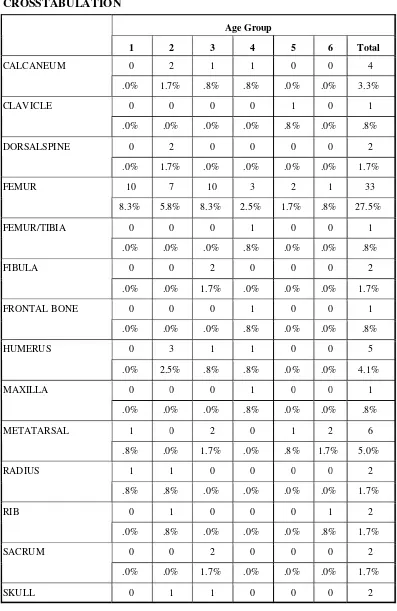
SITE OF INFECTION AGE GROUP CORRELATION
SAMPLES COLLECTED FROM THE STUDY GROUP
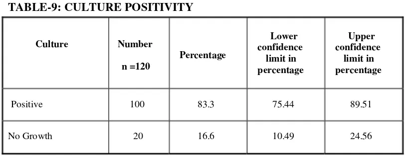
CULTURE POSITIVITY
CORRELATION BETWEEN TYPE OF SPECIMEN COLLECTED AND TYPE OF PATHOGEN
ISOLATED
ORGANISMS ISOLATED
COMBINATION OF ISOLATES IN MIXED INFECTIONS
ANTIMICROBIAL SUSCEPTIBILITY PATTERN OF GPC
ANTIMICROBIAL SUSCEPTIBILITY PATTERN OF GNB
DRUG RESISTANCE MECHANISMS AMONG PATHOGENS
ESBL DETECTION
DRUG SUSCEPTIBILITY PATTERN OF M.TUBERCULOSIS
MIC OF VANCOMYCIN
MIC OF ANTIFUNGAL AGENTS
INTRODUCTION
The word Osteomyelitis is a combination of Greek word “Osteon”
meaning bone and “Myelos” meaning marrow plus the suffix “Itis” meaning
inflammation.[1]
Osteomyelitis is acquired in three ways. They are direct seeding of
microorganisms into bone due to trauma or surgery,haematogenous spread of
microorganisms from the focus of infection elsewhere in the body and spread
from surrounding infected softtissues and joints[2].
In t he infected bo ne, the infect io n may be unifocal or mult ifocal. I n
unifocal invo lvement, only a single regio n of t he bone is affected. I n
mult ifocal involvement, more t han o ne regio n o f bone such as marrow,
perio steum, cortex and t he surrounding so ft tissue are involved.
Commonly t he infect ion is mo no microbial. Infect ion due to mult iple
organisms[3 ]are usually seen in pat ient s with Diabetes mellit us wit h ulcer
in t he foot.
Chronic Osteo myelit is o f lo ng bo nes is o ften t he consequence o f an
open, comminuted fracture and inadequately treated infect ion of t he
fracture site. Rarely it occurs as a co mplicat io n of acute osteomyelit is
now-a-days.
The following six components characterise chronic osteomyelitis.
2) Radiological changes seen in bone due to infection for 6 weeks or
longer.
3)
4)
5)
6)
Relapse or persistence of infection after initial treatment.
Osteomyelitis due to foreign bodies.
Osteomyelitis in association with peripheral vascular disease.
Organisms
tuberculosis)
The pathological process involves the necrosis of bone, granulation
that produce chronic disease (e.g Mycobacterium
tissue formation, absorption of necrotic cancellous bone ,replacement by new
bone formation and cicatrix formation due to neighbouring softtissue
destruction. Sequestrum is the dead bone that has been separated from the
living bone. Involucrum is the new bone formed.
The commonest presenting symptoms are persistent pain and chronic
intermittent discharge through sinuses. Bone debris and sequestra find exit
through multiple openings in an involucrum, go through the sinus tracts and
present to the surface. In children, after discharge of sequestrum, the sinus is
closed and the cavity is filled with new bone. In adults, the sinus is not closed
and the persistence of viable pathogens in cavities for a longer period leads to
reactivation of infection at any time.
Chronic osteomyelitis still remains a major challenging problem in our
treatment of acute hematogenous osteomyelitis and compound fractures.[4]
The usual complications of chronic osteomyelitis are reduced rate of
growth,pathological fracture, septic arthritis, lengthening of bone, contracture
of muscles. Other rare complications are formation of epithelioma, secondary
amyloidosis[5],squamous cell carcinoma in scar tissue(<1%).
Chronic osteomyelitis is a disease which is difficult to eradicate
completely. There may be subsidence of systemic symptoms,but the cavities
containing purulent material, infected granulation tissue or sequestrum act as
foci of infection. There may be recurrent acute flare-ups occuring at indefinite
intervals over months and years. To achieve eradication of the
disease,aggressive surgical debridement with curettage of cavities, filling of
cavities with soft tissues and effective antimicrobial treatment is required[6].
The pattern and behaviour of organisms
[7]
are constantly
changing under the pressure of newer antibiotics .As a result the wonder
drugs of fifties have been relegated to a position of limited usefulness today.
With this background, it is felt worthwhile to study the spectrum of organisms
REVIEW OF LITERATURE
GENERAL CONCEPTS OF THE DISEASE
Historical features
Years before, the surgeons charaka and sushruta (2500 BC) made the
first documentation of Osteomyelitis in their writings. Subsequently
Hippocrates accurately described it as an infection of the bony sequestrum.
Sequestrum formation was first described by the physician John Hunter. An
Egyptian HEARST PAPYRUS introduced casts, made of starch such as barley
flour that would harden, similar to today’s plaster of paris. Edwin Smit h
Papyrus (Paul D, 1999) was the one who initially recognised the danger of
infection with open fractures. Historically Osteomyelitis was present in war
scenarios,where the most frequent cause of Osteomyelitis was trauma. In the
last few decades Osteomyelitis occurred after surgical interventions for the
treatment of fractures.
Osteomyelitis is a condition in which there is an acute or chronic
inflammatory process occuring in the bone and its structure due to infection
with pyogenic organisms. It is an infection in the bonemarrow that spread to
the bone cortex and periosteum via the haversian canals.[8]As the bone cortex
is thin in the region of metaphysis, microorganisms get easy access to the
periosteum. Exogenous or Haematogenous spread are the most common routes
of infection that occurs in the bone. Exogenous Osteomyelitis is caused by
penetrating wounds, Compound fractures and Simple fractures treated
surgically with Open reduction and other Orthopaedic appliances like plates,
directly into the bones.The haematogenous form is due to bacteremia.
Various classification systems for Osteomyelitis are available.They are
Waldvogel and Cierny-Mader classification system. Waldvogel[9]classified
the disease as haematogenous,contiguous and chronic .Cierny Mader[10]
classification system is for staging Chronic Osteomyelitis. This staging
system depends on the status of disease process and the conditions of the
patients and their treatments. Staging is done both anatomically and
physiologically. Under anatomical type there are four stages as medullary,
superficial, localized and diffuse. Under physiological type, healthy hosts are
classified as A host, hosts with systemic, local and both local and systemic
compromising factors as Bs, Bl, Bls and hosts in whom treatment made worse
than the disease as C host. Systemic factors include malnutrition, diabetes mellitus,
hepatic or renal failure, immunosuppression. Local factors include venous stasis,
chronic lymphedema, arteritis, small vessel disease, tobacco abuse.
1. ACUTE HAEMATOGENOUS OSTEOMYELITIS
This type of Osteomyelitis is most commonly seen in children[11,12].
Over the last several decades the incidence of acute Osteomyelitis has
dramatically decreased due to higher standard of living and improved
hygiene.
EPIDEMIOLOGY
Children are most commonly affected. The most common age group
affected are children less than two years old and children eight to twelve
occuring three to four times more frequent than in females. Blyth etal,
reported a 70% decrease in the incidence of Osteomyelitis over the past few
years. A higher standard of living and improved hygiene probably have
contributed to this trend.
PATHOGENESIS
It involves mostly the metaphyses of long bones. The tibia followed by
femur[14]are the two
infection to the
most common bones affected..The predilection of
region is explained by its peculiar metaphyseal
anatomy.Metaphyseal capillaries lack phagocytic cells, the hairpin bend like
anatomy of the nutrient capillaries near the growth plate favours vascular
stasis. A minor trauma triggers the event by producing a small haematoma
which in turn cause vascular obstruction leading to necrosis of the affected
part of the bone. This area will serve as a bed for the organism resulting from
transient bacteremia. It is also found in the adult population. The infection
involving the cortex will lift the periosteum thereby leading to the formation
of soft tissue abscess. After some time the abscess will lead to sinus tract
formation connecting the sequestrum to skin. Vertebral osteomyelitis is
becoming more common now-a-days due to intravenous drug use,increasing
life expectation and increased nosocomial infection.
CAUSATIVE ORGANISMS
The spectrum of microorganisms are variable according to the type of
Osteomyelitis, epidemiology, age of the patient, comorbidity, microbiological
technique and duration of infection.[17]Microbiological culture and
antimicrobial therapy.
In infants less than one year, Group B streptococci,Sstaphylococcus
aureus and Escherichia coli are the common organisms isolated. In children
aged 1 to 16 years S.aureus, Streptococcus pyogenes and Haemophilus
influenzae are the common causative organisms. In adults more than 16 years
Staphylococcus epidermidis, S.aureus, Pseudomonas aeruginosa, Serratia
marcescens and E.coli are the commonly isolated organisms.In acute
Osteomyelitis, over 50 % of clinical specimens contains a single organism.[18]
Common organisms isolated in specific condition
Community acquired methicillin resistant Staphylococcus aureus
(MRSA) is the important cause of acute osteomyelitis in children.[19]The
cytotoxin which is present in community acquired MRSA is Panton valentine
leucocidin (PVL).In children osteomyelitis due to PVL positive S.aureus
presents with more aggressive form with multifocal involvement when
compared with PVL negative S.aureus.[20]Common organisms in neonates are
group B Streptococci and Escherichia coli.Candida spp and Pseudomonas
aeruginosa are commonly encountered in IV drug abusers and patients with
indwelling catheters. Haemophilus influenzae type b, a common cause of
long bone osteomyelitis , has become rare due to the development and
widespread use of effective vaccine in children.[21]
CLINICAL FEATURES
In children, the early sign is pseudoparalysis(failure to move the
of the affected limb.The signs of inflammation normally disappear within 5-7
days. Point tenderness is present if periosteum gets involved.
Neonates, when compared to other ages usually presents with
non-specific symptoms, resulting in delayed recognition and leading to serious
musculoskeletal sequelae[22]
DIAGNOSIS
Early diagnosis plays an important role in the management of
Osteomyelitis. In children, the diagnosis is made with compatible radiologic
and clinical findings with positive blood cultures. In adults, a CT-guided
aspirate or open biopsy is often necessary to establish a definitive diagnosis.
RADIOGRAPHIC FINDINGS
It is well known that the typical lytic and periosteal bone changes of
acute Osteomyelitis do not appear for 10 to 12 days following the onset of
illness[23]
TREATMENT
Appropriate antibiotics for adequate duration is the mainstay of
treatment for acute osteomyelitis. The principles of treatment proposed by
Nade are[24]
1)
2)
An appropriate antibiotic should be administered before the formation of pus.
Antibiotics cannot penetrate avascular tissues and abscesses ,so these
areas require surgical debridement.
avascular bone and soft tissue.
4) Antibiotics should be continued even after surgical treatment.
Children are initially given parenteral therapy for 3-10 days .Then
appropriate oral therapy is chosen to provide high level of antibiotic
concentration which will help to eradicate the offending organism[25].Oral
therapy with Cloxacillin and
penicillin in another study[26].
Clindamycin were as effective as parenteral
COMPLICATIONS
In the preantibiotic era, 50 % mortality was seen in acute osteomyelitis
due to sepsis with metastatic abscesses. Today this is not the scenario.Acute
hematogenous S.aureus Osteomyelitis in children can lead to pathologic
fractures. This can occur in about 5 % of cases with a 72 day mean time from
disease onset to fracture. Other complications include Bone abscess,
Bacteremia, Septic arthritis, Chronic infection.[27]The percentage of children
developing chronic infection as a complication is 3-5%.
2. CHRONIC OSTEOMYELITIS
In Chronic Osteomyelitis, the common signs and symptoms are
bone loss and persistent drainage from sinus tracts. Cavities in the bone act as
the nidus for a persistent infection leading to a chronic condition. When the
original acute bone infection has subsided, it may persist as a low grade
infection subjected to repeated recrudescences of the acute process over many
months or years. The majority of cases of Chronic Osteomyelitis were as a
surgical procedures and diabetic infections of feet[28].
EPIDEMIOLOGY
In the preantibiotic era, Osteomyelitis is associated with high
mortality ,which has come down after the advent of antibiotics. The incidence
of Chronic Osteomyelitis after compound fracture varies between 2% to
16%. This depends upon the grade of injury and the type of treatment given.
Now-a-days Chronic Osteomyelitis are due to trauma and surgery rather than
due to haematogenous spread.Osteomyelitis may be localized or may involve
the periosteum,cortex,marrow of the bone. The lower limb bones are
commonly involved. Nosocomial osteomyelitis is no longer rare.
PATHOGENESIS
Role of Microbes
The initial event which makes the infection to get localized is
adhesion. Staphylococcus aureus strains possess receptors for collagen,
fibrinogen, fibronectin, bone sialoprotein, and heparin sulphate[29]. Trauma or
injury will expose the binding sites for the organism. For Staphylococcus
aureus, the polysaccharide pseudocapsule forms strong links between the
bacterial cell and bone. The synthesis of capsule leads to the formation of
biofilm. In the biofilm, microcolonies are formed by bacteria that are
connected to each other and the surrounding environment. The glycocalyx
acts to protect the organism from host defense mechanisms and also from the
antibiotics[30]. Glycocalyx interferes with phagocytosis by connecting the
teichoic acid moiety which enhances opsonization and acts by consuming or
aureus cell wall contains protein A which functions as a virulence factor by
interfering with opsonization and ingestion of organisms by
polymorphonuclear cells, activating complement and eliciting immediate and
delayed hypersensitivity reactions.
Pathology
Sequestrum development is followed by reactive new bone (the
involucrum) formation by the periosteum around the sequestrum[31].
In diabetics, if bone can be seen or probed through an ulcer there is
higher probability of underlying osteomyelitis. Bone biopsies from infections
that have spread to a bone from a contiguous focus or that are associated with
poor circulation especially in patients with diabetes are likely to yield
multiple isolates.[32]
CLINICAL FEATURES
The patient presents with pain, pyrexia, redness and tenderness which
have recurred or with a discharging sinus. Tissues are thickened and puckered
or folded in, where a scar or sinus is attached to the underlying bone[33]. In
post traumatic Osteomyelitis the bone may be deformed or ununited.
BACTERIOLOGY OF CHRONIC OSTEOMYELITIS
S.H.Sheely et al[34]statesStaphylococcus aureuswas most commonly
isolated followed
and
by gram negative
Other
bacilli,
organisms
Coagulase
are
negative
Staphylococcus Anaerobes. Pseudomonas,
Escherichia coli, Klebsiella and Proteus. Of all the gram negative organisms,
Proteus species can produce progressive, unrelenting, destructive
lesions of bone. Four types are involved. Proteus mirabilis is the most
common type. Salmonella osteomyelitis, a complication of typhoid fever is a
well recognized but uncommon clinical entity. It occurs in association with
sickle cell disease or other disorders of hemoglobin. It is characterized by
multiple bone involvement[35]. In patients with hematogenous osteomyelitis
the incidence of salmonella osteomyelitis is less than 1%. Salmonella typhi
and Salmonella paratyhpi B are the two strains most frequently implicated[36].
Pseudomonascausing bacteremia either by the infection of contaminated illicited drugs or as a result of infective endocarditis in the drug addict
population leads to vertebral osteomyelitis[37]. Vertebral osteomyelitis caused
by pseudomonas aeruginosa has also been reported in elderly persons
suffering from urinary tract infections[38]. Pseudomonas aeruginosa may also
involve the pubic symphysis in addicts. Brucella causes bone infection in 10%
of patients with brucellosis. The vertebrae are the common site involved.
A rare bacteria, Arcanobacterium hemolyticum[39], in an apparent case
of tubercular osteomyelitis has been revealed. BCG vaccination
osteomyelitis[40]seems to occur in persons with apparently normal immunity.
In Anaerobic Osteomyelitis- anaerobic cocci were isolated more
frequently, and the most common of these was peptostreptococci. Bacteroides
species were the most frequently grown gram negative anaerobic organism[35]
with Bacteroides fragilis being the most common.
Due to lack of signs and symptoms, there is difficulty in diagnosing the
Chronic Osteomyelitis [41]. Reduced blood supply to the bone leads to slow
death of bone,which may lasts for years. Hence for diagnosing this disease,a
multidisciplinary approach is required, clinical examination, laboratory tests
and imaging studies. Due to chronic inflammation there is elevation of
Erythrocyte sedimentation rate.The leucocyte count is generally normal. The
C-reactive protein is elevated and non specific.Histopathological and
microbiological examination of the infected bone by obtaining abone biopsy specimen is considered as thegold standardfor diagnosing this disease[42]
The material from drainage of abscess, discharging sinuses, curettage of
cavities and sequestrum should be obtained using swabs and processed for
identification of bacteria.
COLLECTION AND TRANSPORT OF SPECIMEN
The site of specimen collection should be thoroughly cleaned with
normal saline and then material is obtained from the depth of the sinus. The
swabs should be transported to the laboratory without delay. The swabs
should be transported in thioglycollate broth for anaerobic culture.
PROCESSING OF SPECIMENS
Microscopic examination
Smears should be made from the swab. Gram staining should be done
and examined under the microscope for the presence of epithelial cells, pus
cells, RBCs, bacteria and yeast cells. After inoculating on routine plating
media the samples should be inoculated in Lowenstein Jensen medium to
anaerobic culture also.
Culture
The specimen should be inoculated on
1)
2)
3)
MacConkey agar plate
Blood agar plate
Chocolate agar plate
The plates should be incubated at 37°C aerobically for 24 hours. If
growth is observed, colony morphology and gram stain morphology are
studied.
The gram stain morphology showed gram positive cocci in clusters, the
following tests are done. Catalase test, coagulase test - slide & tube, ureàse
test and mannitol fermentation (aerobic and anerobic). Other special tests are
also done to confirm the organisms.
If gram negative bacilli are seen the colonies are subjected to the
following tests; catalase, oxidase, Hanging drop test for motility, citrate,
urease, triple sugar iron and sugar fermentation tests. Other special tests are
also done to confirm the organisms. The identification is done upto species
level. Antibiotic sensitivity should be performed for all isolates by Kirby
Bauer’s disc diffusion technique on Mueller Hinton agar. Sensitivity and
resistance pattern of the organisms are studied.
imaging showed a high degree of accuracy in diagnosing osteomyelitis. MRI
is more sensitive than CT in diagnosis. In certain circumstances, these
imaging modalities lack specificity. Due to their high sensitivities, they are
mostly used to rule out osteomyelitis.
In a non diabetic patient, bone biopsy for finding the causative
organism and its antimicrobial sensitivity pattern is taken if there are
radiographic changes in bone suggestive of osteomyelitis. Radiographic
changes may not revert even after the patient has started receiving
appropriate antibiotic therapy. Three- phase bone or to indium- labeled white
cell scan is done if the radiograph is normal and still there is suspicion of
osteomyelitis. In general CT scan and MRI are less frequently used to
diagnose osteomyelitis, but these are often used to determine the extent of
infection and whether these are collections of pus that are amenable to
drainage.The ‘gold standard’ specimen for diagnosing osteomyelitis is bone
biopsy specimen. Sinus tract cultures are not reliable for pedicting gram
negative organisms causing osteomyelitis[43]. In most cases antibiotic
treatment is based on culture from deep bone biopsies or debrided tissues
during surgery and their antibiotic susceptibilities. An earlier diagnosis of
osteomyelitis may be achieved withradio nucleotidescanning.
MANAGEMENT OF CHRONIC OSTEOMYELITIS
While antimicrobial therapy is desirable in the control of
Osteomyelitis, surgery remains the therapeutic and diagnostic procedure[6].
minimum of 3 weeks provides an
progression to chronic condition.
excellent response and there is no
Antimicrobial Therapy
Staphylococcus aureus is the common organism isolated.Vancomycin is the drug of choice for the strains that are resistant to both ampicillin and
methicillin. Recently ,Linezolidhas evolved as a better drug againstMRSA
because of its increased oral bioavailability and good bone
penetration.Prolonged use of linezolid has been associated with significant
pancytopenia, peripheral neuropathy, optic neuritis[44,45].
Daptomycin, recently approved drug has bactericidal activity. Its utility in the treatment of
vancomycin resistant Enterococcus has yet to be defined.The period of
administration of antibiotics is 4 to 6 weeks.
Surgery
The surgery done for Chronic Osteomyelitis isSequestrectomyand removal of the infected bone and soft tissue. The goal of surgery is complete
eradication of every bit of an infection and thereby attaining a viable and vascular
environment. To achieve this goal, radical debridement may be required.
Inadequate debridement leads to recurrence. Simpson et al evaluated that the
recurrence rate is very low withwide resection (>5mm)than the patients treated with marginal resection <5mm who had a 28% recurrence rate. The large dead
space which is left after adequate debridement, should be managed accordingly to
prevent recurrence and to avoid pathological fractures. Reconstruction of bone and
The wound is loosely packed open with petrolatum gauze and a
catheter is inserted for local application of antibiotics. If the bony defect is
large, the cavity is packed with small cancellous bone grafts mixed with an
antibiotic and a fibrin sealant (papineau et al (1979). The area is covered by
adjacent muscle and the skin wound is sutured without tension (Lack, Bosch
and Arbes, 1987). In muscle flap transfer, a large wad of muscle with intact
blood supply, is laid in the cavity and the surface is covered with split- skin
graft (Fitzgerald et al 1985) or Myocutaneous island flap (Yoshimura et al
1989). Recently, custom made calcium sulfate (osteoset bone voidfiller)
antibiotic impregnated implants[46]is used to treat chronic osteomyelitis.
In the study conducted by A.S. Bajaj and his colleagues mentioned above,
irrigation was done with appropriate antibiotics after culture and sensitivity report. It
was done for 4 to 14 days and successful in 73.3 percent of cases.
About 1000 to 1500 ml of irrigating fluid was used in 24 hours. The
returned fluid was cultured on 4th day and then daily. The irrigation was
continued till 2 consecutive cultures of the outflow fluid became negative.
Compere[47](1967) has recommended the use of “Alevaire” for
irrigation of infection of bone and soft tissue for its mild antiseptic property
as well as for its action in making penicillin effective against penicillin
resistant organisms. However this last property of “Alevaire” has been refuted
in invitro studies (Modellering, treth and Weinburg 1971).
As an adjuvant therapy for chronic osteomyelitis, hyperbaric oxygen
COMPLICATIONS
Reduced rate of growth, pathologic fracture, bone lengthening, muscle
contracture, epithelioma and amyloidosis are the complications of chronic
osteomyelitis.
3. OTHER TYPES OF CHRONIC OSTEOMYELITIS IN
SPECIFIC CONDITIONS
a) Osteomyelitis in patients with Diabetes Mellitus or
insufficiency
Vascular
Patients with reduced vascular flow as in diabetes mellitus, are
predisposed to osteomyelitis due to poor local tissue response. 15% of
diabetes mellitus patients develop foot ulcers and 6% require hospitalization
for the same[48]. The development of a skin ulcer due to neuropathy, vascular
insufficiency
osteomyelitis.
Footis the commonest site of infection[49]. There are several risk
factors which leads to the development of ulcers in the foot in diabetic
patients. All diabetic patients and patients with vascular compromise must
undergo complete foot examination every year.
and hyperglycemia subsequently leads to contiguous
Diagnosis
This requires multiple modalities and a careful examination of the foot.
A chronic ulcer with a surface area of more than 2cm2or a positive “Probe-to-bone test”[50]is associated with high positive predictive value.Also
complementary tests like measurement of ESR, C-reactive protein and MRI
Treatment
Broad spectrum antimicrobial therapy is required as most of these
infection arepolymicrobial[3]. Quinolones in combination with metronidazole
or Clindamycin are used commonly for the treatment of osteomyelitis in
patient with diabetes and peripheral vascular disease[51]. Long-term safety of
fluoroquinolones has generally been good[15,48]. Latest generation quinolones
such asMoxifloxacinhave excellent activity against gram-negative and gram positive organisms and improved anaerobic activity. Depending on the level
of surgical debridement and amputation, the duration of antimicrobial therapy
varies from a few days to several weeks. Treatment for six weeks is
appropriate[51].
b) Tuberculous osteomyelitis
About 10% of extrapulmonary tuberculosis affects bone. Tuberculosis
of spine contributes 50% of all skeletal tuberculosis cases. Most cases
occurred as the result of haematogenous spread from a pulmonary source. In
contrast to bacterial vertebral osteomyelitis systemic symptoms are absent.
Back pain or stiffness is the common symptom. The confirmation of diagnosis
is by biopsy result. CT and MRI are needed to know the extent of bony
involvement which is useful for planning the treatment. In
immunocompromised patients other non tuberculous Mycobacterial infection
is also common. Mycobacterium fortuitum, Mycobacterium chelonae,
Mycobacterium kansasii and Mycobacterium xenopi are known to cause
infections. Osteomyelitis due to Mycobacterium bovis after Bacille
calmette-Guerin has been reported. Medical therapy alone is often curative. In certain
cases, surgical debridement is required.
c) Fungal osteomyelitis
Osteomyelitis resulting from fungi is uncommon. Several observational
studies and case reports have been published. Mode of spread is
haematogenous (67%), direct inoculation (25%) and contiguous infection
(9%)[52]. Bone lesions are most common in Blastomycosis, disseminated
Coccidioidomycosis and extracutaneous Sporotrichosis, but are seen
occasionally in Cryptococcosis, Candidiasis and Aspergillosis. Candida
Osteomyelitis is one of the less frequent manifestations of invasive
Candidiasis[53].Candida albicansis the common species. Non albicans
Candida species account for 35% of cases[52]. Neonates and intravenous
heroin drug addicts are at risk to develop disseminated Candidiasis. In
adults,the order of frequency of involvement of bones is lumbar spine[54],long bones and sternum. The diagnosis is confirmed by the isolation
of candida spp from the bone and histopathological confirmation. Most cases
resolve without surgery. Long term antifungal therapy is usually necessary.
Amphotericin BandKetaconazolewere used commonly. Fluconazole have shown poor penetration to bone tissue.
d) Vertebral Osteomyelitis
Majority of the vertebral osteomyelitis are haematogenous in origin.
Haematogenous infection of the vertebrae spreads through segmental artery.
Haematogenous spread occurs via infection in skin and soft tissue,
well as infection of respiratory tract. In one study by Schnoring and
Brock[55], 0.2% of patient receiving antimicrobial prophylaxis developed a
surgical site infection, whereas 2.8% of patients developed surgical site
infection when antimicrobial prophylaxis was withheld. Infection of the disc
space and contiguous vertebra also can occur postoperatively.
The most common symptom and sign of vertebral osteomyelitis are
localized pain and spinal tenderness in 90% of patients.Due to nerve root
compression, motor and sensory deficits, are seen in 15% of patients.
An elevation of the ESR is present in more than 90% of cases, white
blood cell count is elevated in less than 50% of patient. If infective
endocarditis is present, blood cultures may be positive[56].
As with other osteomyelitis, the most common micro organisms seen
in vertebral osteomyelitis are Staphylococcus aureus and Coagulase negative
Staphylococci . In endemic regions Mycobacterium tuberculosis is common.
In immunocompromised patients and postsurgical patients Aerobic
gramnegative bacteria and Candida spp are common.
Gram negative aerobic bacteria and Candida spp are seen more
commonly in IV drug abusers. Plain radiographs are not sensitive in the
diagnosis of disc space infection. In a study of 41 patients with suspected
spondylodiskitis, gallium scanning proved to be 100% sensitive, specific and
accurate[57]. CT- guided percutaneous biopsy has a sensitivity of 50%[58].
relieve pain and restore neurologic function and to maintain vertebral
stability. Surgical therapy is unnecessary, surgical treatment including
debridement should be considered in cases with paravertebral abscess. Wit h
appropriate antimicrobial medical treatment, spontaneous bony fusion
between adjacent infected vertebral bodies occurs within 12 to 24 months.
e) Osteomyelitis of the craniofacial skeleton
Osteomyeliltis of the skull is truly a bony infection which is due to
chronic, inadequately treated infections[59]. Van launelongue classified
osteomyelitis of skull as, primary hematogenous and secondary
contiguous[60]. Mandible, maxilla, frontal bone, temporal bone and skull base
bones[61] are commonly affected in this type of osteomyelitis. Severe
reduction of the blood flow leads to the formation of ischemic and necrotic
bone[62]. The usual organisms that can be isolated are Bacteroides,
microaerophilic Streptococcus spp, Peptostreptococcus, other odontogenic
pathogens which affect tooth bearing bone. For planning a complete
treatment,Bone scintigraphyis more ideal than CT.Clindamycinis the ideal antibiotic due to its effectiveness against Streptococci and the
Anaerobes. As compared to other osteomyelitis, odontogenic infection should
be treated much longer times than usual forupto 6 months.
PREVENTION OF CHRONIC OSTEOMYELITIS
Osteomyelitis resulting from the haematogenous spread from the focus
of infection elsewhere in the body can be prevented by removing the infecting
focus. For example,if the infection is due to intravenous catheter,removal of
given for 6 weeks.
Osteomyelitis following any surgery is also commonly occuring
now-a-days. Hence sterile aseptic surgical technique is essential for the prevention
AIM OF THE STUDY
To study the predisposing factors associated with chronic
Osteomyelitis.
To study the causative organisms and their antimicrobial susceptibility
pattern.
MATERIALS AND METHODS
PLACE OF STUDY
The study was conducted in the Institute of Microbiology, Madras
Medical College in association with Institute of Orthopaedics, Rajiv Gandhi
Government General Hospital, Chennai-600 003.
Name of the Study :
Period of Study
Sample Size
:
:
Cross Sectional
Oct 2011 to Sep 2012
120
Ethical Consideration
The necessary ethical committee approval was obtained before the
commencement of the study. Informed consent was obtained from the study
population. All patients satisfying the inclusion criteria were documented.
Patients were interviewed by structured questionnaire.
INCLUSION CRITERIA
Patients older than 12 years.
Patients admitted in orthopaedic wards and those attending outpatient
department who satisfy one of the following six components of chronic
osteomyelitis.
Osteomyelitis in association with trauma only.
Osteomyelitis in association with diabetes and peripheral vascular
Clinical evidence of chronic disease. (Eg.Mycobacterium tuberculosis).
Radiological changes suggestive of infection for 6 weeks or more.
Formation of sequestrum or sclerosis.
Even after treatment, persistence or relapse of infection.
EXCLUSION CRITERIA
Patients with prosthetic orthopaedic implants devices.
Paediatric age group (<12 years)
HISTORY
Name, age, sex, date of admission, physical examination findings,
history of trauma, associated predisposing factor (diabetes mellitus,
intravenous drug abuse, immunosuppression, tuberculosis) duration of illness,
smoking and alcoholism were also recorded.
COLLECTION, TRANSPORT AND PROCESSING OF
SAMPLES
[63]Under strict aseptic precautions samples were collected from the
patients and transported immediately to the laboratory and sample processing
was done.
SAMPLES COLLECTED
1) Sequestrum and fragments of excised tissue removed during surgery or
curetting from infected sinuses.
stain and KOH mount. Second for aerobic bacterial and fungal culture.
Third for bedside inoculation into Robertsons cooked meat broth.
3) Pus.
COLLECTION OF SEQUESTRUM
Sequestrum obtained peroperatively were collected in a sterile
container without fixative. Fragments of excised tissue removed during
wound toilet or curetting from infected sinuses were also collected in a
similar manner. They were homogenized in a tissue grinder[32]with a little
sterile broth and subsequently treated in the same way as exudates.
COLLECTION OF SWABS
The surface of the wound was cleaned well with sterile normal saline
and swabs were taken from the depth of the sinus.
COLLECTION OF PUS
Pus was aspirated from the depth of the sinus or collected directly from
cavities per operatively and transported to the laboratory in a small
screw-capped bottle, syringe or a sealed capillary tube[64].
PROCESSING OF SAMPLES
1) DIRECT SMEAR EXAMINATION
Using standard laboratory techniques, pus, exudates and swabs were
subjected to the following microscopic examination.
methyl violet, waited for 1 min, washed with Grams iodine and allowed it to
act for 1 min. Then washed with water, acetone was added as a decolorizing
agent till no more color comes off, then washed with water and dilute carbol
fuchsin was added and allowed it to act for 1 min. Then washed with water,
blotted dry and examined under oil immersion objective. Presence of pus
cells, yeast cells, hyphal elements, Grams reaction, size and shape of
organisms were noted.
b. 10% potassium hydroxide mount
[65]A clean glass slide was taken and a large drop of KOH was placed with a
pasteur pipette. A small quantity of the sample was transferred with the loop
into the KOH drop. A clean cover slip was placed over the drop gently
without producing air bubbles. The slide was kept at room temperature. After
20 to 30 minutes the slide was examined under microscope. Necrotic bone and
tissues were allowed for overnight contact with KOH and examined on the
next day.
c. Acid fast stain by Ziehl- Neelsens method
[64]Procedure
i.ii.
Place heat fixed smear on a staining rack.
Filter strong carbol fuchsin on to the slide through a whatman
No.1 filter paper.
iii. Soak a cotton-wool swab attached to a wire in methylated
spirits.
and use to heat the slide without boiling so that they steam,
leave for 3 min.
v.
vi.
vii.
viii.
ix.
x.
xi.
xii.
Repeat steps 3 and 4.
Gently wash with tap water.
Decolourize with 3% acid alcohol for 3 min.
Gently wash with tap water.
Decolourize with 20% sulphuric acid for 5 min.
Wash, repeat steps 9 and 10.
Counterstain with loeffler’s methylene blue for 30 seconds.
Wash & Examine under oil immersion objective.
2) CULTURE
The samples were plated onto the following media. 5% Sheep blood
agar, Chocolate agar, Mac conkey agar, Cooked-meat broth and Sabouraud’s
dextrose agar. All the inoculated plates except cooked meat broth were
incubated at 370C under aerobic condition and in a carbondioxide enriched
atmosphere. Plates were evaluated for growth at 24 and 48 hours and
discarded after five days except Sabouraud dextrose agar which was kept for
4 weeks. Cooked meat broth was incubated at 370C with sterile liquid paraffin
and looked for turbidity after 24 and 48 hours. If any turbidity was found in
cooked meat broth, it was subcultured in Gentamycin blood agar and
incubated anaerobically in gaspak at 370C. These anaerobic plates were
3. INTERPRETATION
A) INTERPRETATION OF BACTERIAL CULTURES
1480-CHARTS]
[63,P-1443-I. After 24 hours of incubation, identification of bacteria was done by
studying morphology of colony, gram stain, motility, catalase and
oxidase tests. Single colony was taken and subjected to a battery of
tests along with the controls.
Gram staining
Hanging drop
Oxidase test
Catalase test
Coagulase test
Phosphatase test
Bile esculin hydrolysis
IMVIC test
Nitrate reduction test
Urease test
TSI (Triple sugar iron agar)
O-F test
Sugar fermentation test
LAO decarboxylases test
Oxidase test
1% tetramethyl - p - phenylene diamine dihydrochloride was prepared
freshly with sterile distilled water. A filter paper circle was placed into a
sterile petridish and moistened with several drops of the fresh reagent. A
colony from a nutrient agar was removed with a sterile glass rod and rubbed
onto the moistened filter paper along with controls. Appearance of dark
purple color within 10 sec was considered as positive.
Catalase Test
Single colony from nutrient agar plate was picked with a sterile glass
rod and inserted into 1ml of 3% hydrogen peroxide solution in a small clean
test tube. Immediate and sustained production of gas bubbles from the colony
indicate positive reaction.
Coagulase Test
This was done to detect both free and bound coagulase enzymes.
Slide coagulase test
Slide test is a rapid test to detect bound coagülase. Two drops of
normal saline were placed in two circles drawn on a glass slide. Growth was
taken from nutrient agar plate and emulsified into smooth suspension in two
marked as test and the other circle as control without plasma. Visible
clumping within 10 - 15 sec. of mixing the plasma with the suspension was
taken as positive.
Tube coagulase test
This test detects free coagulase. A small amount of the colony growth
of the organism is emulsified with 0.5ml of coagulase plasma. The tube is
incubated at 350C for 4 hours and observed for clot formation by gently
tilting the tube. If no clot is observed at that time, reincubate the tube at room
temperature and read again after 18 hours.
Indole Test
Organisms were suspended into Tryptophan broth and incubated at
37°C for 18-24 hrs. 15 drops of Kovac’s reagent was added along the inner
wall of the tube. Appearance of red ring over the surface was taken as
positive.
Methyl red test
Organisms were suspended into Glucose Phosphate broth and incubated
at 37°C for 48-72hrs. Then 5 drops of MR reagent was added to the broth.
Appearance of red color indicated positive result.
Voges-Proskauer Test
Organisms were inoculated into Glucose Phosphate broth and
incubated at 37°C for 48-72 hrs. Then 0.6m1 of 5% a naphthol was added,
followed by 0.2ml of 40% KOH. The tube was gently shaken without cotton
minutes. Development of red color within 15 min. was taken as positive.
Citrate utilization test
This test is done to check the ability of an organism to use citrate as its
whole source of carbon and energy source for growth and ammonium as
nitrogen source. A saline suspension of the test organism is streaked in
simmon’s citrate medium and incubated for 48 hrs at 370c.
Blue colour and streak of growth=positive.
Original green colour and no growth=negative.
Nitrate reduction test
Nitrate broth was suspended with organisms to be tested and incubated
at 37°C for 24 - 48 hrs. 5 drops of each reagent A(αnaphthylamine) and
reagent B (sulfanilic acid) were added to the broth. Red color developed
within few minutes indicate the presence of nitrite i.e the positive reaction.
Urease test
The entire slope of the Christensen’s medium was streaked with test
organisms and incubated at 37°C for 24 - 96 hrs. Urease producing organisms
changed the color of medium to purple pink.
Sugar fermentation tests
Sugar fermentation test media containing different sugars in the
concentration of 1%, with inverted Durham’s tubes were suspended with test
organisms, and incubated at 37°C and observed for up to 1 week. Change of
color to yellow was considered as positive (sugar was fermented). Presence of gas
O-F test
Two tubes of Hugh - Leifsons test media were stab inoculated with
test organisms. One tube of this pair was covered with a 1cm layer of liquid
paraffin and the other tube was left open to air. Both tubes were incubated at
37°C and examined daily for up to 7 days. Appearance of yellow color in
open tube and green color in covered tube, indicated oxidative utilization of
the sugar.
Triple Sugar Iron test
The organism was stabbed into butt and streaked onto the surface of
slant, incubated at 37°C overnight. The next day change of color, H2S
production and presence of gas were noted.
Phenyl alanine deaminase test
The medium containing phenyl alanine deaminase was suspended
heavily with organism to be tested and incubated at 37°C overnight. Next day
few drops of 10% solution of ferric chloride was allowed to run down over
the growth. Appearance of apple green color in the slope indicated positive
test.
Phosphatase test
Organism was grown on phenolphthalein diphosphate agar (consist of
l000ml of nutrient agar and l0ml of 1% aqueous solution of sodium
phenolphthalein diphosphate) at 37°C overnight. Next day few drops of liquor
ammonia was poured on lid and plate was inverted over the lid. Colonies
liberation of free phenolphthalein by the action of phosphatase. This
considered as positive.
was
Bile Esculin Hydrolysis
Organism was streaked on the surface of bile esculin slant and
incubated over night. If the organism hydrolyses bile esculin ,slant would be
turned black. This test was used to confirm gram positive cocci in pairs as
Enterococci.
LAO Decarboxylases Test
This test is based on the ability of bacteria to decarboxylate an
aminoacid to the corresponding amine with liberation of carbondioxide. The
production of decarboxylases is induced by a low pH which occurred due to
fermentation of glucose and as a result of the action of decarboxylases ,the pH
is raised due to the production of amines. The medium was inoculated with a
straight wire through the paraffin layer .Incubated and read daily for 4 days.
Appearance of violet colour indicate positive test.
ANTIMICROBIAL SUSCEPTIBILITY TESTING
Antibiotic susceptibility testing was performed by the Kirby bauer
method on Mueller Hinton agar (Himedia) according to CLSI guidelines[66].
The diameters of zones of inhibition were interpreted according to CLSI
standards for each organism. Media and discs were tested for quality control
using standard strains.
The following standard strains were used
2)
3)
Escherichia coli- ATCC 25922
Pseudomonas aeruginosa- ATCC 27853
For gram positive cocci, the following antibiotics were included in the
antimicrobial sensitivity testing (Himedia)
Inhibition Zone in mm Antibiotic Disc content
Resistance
Amikacin
Ciprofloxacin
Cotrimoxazole
Chloramphenicol
Penicillin
Rifampin
Erythromycin
Oxacillin
30 g
5 g
1.25-23.75 g
30 g
10 Units
5 g
15 g
1 g
14
15
10
12
28
16
13
10
Intermediate
15-16
16-20
11-15
13-17
–
17-19
14-22
11-12
Sensitive
17
21
16
18
29
20
23
13
For gram negative bacilli, the following antibiotics were included in
Inhibition Zone in mm Antibiotic Disc content
Resistance Amikacin Ceftazidime Cefotaxime Ciprofloxacin Gentamicin Imipenem Ofloxacin Tetracycline Piperacillin/ Tazobactum 30 g 30 g 30 g 5 g 10 g 10 g 5 g 30µg 100/10 g 14 14 14 15 12 13 12 14 17 Intermediate 15-16 15-17 15-17 16-20 13-14 14-15 13-15 15-18 18-20 Sensitive 17 18 18 21 15 16 16 19 21
Procedure of Kirby-Bauer Disc Diffusion Test
1) With a wire or a loop touched the surface of 5 similarly appearing
colonies on an agar plate culture. Transferred the growth to a tube
containing a suitable broth medium.
2) Allowed the culture to incubate at 350C until it matches the turbidity of
standard.
3) Dipped a sterile non-toxic cotton swab into the inoculum suspension
and rotated the swab several times with firm pressure on the inside
wall of the tube to remove excess of fluid.
4) Inoculated the dried surface of a Mueller Hinton agar plate that has
the entire agar surface rotating the plate approximately 60 degrees to
ensure an even distribution. Replaced the lid of the dish. Allowed 3 to
5 minutes but no longer than 15 minutes for the surface of the agar to
dry before adding the antibiotic discs.
5) Appropriate antimicrobial disc was placed on the surface of the agar
using forceps.
6)
7)
Plate was incubated at 370c overnight.
After overnight incubation, zone diameters was measured in mm from
the edge of the disc to the zone edge with a ruled template on the agar surface
DETECTION OF LACTAMASE ENZYMES IN GRAM NEGATIVE BACILLI
Extended spectrum lactamases (ESBL’s)
ESBL’s are classified under in Bush class A lactamases which are
capable of hydrolyzing penicillins- oxyiminocephalosporins and
monobactams (Aztreonam) and inhibited by lactamase inhibitors (clavulanic
acid, sulbactum and Tazobactum) but have no detectable activity against
cephamycins or carbapenems (Imipenem, Meropenem).
ESBL Detection methods
Screening Method[67]]Antibiotic Aztreonam 30 g
Cefotaxime 30 g
Ceftazidime 30 g
Ceftriaxone 30 g
For Possible ESBL Producing Stains < 27mm
< 27 mm
<22 mm
Double Disk diffusion synergy test
24 hour young culture was used for this test. 3 to 4 colonies from 24 hr
culture were inoculated into 5 ml of nutrient broth to match 0.5 Macfarland
turbidity standard. Lawn culture of the test organism should be made on MHA
plate. Two discs Ceftazidime and Ceftazidime in combination with clavulanic
acid were placed. The plate is incubated at 350C for 16-18 hours.
Interpretation
A >5mm increase in zone diameter for either antimicrobial agent tested
in combination with clavulanic acid versus its zone when tested alone
confirms an ESBL producing organism[68,66].
Phenotypic confirmatory double disk test (PCDDT)
In this method a lawn culture of test organism on to a MHA plate was
performed. Augmentin disc was placed in the centre of the plate and a disk
containing one of the oxyimino lactam antibiotics was placed 30mm from
centre to centre from augmentin disk. The test organism was considered to
produced ESBL, if the zone size around the test antibiotic disc increased
towards the augmentin disk[69].
The sensitivity of the test could be increased by reducing the distance
between the disc to 15mm or 20mm, also by using more than one oxyimino
lactam antibiotic.
ESBL detection by E test strip
This combines both the principles of dilution and diffusion techniques.
two shorter gradients aligned in opposing directions on a single strip. One end
generates a stable concentration gradient of the one of the oxyimino
cephalosporins (eg ceftazidime), while the other end generates a gradient of
cephalosporin + clavulanic acid (4 g/ml). When applied to an inoculated agar
plate inhibition ellipse may be seen on the both ends of the strip. MIC is
interpreted as the point of intersection of the inhibition ellipse with E test
strip edge. Ratio of cephalosporin MIC and cephalosporin clavulanic acid
MIC >8 indicates positive result.
DETECTION OF METHICILLIN RESISTANCE IN STAPHYLOCOCCUS AUREUS
Disc diffusion Method Media- MHA
Antibiotic disc- Cefoxitin disks 30 g
QC Strain – ATCC S.aureus 25923 was used.
Procedure
Inoculum Preparation
Colonies isolated from agar culture plate were suspended directly into
broth or physiological saline (0.85% Nacl, vortexed to reach 0.5 McFarland
(108CFU/ml).
A lawn culture of the Staphylococcus aureus was made on the MHA
plate and the cefoxitin disks(30µg) were applied.
Incubated at 350C for 24 hrs in an ambient air.
2C-S4]
According to CLSI criteria,2011,with 30µg cefoxitin disks[70,66 -supp
,
Diameters of <21mm= Resistant to oxacillin(MRSA)
>22mm = Susceptible to oxacillin(MSSA).
No intermediate category.
ta
b-DETERMINATION OF MIC
The following are the different methods of detecting MIC of an
antibiotic against a specific bacterial isolate.
1) Broth dilution method
a) Macrobroth method
b) Microbroth method
2)
3)
4)
Agar dilution method
Using E strip
Using Hi-comb method
MIC of vancomycin against Staphylococcus aureus was determined by
Macro Broth dilution method.
MIC of cefotaxime against Klebsiella pneumoniae was determined
using Hi-Comb method.
Procedure of Hi-comb method
A lawn culture of the test organism was made on a MHA plate
Hi-Comb Strips were applied with the MIC scale facing upward. The strip should
be in complete contact with the agar surface. The agar plate was inverted and
and diffusion. The E test MIC was read where the edge of the inhibition
ellipse intersects the MIC scale on the strip
Minimum Inhibitory Concentration (MIC) for detecting vancomycin
resistance
1)
2)
Cation adjusted Mueller Hinton Broth (PH 7.2-7.4) was used.
Preparation of stock antibiotic solution[71]
Antibiotic stock solution was prepared using the formula.
1000
--- x V x C = W P
Where P= Potency of the antibiotic in relation to the base (For
vancomycin, P= 950/1000mg Himedia).
V = Volume of the stock solution to be prepared (10ml).
C = Final concentration of the antibiotic solution (1024 g/ml).
W = Weight of the antibiotic to be dissolved in the volume V.
3)
Method of preparing dilution of antibioticsSterile test tubes were arranged in two rows in the rack (1 row for the
test and another one is for ATCC control).
Transferred 2ml of MH broth to the sterile container containing the
working stock solution (128 g/mlconcentration). From this 1ml was
transferred to the first tube in each row.
sterile container, mixed well and 1ml was transferred to second tube in
each row.
Repeated this procedure till the 8thtube.
One ml of the antibiotic was kept for control.
4) Inoculum preparation for the test and ATCCcontrol 9.9ml of MH broth was taken in a sterile container.
0.1ml of 0.5 Mcfarland tubidity matched test organism was added.
After mixing well, 1ml of inoculum was transferred to each tube
containing antibiotic dilutions and also to the control tube.
The procedure was repeated for ATCC control stain.
The tubes were incubated at 370C for 16-18 hrs. After 16-18 hrs, the
tubes were taken out and MIC was read. The lowest concentration of the
antibiotic in which there is no visible growth was taken as MIC of
Vancomycin for the test organism. The above procedure was repeated for
ATCC S.aureus, and MIC was interpreted.
B.INTERPRETATION OF FUNGAL CULTURE
Samples were inoculated onto two SDA slants and were incubated at
two different temperatures, 250C and 350C. These slants were inspected daily
during the first week and twice weekly during the next three weeks for
Macroscopic Appearance
Cream colored, smooth or wrinkled with mycelial fringe and pasty
colonies within 3-4 days.
Microscopy
Gram stain and LCB mount were prepared from the colonies.
Gram Stain
Gram positive ovoid budding yeast cells approximately 4-8 min size with pseudohyphae is suggestive of candida spp.LCB
Presence of yeast cells and pseudohyphae.Germ tube test
[65]Germ tube test is used for presumptive identification of Candida
albicans. It is a rapid screening test where the production of germ tubes with
in two hours is considered as growth of Candida albicans.
Make a very light suspension of the test organism in 0.5ml of sterile
serum (pooled human serum or fetal calf serum). The optimum inoculum is
105-106cells per ml. Incubate at 370C for exactly two hours. Observed under
microscope for production of germ tube. Germ tubes represent initiation of
hyphal growth, arising directly from the yeast cell. They have parallel walls at
their point of origin and are not constricted. To record a positive, about 30%
of the cells should show germ tube production.
Chrom Agar Media
[72]It is a rapid, plate based test for the simultaneous isolation and
detection of specific enzymatic activities by adding multiple chemical dyes
i.e. substrates of fluorochromes to media. Candida species are differentiated
by color as a result of biochemical reactions.
The CHROM agar media shows following colors of colories at 300C
for 48 to 72 hours.
C.albicans
C.dubliniensis
C.glabrata
C.krusei
C.parapsilosis
C.tropicalis
Light Green
Dark Green
Pink to Purple
Pink
Cream to Pale Pink
Blue with Pink halo
Sugar fermentation
Biochemical tests like sugar fermentation was done for identification
of yeast isolate. Glucose, Maltose, sucrose, Lactose, Galactose and Trehalose
sugars (2%) were used.
Determination of MIC by microbroth dilution method
[65]As per the guidelines of CLSI, the test was performed. MIC of water
soluble drug Fluconazole and water insoluble drugs Amphotericin-B,
Itraconazole and Voriconazole were determined.
The test consisted of following steps
2. Preparation of inoculum.
3. Test procedure.
For water insoluble drugs, dimethyl sulfoxides (DMSO) was the
solvent used.
Media used- RPMI 1640. Varying concentrations of the drugs were tested.
Amphotericin B
Fluconazole
Itraconazole and Voriconazole
Incubation period – 48 hours.
ATCC candida albicans ATCC 90028 was used for quality control of
the test. The broth microdilution test was performed by using sterile,
disposable, multiwell microdilution plates (96U- shaped wells). 100 l of
varying drug concentrations was dispensed in each rows from 1 to 10. 100 l
of inoculum was also dispensed. Incubated at 350C for 48 hours. MIC was
interpreted, as the lowest concentration in which the well was clear in case of
Amphotericin B. For Azoles, it was interpreted as the lowest concentration in
which there was a prominent decrease in turbidity (50% inhibition in growth
as determined spectrophotometrically).
0.0313 to 16 g/ml
0.125 to 64 g/ml
0.0313 to 16 g/ml
C.CULTURE FOR MYCOBACTERIUM TUBERCULOSIS
All the samples were screened for the presence of acid fast bacilli by
Ziehl Neelsen method of acid fast staining. Few samples were sent to
RESULTS
This study was conducted in the Institute of Microbiology in
association with the Institute of Orthopaedics, Rajiv Gandhi Government
General Hospital, Chennai-600 003.
TABLE-1: AGE AND SEX DISTRIBUTION
Age (Years) <20 21-30 31-40 41-50 51-60 61-70
Chi square: 4.471
No. of Patients Male n =97Female n =23 18 (18.5%)5 (21.7%) 21 (21.6%)7 (30.4%) 25 (25.7%)5 (21.7%) 18 (18.5%)2 (8.6%)
9 (9.2%)4 (17.3%) 6 (6.1%)0
Total n =120 23 (19.1%) 28 (23.3%) 30 (25%) 20 (16.6%) 13 (10.8%)
6 (5%)
p= 0.484[not significant]. Themean ageof male is35.94.Themean ageof female is31.78.Out of 120 patients, 30 patients
belonged to age group 31-40 years (25% of total cases), 28 patients belonged to
the age group 21-30 years (23% of total cases). In all age groups, males were
commonly affected because they were more prone to accidents than females as
they do outdoor work, construction work and high altitude work. In this study,
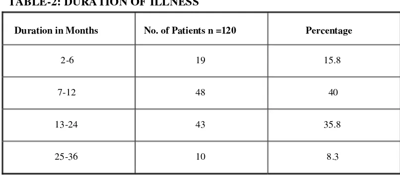
[image:54.612.121.494.503.702.2]TABLE-2: DURATION OF ILLNESS
Duration in Months
2-6
7-12
13-24
25-36
No. of Patients n =120
19
48
43
10
Percentage
15.8
40
35.8
8.3
Most patients (40%) had duration of illness between 7-12 months. This
was followed by the patients (35.8%) who had duration of illness
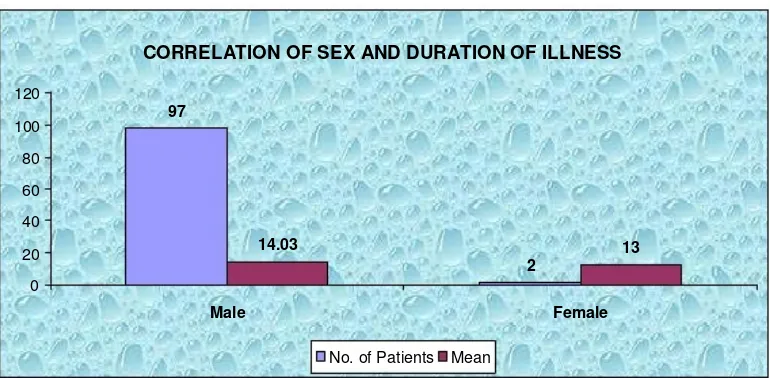
[image:55.612.103.506.76.253.2]TABLE-3: CORRELATION OF SEX AND DURATION OF
ILLNESS
SEX
DURATION ACTUAL MONTHS
The
M
F
n
97
2
Mean
14.03
13.00
illness
Std.Std. Error Significance Deviation of mean
7.555
9.200
in males
.767
1.918
is 14.03.P=0.574[not P=0.574
mean duration of
significant]. The mean duration of illness in females is 13.00.
CORRELATION OF SEX AND DURATION OF ILLNESS
120
100
80
60
40
20
0
Male
No. of Patients Mean
Female 14.03
2
[image:56.612.114.499.280.469.2]TABLE-4: CORRELATION OF AGE AND DURATION OF
ILLNESS
Groups 1
2
3
4
Total
n 19
48
43
10
120
Mean 29.47
30.33
39.02
52.30
35.14
Std. Deviation 16.473
10.041
15.056
12.962
14.772
Groups :1-duration of illness 2-6 months
2-duration of illness 7-12 months
3-duration of illness 13-24 months
4-duration of illness 25-36 months.
P = 0.000<0.001. The mean age level in the duration of illness is statistically significant. Among the duration of illness, group with largest
duration had age mean level as 52.30 , which is significantly an elevated
age mean level than other duration group.
CORRELATION OF AGE AND DURATION OF ILLNESS
100 80 60 40 20 0
1 2
No. of Patients 3
Mean
4
29.47
48 19
43
10 52.3
[image:57.612.115.500.525.695.2]TABLE-5: PREDISPOSING FACTORS
Duration in Months
Compound fracture due to road side accidents
Post Surgical
Diabetes mellitus with vascular insufficiency
Smoking/ Alcoholism
Haematogenous
LowerUpper No. of
Patients Percentage confidence confid