ITS CORRECTION USING PALATOGRAPHY- AN IN-VIVO
STUDY
DISSERTATION
SUBMITTED TO THE TAMILNADU DR MGR MEDICAL UNIVERSITY
IN PARTIAL FULFILLMENT OF THE REQUIREMENT FOR THE AWARD OF DEGREE
OF
MASTER OF DENTAL SURGERY
Branch I
Prosthodontics and Crown & Bridge
DECLARATION
I hereby declare that this dissertation entitled “Evaluation of speech in
maxillary defects and its correction using palatography An In-Vivo Study’’ is a bonafide work undertaken by me and that this thesis or a part of it has not been presented earlier for the award of any degree, diploma, fellowship or similar title of recognition.
Dr. Nikhil.S.Rajan, MDS Student,
Department of Prosthodontics,
ACKNOWLEDGEMENT
I walked in here with an empty mind, trembling hands, unsure footsteps and a lot of fear. I came here from darkness not knowing what to do. A vibrant light entered my life and showed me the way. She taught me to fill my empty mind with gratitude, hold up my trembling hands in prayer, take my unsure footsteps towards God and replace my fear with love. When I celebrated my meager victories, she congratulated me but reminded me about the virtue of humility.
When I was depressed and thought that I was not good enough she told me that I was the best in this world even though I was not even near.
The truth that set me free is the realization that my teacher loves me unconditionally
My heart is filled with gratitude for my teacher Dr.Lovely.M. Professor, Dept of Prosthodontics, Sree Mookambika Institute of Dental Sciences, Kulashekharam.
I humbly bow my head in acknowledgement of your support and love madam. By the grace of God whatever I accomplish in life will always be dedicated to you.
I thank Dr .T. Sreelal , Professor and HOD, Department of Prosthodontics, Sree Mookambika Institute of Dental Sciences , Kulashekharam for teaching me that truthfulness and sincerity might not give us rapid gains in the short run , but these are indispensable qualities which we should possess if we are to accomplish anything worthwhile in life.
“Sir, you don’t use much words , but whatever u say sticks .”
I thank Dr Shibu A, MDS Dr Sangeeth K Cherian, MDS Dr Sudheer.A, MDS and Dr James J Rex BDS (Readers), Dr Kavitha Janardhanan,MDS, Dr Anuroopa A, MDS, and Dr Vini K Varkey, MDS, (Senior Lecturer) for their valuable help and guidance.
I thank Mrs Vineetha George and Mrs Praveena Davis of National Institute Of Speech and Hearing ,Trivandrum (NISH , TVM) without their Unstint support and guidance this study would not have been possible.
I gratefully acknowledge my seniors Dr. Aparna. Mohan, Dr Priya M S and my batch mate Dr Arun R, fellow post graduates Dr Aravind Krishnan, Dr Anjana S, Dr Amal Shajahan and Dr Nissy Elizabath George for their motivation and encouraging words.
I thank my Parentsfor the blessings they have been showering on me throughout my life and telling me that “Money doesn’t matter-Love does”
I thank each and every one of my Patients who participated in this study and all others for teaching me human virtues like patience, forgiveness, respect and love... “Without you, I don’t exist”
LIST OF FIGURES
LIST OF TABLES
LIST OF GRAPHS
ABSTRACT
CHAPTER1. INTRODUCTION
1-101.1 SURGICAL CORRECTION OF MAXILLARY DEFFECTS 1
1.1.1 Armany’s classification for partially edentulous maxillectomy
dental arches 2
1.1.2 The rehabilitative procedure required for each age group of
cleft lip and palate patients 3
1.2 PROSTHODONTIC CORRECTION WITH OBTURATOR
PROSTHESIS 4
1.3 HISTORY 4
1.4 SPEECH 5
1.4.1 Production of sounds in speech 6
1.4.2 Speech phonemes 6
1.4.3 Consonants 7
1.5 CONDITIONS AFFECTING SPEECH 7
1.6 SPEECH ANLYSIS 8
1.7 PALATOGRAPHY 9
CHAPTER2. AIMS AND OBJECTIVES
11CHAPTER3. REVIEW OF LITERATURE
12-283.1 HISTORY 12
3.2 SURGICAL PHASE 12
CHAPTER4. MATERIALS AND METHODS
29-344.1. MATERIALS 29
4.1.1 Equipments 29
4.2. METHODOLOGY 30
4.2.1. Introduction 32
4.2.2. Clinical procedure 33
4.2.3. Making of the Impression and Fabrication of prosthesis 33
4.2.4. Palatography 33
4.3. FLOW CHART 35
4.4. FIGURES
CHAPTER5. RESULTS
36-605.1.1. Results for Intelligibility 36-37
5.1.2. Results for Nasalance 38-47
5.1.3. Results for Formant Evaluation 48-60
5.2. STATISTICAL ANALYSIS 61-65
5.2.1. Intelligibility: PCC (Percentage of Consonants Correct) 61
5.2.2. Nasalance Using Nasometer 62
5.2.3. Formant Evaluation using PRAAT Software 62
CHAPTER6. DISCUSSION
66-73CHAPTER
7
. SUMMARY AND CONCLUSION
74-75REFERENCES
Evaluation of speech in Maxillary defects and its correction using palatography- An In Vivo Study
Abstract
Introduction:
Most of the literature review is on speech analysis and correction of palatal defects. These defects are either corrected by surgical procedures or prosthetic rehabilitation. Even after surgical correction some patients need prosthesis for speech correction and to prevent nasal regurgitation of food. Few of the patients are entirely dependent on prosthesis rehabilitation alone since, surgical correction is not possible when the defects are large due to excision as in cases of oral cancer, trauma or unrepaired clefts. This study is an attempt to present that the prosthesis alone may not totally correct speech defects and palatography is an inevitable tool for correcting articulatory defects thereby improving speech. It was observed that the prosthesis alone can correct speech defects, yet the clarity of certain vowels and volume of mouth was found to be defective since with the help of palatography and modification of the existing prosthesis the exact articulation of these sounds can be corrected.
Materials and Methods
1. Percentage of consonants correct for 20 words (regional language) was used to assess the intelligibility at three clinical Intervals (Before correction of the defect , after correction of the defect with prosthesis and after modifying the prosthesis using palatography)
Evaluation of speech in Maxillary defects and its correction using palatography- An In Vivo Study
3. Formant evaluation (f0-f5) for three vowels |a|, |i| and |u| was done using PRAAT software at three clinical visits (Before correction of the defect , after correction of the defect with prosthesis and after modifying the prosthesis using palatography)
Results
Modified prosthesis using palatography was found to be significantly superior in terms of intelligibility, nasality and formant evaluation showing a higher significance for all the three methods namely Percentage of consonants correct, Nasometer and PRAAT software.
Conclusion
Evaluation of speech in maxillary defects and its correction using palatography An In-Vivo study
1
Introduction
The most common intraoral defects are in the maxilla, in the form of an opening into the antrum and naso-pharynx. Defects in the maxilla may be divided into those resulting from congenital malformations and acquired defects resulting from surgery of oral neoplasms. The opening produced may be small or big/large and may include any portion of the hard and soft palate, the alveolar ridges, and the floor of the nasal cavity (Chalian et al., 1971). Post-surgical maxillary defects pre-dispose the patient to hypernasal speech, fluid leakage into the nasal cavity, and impaired masticatory function. These defects can be corrected by surgical intervention, prosthetic rehabilitation or a combination of both surgical and prosthetic intervention1.
Treatment options are: 1. Surgical correction
2. Prosthodontic rehabilitation
1.1 Surgical correction of maxillary defects
Evaluation of speech in maxillary defects and its correction using palatography An In-Vivo study
2
1.1.1 Armany’s classification for partially edentulous maxillectomy dental arches 2
Class IMidline resection. Class II Unilateral resection. Class IIICentral resection.
Class IV Bilateral anterior-posterior resection. Class V Posterior resection. Class VI
Evaluation of speech in maxillary defects and its correction using palatography An In-Vivo study
3
1.1.2The rehabilitative procedure required for each age group of cleft lip and palate patients.3
Age of patient Operative procedure
0 - 3 days
4 - 6 months
11/2 - 2 years
1 - 3 years
3 - 6 years
9 – 11 years
14 years
18 years
Counseling of parents Feeding plate fitting Surgical closure of lip
Surgical closure of palate
Indirect speech therapy – by parents under guidance of speech therapist
speech therapy
Nasendoscopy ± Pharyngoplasty Alveolar bone grafting ± pre-grafting orthodontic therapy
Comprehensive orthodontic therapy
Evaluation of speech in maxillary defects and its correction using palatography An In-Vivo study
4
1.2 Prosthodontic correction with obturator prosthesis
Definition. An obturator (Latin: obturare,to stop up) is a disc or plate, natural or artificial, which closes an opening or defect of the maxilla as a result of a cleft palate or partial or total removal of the maxilla for a tumour mass (Chalian et al., 1971)1.
Types
Surgical obturator Temporary obturator Definitive obturator
1.2.1 Significance of restoration.
The main objective of an obturator patient is to restore the function of mastication, deglutition, speech and to achieve normal oro-facial appearance (Beumer III et al., 1979).The goals of prosthetic rehabilitation for total and partial maxillectomy patients include separation of oral and nasal cavities to allow adequate deglutition and articulation, possible support of the orbital contents to prevent enophthalmos and diplopia, support of the soft tissue to restore the midfacial contour, and an acceptable aesthetic results (Wang, 1997). Prosthodontic management of palatal defects has been employed for many years.
1.3 History
Evaluation of speech in maxillary defects and its correction using palatography An In-Vivo study
5
techniques for the fabrication of hollow obturators to decrease the weight of the prostheses and to make comfortable and well-tolerated prostheses (Payne & Welton, 1965; Ampil et al., 1967; Brown, 1969; Chalian& Barnett, 1972; Buckner, 1974; Benington & Clifford, 1982; Orr, 1986; Benington, 1989; Wu & Schaaf, 1989; Schmaman & Carr, 1992; Didier et al., 1993; Wang & Hirsch, 1997; McAndrew et al 1998).
The construction of the definitive obturator will vary with the type of resection and the presence or absence of teeth. If the obturator is not properly designed and constructed, the stress on the remaining hard and soft tissues may be pathological and may lead to premature loss of abutment teeth and chronic irritation of soft tissues. Even after rehabilitation of these maxillary defects the patient reports with some amount of hyper-nasality and unintelligible speech.
1.4 Speech
Speech is the use of systematized vocalization to express verbal symbols or words." (Sheridan: 1964).Speech is a very sophisticated autonomous and unconscious activity.
Speech in matured man is a learned habitual neuromuscular pattern which makes use of anatomical structures designed primarily for respiration and deglutition.
Evaluation of speech in maxillary defects and its correction using palatography An In-Vivo study
6 The acoustic theory of the speaking process refers to the sound source and to the way of its production. Exhalation with little compressions and dilatations of the surrounding medium is recognized as a source of sound formation. In this way the resonance room is constituted. 4
The organs involved in mastication, deglutition and respiration take part in speech. Air from the lungs is modulated as it passes through the respiratory passages, oral cavity and nasal cavity producing speech. Thus – Surd, Sonant, Consonants, Respiration, Phonation, Resonations, Articulations, Neurologic Integrations, Audition can be regarded as components of speech.
1.4.1 Production of sounds in speech:
The sound produced can be divided-According to place of production as: Bilabial – p, b, w, m, Labio dental – f, v,
Linguo dental – th ,
Linguo alveolar – t, d, s, z (azure), l, n,
Palatal – ch, j, z (zeal),
Velar – k,g, ng.
1.4.2 Speech phonemes
as-Vowels – a e i o u (Laryngeal sounds),
Evaluation of speech in maxillary defects and its correction using palatography An In-Vivo study
7
1.4.3 Consonants
They are further divided by the duration of the sound, the principal resonating chamber and the articulators used to form the sound-According to manner of production: Plosive – p, b, t, d, k, g,
Fricative – f, v, th, s, z, Affricative – ch, j, Semivowel – w, l, Nasal – m, n, ng .
1.5 Conditions affecting speech
:When these structures are affected automatically speech is affected-Lungs, Larynx, Pharynx, Soft palate, Nasal cavity, Hard palate, Tongue, Mandible, and Cheeks.
Phonetics – The science of sounds used in speech. Phonetic value – The character or quality of vocal cords.
The surd is any voiceless sound and is produced by separation of the vocal folds (glottis open) with no marginal vibration.
The sonants are voiced sounds and include all vowels and vowel like sounds.
Consonants are articulated speech sounds, and all require articulation to impede, constrict, divert, or stop the air stream at the proper place and time to produce the desired sound...
The spoken sound modulation is connected to: - Static sound forming components:
Teeth ("S", "S"),
Hard palate - (anterior area: "T", "D"),
Alveolar bone; - Dynamic articular components: Tongue ("L", "T", "D"),
Evaluation of speech in maxillary defects and its correction using palatography An In-Vivo study
8 Soft palate, the mandible's movement.
The phonetic adaptation of a patient with complete denture is achieved depending on: Selection and placing of the artificial teeth, Thickness of maxillary prosthetic base in the frontal area, optimal space for the tongue, Individual adaptation capacity, and Patients’ sound recognizing capacity.4
1.6 Speech Analysis:
Speech outcomes are relevant in guiding treatment and to evaluate the efficiency of the maxillary obturator prosthesis provided to correct the defect. The maxillofacial prosthodontist often receives referred patients who have experienced partial surgical resection of the maxilla (maxillectomy) as a part of cancer therapy or a congenital defect as in cleft palate. Maxillary defects often results in a high level of morbidity with significant psychological and functional implications for the patient. Such disabilities include inability to masticate and disturbances of deglutition and speech.
Phonetic dentistry or gnathophonics (the new field) is connected, on one side, to dentistry, on another side to articulatory, acoustical and perceptive phonetics, on the third side, to speech technology and artificial intelligence. Dentistry treatment and denture wearing may significantly alter the upper part of the vocal tract and the ability of articulating the complex sound sequences used in speech5.
Based on reasoning backed by articulatory phonetics, put forward several hypotheses, including that that the temporo-mandibular articulation state may significantly affect the pronunciation of the diphthongs and plosives and the hypothesis that the lack of inter-dental spaces between the incisors is equally detrimental, as to the too large spaces, as in case of diastema or lack of teeth.5
Evaluation of speech in maxillary defects and its correction using palatography An In-Vivo study
9
1.7 Palatography:
Palatography is a technique used to identify which parts of the mouth are used when making different sounds. This technique is often used by linguists doing field work on little- known natural languages. It involves painting a colouring agent, such as a dye or a mixture of charcoal and olive oil on the tongue or the roof of a person's mouth and having that person pronounce a specific letter or sound. A photograph is then made of the mouth roof and tongue in order to determine how the sound was articulated. The technique can also be performed electronically using a tool called a pseudo-palate, which consists of a retainer-like plate lined with electrodes that is placed on the roof of the mouth while the speaker pronounces a sound. A record made through palatography is called a palatogram6.
Palatography has been used to determine the optimum thickness and shape of the palatal surfaces. This approach was developed in a study of phonetics to determine the contact position of the tongue relative to the palate in the production of specific sounds essentially, application of these techniques ensured contact between the tongue and palate during articulation of these sounds.
The “s” and “sh” phonemes have received particular attention. Palatography frequently has served as the basis for determining the shape of the anterior palatal vault most conducive to satisfactory sound articulation.7
Evaluation of speech in maxillary defects and its correction using palatography An In-Vivo study
10
Early diagnosis of sub mucosal cleft palate is important. In children too young to tolerate naso-endoscopy and video-fluoroscopy, the diagnosis depends on the patient’s clinical history and intraoral examination findings. Kratzsch and Opitz investigated the relationship of palatine rugae to points (landmarks) and distances on the cleft palate during the period from birth to the time of early mixed dentition. The authors identified changes in the distances from the lateral palatine rugae points of the first and third rugae to the incisal point, the canine point and the tuberosity line. The results of their study indicated that a comparison of distances from the palatine rugae with distances between equivalent points revealed the changes that occurred in the anterior palate during various stages of growth.
Static palatography has been widely used to investigate consonant articulation. The method is based on the observation of the tongue print (a black paste is spread on the tongue before the production) either directly onto the palate (direct palatography) or onto a pseudo palate (indirect palatography).8
Static palatograms have been obtained by tongue modeling of an impression material on the external surface of palatal plate and oral surface of the artificial teeth during repetition of phonemes and words.4
Evaluation of speech in maxillary defects and its correction using palatography An In-Vivo study 11
Aims:
To determine the efficiency of modified palatal obturator prosthesis using
palatography in terms of intelligibility of speech, nasality and change in
formants.
Objectives:
1.
To access the intelligibility of speech for 20 words using Percentage of
consonants correct by listener scale analysis before correction of the defect,
after correction of the defect with prosthesis and after modification of the
prosthesis with palatography.
2.
To evaluate the nasalence for three vowels |a|, |i| and |u| (a, e and u) before
correction of the defect, after correction of the defect with prosthesis and
after modification of the prosthesis with palatography.
3.
To access the change in formants (f0-f5) for three vowels |a|, |i| and |u| (a, e
and u) before correction of the defect, after correction of the defect with
Evaluation of speech in maxillary defects and its correction using palatography An In-Vivo study 12
3.1 History
Cleft lip (cheiloschisis) and cleft palate (palatoschisis), which can also occur together as
cleft lip and palate, are variations of a type of clefting congenital deformity caused by abnormal facial development during gestational period. Approximately 1 in every 600 newborn babies worldwide is born with this defect. This means that, assuming 15 000 children are born per hour worldwide (United States Bureau of the Census, 2001); a child is born with a cleft somewhere in the world approximately every 2½ minutes.
3.2 Surgical Phase
The knowledge of cleft lip and the surgical correction received a big boost during the period between the Renaissance and the 19th century with the publication of Pierre Franco's Petit Traite and Traite des Hernies in which he described the condition as "lievre fendu de nativite" (cleft lip present from birth). The first documented Cleft lip surgery is from China in 390 BC in an 18 year soldier, Wey Young-Chi.
The treatment of cleft lip and palate should be initiated soon after birth and continues up to adulthood. The morphological rehabilitation of clefts involves plastic lip surgery at 3 months of age and palate surgery around 1 year of age, as well as secondary alveolar bone graft performed between 9 and 12 years of age46.
Evaluation of speech in maxillary defects and its correction using palatography An In-Vivo study 13
surgery to the lip and palate. With the help of nasendoscopy, nature of the speech abnormality and appropriateness of additional palatal surgery can be assessed, and also nasendoscopy is used in case of osteotomy surgery where in again speech is compromised.
There was no previous attempt made to classify the dental arches for patients who have had partial resection of the maxilla. There were no proper framework designs for maxillary obturators. In 2001 Mohamed A. Aramany2 proposed a classification for partially edentulous maxillectomy dental arches based on the frequency of occurrence of maxillary defects in a population of 123 patients.
A brief overview of the progress and evolution of philosophies of obturator framework designs was accomplished by Gregory R. Parr et al9 in 2003. It begins in 1530 AD with Ambrose Pare´ who described the first button-shaped sponge and metal obturator, and continues through the formation of the American Academy of Maxillofacial Prosthetics and concludes with a simplified discussion of complex surgical-prosthetic coordination and the use of vascularized free flaps with osseointegrated dental implants.
Evaluation of speech in maxillary defects and its correction using palatography An In-Vivo study 14
3.3 Prosthetic Phase
A new type of mechanical palatal exerciser, called the Lubit Palatal Exerciser (LPE), was described. Erwin C Lubit et al 11 in 1970 presented the case report of 28 patients involved in the therapeutic program (with LPE) to demonstrate the changes in the velopharyngeal structures and speech proficiency. The beneficial changes had been observed in all of them.
In 1991 M. El-Dakkak12 studied ten patients in whom the surgical defect involved the posterior margin of the soft palate which lead to velopharyngeal insufficiency were given with speech aid obturator one month before the evaluation. Prosthetic management of each subject was evaluated as reflected in adequacy of velopharyngeal closure and speech competency.
The purpose of their electromyographic study done by Takashi Tachimura et al13 in 2000 was to examine whether levator veli palatini muscle activity during speech can be changed with placement of a speech appliance and to clarify whether or not the change is related to the type of speech appliance used.
A prosthesis used to close a palatal defect in a dentate or edentulous mouth is referred to as an obturator. In 2001 Filiz keyf1 provided obturator prosthesis which restored masticatory functions and improved speech, deglutition and cosmetics for maxillary defect (hemimaxillectomy) patients.
Evaluation of speech in maxillary defects and its correction using palatography An In-Vivo study 15
alternative Impression Technique a Speech-Aid Prosthesis with a speech bulb was adapted to the patient. Following placement of the special prosthesis excellent restoration of speech and improvement of velopharyngeal function was achieved.
In 2005 A. Nekora-Azak et al15 presented a clinical report describing the prosthodontic rehabilitation of an edentulous patient with a maxillary defect using the biofunctional prosthetic system. The advantage of this technique was to provide patients with optimal form, function, and aesthetics in complete dentures.
Definitive obturation is initiated approximately 3 to 4 months after surgery when healing is complete. According to Won-suck Oh et al16 (2006) the impression for definitive obturator prosthesis should include the skin-graft mucosal junction, lateral aspect of the orbital floor, and the dynamic physiology of the velopharyngeal mechanism during speech and swallowing. The obturator bulb must also be contoured to prevent obstruction of nasal breathing and to maintain nasal resonance during speech.
In 2006 M. Oki et al17 conducted a study, the aim was to investigate whether the vibratory characteristics of obturator prostheses are affected by bulb design, i.e.: the hollow or buccal flange type, and different lateral and medial bulb heights. And the vibration analysis suggested that buccal flange obturator prosthesis with high lateral and low medial walls is preferable.
Evaluation of speech in maxillary defects and its correction using palatography An In-Vivo study 16
reported a case about using the hollow obturator with resilient denture liner on post hemimaxillectomy to overcome pain occurring with intimate contact with the mucosa, and also in preventing the fluid entering into the cavum nasi and sinus.
In 2007 a patient underwent treatment for malignant melanoma which necessitated the excision of all his teeth, the alveolar bone, and the hard palate. Yohsuke Taira et al19 fabricated a silicon obturator with a soft denture liner material, and inserted into the maxillary defect, and an acrylic denture was mechanically connected to the silicon obturator. And the report suggested that a detachable silicon obturator could be an option in the recovery of postsurgical maxillary defect.
Fabricating a successful obturator prosthesis used for the prosthetic rehabilitation of congenital or acquired defects in maxilla depends on making a detailed impression and constructing the prosthetic parts compatible with the oral tissues. The clinical report presented by Bora Bagis et al20 in 2008 described an intraoral technique for impression making and fabrication of open hollow obturator prosthesis.
Evaluation of speech in maxillary defects and its correction using palatography An In-Vivo study 17
In 2009 Suha Turkaslan et al22 explained that avoiding immediate obturator construction may cause serious facial appearance problems due to soft tissue contracture and disfigurement that may have a negative effect on the patients' psychology. When wearing the permanent obturator is neglected, the dynamics of non-supported soft tissue changes towards serious contracture and facial disharmony.
Pharyngeal obturator is a prosthesis which closes the palatal and pharyngeal defects improving the speech and other function. K.Kasim Mohamed et al23 in 2010 discussed a case report with palatopharyngeal insufficiency, its impression procedures, fabrication of prosthesis and improvements in speech.
In 2010 Ramaraju A V et al24 described a clinical case of sub-total maxillectomy due to osteomyelitis, which was successfully rehabilitated with a hollow bulb obturator, retained in a unique combination of a cast clasp and zest anchor type of radicular ball attachment. The placement of radicular attachments and the process of making the prosthesis hollow had a significant effect on the stability and retention of the obturator prosthesis in partially edentulous maxillectomy patients.
Evaluation of speech in maxillary defects and its correction using palatography An In-Vivo study 18
In 2011 subramaniam elangovan et al26 described how to achieve the goal for esthetics and phonetics and also described the fabrication of a hollow obturator by two piece method, which is simple and may be used as definitive obturator for maximum comfort of the patient.
Patients with extensive head and neck injuries due to trauma and/or extensive surgical procedures often exhibit restricted mouth opening. A modification of the standard impression procedure is often necessary. Shuchi Tripathi et al27 in 2011 made an alteration in the final impression procedure using altered cast technique for fabricating an obturator prosthesis with soft palate extension, and the result obtained was quite satisfactory.
Malignancies treated through surgical intervention creates anatomic defect which forms communication among the oral cavity, nasal cavity and maxillary sinus, in which case it is very difficult for the patient to perform functions like mastication, swallowing, and phonation. Naveen YG et al28 in 2011 provided Definitive obturator prosthesis which restored the missing structures and acted as a barrier between the communications among the various cavities.
In 2011 Prakash Somani et al29 reported a case of rehabilitation of a compromised Armany class I maxillectomy defect with a definitive hollow bulb obturator.
Evaluation of speech in maxillary defects and its correction using palatography An In-Vivo study 19
unconventional, modified, and hard to build prosthetic devices such as the design of a hybrid retention prosthetic obturator (swing lock).
Post surgical maxillary defects predispose the patients to hypernasal speech, fluid leakage into the nasal cavity, impaired masticatory function, recurrent upper respiratory tract infection and facial disfigurement. Nayana Anasane et al31 in 2011 in his case report revealed that these disabilities are minimized or eliminated almost immediately with the help of Definitive Hollow Bulb obturator.
In 2011 Sunit K et al32 presented an article which focused on an innovative method of fabricating a palatal obturator which aims at restoring the normal functions along with improving aesthetics. It also enables to devise the fabrication of prosthesis in two parts (Split obturator) for easy insertion, removal and be self-cleansable.
Palatal defect of any extent causes multiple problems in speech, mastication and esthetics. Palatal obturator is the only substitute which covers the defect, eliminates hypernasality and improves the communication. Rajani Dable33 in 2011 in her study discussed about hollow bulb Obturator prosthesis for an edentulous patient which becomes more critical in terms of its movements as there is no mechanical retention available.
Evaluation of speech in maxillary defects and its correction using palatography An In-Vivo study 20
sealing the defect and allowing the patient to acquire better speech quality, and improve her nutrition and well-being.
In velopharyngeal dysfunction, hypernasality and regurgitation of food and liquids is common if defect is not obturated. Ramya R et al35 in 2011 presented a case report of speech aid prosthesis with recording the contours of a partial soft-palate defect for prosthetic obturation.
Defects created in the maxillary bone, principally after resection of malignant tumors surgical, trauma or congenital defects can be corrected either by surgical reconstruction or by placement of maxillary obturator prosthesis. RT-Cayon Velazquez36 in 2011 reviewed the recent and classic literature on palatal obturators and five clinical cases treated with a palatal obturator were reported.
In 2011 Shyammohan .A et al37 in his article suggested a protocol for speech therapy in
cases of velopharyngeal insufficiency to be done in union with a prosthodontist. Speech therapy in
obturator cases demands a team approach comprising the patient, speech therapist, prosthodontist,
and parents/relatives for an effective outcome and the absence of any one can scuttle the result.
Evaluation of speech in maxillary defects and its correction using palatography An In-Vivo study 21
W. Hardcastle et al38 in 1989 reviewed recent developments in Electropalatography (EPG) as a technique for investigating spatio-temporal details of tongue contacts with the hard palate in both normal and pathological speech.
The purpose of the paper presented by John E. Riski et al39 in 1989 was to illustrate the value of combined aerodynamic and endoscopic examination of velopharyngeal function in the revision of prosthetic speech appliances. Combined measures enabled the clinician to identify accurately the site of under or over obturation, and any needed revision was completed accurately and efficiently.
In 1997 an investigation of speech adaptation to palatal modification (to an artificial palate) in [s] production was conducted using acoustic and perceptual analyses by Shari R. Baum et al40. Productions of [sa] were elicited at five time intervals, 15 min apart, with an artificial palate in place. And the results revealed improvement in both acoustic and perceptual measures at the final time interval relative to the initial measurement period.
Evaluation of speech in maxillary defects and its correction using palatography An In-Vivo study 22
In 2000 H. Yoshida et al42 conducted a study to reveal the acoustic characteristics associated with hypernasality and to ascertain their correlation to the severity of hypernasality, 30 speech samples produced by 15 maxillectomy patients were acoustically analysed with and without obturator prosthesis in place. Normalized 1/3-octave spectral analysis demonstrated the spectral characteristics of hypernasality.
Retrospective study conducted by M. Brent Seagle et al43 spans the years 1988 to 2000 and looks specifically at the treatment procedures and outcomes for the correction of velopharyngeal insufficiency(VPI) and the results revealed VPI resolution and the establishment of normal nonnasal speech in more than 95% of the 75 patients for whom outcomes were determined.
A strong association was found between ratings of ‘velopharyngeal function’ and ‘hypernasality’ and the pattern of nasal airflow during the bilabial nasal-to-stop combination. Hans Dotevall et al44 in 2002 assessed the nasal airflow dynamics during the velopharyngeal closing phase in speech which presented with quantitative, objective data that appear to distinguish between perceptually normal and deviant velopharyngeal function with high sensitivity and specificity.
Jana Rieger et al45 in 2002 in his study concluded that Rehabilitation with a maxillary obturator is successful in restoring preoperative speech function but rehabilitation of individuals with involvement of the soft palate may be more challenging.
Evaluation of speech in maxillary defects and its correction using palatography An In-Vivo study 23 bulb height, bulb parts with high (H), middle (M), and low (L) lateral walls were prepared and concluded that the L type is preferable from the standpoint of its vibratory characteristics.
Obturators have been developed for surgical defects caused by cancer of the maxillary sinus and alveolar ridge. Outcome research is necessary to develop evidence-based practice guidelines.
Marsha Sullivan et al47 in 2002 in his study concluded that obturation is an effective intervention for defects of the maxillary sinus and alveolar ridge on speech performance.
Some patients presenting velopharyngeal dysfunction need treatment with a palatal prosthesis, and few researches attempt to evaluate the judgement of its efficacy. In the same way
Joao Henrique Nogueira PINTO et al48 in 2003 in his study concluded that the prosthetic treatment of velopharyngeal dysfunction demonstrated efficacy in improving speech, despite of the heterogeneous sample.
Mean nasalance magnitudes and mean nasalance distances were obtained with three devices the Nasometer, the NasalView, and the OroNasal System. Tim Bressmann et al49 in 2005 in his study used these systems and explained that these three systems measure nasalance in different ways and provide nasalance scores that are not interchangeable.
Evaluation of speech in maxillary defects and its correction using palatography An In-Vivo study 24
clinical correlation exists supporting the efficacy of speech bulb– obturator intervention after velopharyngeal insufficiency for improved intelligibility of both words and sentences.
Viviane de Carvalho-Teles et al51 in 2006 conducted a study, the objective of which was to
evaluate the efficacy of the palatal obturator prosthesis on speech intelligibility and resonance of patients who had undergone inframedial structural maxillectomy. The results of this study indicated that maxillary obturator prosthesis was efficient to improve the speech intelligibility and resonance in patients who had undergone maxillectomy.
The aims of the study done by A.O.Arigbede et al52 in 2006 were to assess the effectiveness of the maxillary obturator as a speech rehabilitation aid and to examine the influence of the classes of surgical defects on speech intelligibility (SI). Results support the widely held view that the maxillary obturator is a useful speech rehabilitation aid and moreover SI is affected by the class of defect.
Sanskrit, an ancient language, has an arrangement of alphabets that is orderly and scientific and therefore provides a simple means to understand the production of phonemes and memorize them. In 2007 Kalpesh Gajiwala53 in his article demonstrates the inherent advantage of this arrangement of Sanskrit alphabets to effectively analyze defective cleft palate speech and provides a tool for surgeons to decide a course of action in their routine clinical practice.
Evaluation of speech in maxillary defects and its correction using palatography An In-Vivo study 25
transform analysis revealed a change in the formantic spectrum and the noise/harmonis ratio decreased and the formants were more meaningful after the implantation in the bisyllabic word.
The study done by Triona Sweeney et al55 in 2008 aimed to evaluate the relationship between perceptual assessment and acoustic measurements of nasality using controlled speech stimuli. The strong relationship between perceptual and acoustic assessments of nasality indicated that the Temple Street Scale and the Nasometer are both valid clinical tools for the evaluation of nasality when a carefully constructed speech sample is used.
The aim of the study done by Suha Turkaslan et al56 in 2009 was to evaluate the articulation performance of obturator patients with three different buccal extension designs and they concluded that Obturators improve speech intelligibility irrespective of their buccal extension levels, nevertheless, medium size buccal extension enables the optimum sealing for better articulation.
The aim of the study done by Aveliny Mantovan Lima-Gregio et al57 in 2010 was to investigate frequency spectral aspects of F1, F2, F3, nasal formant (FN) and anti-formant, in
Evaluation of speech in maxillary defects and its correction using palatography An In-Vivo study 26
The correction of VPI was temporarily done with a pharyngeal obturator since the child presented with a very little movement of the pharyngeal walls during speech, compromising the outcome of a possible pharyngeal flap procedure (pharyngoplasty). The program of intensive speech therapy was conducted in three phases. After the program involving the use of a pharyngeal obturator, Nachale Helen Maciel BISPO et al58 in 2011 observed absence of hypernasality and compensatory articulation with improved speech intelligibility.
Separation of the nasal and oral cavities by dynamic closure of the velo-pharyngeal port is necessary for normal speech and swallowing. Velo-pharyngeal dysfunction (VPD) may either follow repair of a cleft palate or be independent of clefting. Jeffrey L. Marsh59 in 2011 in his study explained that matching the specific intervention for management of VPD with the type of dysfunction, that is, differential management for differential diagnosis, maximizes the result while minimizing the morbidity of the intervention.
3.5 Palatography
Evaluation of speech in maxillary defects and its correction using palatography An In-Vivo study 27 which evaluated the most important spaces for the phonetic modulation, in relation to various settings of the artificial teeth.
The objectives of the study done by Thierry Legou et al8 in 2008 was to provide an automatic analysis of the tongue print characteristic (size, shape, position onto the palate…) in order to allow an objective description of the linguopalatal contact and the other one concerns the shape of the palate. The knowledge of the palate shape can be a key point to understand certain consonant productions and can be used for static direct and indirect palatography.
The paper presented by H.-N. L. Teodorescu5 in 2010 explains the use of indices such as SQI, SII and of the distances which provides the practitioner with the grounds of an automated method for assessing the quality of the performed prosthetic act. From the speech rehabilitation perspective, this is a major step ahead in the current technology and solves a fundamental problem in dentistry.
Evaluation of speech in maxillary defects and its correction using palatography An In-Vivo study 29
4.1 MATERIALS
Materials used for the study.
1. Polyvinyl Siloxane Impression material-Putty and light body (Elite H.D+) (Fig1) 2. Type III Dental Stone ( Gold stone, Asian Chemicals India) (Fig 2)
3. Impression Compound (Ashlate, Asian Acrylates , Mumbai India ) (Fig 3) 4. Green Stick Compound (DPI PINNACLE , Dental Products of India Ltd) (Fig 4) 5. Korecta no.4 wax (Factor II USA) (Fig5)
6. Betadiene Solution( Betadiene Germicide Gargle 2% , Win Medicare )`(Fig 6) 7. Heat cure acrylic resin (Acralyn-H , Asian Acrylates , Mumbai ) (Fig 7) 8. Cold mould seal (Acralyn-H, Asian Acrylates, Mumbai) (Fig 8)
9. Autopolymerizing acrylic resin (DPI RR, Dental Products of India Ltd) (Fig 9) 10.Gauze( Surgicom, Tamilnadu) (Fig 10)
4.1.1 Equipments
1. Nasometer ( Dr Speech ) (Fig 11) 2. Sony Vaio (vpceb14en) Laptop 3. PRAAT software (Fig 12)
Evaluation of speech in maxillary defects and its correction using palatography An In-Vivo study 30
4.2 METHODOLOGY:
Selection of patients
Ten patients aged between 6-25 years, eligible for treatment criteria were selected.
Inclusion Criteria
i) Age group 6 – 35 years (Speaking age group) ii) Subjects desire for a palatal obturator
iii) Willingness to comply with the study requirements
Exclusion Criteria
i) Patients aged above 35 years
ii) Patients with neuromuscular disorders
iii) Patients with having congenital and acquired defects of the vocal chord
Ethical committee clearance number:SMIMS/IHEC/2012/A1
STUDY DESIGN
Evaluation of speech in maxillary defects and its correction using palatography An In-Vivo study 31 STUDY SETTINGS
The study was conducted at the department of Prosthodontics, Sreemookambika Institute of Dental Sciences, Kulasekharam. The speech outcomes were recorded in a Sound proof room (20db max) at NISH (National Institute of Speech and hearing, Trivandrum).
STUDY PERIOD
6 months
STUDY GROUP
A group of ten patients having congenital maxillary defects were selected
A total of ten patients were included after evaluation of the patient to fit within the inclusion category. A detailed case history was taken and evaluated (Appendix 1). All treatment options were explained to the patient. Once the treatment was found feasible, the procedures, advantages, precautions, maintenance and care were explained to the patient and the patient’s informed consent for the treatment was obtained on paper (Appendix 2).
The following diagnostic aids were used to evaluate the patient’s nasalance and the type of obturator to be planned for the treatment purpose.
1 Diagnostic cast
Evaluation of speech in maxillary defects and its correction using palatography An In-Vivo study 32
(Elite H.D) using perforated stock impression trays. The impressions were poured in type III dental stone.
2 Nasometer
Acoustic data obtained with Nasometer [Dr Speech] helps to evaluate the percentage of nasalance in patients with congenital maxillary defects which in turn helps in the treatment planning of the patient.
Nasalance is measured in terms of frequency by time
Nasalance =
3. PRAAT Software
Digital acoustic analysis (or spectrogram) was done with PRAAT Software using vowels a, e, u, and the formants were evaluated from f0 – f5 at three clinical visits.
4. Sound Lab:
The speech outcomes were recorded in Sound proof room (20db max).
4.2.1 Introduction
Evaluation of speech in maxillary defects and its correction using palatography An In-Vivo study 33
evaluate the extent of the palatal defect). Then the treatment plan was made to provide obturator prosthesis in order to close the defect.
Speech outcome measurements were collected prospectively at three clinical visits- (1) before correcting the defect, (2) after correcting the defect with prosthesis (3) after modification of the prosthesis with palatography.
Acoustic data were obtained using Nasometer to determine nasalance. Digital acoustic analysis (or spectrogram) were done using PRAAT Software using vowels a, e, u, and the formants were evaluated from f0 – f5 at the above mentioned three clinical visits.
4.2.2 Clinical procedure (Fig 15 - Fig 36)
A detailed case history was taken and evaluated. Speech outcomes were measured before correction of the defect in terms of intelligibility (Using PCC), Nasalance (Using Nasometer Dr Speech), Formant Evaluation (Using PRAAT software)
4.2.3 Making of the Impression and Fabrication of prosthesis
Impressions were made with Perforated Stock Impression trays (Zhermeck) using polyvinyl Siloxane Impression material-Putty and light body (Elite H.D) and palatal obturator prosthesis was constructed using heat cure acrylic resin.
Evaluation of speech in maxillary defects and its correction using palatography An In-Vivo study 34
4.2.4 Palatography
Palatography was done using mouth temperature flowing wax-Korecta no:4 wax (Factor II USA). The wax was double boiled and painted onto the chameo surface of the prosthesis and molded by asking the patient to repeat few words (like Cha, Ta, Da, Sa, Saraswathy, Mississippi) to elicit the type of articulatory deficiency which was corrected by molding the wax in the deficient areas where by intelligibility of speech was improved markedly.
After palatography the modifications were incorporated onto the prosthesis with self cure acrylic resin and again the speech outcomes were evaluated, thus the values elicited after the procedure had a marked increase in intelligibility of speech.
Speech evaluation and acoustic data analysis were done in sound proof room of 20db.
Parameters
EVALUATION OF SPEECH IN MAXILLARY DEFECTS AND ITS CORRECTION-AN INVIVO STUDY
using 20 words INTELLIGIBILITY
OF WORDS (Pcc) (Assessed @ 3
clinical visits NASALANCE
(Measured using nasometer using 3vowels │a│e│u│ @ 3
clinicalvisits
CHANGE IN VOLUME OF MOUTH
(Evaluation of formants f0-f5 for3 vowels │a│e│u│@ 3
Evaluation of speech in maxillary defects and its correction using palatography An In-Vivo study 36
RESULTS
5.1.1 Results For Intelligibility by (PCC) Percentage of Consonants Correct
Association between t-value Degrees of
freedom
P value Inference
Before Prosthesis and After Prosthesis
8.78 9 P<0.001 Highly Significant
Before Prosthesis and after modified prosthesis
29.06 9 P<0.001 Highly Significant
After Prosthesis and after Modified prosthesis
[image:47.595.171.428.390.507.2]29.06 9 P<0.001 Highly Significant
Table I(Intelligibility assessment with Student’s t paired test)
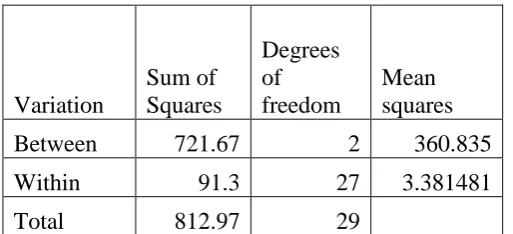
Table II(Intelligibility assessment with ANOVA One Way table)
Fobserved 106.709146
From the table for 0.01 significant level the F value is 99.46
Calculated value is greater than tabled value, which shows that the there is difference in treatment and the variability is highly significant
Evaluation of speech in maxillary defects and its correction using palatography An In-Vivo study 37
Table III(Intelligibility with ANOVA Two way table)
Therefore the difference between the treatment and the difference between the persons are highly significant and the significance level is less than 0.001
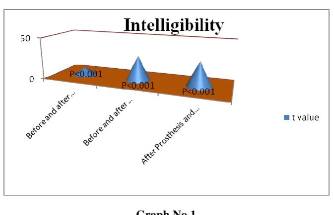
Intelligibility by (PCC) Percentage of Consonants Correct
Graph No.1
Tables I, II, III and Graph no1 shows the difference between before prosthesis and after prosthesis is highly significant with P <0.001, the difference between before prosthesis and after modified prosthesis is highly significant with P< 0.001 and the difference between after prosthesis and after modified prosthesis is highly significant with P< 0.001.
ANOVA Source of Variation Degrees of freedom Sum of Squares Mean Sum of
Squares F ratio
Degrees of
freedom F0.001 1 Persons 2 721.67 360.84 299.7245039 2,18 10.390 2 Treatment 9 69.6 7.73 6.423627134 9,18 5.760 3 Error 18 21.67 1.20
[image:48.595.130.467.355.571.2]Evaluation of speech in maxillary defects and its correction using palatography An In-Vivo study 38
5.1.2
Results of Nasalance Using Nasometer
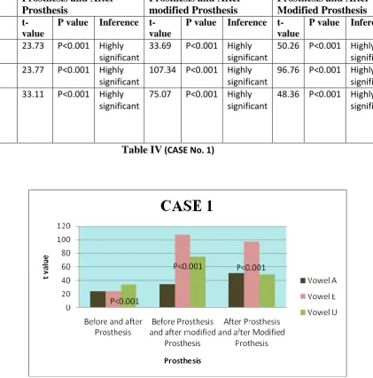
Table IV (CASE No. 1)
Graph No.2
Table IV and Graph No 2 shows that for case 1 there is significant difference between before prosthesis and after prosthesis, before prosthesis and after modified prosthesis and after prosthesis and after modified prosthesis, where the P value is less than 0.001respectively.
Vowel Difference b/w Before
Prosthesis and After Prosthesis
Difference b/w Before Prosthesis and After modified Prosthesis
Difference b/w After Prosthesis and After Modified Prosthesis t-
value
P value Inference t- value
P value Inference t- value
P value Inference
A 23.73 P<0.001 Highly significant
33.69 P<0.001 Highly significant
50.26 P<0.001 Highly significant E 23.77 P<0.001 Highly
significant
107.34 P<0.001 Highly significant
96.76 P<0.001 Highly significant U 33.11 P<0.001 Highly
significant
75.07 P<0.001 Highly significant
[image:49.595.103.525.184.611.2] [image:49.595.128.499.395.614.2]Evaluation of speech in maxillary defects and its correction using palatography An In-Vivo study 39
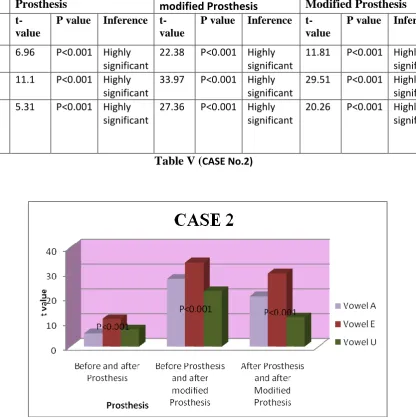
Table V (CASE No.2)
[image:50.595.107.524.155.574.2] [image:50.595.133.497.351.576.2]Graph No.3
Table V And Graph No 3 shows that for case 2 there is significant difference between before prosthesis and after prosthesis, before prosthesis and after modified prosthesis and after prosthesis and after modified prosthesis, where the P value is less than 0.001respectively.
Vowel Difference b/w Before
Prosthesis and After Prosthesis
Difference b/w Before Prosthesis and After modified Prosthesis
Difference b/w After Prosthesis and After Modified Prosthesis t-
value
P value Inference t- value
P value Inference t- value
P value Inference
A 6.96 P<0.001 Highly significant
22.38 P<0.001 Highly significant
11.81 P<0.001 Highly significant E 11.1 P<0.001 Highly
significant
33.97 P<0.001 Highly significant
29.51 P<0.001 Highly significant U 5.31 P<0.001 Highly
significant
27.36 P<0.001 Highly significant
Evaluation of speech in maxillary defects and its correction using palatography An In-Vivo study 40
Table VI (CASE No.3)
Graph No.4
Table VI and Graph No.4 shows that for case 3 there is significant difference between before prosthesis and after prosthesis, before prosthesis and after modified prosthesis and after prosthesis and after modified prosthesis, where the P value is less than 0.001respectively.
Vowel Difference b/w Before Prosthesis and After Prosthesis
Difference b/w Before Prosthesis and After modified Prosthesis
Difference b/w After Prosthesis and After Modified Prosthesis t-
value
P value Inference t- value
P value Inference t- value
P value Inference
A 7.59 P<0.001 Highly significant
27.41 P<0.001 Highly significant
18.59 P<0.001 Highly significant E 11.12 P<0.001 Highly
significant
34.02 P<0.001 Highly significant
29.54 P<0.001 Highly significant U 6.79 P<0.001 Highly
significant
20.73 P<0.001 Highly significant
Evaluation of speech in maxillary defects and its correction using palatography An In-Vivo study 41
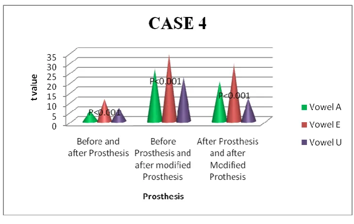
Table VII (CASE No. 4)
Graph No. 5
Table VII and Graph No.5 shows that for case 4 there is significant difference between before prosthesis and after prosthesis, before prosthesis and after modified prosthesis and after prosthesis and after modified prosthesis, where the P value is less than 0.001 respectively.
Vowel Difference b/w Before Prosthesis and After Prosthesis
Difference b/w Before Prosthesis and After modified Prosthesis
Difference b/w After Prosthesis and After Modified Prosthesis t-
value
P value Inference t- value
P value Inference t- value
P value Inference
A 5.32 P<0.001 Highly significant
27.41 P<0.001 Highly significant
20.31 P<0.001 Highly significant E 11.16 P<0.001 Highly
significant
34.06 P<0.001 Highly significant
29.58 P<0.001 Highly significant U 6.81 P<0.001 Highly
significant
22.38 P<0.001 Highly significant
[image:52.595.71.560.135.316.2] [image:52.595.132.497.376.607.2]Evaluation of speech in maxillary defects and its correction using palatography An In-Vivo study 42
Table VIII (CASE No.5)
Graph No.6
Table VII and Graph No.6 shows that for case 5 there is significant difference between before prosthesis and after prosthesis, before prosthesis and after modified prosthesis and after prosthesis and after modified prosthesis, where the P value is less than 0.001 respectively. For vowel E and U the difference between before prosthesis and after prosthesis is significant and the P value is only less than 0.01.
Vowel Difference b/w Before Prosthesis and After Prosthesis
Difference b/w Before Prosthesis and After modified Prosthesis
Difference b/w After Prosthesis and After Modified Prosthesis t-
value
P value Inference t- value
P value Inference t- value
P value Inference
A 22.77 P<0.001 Highly significant
33.82 P<0.001 Highly significant
49.74 P<0.001 Highly significant E 3.65 P<0.01 Highly
significant
85.39 P<0.001 Highly significant
89.26 P<0.001 Highly significant U 3.22 P<0.01 Highly
significant
51.16 P<0.001 Highly significant
[image:53.595.110.518.184.541.2]Evaluation of speech in maxillary defects and its correction using palatography An In-Vivo study 43
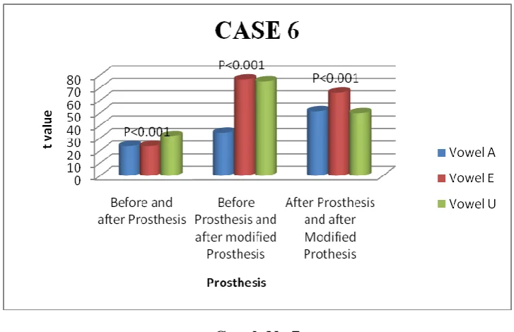
Table IX (CASE No.6)
Graph No.7
Table IX and Graph No.7shows that for case 6 there is significant difference between before prosthesis and after prosthesis, before prosthesis and after modified prosthesis and after prosthesis and after modified prosthesis, where the P value is less than 0.001 respectively.
Vowel Difference b/w Before Prosthesis and After Prosthesis
Difference b/w Before Prosthesis and After modified Prosthesis
Difference b/w After Prosthesis and After Modified Prosthesis t-
value
P value Inference t- value
P value Inference t- value
P value Inference
A 23.57 P<0.001 Highly significant
34.17 P<0.001 Highly significant
51.23 P<0.001 Highly significant E 23.56 P<0.001 Highly
significant
76.62 P<0.001 Highly significant
66.08 P<0.001 Highly significant U 31.52 P<0.001 Highly
significant
75.19 P<0.001 Highly significant
[image:54.595.102.522.118.546.2] [image:54.595.133.497.314.549.2]Evaluation of speech in maxillary defects and its correction using palatography An In-Vivo study 44
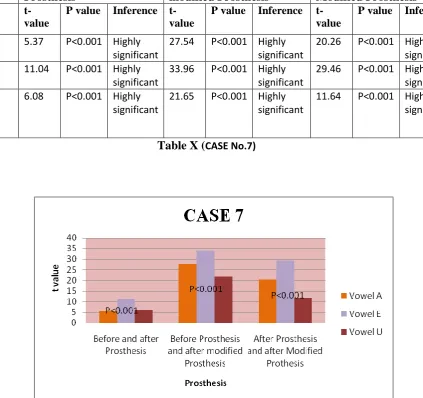
Table X (CASE No.7)
Graph No. 8
Table X and Graph No.8 shows that for case 7 there is significant difference between before prosthesis and after prosthesis, before prosthesis and after modified prosthesis and after prosthesis and after modified prosthesis, where the P value is less than 0.001 respectively.
Vowel Difference b/w Before Prosthesis and After Prosthesis
Difference b/w Before Prosthesis and After modified Prosthesis
Difference b/w After Prosthesis and After Modified Prosthesis t-
value
P value Inference t- value
P value Inference t- value
P value Inference
A 5.37 P<0.001 Highly significant
27.54 P<0.001 Highly significant
20.26 P<0.001 Highly significant E 11.04 P<0.001 Highly
significant
33.96 P<0.001 Highly significant
29.46 P<0.001 Highly significant U 6.08 P<0.001 Highly
significant
21.65 P<0.001 Highly significant
[image:55.595.73.563.127.313.2] [image:55.595.101.525.166.564.2]Evaluation of speech in maxillary defects and its correction using palatography An In-Vivo study 45
Table XI (CASE No.8)
Graph No. 9
Table XI and Graph No.9shows that for case 8 there is significant difference between before prosthesis and after prosthesis, before prosthesis and after modified prosthesis and after prosthesis and after modified prosthesis, where the P value is less than 0.001 respectively and the difference between before Prosthesis and after prosthesis is not significant for Vowel A, where the P value is greater than0.05.
Vowel Difference b/w Before
Prosthesis and After Prosthesis
Difference b/w Before Prosthesis and After modified Prosthesis
Difference b/w After Prosthesis and After Modified Prosthesis t- value P value Inference t-
value
P value Inference t- value
P value Inference
A 1.203 P>0.05 Not Significant
27.36 P<0.001 Highly significant
23.29 P<0.001 Highly significant E 11.1 P<0.001 Highly
significant
33.98 P<0.001 Highly significant
29.55 P<0.001 Highly significant U 6.79 P<0.001 Highly
significant
20.66 P<0.001 Highly significant
[image:56.595.97.531.106.534.2] [image:56.595.133.498.319.541.2]Evaluation of speech in maxillary defects and its correction using palatography An In-Vivo study 46
Table XII (CASE No.9)
Graph No. 10
Table XII and Graph No.10 shows that for case 9 there is significant difference between before prosthesis and after prosthesis, before prosthesis and after modified prosthesis and after prosthesis and after modified prosthesis, where the P value is less than 0.001 respectively.
Vowel Difference b/w Before Prosthesis and After Prosthesis
Difference b/w Before Prosthesis and After modified Prosthesis
Difference b/w After
Prosthesis and After Modified Prosthesis
t- value
P value Inference t- value
P value Inference t- value
P value Inference
A 23.66 P<0.001 Highly significant
33.71 P<0.001 Highly significant
50.34 P<0.001 Highly significant E 23.76 P<0.001 Highly
significant
106.47 P<0.001 Highly significant
96.53 P<0.001 Highly significant U 33.18 P<0.001 Highly
significant
75.25 P<0.001 Highly significant
Evaluation of speech in maxillary defects and its correction using palatography An In-Vivo study 47
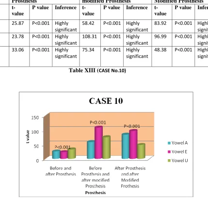
Table XIII (CASE No.10)
[image:58.595.96.526.129.539.2]Graph No.11
Table XIII and Graph No.11 shows that for case 10 there is significant difference between before prosthesis and after prosthesis, before prosthesis and after modified prosthesis and after prosthesis and after modified prosthesis, where the P value is less than 0.001 respectively.
Vowel Difference b/w Before Prosthesis and After Prosthesis
Difference b/w Before Prosthesis and After modified Prosthesis
Difference b/w After Prosthesis and After Modified Prosthesis t-
value
P value Inference t- value
P value Inference t- value
P value Inference
A 25.87 P<0.001 Highly significant
58.42 P<0.001 Highly significant
83.92 P<0.001 Highly significant E 23.78 P<0.001 Highly
significant
108.31 P<0.001 Highly significant
96.99 P<0.001 Highly significant U 33.06 P<0.001 Highly
significant
75.34 P<0.001 Highly significant
[image:58.595.132.498.313.540.2]Evaluation of speech in maxillary defects and its correction using palatography An In-Vivo study 48
5.1.3 Results for Formant Evaluation using PRAAT software
Table XIV (CASE No.1)
Graph No. 12
Table XIV and Graph No.12 shows that for Case 1 the difference between after prosthesis and after modified prosthesis is significant and the P value is less than 0.05 for vowels [a] and [u].
Vowel Difference b/w Before
Prosthesis and After Prosthesis
Difference b/w Before Prosthesis and After modified Prosthesis
Difference b/w After Prosthesis and After Modified Prosthesis t- value P
value
Inference t- value P value
Inference t- value P value
Inference
A 1.94 P>0.05 Not Significant
1.68 P>0.05 Not Significant
3.27 P<0.05 Significant
E 2.068 P>0.05 Not Significant
2.54 P>0.05 Not Significant
3.18 P<0.05 Significant
U 2.11 P>0.05 Not Significant
2.34 P>0.05 Not Significant
Evaluation of speech in maxillary defects and its correction using palatography An In-Vivo study 49
Table XV (CASE No.2)
[image:60.595.56.541.142.317.2] [image:60.595.105.492.281.569.2]Graph No.13
Table XV and Graph No.13 shows that for Case 2 in Vowel [a] the difference between before prosthesis and after prosthesis is significant, where P<0.05 and the difference between after prosthesis and after modified prosthesis is significant, where P<0.02. In vowel [u] the difference between before prosthesis and after prosthesis is not significant, where P>0.05 and the difference between after prosthesis and after modified prosthesis is just significant, where P=0.05.
Vowel Difference b/w Before Prosthesis and After Prosthesis
Difference b/w Before Prosthesis and After modified Prosthesis
Difference b/w After Prosthesis and After Modified Prosthesis t- value P
value
Inference t- value P value
Inference t- value P value
Inference
A 2.55 P>0.05 Not significant
3.22 P<0.05 Significant 3.51 P<0.02 Significant
E 1.92 P>0.05 Not significant
2.23 P>0.05 Not Significant
1.79 P>0.05 Not Significant U 2.36 P>0.05 Not
significant
1.99 P>0.05 Not Significant