A CLINICAL STUDY OF ULCERS OF THE FOOT
A dissertation submitted to the TAMILNADU DR. M.G.R. MEDICAL
UNIVERSITY,CHENNAI -10.
in partial fulfillment of the requirements for the degree of the
M. S. (GENERAL SURGERY)
DEPARTMENT OF GENERAL SURGERY TIRUNELVELI MEDICAL COLLEGE HOSPITAL
DECLARATION BY THE CANDIDATE
I hereby declare that this dissertation entitled “A CLINICAL STUDY OF
ULCERS OF THE FOOT” is a bonafide and genuine research work carried
out by me under the guidance of Dr. V.PANDY M. S., Professor, Department
of Surgery, TIRUNELVELI MEDICAL COLLEGE HOSPITAL, Tirunelveli
Medical College, Tirunelveli.
Place: TIRUNELVELI Signature of the Candidate
Date: Name: Dr. P. SHASHIDHARA
Prof. Dr. S. Soundararajan M.S,
Professor and Head of the Department, Department of General Surgery,
Tirunelveli Medical College Hospital, Tirunelveli.
CERTIFICATE
This is to certify that this dissertation titled “STUDY OF THE ULCERS OF THE FOOT” is a bonafide work of Dr. P. SHASHIDHARA, and has been prepared under my guidance, in partial fulfillment of regulations of
The Tamilnadu Dr. M.G.R. Medical University, for the award of M.S. Degree in
General Surgery during the year 2013.
Place: Tirunelveli Prof. Dr. S. Soundararajan M.S.
Date:
The Dean
ACKNOWLEDGEMENT
So many people have contributed to the successful completion of this
dissertation that I cannot list them all and must ask the forgiveness of those
whom I omit. This study required the cooperation of many people. My sincere
acknowledgements to all the participants in this study of mine.
With the most sincere and deepest sense of gratitude, I thank Dr.S. SOUNDARAJAN M.S., Professor and Head, Department of General Surgery, Tirunelveli Medical College Hospital, Tirunelveli Medical College, for his
patience, constant encouragement, guidance and constructive criticism. His
valuable suggestions and timely advice were of immense help to me throughout
all phases of this study.
I extend my sincere thanks to Dr. V. PANDY, M.S.,Professor,
Department of General Surgery, Tirunelveli Medical College, Tirunelveli, for
his valuable guidance, encouragement and suggestion during this dissertation.
I express my heartfelt gratitude to Prof. Dr. MAHESHWARI, Prof.
Dr. M.S. VARADARAJAN, Prof. Dr. ALEX EDWARDS, Assistant Professors and all the teaching faculties for their timely advice and encouragement in preparing this dissertation.
I thank Dr.S. Senthil Arumugam M.S., Dr. Amalan M.S., Dr.B. M.
M.S.,,for helping me to complete this study without whose help thestudy could have not been completed.
I sincerely acknowledge the help and assistance rendered by my fellow
postgraduates, junior postgraduates, CRRI’s, and SOT staff. Last but not the
least; I thank all the patients for their kind co-operation in carrying out the study
successfully.
Date: Signature of the Candidate
LIST OF ABBREVIATIONS USED
TAO Æ Thromboangiitis Obliterans
HLA Æ Human Leucocyte Antigen
ESR Æ Erythrocyte Sedimentation Rate
TC Æ Total Count
DC Æ Differential Count
LDL Æ Low-density Lipoprotein
VDRL Æ Veneral Diseases Research Laboratory
RBC Æ Red Blood Corpuscles
ABSTRACT BACKGROUND
A chronic ulcer of the foot is a frequent condition, with prevalence in
the population over 60 years of age. The incidence of ulcers is rising as a result
of the ageing population and increased risk factors for atherosclerotic occlusion
such as smoking, obesity and diabetes.
Ulcers can be defined as ‘break in the continuity of the covering
epithelium either skin or mucous membrane due to molecular death’. In general,
the slow healing tendency is not simply explained by depth and size, but caused
by an underlying pathogenetic factor that needs to be removed to induce healing.
The main causes are varicose veins, lower extremity arterial disease and
diabetes, less frequent conditions are infection, vasculitis, skin malignancies and
ulcerating skin diseases such as pyoderma gangrenosum. For a proper treatment
of patients with foot ulcers it is important to be aware of the large differential
diagnosis of foot ulceration and to effectively manage the conditions
AIMS AND OBJECTIVES
• To compare and analyze the distribution of age, sex, systemic disease and
location of the ulcer among 100 cases of the study group.
• To study the clinical features of various types of foot ulcers.
• To identify the methodology to effectively manage the condition.
• To identify the steps to prevent as far as possible foot ulcers in high-risk
individuals prone to the condition.
METHODS
Prospective study of 100 cases of chronic foot ulcers admitted at
Tirunelveli medical college Hospital, Tirunelveli, during the period March 2011
to March 2012, with regular dressing, debridement, treating the underlying
systemic disease, skin grafting and amputation were done.
RESULTS
In a study group of 100 cases, most of the patients with leg ulcers had an
underlined systemic disease such as diabetes mellitus, varicose veins, arterial
occlusion secondary to atherosclerosis, leprosy and malignancy.
KEYWORDS
Chronic non-healing ulcer; Diabetic leg and foot ulcer; varicose ulcer; Tropic
TABLE OF CONTENTS
Sl. No. Particulars Page Nos.
1 INTRODUCTION 1
2 AIMS AND OBJECTIVES 3
3 REVIEW OF LITERATURE 4
4 METHODOLOGY 82
5 ANALYSIS AND OBSERVATIONS 86
6 DISCUSSION 101
7 OBSERVATION 106
8 SUMMARY 107
9 BIBLIOGRAPHY 109
LIST OF TABLES
Sl. No.
Particulars Page No.
1 Distribution of various types of chronic foot ulcers 86
2 Sex distribution of various types of chronic foot ulcers 88
3 Age distribution of various types of chronic foot ulcers 89
4 Distribution of diabetic ulcers in the limbs 90
5 Sex distribution of diabetic ulcers 91
6 Age distribution of diabetic ulcers 92
7 System affected in venous leg ulcers 93
8 Age distribution of venous ulcers 94
9 Sex distribution in venous ulcers 95
10 Age distribution of various types of arterial ulcers 96
11 Pathology in arterial ulcers 97
12 Location of the ulcer according to its types 98
LIST OF FIGURES
Sl. No. Particulars Page
No.
1 Posterior tibial, popliteal and lateral plantar artery 10
2 Anterior tibial and Dorsalis pedis artery 10
3 Arteries of the foot 10
4 Muscles of the leg 16
5 Superficial and deep veins of the leg and foot 24
6 Layers of the skin 27
7 Squamous cell carcinoma – (high power field) 27
8 Thrombus in popliteal vein 67
9 Popliteal vein with thrombus near the valve 67
10
With distal compression of foot, flow is normal from
superficial to deep vein (coded red), on release flow is
retrograde through the perforator (coded blue)
67
11 Superficial artery occlusion 67
12 Popliteal artery with collateral flow around 67
13 Venous ulcer 83
14 Diabetic ulcer 83
15 Arterial ulcer (TAO) 84
16 Malignant ulcer (Squamous cell Ca) 84
LIST OF GRAPHS
Sl.
No. Particulars
Page No.
1 Showing distribution of various types of foot ulcers 86
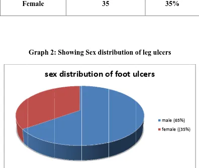
2 Showing Sex distribution of foot ulcers 88
3 Age distribution of various ulcers 89
4 Distribution of diabetic ulcers in the foot 90
5 Showing sex distribution of diabetic ulcers 91
6 Showing age distribution of diabetic ulcer 92
7
Showing distribution of venous ulcers with reference to
system affected
93
8 Age distribution of venous ulcers 94
9 Showing sex distribution of venous ulcers 95
10 Showing Age distribution of arterial ulcers 96
11 Showing pathology of arterial ulcers 97
INTRODUCTION
Chronic ulcers of the lowerlimb is frequent condition and wide in
distribution they may be associated with a number of Medical, Surgical &
Dermatological condition,most commonly dealt with and treated in the
surgical wards and OPD.
The incidence of ulcers is more in aging population and increased
risk factor for atherosclerotic occlusion such as smoking, obesity and
Diabetics.
Ulcer can be defined as ‘break in the continuity of the covering
epithelium either skin or mucous membrane due to molecular death’, In
general the slow healing tendency is not simply explained by depth and size.
But caused by a underlying pathologic fact that needs to be removed to induce
healing.
The problems of lower limb ulcer represent a wide spectrum of
etiology, pathology, severity and morbidity. The main causes are varicose
veins, lower extremity arterial disease and diabetes. Less frequent conditions
are infections, vasculitis, skin malignancies and ulcerating skin diseases such
as pyoderma gangrenosum. But even rare condition exists such as recently
For a proper treatment of patients with lower limb ulcers, it is
important to be aware of the large differential diagnosis and different causess
of foot ulcers. The causes may be various but the anatomical situation of
ulcers in the foot by itself can give rise to problems that can at times test the
ingenuity and patience of the surgeons
Various studies have been conducted and a number of procedures
and techniques have evolved with varying degree of success. It is common to
see patients with different types of ulcers due to various etiology and
underlying systemic diseases. Moreover, foot ulcers form a good bulk of
patients in our hospital. Treatment of these ulcers forms a challenging task as
well.
I have therefore in my present study attempted to analyze the ulcers
of the foot.
This study comprises of:
Review of literature with regard to historical aspects, etiology,
anatomy, pathology, pathophysiology, clinical features and diagnosis of
chronic foot ulcers along with the various modern investigative studies
required for the diagnosis.
Clinical investigation and histopathological study of patients of
chronic foot ulcers are done in patients who are admitted in Tirunelveli
AIMS AND OBJECTIVES
To compare and analyze the distribution of age, sex, systemic disease
and location
of the ulcer among 100 cases of the study group.
To study the clinical features of various types of foot ulcers.
To study the usefulness of applied investigations.
To identify the methodology to effectively manage the condition.
To identify the steps to prevent as far as possible foot ulcers in
REVIEW OF LITERATURE
I. HISTORICAL ASPECT OF LEG ULCERS
The incidence of chronic foot ulcer is as old as history, as with any
disease of mankind. The most common and noted ulceration of the chronic
foot ulcers for many years is Stasis ulcers.Hippocrates, the legendary father of
medicine himself had a leg ulcer. He was against treating various ulcerations
by surgical means. He treated multiple varicose veins by puncturing them at
different levels to avoid non-healing of ulcers and about 400 years B.C. He
wrote - " In case of an ulcer, it is not expedient to stand, especially if the ulcer
be situated on the leg" (Sarkar P. K. Ballantynes).1
In 4th Century B.C, healing by faith was practiced as seen in clay
models, which are taken from Temples of Athens.
Avicenna (982 - 1027 AD) gave a good account of diabetes and was the
first oneto describe diabetic gangrene.
Mollers Christenses during the period 1250 - 1500 A.D reported
changes in thelong bones of the leg due to leprosy. He found these changes in
the skeletons in burial grounds of Lazar Hospital. Earlier, pathological
evidences to date are in Mummies of 2nd century B.C. Sir Benjamin Collin
blood by means of a clinical test. Half a century later, this test was termed as
"Trendlenberg's test".
In 1828 Marjolin described the carcinomatous ulcers originating from
degenerative burns scars - this ulcers bears his name.
Celsus, the Roman physician & Hippocrates described treatment by
bandaging for chronic ulcers in the leg and related them to varicose veins.
John Hunter (1878) attempted to explain these ulcers in terms of venous stasis
due to the pressure of the column of blood while in upright posture.
Gay & Spencer’s in 1868 wrote 2 important books on venous ulcers,
which stressed the role of deep vein thrombosis and other lesions of arteries
and veins (both superficial and deep) in etiology of lower limb ulcerations.
Gay also described perforating ankle veins and suggested use of the term
venous ulceration (M. Wayne Flye).2
Linton drew attention to incompetence of communicating calf veins as
potential cause of venous ulceration (Linton RR).3
Pryce (1887) described the association of ulceration and vascular
diseases in diabetes.
From the 10th to 18th centuries various physicians including Halu,
Abbas, Avicenna, Falopio and Pare attributed ulceration of the leg to
leg served useful purpose in getting rid of these live Substances (Shami S. K.
Shield).4
In 1909 Burger described a syndrome of vascular occlusion in which
arteries, veins, and nerves of the extremities were involved in extensive
fibrosis that resulted in ulceration.
Although a minor traumatic incident is often the immediate cause of the
ulcer, the underlying pathology is usually vascular. Callam et al (1985)
estimated that 70 percent of leg ulcers are venous in origin, 10 per cent are
arterial and 10-15 per cent are of mixed arterial/venous origin. More unusual
causes include malignancy, vasculitis, neuropathy, metabolic disorders such
as pyoderma gangrenosum, and ulcers associated with disorder such as sickle
cell ulceration (Moffatt and Harper 1997)
II. INCIDENCE AND OCCURRENCE OF FOOT ULCERS
Accurate data concerning the incidence of non-fatal diseases are
difficult to obtain and statistics are usually derived from hospital attendance
records and general practice surveys. In under developed countries, the
incidence of ulcer may be greatly under estimated and apparent differences
between populations may be affected by differences in age structures.
The site of ulceration is recorded using the method of Callum; 90% of
the ulcers were present in the gaiter area, 2 % in the foot and 8% in the leg
A basic study of 4422 healthy working adults aged between 20 and 70
years in Europe resulted in detection of chronic venous insufficiency in 19%
of men and 25% of women. Other surveys conducted in Europe and America
came out with similar results and surveys confirmed the increased incidence
of ulcerations in females.
Information from other parts of the world other than Europe and
America is sparse. Schulz, Finaly and Scott (1962), in Transvaal, found that
0.1% of 2000 consecutive Bantu patients attending surgical outpatient
department had venous ulcers, but do not give figures for the incidence of
non-venous leg ulcers, while Dogliotti (1970) in Johannesburg found the
incidence of leg ulcers (unspecified) in Bantu patients referred to hospital to
be 1.75%. Finally and Park (1969) compared the incidence of varicose
eczema in their middle aged Indian patients (7%) with that in comparable
"white" patients (3%) and Bantu (1%) but give no figures for the incidence of
venous ulcerations.
III. ANATOMY OF THE LEG AND FOOT
The leg is that part of the lower limb below the knee and the terminal part of
the leg below the ankle is the foot.
Front of the leg and dorsum of foot
Superficial fascia of the front of the leg and the dorsum of the foot
Superficial veins.
Cutaneous Nerves
Lymphatics
Small unnamed arteries.
The subcutaneous bony surfaces are not covered by the deep fascia of
the leg but are attached to it at its borders. It is thick in the upper part of the
leg and gives origin to the underlying muscles while in the lower part is thin
and forms retinacula around the ankle.
Intermuscular septa are formed in the deep fascia, which tends to divide
the leg into compartments. The anterior and the posterior intermuscular septa
are attached anterior and posterior borders of the fibula and divide the leg into
anterior, lateral and posterior compartments. In the posterior compartment a
superficial transverse facial septum separate tibialis posterior from the long
flexors of the toe.
Muscles of the anterior compartment are – Tibialis anterior,
Extensor halucis longus, Extensor digitorum longus and Peroneous tertius.
Anterior Tibial Artery
The main artery that is found in the anterior compartment of the leg is
the anterior tibial artery. The perforating branch of the peroneal artery feeds
this artery. Anterior tibial artery is the smaller terminal branch of popliteal
Surface Anatomy
In surface projection of the anterior tibial artery begins 2.5 cm below
the medial side of fibula and ends at the midpoint between the malleoli. The
vessel can be felt pulsating lateral to the tendon of extensor halucis longus at
the ankle.
Course
The originating point of the anterior tibial artery is at the back of the
leg at the lower border of the popliteus muscle and it penetrates the anterior
compartment of the leg through an opening in the upper part of the
interosseous membrane.
In the anterior compartment it runs vertically downwards to a point
midway between the two malleoli, where it ends by becoming continuous as
the dorsalis pedis artery. Anterior tibial artery is accompanied by two venae –
commitantes and the deep peroneal nerve is lateral to it in the upper and lower
third and anterior to it in the middle 1/3rd of the leg.
Variations
The perforating branches of the peroneal artery and the perforating
branches of the posterior tibial artery may partially or completely replace the
Branches
a) The adjacent muscles are supplied by the muscular branches.
b) Anastomotic branches feed the knee and the ankle. It is formed by:
Medial tarsal branches of dorsalis pedis.
Medial malleolar branches of posterior tibial.
Calcaneus branches of posterior tibial and
Twigs from the lateral plantar artery.
Lateral malleolar network lies just below the lateral malleolus. It is formed
by:
Anterior lateral malleolar branch of anterior tibial.
Lateral branches of dorsalis pedis.
Perforating branch of peroneal artery.
Twigs from the lateral plantar artery.
Dorsalis Pedis Artery
The direct continuum of the anterior tibial artery, which forms the
chief artery of the dorsum of the foot, is the dorsalis pedis artery.
Surface anatomy
Being superficial the vessel can be felt pulsating along a line from the
midpoint between the malleoli to the proximal end of the first intermetatarsal
space.
Course
The originating point of this artery is in front of the ankle between the
two malleoli. The artery lies along the medial side of the dorsum of the foot
downwards between the two heads of the first dorsal interosseous muscle and
ends in the side by completing the plantar arterial arch.
Relations
a) Superficial:
- Skin and fascia with interior and exterior retinaculum.
- Extensor hallucis brevis, crosses artery from lateral to medial side.
b) Deep:
- Capsular ligament of ankle joint
- Talus, navicular and intermediate cuneiform
c) Medial:
- Extensor hallucis longus
d) Lateral:
- First tendon of extensor digitorum longus
- Medial terminal branch of deep peroneal artery.
Branches
1. Lateral tarsal artery supplies extensor digitorum brevis, tarsal joints and
ends in lateral malleolar network.
2. Medial tarsal branches joints medial malleolar network.
3. Arcuate artery is a large branch that arises opposite the medial
artery each of which divides into dorsal digital branches for the
adjoining toes.
4. First dorsal metatarsal artery gives a branch to medial side of big toe
and divides digital branches for adjacent sides of first and second toes.
Cutaneous Innervation
1. Saphenous nerve which is a branch of the posterior division of femoral
nerve pierces the deep fascia an the medial side of the knee, runs
downwards in front of great saphenous vein and supplies skin of the
medial side of the leg and medial border of the foot up to the ball of big
toe.
2. Infrapatellar branch of saphenous nerve pierces the sartorius and deep
fascia on the medial side of the knee and supplies the skin over the
ligamentum patellae.
3. Lateral cutaneous nerve of calf, which is a branch of the common
peroneal nerve, supplies skin of their upper 2/3rd of lateral side of leg.
4. Superficial peroneal nerve is a branch of common peroneal nerve on
the lateral side of the neck of fibula. It pierces the deep fascia at the
junction of the upper 2/3rd and the lower 1/3rd of the lateral side of the
leg and supplies skin over the lower 1/3rd of the lateral side of the leg
5. Sural nerve is a branch of the tibial nerve given of in the popliteal
fossa. It pierces the deep fascia in the middle of the back of the leg
accompanies the small saphenous vein and supplies the skin of the
lower ½ of the back of the leg and lateral border of the foot upto the tip
of the little toe.
6. Deep peroneal nerve supplies the cleft and adjacent borders of the first
and second toe.
7. Digital branches of the medial and lateral plantar nerve supply the
distal parts of the toes.
Deep Peroneal Nerve
This is the nerve of the anterior compartment of the leg and the dorsum
of the foot. It is one of the two terminal branches of peroneal nerve.
Course
Originating on the lateral side of the neck of the fibula, it enters the
anterior compartment by piercing through the anterior intermuscular septum
from where it accompanies the anterior tibial artery in to the leg. It is the main
supplier to the anterior muscle compartment. The nerve ends on the dorsum of
the foot close to the ankle joint by dividing into lateral and medial terminal
branches. The extensor digitorum brevis, tarsal joints and the second dorsal
terminal branch ends by supplying the first interdigital cleft, proximal joint of
the big toe and often the first dorsal interosseous muscle.
Branches
1. The adjacent sides of the first and the second toes are supplied by the
cutaneous branches.
2. The muscles of the anterior compartment of the leg and the extensor
digitorum brevis on the dorsum of the foot are supplied to by the
muscular branches.
3. Articular branches supply the ankle joint, tarsal joint, tarsometatarsal
and metatarsophalangeal joint of the big toes.
Lateral Side of the Leg
The lateral compartment of the leg is a bound unit. There are many
muscular membranes involved in binding this compartment, they are:
Anteriorly by anterior intramuscular septum
Posteriorly by posterior intramuscular septum
Medially by lateral surface of fibula.
Laterally by deep fascia of the leg.
Contents
Muscles
Peroneus longus
Actions
The peroneus longus and the brevis are evertors of the foot. The
movements of inversion and eversion help to adapt the foot to uneven
ground.
Along with the other muscles present around the ankle the peroneal
The peroneus longus helps to maintain the lateral longitudinal arch of
the foot.
Vessels
Arterial supply is derived from the branches of peroneal artery; veins
drain into short saphenous vein.
Nerve
Superficial peroneal nerve
Superficial Peroneal Nerve
One of the common peroneal nerve’s two terminal branches which is
the main nerve to the lateral compartment of the leg is the superficial peroneal
nerve.
Course and Relation
It begins on the lateral side of the neck fibula, descends through the
substance of peroneus longus in the upper 1/3rd, lies between the peroneus
longus and brevis muscle in the middle 1/3rd and then descends to a groove
between the peroneus brevis and extensor digitorum longus. At the junction of
the upper 2/3rd and lower 1/3rd of the leg it pierces deep fascia and divides
into medial and lateral branches, which descend into the foot.
Branches and distribution
b) Cutaneous branches – through the terminal branches supplies 1/3 of
the lateral side of the leg and the dorsum of the foot. The lateral
branch supplies the third and the fourth clefts of the toes.
c) Communication branches – communication between the saphenous
and the deep peroneal nerves takes through the medial branch and
the lateral branch with the sural nerve.
Medial side of the leg
The medial surface of the shaft of the tibia is the originator of this part
of the leg. A large part of the medial surface of the leg is covered only by the
skin and the superficial fascia and hence this part is largely subcutaneous. The
surface at the upper end provides anchorage to the tibial collateral ligament
and also acts as the insertion point of the sartorius, gracilis and
semitendinosus, all of which are covered by a thin layer of deep fascia. The
great saphenous vein lies in the superficial fascia as they cross the lower 1/3
of this surface. This skin, fascia and the periosteum of this surface are
supplied by saphenous nerve.
Rear of the leg
The hind leg contains the posterior compartment of the leg. This
posterior compartment is bound by the following:
a) Anteriorly by the rear surface of the Tibia, Interosseous membrane, Fibula,
b) Posteriorly by the deep fascia of the leg. Superficial fascia of the back of
the leg contains:
• The small and the great saphenous vein
• Several cutaneous nerves
• Medial and lateral calcaneal arteries.
The muscles of the posterior compartment are divided into superficial and
deep Group. The superficial muscles are the gastrocnemius, Soleus and the
Plantaris. The deep muscles are the Popliteus, Flexor digitorum longus,
Flexor hallucis longus and the Tibialis posterior.
Posterior Tibial Artery
This is the larger terminal branch of the popliteal artery.
Surface anatomy
The posterior tibial artery from the middle line of the calf at the level
of neck of fibula to the midpoint between the medial malleolus and the
prominence of the heal, where its pulsations can be felt.
Course
Originating at the lower border of the popliteus between the tibia and
the fibula deep to gastrocnemius, it enters the back of the leg and passes deep
into the soleus. It then runs downwards and medially to reach midway
deep into the flexor retinaculum by dividing into a large lateral and a small
medial plantar artery.
Branches
Peroneal artery
Muscular branches for the posterior compartment.
Nutrient artery to tibia is the largest nutrient artery in the body.
Anastomotic branches – Circumflex fibular for the knee,
communicating branch
for peroneal, medial malleolar and calcaneal artery for ankle and heel.
Terminal branches – medial and lateral plantar.
Peroneal Artery
The lateral and the posterior compartments of the leg are supplied by
the largest branch of the posterior tibial artery and are called the peroneal
artery.
Course
Beginning just 2.5 cm below the lower border of the popliteus, it
descends along the medial crest of the fibula. It is found in a fibrous canal
between the tibialis and the flexor hallucis longus, it further continues
downward behind the ankle joint and terminates into the lateral calcaneum
Branches
1. Muscular branches to posterior and lateral branches.
2. Nutrient artery to the fibula.
3. Anastomotic branches.
Perforating branch joins the lateral malleolar network.
Communicating branch anastomosis with a similar branch from
posterior tibia.
Calcaneous branches join the lateral malleolar network.
Cutaneous Innervation
1. Saphenous nerve is a branch of posterior division of femoral nerve and
supplies the skin of medial side of the leg and medial border of foot
upto the ball of the big toe.
2. Posterior division of the medial cutaneous nerve of thigh supplies the
skin of the uppermost part of medial area of calf.
3. Posterior cutaneous nerve of the thigh supplies the skin of the upper ½
of the central area of the calf muscle.
4. Sural nerve is a branch of the tibial nerve and supplies the skin of the
lower half the central area of the leg, lower 1/3rd of the lateral area of
5. Lateral cutaneous nerve of calf is branch of the common peroneal nerve
and supplies the skin of the upper 2/3rd of the lateral area of the leg
both in front and behind.
Sole of the foot
The skin of the sole of the foot is thick, firmly adherent to the
underlying plantar aponeurosis and is creased.
Cutaneous innervations
a) Medial calcaneal branches of tibial nerve supplies the skin of posterior
and medial portions of the sole.
b) Branches from medial plantar nerve of the anteromedial portions with
medial 3 ½ digits.
c) Branches from lateral plantar nerve supplies the anterolateral portions
with 1 ½ lateral digits.
Superficial fascia
This is more fibrous and dense. The fibrous brands bind the skin to
the deep fascia and divide the subcutaneous fat into small tight compartments.
The fascia is very thick and dense over the weight bearing points.
Deep Fascia
Deep fascia of the sole forms:
b) Deep transverse metatarsal ligaments between the metatarsophalangeal
joints.
c) Fibrous flexor sheaths in the toes.
Muscles of the first layer of the sole are Flexor digitorum brevis,
Abductor hallucis and Abductor digitorum. The muscles of second layer are
Flexor digitorum accessories and lumbricals, which are four in number.
Muscles of third layer of sole are Flexor hallucis brevis, Adductor hallucis.
And Flexor digiti minimum brevis. The fourth layer of the sole contains the
muscles – the Dorsal interossei which are four in number and lie between
metatarsal bones and the Plantar interossei which are three in number and lie
below the metatarsal bones.
The main arteries of the sole are the medial plantar artery and the
lateral plantar artery.
The main nerves of the sole are medial and lateral plantar nerves.
Venous drainage of the leg and foot Superficial veins
These are the long and short saphenous systems and their tributaries.
They lie in the superficial fascia. The presence of smooth muscles and fibrous
tissue makes them thick walled. They contain valves, which are more in
number at the distal part than proximally. A large portion of blood from the
The Dorsal venous arch
This is formed by the four dorsal metatarsal veins, each of which is
formed by two digital veins. It lies on the dorsum of the foot over proximal
part of the metatarsal bones.
The long saphenous vein
This is formed by the union of medial end of dorsal veins are which
in front of the medial malleolus and runs along the medial border of the tibia
to reach the back of the knee. From here it passes upwards and slightly
anteriorly to reach the cribriform fascia and pierces it and drains into the
femoral vein.
The short saphenous vein
This is formed by union of the lateral end of the dorsal venous arch
with lateral marginal vein from the lateral side of the little toe. It passes
upwards behind the lateral mallelous to reach the back of the leg and ascends
first lateral to the tendoachilles and then along the midline of the calf to pierce
the deep fascia of the popliteal fossa and opens into popliteal vein, It drains
the lateral border of the foot, the heel and back of leg. Sometimes the whole
short saphenous veins open into the long saphenous system through an
accessory saphenous. Also occasionally it can either end in the long
saphenous system just below the knee or in the deep muscular veins.
The deep veins
These are the anterior tibial, posterior tibial, peroneal, popliteal veins.
These veins accompany their corresponding arteries, supported by the
surrounding powerful muscles. They also have valves, which are more
numerous than in superficial veins. The muscular veins are also valved except
for soleal vein. Deep veins are more efficient channels than superficial veins
Perforating Veins
These are of two types:
a) The direct perforators – these connect the superficial veins to the deep
veins. The long short saphenous veins are the large direct perforators.
There are about five small perforators along the long saphenous vein
and one perforator along the short saphenous vein.
b) The indirect perforators – these connect the superficial veins to the
deep veins through muscular veins.
LYMPHATIC DRAINAGE Superficial lymphatics
These form large trunks and are more numerous than the deep
lymphatics. They run in the superficial fascia in two streams.
a) The main stream follows the long saphenous vein and ends in the
lower vertical group of the superficial inguinal lymphnodes.
b) The accessory stream follows the short saphenous vein and ends in the
popliteal lymph nodes.
Deep Lymphatics
These are fewer than superficial lymphatics and drain all structures
lying deep to the deep fascia. They terminate mostly into the deep inguinal
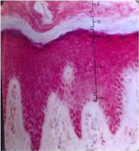
Fig 6: Layers of the skin
a. Stratum germinitivum
b. Stratum spinosum
c. Stratum granulosum
d. Stratum lucidum
e. Stratum corneum
FIG 7 :Squamous cell
[image:39.595.91.359.413.602.2]IV. HISTOLOGY OF THE SKIN
The skin is anatomically composed of three distinct strata and they are
named as (From the outermost to the inner layer). Epidermis, Dermis and
Subcutaneous fat.
The Skin
Its development
The ectoderm and the mesoderm act as the originator of all the
constituents of the skin. Or to put it in simple terms the skin is
developmentally derived from the ectoderm and the mesoderm.
The Epidermis
The epidermis is composed mostly of keratinocytes, and stratified
squamous epithelium. The epidermis is made – up of the five layers.
The stratum corneum is the first layer and this is thickest in palmar
and plantar areas. The second layer is stratum lucidium which is an even,
colourless, translucent and a shining band just below stratum corneum. The
third layer is stratum granulosum. The forth layer is stratum malphigii
(Stratum spinosum) which consists of row of mosaic like keratinocytes. The
last and the fifth layer of the epidermis is the stratum germinatum (Basal
layer). This layer consists of rectangular columnar cells arranged vertically
Cell differentiation in Keratinocytes takes place in the direction of the
external environment and they yield a cornified surface membrane. The
process of differentiation whereby germinative keratinocytes move outward to
become fully cornified cells takes approximately 14 days and are present in
transit through the cornified layer. The replacement of the epidermis and in
very thick epidermis, in the second layer as well. In the basal layer 3-5 % of
the cells synthesize DNA at any given time. During upward migration
towards maturation, keratinocytes undergo characteristics changes. Basal,
spinous, granular and cornified layers are the strata that reflect stages in the
conversion of a germinative keratinocyte from the beginning to the end and
produce the epidermal differentiation.
In the upper malphigian layer dendritic cells resembling monocytes are
found and are called as Langerhan’s cells.
Melanin pigment is synthesized and secreted by melanocytes that are
dendritic cells. The melanocytes are present all over the body but their
distribution is not even. The intensity of coloration is further determined by
the total number of cells and intercellular distribution of melanin granules
within the epidermal keratinocytes.
The Dermis
The dermis or the corium is a dense fibrous or elastic layer, which
fibrous tissue,some yellow elastic fibres and cement like substances called the
cellular elements. Blood vessels, glandular structures, lymph vessels, hair
follicles, muscle elements prolongations of fatty tissue and nerves with
terminal organs of touch and sensation are all contained and supported in this
layer. The dermis is covered over its entire surfaces with numerous conical
papillae. This stratum is called ‘The papillary layer’, whereas the deep
stratum is termed ‘The reticular layers’. The combined anatomical unit of
papillary and reticular dermis is called ‘The adventitial dermis’.
The level of intimacy between the papillary dermis and the epidermis
is easily visible in their alterations as a result of inflammatory skin diseases.
They form a morphological and a functional unit, which work together.
Subcutaneous Fat
Subcutaneous fat, like the dermis is derived from mesenchyme and
mesenchymal cells. These cells give rise to lipocytes (fat cells) as well as
fibrocytes. The lipocytic cells manufacture so much fat inside themselves that
in the process they tend to flatten the nucleus against the wall of the cell.
Strands of collagen divide the population of the fat cells into lobules. These
fibrous elements known as trabeculae have major vascular networks,
lymphatics and nerves and are analogous to the adventitial dermis. The
subcutaneous fat acts as an insulator, shock absorber and a chronic reserve
depot.
Nerves
Sensations such as touch, pressure, temperature, pain and itch are
received by millions of microscopic dermal nerve endings. They are most
numerous on hairless parts like palms, soles of the feet, fingers and clitoris,
the erogenous zones. These tiny end organs terminate principally in the
papillary dermis and around hair follicles. These nerve endings can be free or
encapsulated, myelinated or unmyelinated and most of these are visualized
with difficulty using the heamotoxylin and eosin stain.
V. BIOLOGICAL PROCESS OF HEALING
In the wound healing process three distinct phases can be visualized.
1. The phase of inflammation (Day 1-4)
2. The phase of proliferation (Day 5-20)
3. The phase of differentiation (Day 20 onwards).
The phase of inflammation
A wound causes a change in the tension prevalent in the tissue and this
causes a change in the charge, in the collagen molecule. Contract of the
collagen with the blood flowing in the wound causes the activation of kinin
and complement cascade, which is the initial phase in the clotting process. A
undergo brief construction followed by vasodilatation under the influence of
histamines from the platelets and mast cells. An increase in the capillary
permeability is noted. Serotonins, the kinins, and the prostaglandins maintain
capillary engorgement. White and red blood cells escaping from the walls of
the blood vessels form a network of fibrins over the wound site, which within
three hours is surrounded by a few lymphocytes and an increasing number of
polymorphs. A distinctive lytic behaviour is observed on part of these
neutrophils because of their lysosome content. By the fifth day monocytes
would have become the dominant cell type because they are observed in
maximum number. They are phagocytic and ingest cellular debris. Reduction
or delay in this macrophage function will delay wound healing. By the end of
the first phase new capillaries bud from the endothelial cells in the capillaries
near the wound edge, while in connective tissue around the vessels
mesenchymal cells differentiate to become fibroblasts. Clinically in this phase
the classical features observed that are manifested by the wound are heat,
redness, tenderness, swelling and loss of function.
The phase of Proliferation
By the fifth day fibroblasts have began to synthesize collagen and
Collagen
Collagen is the extra cellular fibrous framework that gives strength
and form to the tissue. Dependent on the amino acid sequence there are
various types of collagen. Proline, hydroxyproline and glycine are the most
predominant types of collagen that are seen. The types of collagen that is
found in the skin, bone, tendon and ligament is the type 1 collagen, type 2
collagen occurs in the cartilage and the type 3 collagen occurs in the fetal
dermis and is replaced by type 1 collagen at the time of birth. Granulation
tissue gives rise to the type 1 collagen. Hydroxylation of immature
protocollagen requires oxygen, ferrous ions and ascorbic acid. Further
maturation involving glycosylation produces more stable tropocollagen.
Tropocollagen is held together by weak electrostatic forces and is soluble in
weak salt solutions. This collagen, though stable, is not inert, and it undergoes
constant turnover under the influence of tissue collagens. Thicker collagen
fibers soon abound and are laid down haphazardly.
Ground Substance:
The Ground substance is an amorphous matrix of connective tissue and
is basically constituted of water, electrolytes, mucopolysaccharides and
glycoprotiens. It is a thin gel, but in cartilage it is elastic. It is produced by
fibroblasts and is involved in the formation and maturation of extra cellular
phase. Complexes occurring between the proteins and the polysaccharides
provide unique property to the ground substance. The types of
mucopolysaccharides include chondoritin, chondroitin-4 sulphate,
chondroitin-6 sulphate, dermatan sulphate, keratin sulphate and hyaluronic
acid.
The phase of differentiation
There is no clear demarcation between proliferation and the
differentiation phase. The later starts in the proliferating granulation tissue
and continues indefinitely. There is a rationalization of copious new blood
vessels and notably a remodeling of the haphazard arrangement of the
collagen fibers. The synthesis of the new collagen is in a more orderly fashion
along the lines of tension in the scar. Collagen turnover and remodeling in the
scar never stops. Indeed, the turnover of the collagen in the scar tissue is
faster than in other tissues.
The epithelial defect in the incised wound is initially plugged with
fibrin collagen and the epidermis turns downwards over the edge of the
underlying dermis. After 24 hours, basal cells are mobilized on the underlying
surface of the epidermis and by about 48 hours the advancing epithelial edge
would have undergone cellular hypertrophy and mitosis. The epithelium
When there is superficial skin loss, the dermal pits that have been left
behind act as islands for regenerating epithelium. However, once lost there is
no regeneration of hair follicles, sweat or sebaceous glands in the new
epidermis.
Healing by first intension
The healing of wounds caused by accident, assault, warfare and
surgical operations has always been a central, consideration in surgical
practice because any breach in the surface of the body – the skin and mucous
membrane – exposes the deeper tissue to the danger of infection. Therefore, it
is necessary to assist the healing process of the body to restore an intact
surface as soon as possible. Immediate closure of a wound (Primary suture)
using sutures, clips and adhesive materials favour healing with minimal
scarring and is called healing by first intension.
Healing by second intension
When the wound edges do not come together or when there is
irreparable skin loss or when the wound becomes infected and breaks apart
and has to be laid open, then in such cases healing takes place by second
intension. In this type of healing, the wound is healed by a filling of
granulation tissue. This process of healing is far slower than wound healing
by first intension and there is more amount of scar tissue left behind (Ulcers
Other methods of providing skin cover – in the presence of devitalized
tissue, swelling tension and skin loss – include delayed primary suture, skin
grafting and second suture.
Factors contributing to rate of wound healing
There are many factors, which govern the rate at which a wound heals.
Some of these factors are discussed below:
¾ Blood Supply
Wounds incorporated on the face and hands may seem horrifying
initially but they tend to heal well because of good blood supply to these
organs. Injuries sustained in areas of relatively less vascular supply such as
areas below the knees, over the shin tend to take a much longer time to heal.
¾ Tension
Tension of the tissue inhibits the local blood supply and leads to
wound failure.Local swelling and therefore tension builds up automatically
during the first 48 hours after the injury as part of the phenomenon of
inflammation. Haematoma, venous stasis (Eg. In a dependent limb) and
infection also increase tension.
¾ Age
The age factor plays an important role in the wound healing process.
turnover rate. As the age increases this capacity of the body to turnover
proteins is reduced considerably leading to slower wound healing rates.
¾ Rests
Granulation tissue has a delicate blood supply that is easily damaged
by movement and shearing forces. Rest only to the part of the body with ulcer
is indicated such as POP cast or slab.
¾ Infection
Once infection is established the fibroblasts must compete with the
bacteria and the inflammatory cells for oxygen and nutrients. Thus, overall
collagen synthesis is inevitably reduced and the collagen breakdown is
enhanced by collagenolytic enzyme activity. Infections are a major factor in
wound healing failure.
¾ Malnutrition
Defective synthesis of both collagen and ground substance can be
directly linked with malnutrition. Severe protein calorie malnutrition has long
been implicated in the failure of wounds to heal, while lesser degrees of
malnutrition depress healing as well. Vitamin C is necessary for the synthesis
of ground substance. Vitamin D is essential for new bone formation and
vitamin A for epithelialisation. Calcium, zinc, copper and manganese are
essential minerals. In patients with burns and intestinal fistulas, in particular,
¾ Uremia
Experimentally the addition of urea to tissue cultures of fibroblasts
inhibits their growth. Clinically uremia is implicated in the retardation of
wound healing in patients with renal failure.
¾ Jaundice
Jaundice is associated with reduction in wound strength. The
appearance of fibroblasts and the formation of new blood vessels are both
delayed. Bioposies of the skin in jaundice patients show a reduction of the
enzyme proline – hydroxylase involved in the collagen maturation.
¾ Steroids
An inflammatory response is essential for wound healing to proceed
normally.Steroids depress wound healing by their anti-inflammatory action.
New vessels are abnormal and sparse, as are fibroblasts. If steroids are given
after the inflammatory phase of wound healing there is little increase in
overall effect of healing.
¾ Radiation
Radiation causes cell death by damaging both DNA and disrupting
intracellular metabolism.
VI. AETIOLOGY AND PATHOLOGY
About 95% of the leg ulcers are due to vascular aetiology (Gilliland)7
(Callam MJ, et al)9. Arterial disease accounts for 5-10%; most of the others are due to neuropathy, usually diabetic or a combination of these diseases
(Yound JR).10 Diabetic ulcers are common on the toe and the heel (Hansson
Carita).11. Arterial insufficiency and / or diabetes may also be the causatives for ulcers below the line of the shoe. Ulcers at the ankles in the gaiter zone
and venous ulcers are mostly caused by varicose veins (Hansson Carita). 11
Primary varicosity of the long saphenous system and / or short saphenous
system is the causative factors for venous ulcerations (Hoare MC et al).13
The elevated ambulatory pressure in peripheral venous system in
venous insufficiency manifests itself not only in form of disturbed
microcirculation but also and particularly in microangiopathic changes. These
include decrease in capillaries, glomerulus like changes and decrease in
oxygen content. (Junger, M Stiens. A).14 It has been also noted that
perivascular fibrin cuffs and skin hypoxia precede lipodermatosclerosis in
limbs at increased risk of developing a venous ulcer (Stacey M. C., Burnand
K.G et al).15 Classification of foot ulcers
a. Venous ulcerations
b. Arterial insufficiency – Thromboangiitis Obliterans, atherosclerosis,
Raynaud’s embolic occlusions (sub acute bacterial endocarditis).
d. Neoplastic ulcers – epithelioma melanoma, basal cell carcinoma and
malignant change in long standing scars (Marjolin’s ulcer).
e. Tropical ulcers including leishmaniasis, fungal infections.
f. Specific infections – tuberculosis, syphilis and AIDS.
g. Blood dyscrasias – severe anemia, sickle cell anemia, thalassemia,
hereditary
spherocytosis and leukaemia.
h. Nutritional and metabolic disturbances.
i. Skin sensitivity or allergy.
j. Trauma.
k. Necrosis by injection of chemicals, insect bites, snakebites or radiation.
l. Repeated trauma.
m.Rheumatoid arthritis.
n. Systemic autoimmune and micro vascular diseases.16
There are various stages in the evolution of the ulcers. They are:
The extension stage: Floor of the wound is covered in slough and the base is indurated.
The transition stage: separation of the slough combined with the clearing of the floor of the wound and appearance of red granulation
The healing stage: transition of the granulation tissue into fibrous tissue is observed, which later contracts and the wound healing by
epithelisation is seen particularly in this stage.
The Venous Ulcer
Histopathologic reasons caused by venous insufficiency, is one of the
main causes of venous ulceration. Changes taking place in the cluster of thick
walled capillaries within a thickened fibrotic papillary dermis, accompanied
by siderophages and extravasated erythrocytes in variable numbers also
contribute to the overall situation of ulceration. Occurrence of perivascular
fibrin cuffs in the skin is seen in venous insufficient areas. They are often
present in the walls of superficial vessels and evident as smudgy eosinophilic
areas. (fibrin cuff theory). Lipodermatosclerosis or sclerosing panniculitis
demonstrates fibrosis and thickening of the subcutaneous septa. An early
lymphocytic infiltrate of the subcutaneous septa is gradually replaced by a
mixed infiltrate with increasing fibroplasia and sclerosis. In the late
Lipodermatosclerosis there is minimal inflammation and subcutaneous
sclerosis.
Dysfunction of the calf muscle pump can result in venous
insufficiency in the deep, connecting or superficial veins; arteriovenous
fistulae, or muscle dysfunction as a result of fibrosis, neuropathy or
to deep venous insufficiency. Venous ulcers are mainly attributed to be
caused by venous insufficiency; multisystem incompetence of the valves is
also a common cause.
Another plausible hypothesis suggests that reduction in the blood flow
rate and endothelial damage allows white cells to form plugs, which in turn
plug the capillaries. These cells release inflammatory mediators increasing
vascular permeability. This causes tissue ischemia and ulceration (white cell
trapping theory).
Arterial Ulcers
Arterial or ischaemic ulcers are most commonly due to
atherosclerosis and hence encountered in older adults. They can be also seen
in younger adults and here usually peripheral arterial disease like
Thromboangiitis Obliterans is the cause. The other rare causes of ischaemic
ulcers are Raynaud’s diseases and phenomenon.
Atherosclerosis develops at twice the frequency in patients who smoke
compared with non-smokers (Coffman J. D.)17 about 50% of the patients
have lipoproteinemia.Patients with diabetes develop the disease at an earlier
age than non-diabetics and have more severe and progressive disease. The
distribution of disease is also different in that diabetics have less aorto-illiac
There is focal accumulation of lipids, mucopolysaccharides, blood and
blood products, fibrous tissue and calcium deposits in the intima of the
arteries. Localized areas of thickening of the intima by fibroblastic
proliferations and phagocytes laden with lipid materials are seen. The media
becomes atrophic with thin strands of muscles, disrupted elastic lamella,
collagen tissue and calcium deposits. Enlarging plaques encroach upon the
lumen despite dilatation of the artery, and plaques may ulcerate. Hemorrhages
occur within the arterial wall. Thrombi are formed and finally occlude the
vessel lumen. Another important feature here which causes sudden gangrene
with ulceration is atheromatous embolisation. It is due to embolisation of
small pieces of atheromatous plaques and debris to the arteries of extremities.
This is called as blue toe or trash foot syndrome.
Another important arterial disease, which can cause ulceration and
gangrene, is Thromboangiitis Obliterans (Buerger’s disease).
The etiology of TAO remains unknown. Almost all patients who
develop this disease are smokers and the syndrome sometimes abates
following cessation of smoking. An increased frequency of HLA-A9 and
HLA-B8/B5 have been reported but not found by all investigators (Millis J. L.
et al).18 The pathogenesis involves production of ischaemia and all its
manifestations by an inflammatory action of medium and small arteries of
In acute stage there is panvasculitis of the arteries and veins; the
diagnostic finding being arterial thrombi with foci of microabscess and giant
cells. In chronic stage only fibrotic obliteration of arteries is seen and
diagnosis can only be surmised since atherosclerosis is absent.
Diabetic Ulcers
The various factors, which are the contributions for the cause of ulcers,
are Hyperglycemia, microangiopathy, Neuropathy, Liability to infection and
Alteration in blood flow.
The precipitating factors are injuries and infection. Injuries could be
physical injuries like penetrating wounds, disruption of skin and injury due to
continuous localized blunt pressure
In a diabetic condition of either type 1 or type 2, hyperglycemia may
lead to surgical complications. In this condition it affects the basement
membrane of the capillaries and cell permeability; interfering with the transfer
of oxygen and nutrients to the tissues. Along with decreased supply of
leukocytes into this area and also high concentration of glucose in the tissue
fluids which help in the growth of pathogenic organisms, it impairs wound
healing. The vascular changes that take place during diabetes are responsible
for ulceration of the foot. The principle finding in the vascular flow
mechanism in diabetes is a marked decrease in blood flow as shown by
flow phase( high ankle / brachial Doppler ratio) found in diabetes. The major
cause of this blood flow change is the distal AV shunting and sympathetic
dysfunction. The other cause being atherosclerotic stiffening of the vessels,
sometimes this is associated with calcification of the vessel walls. The
irregular pattern of blood flow definitely has an influence in the ultimate
delivery of oxygen to the tissues.
Diabetic patients also tend to develop calcifications more commonly
and at a much younger age. This causes some vascular narrowing which may
cause distal ischemia. Calcification of intima may produce trauma to blood
cells and cause development of thrombi which may distally produce localized
areas of ischemia and gangrene.
Histologically the disease is manifested as a thickening of the tunica
intima along with large atheromatous plaques and lumps. The metatarsal
arteries are more often occluded in diabetes.
An important yet a very common factors responsible for producing
neuropathic ulceration is the presence of diffuse distal peripheral
polyneuropathy due to which painless ulcers are predominantly seen in the
lower limbs. During this type of ulceration there is involvement of all types of
nerve fibres. A notable feature is the marked reduction in the amount of
Schawann cells. Neuropathy is predominantly selective for large myelinated
myelinated fibers and non-myelinated fibers sub serving pain and temperature
sense are affected leading to mixed fiber neuropathy. Osmotic and metabolic
derangements are caused due to ischemia from vessel occlusion and altered
capillary permeability; this paves the way for producing neuropathy.
Increased capillary permeability allows the toxic proteins which are
circulating to reach the nerves and tissue oedema which cause the impairment
of nutrition contribution to development of the neuropathy.
Metabolic conditions prevalent are more important in the development
of the neuropathy than are the vascular diseases. Abnormal Schawann cell
function causes segmental demyelination, which is a dominant histological
aspect of diabetic neuropathy. This hinders nerve conduction and the situation
can only be reversed by remyelination. There are number of reasons which
contribute to the production of myelin. Some of these factors are:
1) Synthesis of abnormal myelin
2) Increased activity of polyol pathway in conversion of glucose to
fructose with a resultant increase in concentration of metabolites.
Simultaneous occurrence of axonal damage is also observed, but
segmental demyelination of diabetic neuropathy may exist independent
of axonal damage. Axonal damage is the first manifestation of diabetic
and a disturbance in its metabolism in diabetics is suggested as the
mechanism for axonal damage.
WAGNER’S CLASSIFICATION
GRADE 0:NO ULCERATION(BONY DEFORMITY,HIGH RISK GROUP).
GRADE 1 : SUPERFICIAL ULCERATION.
GRADE 2 : DEEP ULCERATION WITH PENETRATION INTO
TENDONS, BONES, JOINTS.
GRADE 3 : DEEP ABSCESS / OSTEOMYELITIS.
GRADE 4 : GANGRENE OF TOE / TOES / FOREFOOT.
GRADE 5 : EXTENSIVE GANGRENE FOOT / LEG.
The absence of the sweat glands or its malfunction, with the loss of
Lysozymes alters the cutaneous bacterial environment and also decreases the
defense mechanism of the skin. Trophic changes include redness and glossy
shining skin with loss of hair. Thick keratinization of the skin and atrophy of
the local subcutaneous tissue is observed in areas of increased pressure.
Breakdown of such skin produces trophic ulcers.
Tropic Ulcers
Tropic ulcers are particularly perforating ulcers of the foot, which is
associated with Tabes dorsalis, diabetic neuropathy, leprosy, and other
About 10 to 20% of the patients with leprosy all over the world
complain of the most common yet the most serious complication that is of
ulceration on the bottom of their feet.
The second type of ulcer which is seen here is the stasis ulcer which is
seen in the crease of flexor muscles of the ankle, due to autonomic
dysfunction leading to abnormality in regulation of blood supply and absence
of sweating. The last and the third type of ulcer seen in leprosy is the plantar
ulcer, which is seen in the weight bearing areas of the foot where there is
sensory loss (Dharmendra).19
The three modalities of neurological deficit are:
1) Sensory loss.
2) Motor paralysis.
3) Autonomic nerve damage.
Sensory loss
Impending danger to the tissues is preceded by the sensation of pain,
which is the body’s natural alarm system. But in the decreased condition due
to the neurological impairment (sensory loss), repeated trauma may attain
proportions which might actually cause tissue destruction.
Motor Paralysis
Only open injuries on the foot with intrinsic paralysis are usually
well as sensory loss. The pads of the fore foot are protected by the intrinsic
muscles of the foot in two ways, one by straightening the toes, the metatarsal
heads are slightly retracted and the pulps of the terminal segments also take a
small share of the weight. When these muscles are atrophied the bony
prominences are made more obvious.
Autonomic nerve damage a) Blood supply
The regulation of the blood supply to the sole of the foot is largely,
reflex, depending on the integrity of sympathetic supply to the muscular part
of the arteriole. In the nerve trunk lesions, these nerves are also destroyed so
that the arterioles are sympathectomized. In an anesthetic foot, the increase in
blood supply after exercise or slightly excessive stress is not present and
hence there is a relative tissue anoxia ensues.
b) Sweat loss
The second autonomic function that is disrupted is the control of the
sweat glands.Thus the denervated foot is dry. Due to this deprivation in the
supply of the sweat, another one of the skins protective mechanisms is lost,
which causes the skin to loose its suppleness. Deprivation of sweat causes the
skin to go dry and crack, which may cause the introduction of infections into
weight bearing areas where special mechanical strains are supplied during
normal walking.
Studies have shown that the initiation of the ulceration is usually due to
an initial damage in the deep tissue surface. The pressure of the entire body
falls on the foot and during walking, unperceived by sensation and the normal
rotation of the joints is uncontrolled by muscular coordination, the result
being the introduction of torsional stress, which leads to necrosis of the deep
tissues immediately adjacent to the bone. The clinical features follow a
sequence of incidents listed below:
1) Initial deep necrosis.
2) Necrotic sinuses.
3) Necrotic blisters and finally.
4) Plantar ulcer.
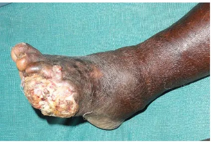
MALIGNANT ULCERS Squamous cell carcinoma
This is the carcinoma of the cells of the epidermis that usually migrate
outwards to the surface. The initiation point of the squamous cell carcinoma is
usually the layer that is formed by the prickle cell layer. This may also occur
in a few preexisting lesion of the skin like:
1. Long-standing chronic ulcers (e.g. Marjolin’s ulcers) following burns,
2. Senile keratosis.
3. Bowen’s disease.
4. Leukoplakia.
5. From skin exposed to irradiation.
6. Chronic skin lesion e.g. Lupus Vulgaris (Cutaneous tuberculosis),
eczema, warts.
7. Exposed to prolonged irritation by various chemicals.
Very rarely squamous cell carcinoma may develop from a basal cell
carcinoma.
Two types are usually seen:
1) Proliferative type.
2) Ulcerative type.
The ulcerative type is found to more commonly prevalent.
Macroscopic Features
The origin of the squamous cell carcinoma is as a small lump or
nodule. These nodules enlarge as they mature and become necrotic and their
centers sloughs out. The shape of the ulcers is usually circular or oval, and
their shape and sizes varies extremely. The edges of the ulcer are raised and
everted; this shows excessive tissue growth just above the surface. The floor
of the wound is covered by necrotic tumor, serum and blood. Presence of pale
structures such as muscle, tendon, cartilage or bone may also be seen. The
base of the wound is usually indurated.
Microscopic Features
Solid columns of epithelial cells, which are seen, growing down into
the dermis, separated from one another by connective tissue. These extend
into bulk like masses, which in section appear to be distinct and detached. In
the course of time in which the wound evolved the cells lying nearest to the
center of the wound, being the oldest undergoes degenerative changes and get
converted into hyaline structure less masses of keratin. This process is called
keratinization. The mass of keratin looks red with eosin stains. This is
surrounded by normal looking squamous cells presenting the characteristic
‘prickle cell’ appearance and these are arranged in collective manner as seen
in ‘onion skin’. This whole arrangement is called as a ‘cell nest’ or ‘epithelial
pearl’.
The appearance of the cell nest is a characteristic feature of
epidermoid carcinoma, but this may be absent in rapidly growing tumor and
in mucous membranes.
Another important feature to be noted in this type of malignant tumor
is the infiltration