Extracellular Interactions between
Hepatitis C Virus and Secreted
Apolipoprotein E
Zhihua Li,
a,b,cYadong Li,
a,bYanwei Bi,
a,b,cHui Zhang,
a,bYufeng Yao,
a,bQihan Li,
a,bWei Cun,
a,b,cShaozhong Dong
a,bInstitute of Medical Biology, Chinese Academy of Medical Sciences and Peking Union Medical College, Kunming, Yunnan, Chinaa; Yunnan Key Laboratory of Vaccine Research and Development of Severe Infectious Disease, Kunming, Yunnan, Chinab; Centre for Tuberculosis Research, Chinese Academy of Medical Sciences and Peking Union Medical College, Beijing, Chinac
ABSTRACT
Interactions between hepatitis C virus (HCV) and lipoproteins in humans
play an important role in the efficient establishment of chronic infection.
Apolipo-protein E (ApoE) on the HCV envelope mediates virus attachment to host cells as
well as immune evasion. This interaction is thought to occur in hepatocytes, as ApoE
plays dual functions in HCV assembly and maturation as well as cell attachment. In
the present study, we found that secreted ApoE (sApoE) can also bind to viral
parti-cles via its C-terminal domain after HCV is released from the cell. Furthermore, the
binding affinity of interactions between the sApoE N terminus and cell surface
re-ceptors affected HCV infectivity in a dose-dependent manner. The extracellular
bind-ing of sApoE to HCV is dependent on HCV envelope proteins, and recombinant HCV
envelope proteins are also able to bind to sApoE. These results suggest that
extra-cellular interactions between HCV and sApoE may potentially complicate vaccine
de-velopment and studies of viral pathogenesis.
IMPORTANCE
End-stage liver disease caused by chronic HCV infection remains a
clinical challenge, and there is an urgent need for a prophylactic method of
control-ling HCV infection. Because host immunity against HCV is poorly understood,
addi-tional investigations of host-virus interactions in the context of HCV are important.
HCV is primarily transmitted through blood, which is rich in lipoproteins. Therefore,
it is of interest to further determine how HCV interacts with lipoproteins in human
blood. In this study, we found that secreted ApoE (sApoE), an exchangeable
compo-nent found in lipoproteins, participates in extracellular interactions with HCV virions.
More significantly, different variants of sApoE differentially affect HCV infection
effi-ciency in a dose-dependent manner. These findings provide greater insight into HCV
infection and host immunity and could help propel the development of new
strate-gies for preventing HCV infection.
KEYWORDS
CRISPR-Cas9, hepatitis C virus, infectivity, secreted apolipoprotein E,
envelope protein
A
pproximately 170 million people are chronically infected with hepatitis C virus
(HCV) worldwide (1). Most HCV infections become chronic in humans and can
progress to hepatic fibrosis, cirrhosis, or even hepatocellular carcinoma (2). This
process is normally accompanied by high morbidity due to extrahepatic
manifes-tations, including mixed cryoglobulinemia, diabetes mellitus, and atherosclerosis
(3). In most developing countries, the current standard treatment for chronic
hepatitis C is a combination of pegylated interferon with ribavirin (4), although
unfortunately, the effects are unsatisfactory. Recently, highly efficacious
direct-acting antiviral drugs (DAAs) have been approved; however, their clinical benefits
Received14 November 2016Accepted4 May 2017
Accepted manuscript posted online24 May 2017
CitationLi Z, Li Y, Bi Y, Zhang H, Yao Y, Li Q, Cun W, Dong S. 2017. Extracellular interactions between hepatitis C virus and secreted
apolipoprotein E. J Virol 91:e02227-16.https://
doi.org/10.1128/JVI.02227-16.
EditorJ.-H. James Ou, University of Southern California
Copyright© 2017 American Society for
Microbiology.All Rights Reserved.
Address correspondence to Wei Cun, cun@imbcams.com.cn, or Shaozhong Dong, dsz@imbcams.com.cn.
Z.L. and Y.L. contributed equally to this article.
VIRUS-CELL INTERACTIONS
crossm
on November 7, 2019 by guest
http://jvi.asm.org/
may be limited by their lack of improvement of HCV-associated end-stage liver
disease, their high cost, the emergence of drug-resistant HCV variants, and the
potential for HCV reinfection in previously cured patients. Therefore, HCV might not
be eliminated in the short term (5), and as a consequence, a prophylactic vaccine
is highly desirable. Because the development of efficient HCV vaccines has proven
difficult, further understanding of the molecular mechanisms underlying HCV
in-fection is necessary.
HCV is a single-stranded, positive-sense RNA virus of the
Flaviviridae
family (6). The
HCV genome is
⬃
9.6 kb in length and encodes a long polyprotein (of more than 3,000
amino acids [aa]) that is proteolytically processed to generate 10 mature viral proteins.
Viral structural proteins reside in the N-terminal third of the polyprotein and include
core or capsid protein (C) and the envelope glycoproteins E1 and E2. p7 (a viroporin)
and nonstructural proteins are encoded in the remaining C-terminal two-thirds of the
polyprotein; these proteins play a variety of roles in virus assembly and RNA replication
(7–9). HCV virions consist of a nucleocapsid containing the viral genome enveloped by
an endoplasmic reticulum (ER)-derived lipid bilayer in which E1 and E2 are assembled
as heterodimers (10, 11).
Highly efficient establishment of chronic HCV infection relies on not only the
effective inhibition of the host’s innate immunity through the activities of viral proteins
(12–17) but also the chimeric formation of lipoviral particles (LVPs) by HCV virions and
blood lipoproteins, which allow HCV to efficiently spread through blood vessels and
effectively escape from host humoral immunity (18, 19). These relationships imply that
lipoprotein components could be involved in the mechanism of HCV escape from
humoral immunity.
Apolipoprotein E (ApoE) is abundant in plasma (20 to 50
g/ml), where it functions
as an exchangeable surface ligand for several classes of lipoproteins to facilitate
receptor recognition and lipid transport regulation, and it is also involved in immune
regulation and nerve tissue regeneration (20, 21). ApoE is polymorphic, with three
common alleles (ApoE2 [Cys112 and Cys158], ApoE3 [Cys112 and Arg158], and ApoE4
[Arg112 and Arg158]) and tens of rare alleles (22). The ApoE isoforms are classified
based on their relative charge. Different mutations causing the same migration
pattern after isoelectric focusing define the different isoform subtypes. Although
these allelic forms differ by only one or two amino acids, the differences often alter
the structure and function of the protein. According to crystallography studies, a
hinge region connects the N- and C-terminal regions of ApoE (23, 24). The
N-terminal region (residues 1 to 167) forms an antiparallel four-helix bundle that
contains a receptor-binding site (residues 136 to 150) (25). The C-terminal domain
(residues 206 to 299) contains three
␣
-helices that form a large exposed
hydro-phobic surface and that interact with residues in the N-terminal helix bundle
domain through hydrogen bonds and salt bridges (26).
In an
in vitro
hepatocyte culture model, the role of ApoE in the HCV replication cycle
was elucidated in several previous studies. For example, small interfering RNA
(siRNA)-induced downregulation of cellular ApoE (cApoE) expression led to decreased HCV
yield and infectivity (27–29). Moreover, interactions of cApoE with the viral protein
NS5A or the viral envelope proteins have also been detected (30–34). As a
conse-quence, cApoE is thought to be involved in HCV virion assembly and maturation (34,
35). Furthermore, immunoelectron microscopy has indicated the presence of unequal
numbers of ApoE molecules on the surfaces of HCV LVPs, and it was found that HCV
LVP attachment to cells is mediated through the binding of ApoE to cell surface heparin
sulfate (36–39).
It has been shown that during the spread of dengue virus through blood vessels, the
virus can associate with host antibodies to alter infectivity
in vivo
. The
antibody-dependent enhancement (ADE) formed by dengue virus is an important factor to
consider when studying the pathogenic mechanism of the virus and when developing
vaccines (40, 41). Notably, both dengue virus and HCV belong to the
Flaviviridae
family.
Secreted ApoE (sApoE) is abundant in blood and can dissociate from the lipoprotein
on November 7, 2019 by guest
http://jvi.asm.org/
surface (42, 43). Dissociated free sApoE is structurally labile and can rapidly bind to
other lipoproteins, become recruited to the plasma membrane, or associate with other
complexes. Therefore, we sought to understand whether sApoE molecules on
lipopro-teins in blood could associate with HCV to affect viral infectivity and whether these
interactions are involved in the immune escape mechanism of the virus.
RESULTS
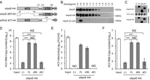
Binding of secreted ApoE to HCV particles.
The liver is the major organ of ApoE
synthesis, and several studies have reported that HCV can associate with cApoE before
being secreted from infected hepatocytes during the HCV LVP assembly and release
process. However, there are high concentrations of sApoE protein present in blood, and
the ApoE molecules found on HCV LVPs are likely obtained from extracellular exchange
of sApoE, although there is currently no clear understanding of the details defining
these extracellular interactions.
To distinguish cApoE carried by HCV virions assembled in the Huh7.5 cell line, we
used sApoE protein tagged with anti-hemagglutinin (anti-HA) secreted by
heterolo-gous cells and its deletion mutant in this study (Fig. 1A). It has been shown that using
a flexible linker (SGGRGG) between the HA tag and the C terminus of ApoE does not
affect the secretion or normal biological function of the protein (44). ApoE expression
was not detected in the cell culture medium or cell lysates of 293T cells, which
eliminates the concern of interference due to endogenous ApoE. sApoE or sApoE-HA
was secreted into the cell culture medium following transient transfection of
pcDNA3-ApoE3 or pCDNA3-ApoE-HA into 293T cells. Density gradient centrifugation analysis
FIG 1Secreted ApoE interacts with extracellular HCV particles in a manner dependent on the C-terminal domain of ApoE. (A) Schematic representation of HA-tagged, full-length, N- or C-terminal domain deletion mutants of sApoE. (B) Isopycnic gradient centrifugation analysis showing the distribution of exogenously expressed sApoE or sApoE-HA in 293T cell culture medium as well as recombinant ApoE (rApoE) protein expressed byE. coliand run through a density gradient as a control. ApoE protein was detected by Western blotting with WUE-4 anti-ApoE antibody. (C) sApoE-HA and concentrated HCV core proteins in cell culture media were detected by Western blotting and are shown at the top. Proteins are specified on the left. The mixtures were separately captured by HA-specific immunoprecipitation, and the presence of sApoE-HA in the immunocomplexes was verified by Western blotting using HA antibody (bottom). (D) HCV genome copies in the immunocomplexes were quantified using qRT-PCR. (E) Determination of the infectivity of immunocaptured HCV particles. Equivalent amounts of HCVcc particles and full-length sApoE-HA or deletion mutants were mixed and subjected to HA affinity capture. Captured particles were eluted with an HA peptide. Infectivity titers in the input and the corresponding eluent were determined by FF-IHC. (F) PHH derived-HCV genomes in the immunocomplexes were pulled down using anti-HA agarose and quantified using qRT-PCR. The data in panels D to F represent the mean values and standard deviations from three independent experiments.**,P⬍0.01;***,P⬍0.001 (unpaired Student’sttest). NS, not significant; ND, not detected.
Extracellular Interaction of HCV and sApoE Journal of Virology
on November 7, 2019 by guest
http://jvi.asm.org/
[image:3.585.42.546.70.341.2]confirmed that sApoE-HA was in a lipoprotein-associated state, equivalent to sApoE
(Fig. 1B), and could therefore be used as a mimic of sApoE in blood.
We mixed sApoE-HA secreted from 293T cells with HCV derived from a cell culture
system (HCVcc) and captured the HA antibody. Compared with 293T cell supernatant,
which did not express sApoE (Fig. 1C), beads coupled with the HA antibody could
efficiently pull down the HCV genome when associated with sApoE-HA protein (Fig.
1D). To confirm that sApoE-HA could associate with infectious HCV virions, we then
used the HA peptide to competitively elute the immunoprecipitate associated with the
HA antibody. Infectious HCV virus was detected in the eluent containing sApoE-HA (Fig.
1E), indicating that extracellularly secreted sApoE-HA protein can associate with
infec-tious HCVcc.
According to crystallography studies, there are two independent domains in the
ApoE protein: the N-terminal domain (NTD) and the C-terminal domain (CTD). We
therefore designed a pulldown assay using the HA antibody to identify which ApoE
domain interacts with HCV by using secreted sApoE-HA mutated so that either the NTD
or CTD was removed (dNT or dCT sApoE-HA, respectively) (Fig. 1A). Our results showed
that the ApoE protein with no NTD interacted with HCV in a manner similar to that of
full-length ApoE protein (Fig. 1D and E), whereas the ApoE protein with no CTD could
not associate with HCV virions. These results suggest that the interaction of sApoE-HA
with extracellular HCVcc virions depends on the CTD of ApoE.
It has been reported that Huh7-derived cells, which are deficient in very-low-density
lipoprotein (VLDL) secretion, do not possess typical lipoprotein production machinery.
Therefore, to reflect the natural behavior of authentic viruses, we also detected whether
HCV particles produced by primary human hepatocytes (PHHs) could bind to sApoE. To
accomplish this, PHH-derived HCV particles were separately mixed with sApoE-HA
mutants (FL-HA, dNt-HA, and dCt-HA) secreted by 293T cells and subjected to pulldown
using anti-HA agarose. HCV viral genome copies were then measured. The results (Fig.
1F) indicated that sApoE can also bind with PHH-derived HCV particles and that this
process is also dependent on the CTD of ApoE.
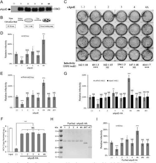
ApoE polymorphisms influence HCV infectivity in a dose-dependent manner.
ApoE exhibits genetic polymorphisms among the population, and these variants differ
in their receptor binding affinities. Clinical studies have shown that HCV susceptibilities
vary among populations with different ApoE genotypes. Therefore, we hypothesized
that differences in sApoE variants may directly affect HCV infectivity.
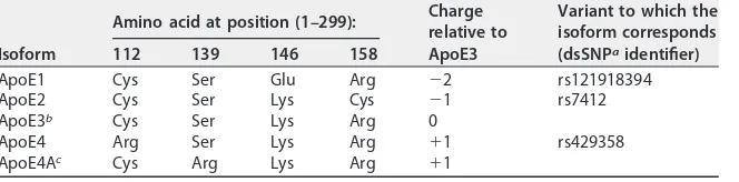
[image:4.585.42.374.84.166.2]To investigate whether different sApoE variants differentially affect HCV infectivity,
we examined four ApoE natural variants in humans, termed ApoE1 (Harrisburg type)
(45), 2, 3 and 4 (Table 1). We obtained cell culture medium from 293T cells individually
containing each of the 4 sApoE variants, as well as a blank control (Fig. 2A). Equal
amounts of HCVcc were separately mixed with the cell supernatants, and the mixtures
were then used to infect Huh7.5 hepatocytes (Fig. 2B). For accurate measurement of
infectivity, HCV viral titers were determined using focus forming assays based on
immunohistochemistry (FF-IHC).
TABLE 1List of ApoE variants used in this study
Isoform
Amino acid at position (1–299): Charge
relative to ApoE3
Variant to which the isoform corresponds (dsSNPaidentifier)
112 139 146 158
ApoE1 Cys Ser Glu Arg ⫺2 rs121918394
ApoE2 Cys Ser Lys Cys ⫺1 rs7412
ApoE3b Cys Ser Lys Arg 0
ApoE4 Arg Ser Lys Arg ⫹1 rs429358
ApoE4Ac Cys Arg Lys Arg ⫹1
adbSNP URL:https://www.ncbi.nlm.nih.gov/SNP/.
bRepresents the prototype ApoE (UniProt accession numberP02649and GenBank accession number K00396).
cRepresents an artificial ApoE mutant.
on November 7, 2019 by guest
http://jvi.asm.org/
FIG 2Infectivity of HCV is influenced by sApoE isoform. (A) Forty-eight hours after transient transfection of plasmids, the blank sample and five sApoE variants were secreted by 293T cells and detected in cell culture media by Western blotting. (B) Schematic diagram of the experimental procedure. (C to E) Equivalent amounts of HCVcc derived from Huh7.5 (C and D) or PHHs (E) were mixed with the same amounts of cell culture media including different sApoE variants and deletion mutants used to infect Huh7.5 cells in a 24-well cell culture plate. The infectivity of HCV was determined by FF-IHC in quadruplicate. (F) The ability of different sApoE isoforms and mutants to interact with HCV. Equivalent amounts of HCVcc particles and different full-length sApoE isoforms and mutants were mixed and subjected to HA affinity capture. HCV genome copies in the immunocomplexes were quantified using qRT-PCR. (G) The same amounts of cell culture media including different sApoE-HA variants or anti-HA agarose as used to remove sApoE-HA were mixed with equivalent amounts of HCVcc and used to infect Huh7.5 cells. The infectivity of HCV was determined. (H) Different sApoE-HA variants immunoprecipitated by anti-HA agarose were eluted with EF solution and assayed by SDS-PAGE utilizing Coomassie blue staining. (I) The effects of purified ApoE variants and deletion mutants on HCV infectivity were detected. The HCV infectivity data are shown as the mean⫾SDs pooled from three independent experiments.*,P⬍0.05;**,P⬍0.01;***,P⬍0.001 (unpaired Student’s ttest). NS, not significant.
Extracellular Interaction of HCV and sApoE Journal of Virology
on November 7, 2019 by guest
http://jvi.asm.org/
[image:5.585.42.543.72.594.2]The supernatant containing sApoE1 exhibited lower receptor affinity than did the
293T culture supernatant without sApoE and led to a significant decrease in HCVcc
infectivity (Fig. 2C and D). Among the three most common ApoE variants (ApoE2, -3,
and -4) in the population, the sApoE3 supernatant led to a slight increase in HCVcc
infectivity, whereas the sApoE4 supernatant decreased HCVcc infectivity.
It has been shown that increasing the positive charge on the ApoE receptor
interaction area (aa 136 to 150) can enhance the affinity of ApoE in binding to heparan
sulfate proteoglycan (HSPG) on the hepatocyte cell surface. We therefore replaced the
serine at amino acid position 139 in ApoE3 with arginine to artificially construct an
ApoE4A mutant with an additional positive charge and therefore higher receptor
affinity for HSPG (Table 1). The sApoE4A mutant secreted by 293T cells was mixed with
an equal amount of HCVcc, and viral infectivity was measured (Fig. 2A and B). As
expected, the results indicated that the sApoE4A mutant significantly increased the
infectivity of HCVcc
in vitro
(Fig. 2C); similar results were obtained upon repeating the
experiment (Fig. 2D).
To measure the effects on infectivity caused by different ApoE variants using
PHH-derived HCV, the same method as that described above was applied. The results
indicated that different ApoE variants altered the infectivity of HCV particles derived
from PHHs (Fig. 2E). To further ensure that these alterations were specifically caused by
sApoE variants in the supernatant, we expressed sApoE1, -2, -4, and -4A as well as
sApoE3-HA, all carrying the HA tag. We found that the sApoE1, -2, -3, -4, and -4A were
all able to associate with HCV particles (Fig. 2F). Furthermore, the infectivity results (Fig.
2G) showed that the presence of sApoE caused fluctuations in HCV infectivity
com-pared to results from experiments using a cellular supernatant control containing no
ApoE. Likewise, when sApoE-HA proteins were removed from the cell supernatant
using anti-HA agarose, HCV infectivity was no longer altered. After this, we allowed HCV
particles to bind to purified ApoE-HA protein (Fig. 2H), and the resultant effects on HCV
infectivity were again observed (Fig. 2I). The results indicated that the presence of
sApoE in the supernatant was the key factor altering HCV infectivity.
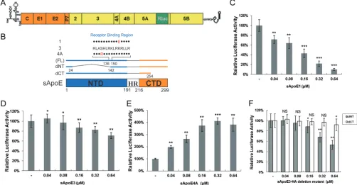
Because the detection of high-concentration HCVcc titers requires continuous serial
dilution, the concentration of sApoE in solution is also diluted during these
measure-ments. Therefore, in order to examine the dose dependency of the relationship
between HCV infectivity and sApoE concentration in the supernatant, we used HCV
reporter virus, which has a wider linear range that allows detection with no serial
dilution (Fig. 3A). We used sApoE1, -3, and -4A proteins secreted by heterologous cells
and the sApoE deletion mutant in this study (Fig. 3B). As sApoE1 has the lowest
receptor affinity, amounts of reporter virus equivalent to those in the experiments
described above were used, with exposure to increasing concentrations of sApoE1. The
results showed a dose-dependent relationship, with increasing sApoE1 concentrations
resulting in continuously decreasing HCV infectivity. When the sApoE1 concentration
approached the level found in human blood (20
g/ml), HCV infectivity was decreased
by 90% (Fig. 3C). Moreover, as the concentration of sApoE3 in the mixture gradually
increased, the HCV infectivity first increased and then decreased (Fig. 3D). In contrast,
as the concentration of sApoE4A in the mixture gradually increased, the infectivity of
HCV quadrupled (Fig. 3E). These results suggest that both the concentration and the
receptor affinity of sApoE in the host alter the infectivity of HCV.
Theoretically, sApoE alters viral infectivity not only by associating with HCV but also
by competing with HCV for receptor binding. It has been shown that ApoE has two
functional domains: the NTD, which can competitively associate with receptors, and the
CTD, which can associate with HCV particles. Notably, each of these domains can
separately inhibit HCV infectivity. Under the experimental conditions used in this study,
the ApoE construct with no NTD exhibited stronger inhibition of HCV infectivity (Fig. 3F)
than the ApoE construct with no CTD, indicating that the effect of CTD of this protein
on HCV infectivity was more notable than that of NTD competitively associated with the
HCV receptor.
on November 7, 2019 by guest
http://jvi.asm.org/
Secreted ApoE binds HCV independent of cellular ApoE.
It has been shown that
ApoE can be expressed in liver cells and secreted extracellularly. ApoE is also able to
form homodimers. Previous reports have indicated that HCVcc can associate with
intracellular cApoE. Therefore, we further investigated whether the interaction between
sApoE and HCVcc depends on the virus associating with cApoE intracellularly.
To test this dependence, it was necessary to examine the behavior of HCVcc
particles that had not been exposed to cApoE. Using siRNA to knock down host cellular
ApoE expression in Huh7-HCV cells would not be sufficient for this purpose, as siRNA
cannot completely eliminate ApoE expression.
To overcome this limitation, we utilized the recently developed genome editing
technology known as CRISPR-Cas (clustered regularly interspaced short palindromic
repeats/CRISPR-associated protein). This system can induce site-specific cleavage of the
cellular genome and preferentially introduce an indel mutation while the genome is
repaired via error-prone nonhomologous end joining (NHEJ). For detection
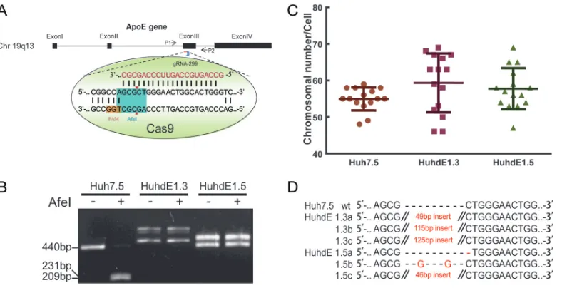
conve-nience, we chose the AfeI restriction site on the third exon of the ApoE gene as our
target (Fig. 4A). The CRISPR-Cas9 system was transfected into Huh7.5 cells for transient
expression. After two rounds of single-cell clone selection and amplification, ApoE
protein expression was undetectable in 2 cell clones, which were named HuhdE1.3 and
HuhdE1.5. To confirm the absence of ApoE, we extracted the genomes of the cells and
subjected them to analysis via PCR and AfeI enzyme digestion. Only 1 PCR product was
amplified from wild-type Huh7.5 cells, and the PCR product could be digested with AfeI.
In contrast, multiple PCR products were amplified from both the HuhdE1.3 and
HuhdE1.5 cells, and AfeI digestion was no longer possible (Fig. 4B). These results
suggested that no wild-type ApoE gene was present in either of the modified cell lines.
Additionally, karyotype analysis showed that the wild-type and derivative Huh7 cells
were hypotriploid (Fig. 4C). Next-generation sequencing analysis showed 3 ApoE
FIG 3Infectivity of reporter HCV is influenced by sApoE in a dose-dependent manner. (A) Genomic organization of the JFH1-derived luciferase reporter construct (HCV-Rluc). (B) Schematic representation of full-length (FL; 1, 3, and 4A) and N- or C-terminal domain deletion mutants of sApoE. (C to F) Equivalent amounts of reporter HCV were incubated with increasing concentrations of sApoE1 (C), sApoE3 (D), sApoE4A (E), or sApoE3 deletion mutants (F) at room temperature for 30 min and subsequently used to infect Huh7.5 cells in 24-well culture plates. At 5 h h.p.i., the mixture of HCV-Rluc and sApoE was removed, and the cells were washed twice with PBS and then incubated with DMEM containing 10% FBS. After 24 h, the cells were lysed for luciferase assays. The quantitative results derived from the data were converted to percentages of the control in the bar graph. The data are shown as the mean⫾SDs (error bars) pooled from three independent experiments.*,P⬍0.05;**,P⬍0.01;***,P⬍0.001 (unpaired Student’sttest). NS, not significant.
Extracellular Interaction of HCV and sApoE Journal of Virology
on November 7, 2019 by guest
http://jvi.asm.org/
[image:7.585.41.543.74.335.2]mutants in both the HuhdE1.3 and HuhdE1.5 cells (Fig. 4D), and the wild-type ApoE
sequence was not detected. Amino acid sequence analysis revealed that the open
reading frame of the ApoE gene was destroyed in both the HuhdE1.3 and HuhdE1.5
cells and that the cells no longer encoded or produced full-length ApoE protein (
⬍
100
aa). Thus, we successfully constructed an ApoE gene knockout hepatocyte cell line
using the CRISPR-Cas9 system.
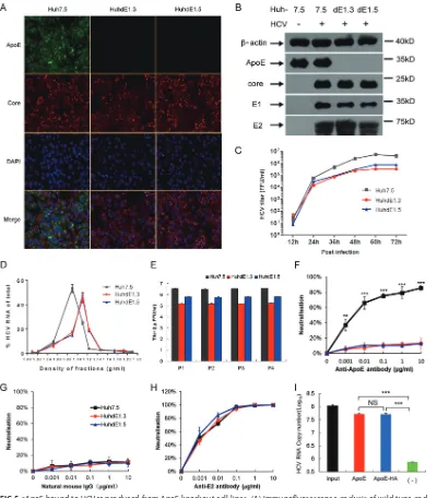
We next used HCVcc to infect Huh7.5, HuhdE1.3, and HuhdE1.5 cells, and all three
cell lines were then infected with HCV (Fig. 5A). HCV protein expression was not
inhibited by ApoE knockout in any of the cell lines (Fig. 5B), but viral infectivity was
reduced (Fig. 5C), and the density distribution of virus particles over an iodixanol
density gradient also shifted toward a higher density (Fig. 5D). HCV was able to be
passaged both in Huh7.5 cells and in the ApoE gene knockout HuhdE1.3 and HuhdE1.5
cells (Fig. 5E). To further confirm the absence of ApoE on the HCV particles, the particles
were subjected to a neutralization assay using ApoE antibody. In contrast to the HCV
produced in Huh7.5 cells, the viral particles produced in the ApoE gene knockout cell
lines could not be neutralized by the ApoE antibody (Fig. 5F and G), whereas viral
particles from all three cell lines could be neutralized using an E2 antibody (Fig. 5H).
After confirming that no cApoE was present on the HCV particles, HCV produced in
HuhdE1.5 cells was mixed with sApoE-HA or sApoE for a pulldown assay using beads
coupled with ApoE antibody, and viral genome copy number was quantitatively
analyzed. Compared with the control group, both sApoE-HA and sApoE could associate
with HCVcc lacking cApoE (Fig. 5I). These results indicate that the interaction of sApoE
with HCV virions occurs independently of the presence of cApoE during intracellular
virus assembly. The results also illustrate that the presence of the HA tag at the
C-terminal domain of sApoE did not affect the ability of HCV virions to interact with
ApoE.
Binding of sApoE to HCV depends on envelope proteins.
Heterodimers of the E1
and E2 viral envelope glycoproteins are present on the surfaces of HCV particles.
Because the extracellular interaction of sApoE with HCVcc occurs independently of
the intracellular interaction of the virus with cApoE during virus assembly, we
hypoth-FIG 4Establishement of ApoE knockout human hepatic cell lines. (A) Schematic showing the CRISPR-Cas9 system and the target site for gRNA 299 in the ApoE gene locus used in this study. Protospacer adjacent motif (PAM) sequences are in orange. Fragments amplified by p1 with p2 and the AfeI restriction site (in aqua blue) were used in a restriction fragment length polymorphism gel assay. (B) Restriction fragment length polymorphism gel analysis. The CRISPR-Cas9 system was transiently expressed in Huh7.5 cells. After two rounds of subcloning and testing for protein expression, two monoclonal ApoE knockout cell lines were isolated and termed HuhdE1.3 and HuhdE1.5. The short lines indicate fragments generated by AfeI digestion. (C) Karyotype analysis of the Huh7.5 and ApoE knockout cell lines, Huh7dE1.3 and Huh7dE1.5. (D) Sequences of indel mutations identified in the ApoE gene using next-generation sequencing for each cell line. Red dashes, deletions; red bases, insertions.
on November 7, 2019 by guest
http://jvi.asm.org/
[image:8.585.43.437.69.271.2]FIG 5sApoE bound to HCVcc produced from ApoE knockout cell lines. (A) Immunofluorescence analysis of wild-type and ApoE knockout Huh7.5 cell lines infected with HCVcc. Cell lines (top) were infected by HCVcc, and 48 h later, immunofluorescence analysis was performed using ApoE (green) and HCV core (red) antibodies and 4= ,6-diamidino-2-phenylindole (DAPI; blue). Three images were merged in the bottom row. (B) Expression of cellular ApoE and viral proteins analyzed by Western blotting. (C) Huh7.5 and ApoE knockout cell lines were infected with HCVcc (multiplicity of infection [MOI]⫽0.01). The supernatants were harvested to quantify virus production by FF-IHC assay. (D) Biophysical properties of HCV particles produced from Huh7.5 and ApoE knockout cell lines. Huh7.5, HuhdE1.3, and HuhdE1.5 cells were infected with HCVcc. After 60 h, the supernatants were concentrated and fractionated by density gradient centrifugation. Twelve fractions were harvested and used for the determination of density and HCV RNA concentration. (E) HCVcc was continuously passaged separately in the Huh7.5, HuhdE1.3, and HuhdE1.5 cell lines. The Huh7.5 and ApoE knockout cell lines were infected with HCVcc (MOI⫽0.01). The supernatants were harvested to quantify virus production 60 h postinfection. Virus titers were determined using focus forming assays at the indicated number of passages (P1 to P4) and are shown as the means⫾SDs (error bars) pooled from three independent wells. (F to H) HCV produced from the Huh7.5 and ApoE knockout cell lines, HuhdE1.3 and HuhdE1.5, were neutralized by anti-ApoE or anti-E2 antibodies. The natural mouse IgG was used as a control. (I) HCVcc produced by the ApoE knockout cell line HuhdE1.5 was mixed with sApoE or HA-tagged sApoE (sApoE-HA) secreted by 293T cells in cell culture media. The negative control was blank 293T cell culture supernatant. The mixtures were separately captured by ApoE-specific immunoprecipitation. HCV genome copies in the immunocomplexes were quantified using qRT-PCR. The data represent the means⫾SDs from three independent experiments.**,P⬍0.01;***,P⬍0.001 (unpaired Student’sttest). NS, not significant.
Extracellular Interaction of HCV and sApoE Journal of Virology
on November 7, 2019 by guest
http://jvi.asm.org/
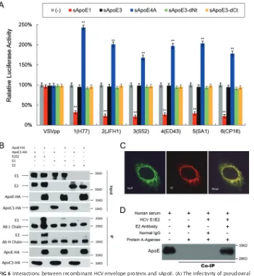
[image:9.585.41.441.69.525.2]esized that the interaction is mediated through the HCV envelope proteins. To exclude
the effects of other HCV viral proteins, we used HCV envelope proteins from six HCV
genotypes to create lentiviral vectors capable of producing HCV pseudoparticles
(HCVpps). The six HCVpps were separately mixed with sApoE1, sApoE3, sApoE4A, and
ApoE3 deletion mutants in the cell supernatant, and infectivity assays were performed.
A vesicular stomatitis virus envelope protein pseudotyped particle (VSVpp) was used as
a control, and the results indicated that the presence of different sApoE variants had no
differential effects on the infectivity of this pseudovirus (Fig. 6A). However, the
infec-tivity of the HCVpps was significantly affected, with ApoE1 causing inhibition of
infectivity and sApoE4A increasing it. As the only difference in the assembly processes
of the VSVpp and HCVpps was the presence of the HCV envelope proteins, these results
suggest that the effects of sApoE on the infectivity of HCV are mediated through these
envelope proteins.
To assess what interactions form between sApoE and the HCV envelope proteins,
we constructed cell lines stably expressing E1E2 dimers, E1 monomers, or E2
monomers. Cell lysates were mixed with sApoE-containing supernatant and
de-tected via coimmunoprecipitation (Co-IP). An obvious interaction was observed
between sApoE3 and the E1E2 dimer, whereas only weak interactions were
ob-served for E1 and E2 monomers (Fig. 6B). The interaction of ApoE with fused
envelope proteins was further evaluating using a two-hybrid system with truncated
envelope proteins missing the transmembrane region. The results showed no
detectable interactions (unpublished data), indicating that sApoE efficiently
inter-acts with HCV envelope proteins only when they are in their native spatial
confor-mation, with a preference for the E1E2 dimer.
As the HCV viral envelope proteins in their native conformation induce the
production of neutralizing antibodies
in vivo
, they are therefore the main candidate
antigens for vaccine development. However, it must be considered that antigen
immunogenicity is influenced by numerous factors. In the present study, we
showed that recombinant HCV envelope proteins can bind to ApoE protein in both
cells and blood. To demonstrate this, we first examined the distribution of
cell-expressed recombinant E1E2 envelope proteins, and the results indicated that the
HCV envelope proteins can colocalize with cApoE in cells (Fig. 6C). Then, we mixed
E1E2 protein expressed by 293T cells with normal human serum and found that the
HCV envelope proteins coimmunoprecipitated with sApoE (Fig. 6D). These results
imply that HCV envelope proteins interact with both cApoE and sApoE proteins in
both cells and blood.
DISCUSSION
Typically, host-virus interactions begin with viral attachment to the cellular
mem-brane. However, a study reported in 1964 showed that blood components can enhance
the infectivity of many viral species, including Japanese encephalitis virus and West Nile
virus (46), through a phenomenon termed ADE. The correlation of ADE with virus
pathogenicity was later clarified in a dengue virus study (47) which reported that when
a virus enters the host through blood, the interaction of the virus with blood
compo-nents is a key determining factor for pathogenicity. It has been shown that HCV
interacts with cApoE in hepatocytes and is secreted into blood for transmission, and
abundant exchangeable sApoE on the surfaces of lipoproteins in blood can dissociate
and bind to other biological complexes. In the present study, the three major ApoE
isoforms and two ApoE variants, ApoE1 and ApoE4A, which have a significantly
decreased or increased affinity for HSPG on hepatocytes than ApoE3, were employed.
We reported that HCV also binds to extracellular sApoE and found that both sApoE
protein concentration and variant type affect virus infectivity. For example, sApoE4
caused a reduction in HCV infectivity, a finding that is consistent with clinical data
indicating that the ApoE4 genotype is associated with a degree of resistance against
HCV infection (48, 49). During the preparation of the manuscript, another similar result
was also reported (50).
on November 7, 2019 by guest
http://jvi.asm.org/
Interactions between HCV and lipoprotein components in blood can affect not only
virus infectivity but also viral immunogenicity. A recent study showed that binding of
ApoE to HCV virions mediates viral evasion from neutralizing antibodies (51). Although
the binding of HCV with ApoE intracellularly or extracellularly may have only a slight
influence on the virus life cycle, the interaction of either pseudo-viral particles or
FIG 6Interactions between recombinant HCV envelope proteins and sApoE. (A) The infectivity of pseudoviral particles influenced by sApoE variants depends on HCV envelope proteins. The HCV or VSV envelope protein pseudotyped lentiviral particles containing a luciferase reporter gene were produced and incubated with cell culture supernatant containing different sApoE variants or mutants secreted from 293T cells at room temperature for 30 min and subsequently used to infect Huh7.5 cells in a 24-well culture plate. At 5 h.p.i., the mixture was removed, and the cells were washed twice with PBS and then incubated with DMEM containing 10% FBS. After 24 h, the cells were lysed for luciferase assays. The quantitative results derived from the data were converted to percentages of the control in the bar graph, and the data are shown as the means⫾SDs (error bars) pooled from three independent experiments. **, P ⬍ 0.01 (unpaired Student’s t test). (B) Interactions between sApoE and HCV envelope proteins were analyzed by coimmunoprecipitation. 293T cells stably expressing the HCV envelope protein E1E2, E1, or E2 were lysed and separately incubated with cell culture supernatant containing sApoE3-HA or sApoC-HA secreted by 293T cells. These mixtures were then captured by HA-specific immunoprecipitation. The proteins (input) or immunocomplexes (IP) in the mixture were analyzed by Western blotting. Proteins are specified on the left, and molecular mass standards are labeled on the right. (C) Colocalization of HCV envelope proteins and cApoE in hepatocytes. Recombinant HCV envelope proteins were expressed in Huh7.5 cells. cApoE (green) and HCV E2 (red) proteins were analyzed by immunofluorescence with specific antibodies and observed via confocal microscopy. (D) Co-IP of recombinant HCV envelope proteins with sApoE in human serum. Lysates from naive 293T cells or cells stably expressing HCV envelope proteins (E1E2) were incubated with human serum and protein G-conjugated agarose beads (Invitrogen) bound with normal mouse IgG or an E2-specific MAb. Upon extensive washing, precipitated proteins were separated by 10% SDS-PAGE and then transferred onto a PVDF membrane. ApoE protein was subsequently detected by Western blotting using an ApoE-specific MAb.
Extracellular Interaction of HCV and sApoE Journal of Virology
on November 7, 2019 by guest
http://jvi.asm.org/
[image:11.585.43.402.70.459.2]recombinant E1E2 protein with extracellular sApoE could have a significant role in
vaccine development. These findings imply that antigens containing HCV envelope
proteins could bind to host sApoE and that this interaction might lead to decreased
antigen immunogenicity. Therefore, disrupting these interactions might be a novel
strategy to enhance the immune response for vaccine antigen design.
The neutralizing antibody level in serum is a key indicator when evaluating humoral
immunity. HCV can form LVPs with lipoproteins to attach to HSPG and very low-density
lipoprotein receptors on hepatocyte membranes. At low neutralizing antibody levels
(resulting from a lack of high dilutions), serum lipoprotein could competitively interact
with receptors to interfere with results. In the present study, we further confirmed that
sApoE, as a component of lipoprotein, can form extracellular interactions with HCV
virions. Furthermore, different ApoE variants and protein concentrations differentially
affect HCV infectivity.
Human ApoE functions in lipid metabolism, immune regulation, and neural
devel-opment, and as a consequence, interactions between HCV and cApoE mainly occur
within hepatocytes and thus cause pathogenic effects through the liver. However,
these interactions could also affect the systemic function of sApoE and thereby cause
indirect effects on other organs. It has been shown that chronic HCV patients have a
high incidence of extrahepatic manifestations, including atherosclerosis, diabetes
mel-litus, and other abnormal lipid metabolism-related disorders. The phenomenon of HCV
forming extracellular interactions with carrier lipoproteins provides a new avenue for
clinical studies on HCV-related extrahepatic syndrome and might impact clinical safety
evaluations of HCV envelope protein-based vaccines.
Because no crystal structures of the E1E2 protein complex exist, we can only
hypothesize that deletion of E2 or the expression of fusion proteins directly disturbed
the ability of the viral proteins to interact with ApoE (unpublished data), and no
interaction domain on the envelope proteins could be defined. Although Lee et al. (34)
showed that the interaction of ApoE with HCV E2 protein was not affected when using
detergent to remove lipids, indicating that a direct interaction exists between the two
proteins, more accurate studies utilizing amino acid mutation are required to further
confirm the residues that mediate the interaction. Lee et al. (34) also indicated that the
interaction of envelope proteins with cApoE depends on the protein’s transmembrane
domain. However, the presence of sApoE on the viral surface is not involved in
intracellular HCV LVP assembly. Therefore, the HCV E2 transmembrane domain may not
act directly on ApoE but rather help maintain the native conformation of the E2 protein
to facilitate its association with ApoE. E1 and E2 assist each other in correct folding, and
as a consequence, when E1 or E2 protein was expressed alone, the binding affinity
toward ApoE markedly decreased (Fig. 6B). Moreover, a recent study also demonstrated
that the HVR1 of the HCV E2 protein modulates the conformation and/or epitope
exposure of virus particle-associated ApoE (51), which also indicates that the structural
conformation of E2 could affect its affinity toward binding with ApoE. Notably,
al-though the association of sApoE with viral particles relies on interactions with the viral
envelope proteins, prior immunoelectron microscopy studies have revealed various
amounts of ApoE proteins on individual HCV virion surfaces (36, 39). On the one hand,
this indicates that a single HCV particle could interact with several ApoE proteins, which
supports our results showing that the interaction of sApoE with HCV is dose dependent.
On the other hand, this finding implies that the interaction efficiency of different HCV
LVPs with sApoE varies. In the present study, we observed that sApoE more markedly
affected the infectivity of HCVccs than HCVpps, although the reason behind this
difference remains unclear.
Several earlier studies have indicated that ApoE can associate with HCV
intracellu-larly and participate in the maturation of HCV viral particles. In our research, we further
proved that HCV can form extracellular associations with secreted sApoE. Thus, the
presence of ApoE on HCV viral particles might result not only from hepatic cells but also
from blood constituents. Therefore, future analyses of HCV infection should consider
that extracellular ApoE may not only competitively associate with HCV receptors but
on November 7, 2019 by guest
http://jvi.asm.org/
also directly interact with HCV particles. Moreover, ApoE could also competitively
associate with several blood constituents. Therefore, when assessing the effects of
ApoE protein on HCV infectivity, factors including cApoE production within hepatic
cells, the secretion of ApoE from hepatocytes, the concentration of sApoE in blood
circulation, HCV concentration, and the concentration of several blood constituents,
including VLDL and LDL, must all be considered with regard to HCV infectivity
in vivo
.
Further probing of the extracellular interactions that exist between HCV and sApoE
could provide new insights into the aspects of the HCV life cycle that affect viral
infectivity.
MATERIALS AND METHODS
Cells and virus.The Huh7.5 cell line was provided by Charles Rice (Rockefeller University). 293T cells were obtained from the Kunming Cell Bank of Type Culture Collection, Chinese Academy of Sciences. All cells were grown in Dulbecco’s modified Eagle’s medium (DMEM) supplemented with 10% fetal bovine serum (FBS), 2 mML-glutamine, nonessential amino acids, 100 U/ml of penicillin, and 100g/ml of streptomycin (HyClone) at 37°C in 5% CO2. Stable cell lines induced to express HCV envelope proteins
were maintained by the addition of 6g/ml of puromycin (Sigma-Aldrich). Primary human hepatocytes (PHHs) (lot number FCL; BioreclamationIVT) were plated at 1.5⫻105/cm2in 6-well collagen I-coated
plates with InVitroGRO CP medium (BioreclamationIVT). Four hours after plating, nonadherent cells were removed, and the PHHs were maintained in InVitroGRO HI medium (BioreclamationIVT). Infectious HCVcc was produced by transfection of Huh7.5 cells within vitro-transcribed RNA derived from JFH1A or JFH1A-Rluc.
Plasmid constructs.Plasmids were constructed according to standard methods. All plasmid vectors were cloned and propagated inEscherichia coliDH5␣, DH10B, or Stbl3 grown at 30°C. The DH10B strain was used for vectors containing HCV genomes, and the Stbl3 strain was used for constructs containing duplicated sequences. Constructs were verified by restriction enzyme digestion and sequencing. De-scriptions of the cloning strategies are provided below.
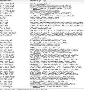
The p94-JFH1A plasmid, which contains the T7 promoter sequence fused to the JFH1 complete genome sequence with adaptive mutations, the hepatitis delta virus genomic ribozyme, and the T7 terminator sequence, was described previously (52). To introduce an MluI monoclonal restriction site in the NS5A C-terminal coding sequence, the PCR primers 7453-RsrIIF/7525-MluR and JFH1-7525-MluF/JFH1-7796-BsrGIR (see Table 2 for primer sequences) were used to amplify two products from the p94-JFH1A template that were combined in a second round of PCR, cleaved by RrsII and BsrGI, and then ligated back into p94-JFH1A digested by SpeI and BsrGI with the SpeI-RrsII fragment from p94-JFH1A to create p94-JFH1A-M420. The p94-JFH1A-Rluc vector containing the HCV reporter genome was created by amplifying theRenillaluciferase-coding region from pRL-SV40 (Promega) using primers NS5A-Rluc-HF and NS5A-Rluc-HR and then inserted into MluI-linearized p94-JFH1A-M420 using the In-Fusion HD enzyme (TaKaRa). The amino acid substitutions T197S, S199G, S200L, and M202H were introduced into E1 by overlapping PCR amplification using synthetic oligonucleotide primers 2a-78F, 2a-1371R, 2a-E1A4F, and 2a-E1A4R. The PCR DNA fragments were digested with the restriction enzymes AgeI and BsiWI and cloned into JFH1A cDNA vectors, which were similarly cut by both AgeI and BsiWI, as previously described (53). The E1 protein containing these substitutions can be recognized by the E1-specific monoclonal antibody (MAb) A4.
The pcDNA3.1-derived vectors ApoE1, -2, -3, -4, and -4A, ApoE dNT, and pcDNA3-ApoE dCT expressing different full-length pcDNA3-ApoE variants and deletion mutants were described previously (32). An HA tag was fused to the C terminus of ApoE (Fig. 1A) using overlap extension PCR. For ApoE-HA and ApoE dNT-HA, PCR products were amplified from pcDNA3-ApoE3 using the primer ApoE3-KasIF paired with ApoE-SG-HAR. For ApoE dCT-HA, PCR products were amplified from pcDNA3-ApoE dCT using primer ApoE3-KasIF paired with ApoE dCT-SG-HAR. These PCR products were gel purified and extended using ApoE3-KasIF and HA-SacI-XbaIR. The resulting products were cloned into pcDNA-ApoE and cleaved using the restriction enzymes KasI and XbaI to create pcDNA3-ApoE-HA, pcDNA3-ApoE dNT-HA, and pcDNA3-ApoE dCT-HA.
The human-actin gene was amplified from Huh7 cell total RNA using primers Hbactin-KpnIF and Hbactin-XbaIR with a GoldScript one-step reverse transcription-PCR (RT-PCR) kit (Life Technologies, Guangzhou, China) according to the manufacturer’s instructions. The RT-PCR product was inserted into a pcDNA3.1 vector between the KpnI and XbaI restriction sites to create the pcDNA3.1-HBactin plasmid. To produce CRISPR-Cas9 bicistronic expression vectors, pCW299 with gRNA 299 and oligonucleotides ApoE129-g20F and ApoE129-g20F were annealed at 95°C for 5 min and then cooled to room temper-ature, followed by ligation to plasmid pX330-U6-Chimeric_BB-CBh-hSpCas9 (a gift from Feng Zhang; Addgene plasmid 42230), which was linearized using the BbsI restriction enzyme.
pUC57-H77C, pUC57-S52, pUC57-ED43, and pUC57-SA1, containing the genomes of HCV geno-types 1a (H77C, GenBank accession no.AF011751[54]), 3a (S52, GenBank accession no.GU814264[55]), 4a (ED43, GenBank accession no.GU814266[55]), and 5a (SA1, GenBank accession no.KJ925146[56]), respectively, and flanked with the EcoRI-T7 promoter and an XbaI restriction site, were synthesized by Sangon Biotech Co. Ltd. (Shanghai, China). Some restriction enzyme sites were deleted via silent mutation. pBR-CP16 contains the genome of HCV genotype 6n, which is prevalent among intravenous drug users in the Yunnan province of southwestern China (GenBank accession no.KY014622). Using the plasmids containing various HCV genotypes as templates, PCR was used to amplify DNA fragments
Extracellular Interaction of HCV and sApoE Journal of Virology
on November 7, 2019 by guest
http://jvi.asm.org/
encoding the last 58 residues of the HCV core and all of the E1 and E2 proteins using the primers H77-741-BglIIF/H77-2579-XbaIR, JFH1-740-BglIIF/JFH1-2590-XbaIR, S52-739-BglIIF/S52-2592-XbaIR, ED43-740-BglIIF/ED43-2575-XbaIR, SA1-739-BglIIF/SA1-2577-XbaIR, and CP16-740-BglIIF/CP16-2584-XbaIR. The DNA fragments were amplified separately and inserted into a pEGFPN2 vector (Clontech) using BglII and XbaI to create plasmids pN-Env HCV1 to pN-Env HCV6.
PCR with primers H77-171-NheF/H77-383-BglIIR, H77-384-NheF/H77-746-BglIIR, and H77-171-NheF/ H77-746-BglIIR was used to amplify E1 (polyprotein residues 171 to 383), E2 (polyprotein residues 384 to 746), and E1 plus E2 (polyprotein residues 171 to 746) from the pN-Env HCV1 template; the products were cleaved by NheI and BglII, ligated with pCW57.1 (a gift from David Root; Addgene plasmid 41393), and digested by NheI and BamHI to create the lentiviral plasmids pCW-E1, pCW-E2, and pCW-E1E2.
pEGFP-puro was a gift from Michael McVoy (Addgene plasmid 45561). pMD2.G and psPAX2 were obtained from Didier Trono (Addgene plasmids 12259 and 122560). pNL4-3.Luc.R⫺.E⫺was obtained through the NIH AIDS Reagent Program, Division of AIDS, NIAID, NIH.
Antibodies.A hybridoma cell line producing the ApoE-specific monoclonal antibody WUE-4 was obtained from the American Type Culture Collection (CRL-2247). A human ApoE enzyme-linked immu-nosorbent assay (ELISA) kit was obtained from Mabtech (number 3212-1A-20). Goat anti-human ApoE polyclonal antibody was purchased from Calbiochem (number 178479). HCV core (number 1868), E1 (number 1879), and E2 (number 1876) monoclonal antibodies were purchased from Virostat. Anti--actin (AC15) and HA monoclonal antibodies (HA-7), an anti-HA immunoprecipitation kit, an HA peptide, and a horseradish peroxidase-conjugated goat anti-mouse IgG were purchased from Sigma-Aldrich. Alexa Fluor 488-labeled donkey anti-goat IgG and Alexa Fluor 594-labeled donkey anti-mouse IgG were purchased from Thermo Fisher Scientific. The mouse natural IgG protein (ab198772) was purchased from Abcam. Mouse anti-E1 MAb A4 (11) was kindly provided by Jean Dubuisson (Institut de Biologie de Lille, France). The mouse anti-E2 MAb AP33 (57) was kindly provided by Arvind H. Patel (Institute of Virology, Glasgow, Scotland).
[image:14.585.41.385.86.432.2]HCV RNA synthesis and transfection of cultured cells.Plasmids carrying full-length HCV genomes were linearized with XbaI and extracted with phenol and chloroform, precipitated with ethanol, and dissolved in RNase-free water. RNA was transcribed from 3g of purified template using a T7 RiboMAX expression large-scale RNA production system (Promega, Beijing, China). Complete removal of the TABLE 2Primers used to generate the viral constructs
Primer name Sequence (5=–3=)a
JFH1-7453-RsrIIF ATCCGGCGGTCCGACGT
JFH1-7525-MluR AGGCTCACGCGTGAGGGGGGGCATAGAGGAGGC JFH1-7525-MluF CTCACGCGTGAGCCTGGAGATCCGGACCTGGAGTC JFH1-7796-BsrGIR TCTTTGATGTTGTACAGTACACCTTG
NS5A-Rluc-HF GCCCCCCCTCACGCGTATGGCTTCCAAGGTGTACG NS5A-Rluc-HR CTCCAGGCTCACGCGTCTGCTCGTTCTTCAGCACGCGC
2a-78F CTAGCCATGGCGTTAGTATGAGTGT
2a-1731R ATGACCTCGGGGACGCG
2a-E1A4F CAGGTGAAGAATTCCAGTGGCCTCTACCATGTGACCAATGACTGC 2a-E1A4R GCAGTCATTGGTCACATGGTAGAGGCCACTGGAATTCTTCACCTG ApoE3-KasIF GAGGGCGCCGAGCGCGG
ApoE-SG-HAR GTATGGGTATCCTCCTCTTCCTCCTGAGTGATTGTCGCTGGGCACA ApoE dCT-SG-HAR GTATGGGTATCCTCCTCTTCCTCCTGAGGCCTGCAGGCGTATCTG HA-SacI-XbaIR GGTCTAGAGCTCAAGCGTAATCTGGAACATCGTATGGGTATCCT
CCTCTTCCTCCTG
Hbactin-KpnIF AGTGGTACCATGGATGATGATATCGCCGCG Hbactin-XbaIR AGTTCTAGACTAGAAGCATTTGCGGTGGACG ApoE129-g20F CACCGCCAGTGCCAGTTCCCAGCGC ApoE129-g20R AAACGCGCTGGGAACTGGCACTGGC
H77-741-BglIIF AATAGATCTGCCACCATGGGGTACATACCGCTCGTCGGCG H77-2579-XbaIR GAATCTAGATCACGCCTCCGCTTGGGATATGAGtaac JFH1-740- BglIIF AATAGATCTGCCACCATGGGGTACATCCCCGTCGTAGGCG JFH1-2590-XbaIR GAATCTAGATCATGCTTCGGCCTGGCCCAACAAGATG S52-739-BglIIF AATAGATCTGCCACCATGGGGTACATCCCGCTCGTCGGCG S52-2592-XbaIR GAATCTAGATCATGCTTCTGCTTGTGATACCATCAGCATC ED43-740-BglIIF AATAGATCTGCCACCATGGGATACATCCCGCTCGTAGGCG ED43-2575-XbaIR GAATCTAGATCACGCCTCAACTTGACTTACCATAAACATCATCC SA1-739-BglIIF AATAGATCTGCCACCATGGGGTACATCCCGCTCGTAGGCG SA1-2577-XbaIR GAATCTAGATCATGCTTCTGCCTGACAAACTAGAAGC CP16-740-BglIIF AATAGATCTGCCACCATGGGGTACATCCCAGTCCTAGGAG CP16-2584-XbaIR GAATCTAGATCAGGCCTCCGCTTGGCCCACCAGGAAC H77-171-NheF TTGGCTAGCATGGGTTGCTCTTTCTCTATCTTCCTTCTGG H77-383-BglIIR TTGAGATCTTACGCGTCGACGCCGGCAAATAG H77-384-NheF TTGGCTAGCATGGAAACCCACGTCACCGGGGGAAATG H77-746-BglIIR TTGAGATCTTACGCCTCCGCTTGGGATATGAG
aThe restriction sites used to facilitate cloning are underlined.
on November 7, 2019 by guest
http://jvi.asm.org/
template DNA was ensured by an additional treatment with RNase-free DNase and purification over Qiagen RNA isolation columns. The quality of thein vitro-transcribed RNA was examined by agarose gel electrophoresis. RNA transcripts were introduced into cells by transfection using a TransIT-mRNA transfection kit (Mirus). For transfection, cells plated in 10-cm cell culture dishes were grown overnight to approximately 80% confluence. The culture medium was replaced with fresh complete growth medium 30 min before transfection. Transfection complexes, prepared by adding 10g of HCV RNA, 20 l of mRNA boost reagent, and 20l of transfection reagent to 500l of Opti-MEM in sterile tubes, were incubated for 3 min at room temperature and added dropwise to each plate.
Isopycnic gradient centrifugation analysis.Media containing sApoE or HCV were layered on top of 9 to 30% OptiPrep (Axis-Shield, Norway) gradients prepared in phosphate-buffered saline (PBS). The gradients were centrifuged at 40,000 rpm (197,000⫻g) for 6 h at 4°C in an SW-41 rotor. Eleven fractions (1 ml each) were collected from the bottom of the tube. The density of the fractions was determined using a refractometer (Abbemat 350; Antton Paar).
Western blot analysis. Protein concentrations were determined using protein assay reagent (Thermo Scientific, Beijing, China). Each sample was analyzed by 10% sodium dodecyl sulfate-poly-acrylamide gel electrophoresis (SDS-PAGE). Separated proteins were transferred onto a polyvinylidene difluoride (PVDF) membrane. After blocking with 5% skim milk, immunoblotting analysis for ApoE, -actin, HA tag, HCV core, E1, and E2 proteins was performed using primary monoclonal antibodies specific to each protein, a secondary goat anti-mouse antibody, and an enhanced chemiluminescence kit (Thermo Scientific).
DNA transfection and ApoE knockout cell line selection.DNA transfection was performed using a lipofection method according to the manufacturer’s instructions. For sApoE production, cell culture dishes were coated with 0.1 mg/ml of poly-L-lysine. The day before transfection, 3.2⫻106293T cells in
8 ml of cell culture medium without antibiotics were seeded onto a 10-cm dish to reach 80% confluence by the time of transfection. A volume of 15g of ApoE expression plasmid was diluted to 1 ml in Opti-MEM medium (Life Technologies). In addition, 37.5l of Lipofectamine 2000 (Life Technologies) was added to 1 ml of Opti-MEM medium, and the DNA and Lipofectamine 2000 were gently mixed, incubated at room temperature for 20 min, and added to the cells in a dropwise manner. Eight hours after transfection, the cells were transferred to DMEM complete medium, and the supernatants were collected after 48 h. For ApoE gene knockout cell line selection, 4⫻105Huh7.5 cells in 6-well plates were
cotransfected with plasmids pEGFP-puro and pCW299 in a 3:1 ratio. Then 2g of DNA was mixed with 5l of DMRIE-C reagent (Life Technologies) in Opti-MEM and transferred to the Huh7.5 cells. At 24 h posttransfection, 10g/ml of puromycin was added and selected for 2 days. Then the cells were split into 10-cm cell culture dishes at various cell densities in growth medium. Well-isolated cell colonies were selected and amplified. sApoE protein expression was detected by ELISA. A cell line not expressing sApoE was subjected to two rounds of subcloning by limiting dilution, and the ApoE knockout cell lines were confirmed by restriction fragment length polymorphism (RFLP) assays or Sanger sequencing of the mutated ApoE gene.
HCVcc particle pulldown by HA-specific immunocapture. HCVcc (500 l) and an equivalent volume of cell culture supernatant containing ApoE-HA were preincubated at 4°C overnight. The mixture was subjected to HA tag-specific immunocapture using 40l of HA affinity gel (Sigma-Aldrich). After washing with 800l of PBS, the captured proteins were eluted by boiling in Laemmli sample buffer and subjected to SDS-PAGE and Western blot analysis. Captured HCV RNAs were extracted using TRIzol A⫹ and quantified by quantitative RT-PCR (qRT-PCR). Immunocaptured HCV particles were eluted with 5 volumes of PBS containing 100g/ml of HA peptide (Sigma-Aldrich). Infectivity titers were determined by immunohistochemical assays.
RNA extraction and quantification by qRT-PCR. HCV RNA was isolated using TRIzol LS (Life Technologies) according to the manufacturer’s protocols. RNA levels were adjusted to internal control RNA input (2 ng of-actin RNA), which was added in excess prior to RNA isolation. The absolute levels of HCV RNA were quantified. Oligonucleotide primers w168 (5=-CTA GCC ATG GCG TTA GTA TGA GTG T-3=) and w169 (5=-CGG CAA TTC CGG TGT ACT CA-3=) and probe w170 (5=-FAM-TCC CGG GAG AGC CAT AGT GGT CTG CG-BHQ1-3=), which are complementary to the HCV 5=untranslated region (UTR), were used. For -actin mRNA, the primers used were w159 (5=-TGC CGA CAG GAT GCA GAA G-3=) and w160 (5=-CTC AGG AGG AGC AAT GAT CTT GAT-3=), and the TaqMan probe was 5=-Joe-ATC ACT GCC CTG GCA CCC AGC A-BHQ1-3=. Reactions were run using an ABI 7500 real-time PCR system (Life Technologies) as follows: 5 min at 42°C, 10 s at 95°C, and 40 cycles at 95°C for 5 s and 60°C for 35 s. Fluorescence detection was performed using the preset channels Fam, Joe, and Rox. RNA quantities were derived by comparison with RNA concentration standards run in parallel. The RNA standards werein vitroT7 transcripts of the JFH1 genome and-actin RNA, which were DNase treated, purified on an RNeasy minicolumn (Qiagen, Shanghai, China), and quantified via spectrophotometry.
FF-IHC. For the focus-forming assay based on immunohistochemistry (FF-IHC), HCV in culture supernatant was 10-fold serially diluted, and 250l of diluted HCV was used per well to infect Huh7.5 cells in 24-well plates. After 6 h of incubation at 37°C, the cells were washed twice with PBS, and 250l of fresh complete medium containing 1% methylcellulose was added. At 72 h postinfection (h.p.i.), the cells were fixed with 10% neutralized formalin. Focus-forming units were determined by immunohisto-chemistry using a core-specific monoclonal antibody, peroxidase-conjugated secondary anti-mouse antibody, and 3,3-diaminobenzidine substrate (Vector Laboratories, Burlingame, CA). Viral titer was determined based on the average number of core-positive focus-forming units in triplicate or quadruplet assays.
Extracellular Interaction of HCV and sApoE Journal of Virology
on November 7, 2019 by guest
http://jvi.asm.org/
Quantification of ApoE. Human ApoE levels in culture medium or normal human serum were quantified using a human apolipoprotein E enzyme-linked immunosorbent assay kit (Mabtech AB, Sweden) according to the manufacturer’s protocol; the recombinant ApoE3 standard protein was also provided by the manufacturer.
ApoE protein purification.A total of 500l of anti-HA affinity matrix beads was washed 3 times with 10 mM PBS (pH 7.4) and mixed with 10 to 15 ml of 293T cell supernatant with or without HA-tagged ApoE protein for 2 h. After centrifugation at 3,000⫻gfor 5 min, the supernatant was collected, and the beads were washed 3 times with PBS; this was followed by the addition of 500l of EF (1 mg/ml HA peptide in PBS) to elute the ApoE-HA protein.
HCV neutralization assays.Antibody inhibition assays were performed using Huh7.5 cells, and virus infectivity was determined by FF-IHC. Briefly, Huh-7.5 cells were plated at a density of 1⫻104per well
in a 96-well plate, and⬃50 FFU of virus was preincubated at 37°C for 1 h with the appropriate inhibitory or control antibody prior to infecting cells. At 3 h postinfection, the inoculum was replaced with fresh medium containing 1% methylcellulose and incubated for 48 h. The infectivity was determined as FF-IHC following immunostaining of the cells for viral core proteins as described above.
Lentiviral pseudoparticles.To generate pseudoparticles for the delivery of inducible HCV envelope protein, 1⫻107Lenti-X 293T cells (Clontech) in a T175 tissue culture flask were cotransfected with
plasmids encoding (i) pCW-E1, pCW-E2, or pCW-E1E2 provirus, (ii) psPAX2, and (iii) pMD2.G in a ratio of 4:3:2. To generate HCV pseudoparticles, 4⫻106Lenti-X 293T cells (Clontech) in a 10-cm cell culture dish
were cotransfected with a pNL4-3.Luc.R⫺.E⫺proviruses vector containing the firefly luciferase reporter gene and plasmids pN-HCV1 to -6, encoding the Env proteins of various HCV genotypes, in a ratio of 3:1. A total of 12g of DNA was transfected using Lipofectamine 2000 reagent. Transfections were carried out for 16 h, followed by a medium change to DMEM containing 10% FBS. The culture medium was collected at 48 h and 72 h, pooled, filtered through a 0.45-mm filter, ultracentrifuged, and stored at
⫺80°C.
Infectivity measurement via luciferase assay.Huh7.5 cells were seeded at a density of 2⫻105per
well in 12-well plates and allowed to adhere overnight. A volume of 100l of HCV-Rlu reporter virus was incubated with 200l of DMEM complete medium containing increasing concentrations of secreted ApoE at room temperature for 1 h, after which the mixture was added to the Huh7.5 cells. At 5 h postinfection, the mixture was removed, and the infected cells were washed twice with PBS and then incubated with DMEM complete medium. The supernatants were removed after a 24-h culture. The cells were then washed with PBS and lysed in 100l of lysis buffer, and 10l of lysate was added to 100l of assay buffer (Promega) in a 96-well assay plate. A microplate reader (Synergy 4, BioTek, Shanghai, China) was then used to perform a 10-s measurement. All luciferase assays were performed at least in triplicate.
Coimmunoprecipitation.293T cells overexpressing HCV H77C E1E2, E1, and E2 were cultured in T225 cell culture flasks for 16 h to become adherent, and doxycycline was added at a final concentration of 1g/ml. After 48 h, the cells were lysed using nondenaturing lysis buffer (20 mM Tris-HCl [pH 8.0], 137 mM NaCl, 2 mM EDTA, 1% Triton X-100, and protease inhibitor cocktail tablets). After 1 h of incubation on ice, cell debris was removed by 10 min of centrifugation at 3,000⫻g. To detect interactions between sApoE in the cell culture medium and HCV envelope proteins, 10l of anti-HA agarose beads (Sigma-Aldrich) and 250l of sApoE-HA secreted by 293T cells transfected with pcDNA3.1-ApoE-HA using Lipofectamine 2000 were added to 250-l aliquots of cell lysates containing E1, E2, or E1E1. ApoC1-HA secreted by 293T cells was used as a negative control. To detect interactions between E1E2 and sApoE in serum, 250l of E1E2 protein in cell lysis buffer was mixed with 250 l of normal human serum and added to protein A agarose bound to anti-E2 antibody (Thermo Scientific). Normal mouse IgG was used as a negative control. The mixtures were incubated overnight by continuously inverting the tubes at 4°C, and the beads were washed three times by resuspension in 20 mM cold PBS followed by centrifugation at 3,000⫻g. The samples were resuspended with 50l of 1⫻SDS-PAGE loading buffer (50 mM Tris-HCl [pH 6.8], 2% SDS, 0.1% bromophenol blue [BPB], 10% glycerol, and 1%-mercaptoethanol), and the samples were incu-bated for 5 min at 95°C, followed by Western blotting.
Statistical analysis.Data are expressed as the means and standard deviations (SDs). Statistical analyses were performed using Student’sttest. APvalue of⬍0.01 was considered highly significant, and a value of⬍0.001 was considered extremely significant.
Accession number(s).Complete sequences for pcDNA3.1-derived vectors pcDNA3-ApoE1, -2, -3, -4, and -4A are available from GenBank under accession numbersKY924484toKY924488.
ACKNOWLEDGMENTS
We thank Charles Rice for kindly providing the Huh-7.5 cell line; Feng Zhang, David
Root, Michael McVoy, and Didier Trono for plasmids; and Arvind Patel and Jean
Dubuisson for antibodies.
W.C. and S.D. designed the study; Z.L., Y.L., Y.B., H.Z., Y.Y., and W.C. performed the
experiments; Q.L., W.C., and S.D. analyzed the data; and W.C. wrote the manuscript.
We have no conflicts of interest to declare.
This work was supported by the Natural Science Foundation of Yunnan Province
(2013FA053 and 2015BC010), Fundamental Research Funds for the Central Universities
on November 7, 2019 by guest
http://jvi.asm.org/
(2015PT310001 and 2016ZX310183-5), and the CAMS Initiative for Innovative Medicine
(2016-I2M-1-013 and 2016-I2M-1-019).
REFERENCES
1. Mohd Hanafiah K, Groeger J, Flaxman AD, Wiersma ST. 2013. Global epidemiology of hepatitis C virus infection: new estimates of age-specific antibody to HCV seroprevalence. Hepatology 57:1333–1342.
https://doi.org/10.1002/hep.26141.
2. Chen SL, Morgan TR. 2006. The natural history of hepatitis C virus (HCV) infection. Int J Med Sci 3:47–52.https://doi.org/10.7150/ijms.3.47. 3. Negro F. 2014. Facts and fictions of HCV and comorbidities: steatosis,
diabetes mellitus, and cardiovascular diseases. J Hepatol 61:S69 –S78.
https://doi.org/10.1016/j.jhep.2014.08.003.
4. McHutchison JG, Gordon SC, Schiff ER, Shiffman ML, Lee WM, Rustgi VK, Goodman ZD, Ling MH, Cort S, Albrecht JK. 1998. Interferon alfa-2b alone or in combination with ribavirin as initial treatment for chronic hepatitis C. Hepatitis Interventional Therapy Group. N Engl J Med 339: 1485–1492.
5. Baumert TF, Fauvelle C, Chen DY, Lauer GM. 2014. A prophylactic hepatitis C virus vaccine: a distant peak still worth climbing. J Hepatol 61:S34 –S44.https://doi.org/10.1016/j.jhep.2014.09.009.
6. Lemon SM, Walker C, Alter MJ, Yi M. 2007. Hepatitis C virus, p 1253–1304.InKnipe DM, Howley PM, Griffin DE, Lamb RA, Martin MA, Roizman B, Straus SE (ed), Fields virology, 5th ed, vol 1. Lippincott Williams & Wilkins, Philadelphia, PA.
7. Bartenschlager R, Ahlborn-Laake L, Mous J, Jacobsen H. 1994. Kinetic and structural analyses of hepatitis C virus polyprotein processing. J Virol 68:5045–5055.
8. Grakoui A, Wychowski C, Lin C, Feinstone SM, Rice CM. 1993. Expression and identification of hepatitis C virus polyprotein cleavage products. J Virol 67:1385–1395.
9. Hijikata M, Kato N, Ootsuyama Y, Nakagawa M, Shimotohno K. 1991. Gene mapping of the putative structural region of the hepatitis C virus genome by in vitro processing analysis. Proc Natl Acad Sci U S A 88:5547–5551.https://doi.org/10.1073/pnas.88.13.5547.
10. Jones DM, McLauchlan J. 2010. Hepatitis C virus: assembly and release of virus particles. J Biol Chem 285:22733–22739.https://doi.org/10.1074/ jbc.R110.133017.
11. Dubuisson J, Hsu HH, Cheung RC, Greenberg HB, Russell DG, Rice CM. 1994. Formation and intracellular localization of hepatitis C virus enve-lope glycoprotein complexes expressed by recombinant vaccinia and Sindbis viruses. J Virol 68:6147– 6160.
12. Ding Q, Cao X, Lu J, Huang B, Liu YJ, Kato N, Shu HB, Zhong J. 2013. Hepatitis C virus NS4B blocks the interaction of STING and TBK1 to evade host innate immunity. J Hepatol 59:52–58.https://doi.org/10.1016/j.jhep .2013.03.019.
13. Li K, Foy E, Ferreon JC, Nakamura M, Ferreon AC, Ikeda M, Ray SC, Gale M, Jr, Lemon SM. 2005. Immune evasion by hepatitis C virus NS3/4A protease-mediated cleavage of the Toll-like receptor 3 adaptor protein TRIF. Proc Natl Acad Sci U S A 102:2992–2997.https://doi.org/10.1073/ pnas.0408824102.
14. Meylan E, Curran J, Hofmann K, Moradpour D, Binder M, Bartenschlager R, Tschopp J. 2005. Cardif is an adaptor protein in the RIG-I antiviral pathway and is targeted by hepatitis C virus. Nature 437:1167–1172.
https://doi.org/10.1038/nature04193.
15. Nitta S, Sakamoto N, Nakagawa M, Kakinuma S, Mishima K, Kusano-Kitazume A, Kiyohashi K, Murakawa M, Nishimura-Sakurai Y, Azuma S, Tasaka-Fujita M, Asahina Y, Yoneyama M, Fujita T, Watanabe M. 2013. Hepatitis C virus NS4B protein targets STING and abrogates RIG-I-mediated type I interferon-dependent innate immunity. Hepatology 57:46 –58.https://doi.org/10.1002/hep.26017.
16. Otsuka M, Kato N, Moriyama M, Taniguchi H, Wang Y, Dharel N, Kawabe T, Omata M. 2005. Interaction between the HCV NS3 protein and the host TBK1 protein leads to inhibition of cellular antiviral responses. Hepatology 41:1004 –1012.https://doi.org/10.1002/hep.20666. 17. Taylor DR, Shi ST, Romano PR, Barber GN, Lai MM. 1999. Inhibition of the
interferon-inducible protein kinase PKR by HCV E2 protein. Science 285:107–110.https://doi.org/10.1126/science.285.5424.107.
18. Huang H, Sun F, Owen DM, Li W, Chen Y, Gale M, Jr, Ye J. 2007. Hepatitis C virus production by human hepatocytes dependent on assembly and
secretion of very low-density lipoproteins. Proc Natl Acad Sci U S A 104:5848 –5853.https://doi.org/10.1073/pnas.0700760104.
19. Yamamoto M, Aizaki H, Fukasawa M, Teraoka T, Miyamura T, Wakita T, Suzuki T. 2011. Structural requirements of virion-associated cholesterol for infectivity, buoyant density and apolipoprotein association of hepa-titis C virus. J Gen Virol 92:2082–2087. https://doi.org/10.1099/vir.0 .032391-0.
20. Weisgraber KH. 1994. Apolipoprotein E: structure-function relationships. Adv Protein Chem 45:249 –302. https://doi.org/10.1016/S0065 -3233(08)60642-7.
21. Mahley RW, Rall SC, Jr. 2000. Apolipoprotein E: far more than a lipid transport protein. Annu Rev Genomics Hum Genet 1:507–537.https:// doi.org/10.1146/annurev.genom.1.1.507.
22. de Knijff P, van den Maagdenberg AM, Frants RR, Havekes LM. 1994. Genetic heterogeneity of apolipoprotein E and its influence on plasma lipid and lipoprotein levels. Hum Mutat 4:178 –194.https://doi.org/10 .1002/humu.1380040303.
23. Aggerbeck LP, Wetterau JR, Weisgraber KH, Wu CS, Lindgren FT. 1988. Human apolipoprotein E3 in aqueous solution. II. Properties of the amino- and carboxyl-terminal domains. J Biol Chem 263:6249 – 6258. 24. Wetterau JR, Aggerbeck LP, Rall SC, Jr, Weisgraber KH. 1988. Human
apolipoprotein E3 in aqueous solution. I. Evidence for two structural domains. J Biol Chem 263:6240 – 6248.
25. Wilson C, Wardell MR, Weisgraber KH, Mahley RW, Agard DA. 1991. Three-dimensional structure of the LDL receptor-binding domain of human apolipoprotein E. Science 252:1817–1822.
26. Westerlund JA, Weisgraber KH. 1993. Discrete carboxyl-terminal seg-ments of apol