Immunological Hallmarks of JC Virus Replication in Multiple
Sclerosis Patients on Long-Term Natalizumab Therapy
Houria Hendel-Chavez,a,bMarie-Ghislaine de Goër de Herve,a,bClaire Giannesini,c,dAnne-Aurélie Mazet,eCaroline Papeix,f
Céline Louapre,fAudrey Chardain,c,dNassima Boutarfa,a,bMarie Théaudin,g,hDavid Adams,g,hJacques Gasnault,b,iBruno Stankoff,c,d Yassine Taoufika,b,h
Service d’Immunologie Biologique, Hôpitaux Universitaires Paris-Sud, Le Kremlin-Bicêtre, Francea
; INSERM 1012, Le Kremlin-Bicêtre, Franceb
; Service de Neurologie, Hôpital Tenon, APHP, Paris, Francec
; Université Pierre et Marie Curie, Paris, Franced
; Laboratoire de Virologie, Hôpital de Bicêtre, APHP, Le Kremlin-Bicêtre, Francee
; Fédération des Maladies du Système Nerveux, Hôpital Pitié-Salpétrière, APHP, Paris, Francef
; Service de Neurologie, Hôpital de Bicêtre, APHP, Le Kremlin-Bicêtre, Franceg
; Université Paris-Sud, Le Kremlin-Bicêtre, Franceh
; Service de Médecine Interne, Hôpital de Bicêtre, APHP, Le Kremlin-Bicêtre, Francei
Progressive multifocal leukoencephalopathy (PML) is the main adverse effect of natalizumab. Detectable JC virus-specific effec-tor memory T-cell (TEM) responses may indicate ongoing JCV replication. We detected JCV-specific TEM responses in blood of patients with multiple sclerosis (MS) treated with natalizumab, including 2 patients with PML. The frequency of detection of these responses increased with the time on natalizumab. Thus, a subset of MS patients exhibit immunological hallmarks of JCV replication during prolonged natalizumab therapy.
N
atalizumab, an antibody used to treat relapsing multiple sclerosis (MS), prevents trafficking of activated lympho-cytes, including autoreactive lympholympho-cytes, through the blood-brain barrier (BBB). The main adverse effect of natalizumab is progressive multifocal leukoencephalopathy (PML), a devas-tating demyelinating disease caused by replication of human JC polyomavirus (JCV) in oligodendrocytes and astrocytes (1,2). Natalizumab inhibition of effector T-cell trafficking from blood to the central nervous system (CNS) might favor local JCV replication (3–7).Here, we examined the presence of JCV-specific effector memory T cells (TEM) in blood of MS patients after overnight antigen activationex vivo.In vivo, specific TEM numbers de-cline once the cognate antigen is cleared (8–10), and their pres-ence may thus point to ongoing JCV replication. TEM rapidly release cytokines such as gamma interferon (IFN-␥) when re-exposed to antigen. A previous enzyme-linked immunosorbent spot assay (ELISPOT) examined IFN-␥ expression in T cells following long-term activation with JCV peptides for up to 14 days (11). However, prolonged activation allows long-term quiescent T-cell memory to be reactivated and to expand, meaning that a positive response does not necessarily signify an ongoing immune response. Here, we first examined the pres-ence of JCV-specific TEM in two MS patients who developed PML. The first patient was diagnosed with PML after 39 months on natalizumab (39 infusions) (Department of Neu-rology, Pitié-Salpétrière Hospital). She was 39 years old and had a 10-year history of relapsing-remitting MS. Prior to na-talizumab, she had received cyclophosphamide and mitoxan-trone. PML was confirmed by MRI and JCV PCR on cerebro-spinal fluid (CSF). When tested for JCV-specific TEM, 8 weeks after PML symptom onset, she had 1,040 JCV genome copies per ml of CSF (JCV Q-PCR Alert kit; Nanogen Advanced Di-agnostics). Her peripheral blood mononuclear cells (PBMC) were activatedin vitroin AIM-V and Albumax medium (Invit-rogen) for 16 h with purified JCV (strain MAD-4; LGC Pro-mochem; 104.550% tissue culture infective dose [TCID
50]/0.2
ml). No recombinant interleukin 2 (IL-2) was added. An
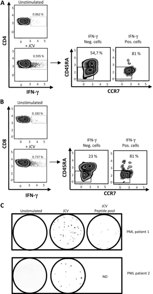
amount of virus corresponding to 5,534 PFU was added to each well. This corresponded to a multiplicity of infection (MOI) of 0.02. Cells were then tested for intracellular IFN-␥ by flow cytometry. Cell mortality following overnight activation was less than 5%. As shown inFig. 1AandB, responding cells were detected in both the CD4 and CD8 T-cell subsets, and most of them had the CCR7⫺CD45RA⫺phenotype characteristic of memory effectors (12). Anti-JCV TEM were also detected by IFN-␥ ELISPOT (capture and detection antibodies, clones 1-D1 and K7-B6-1, respectively; Mabtech) following 16 h of activation with purified JCV or VP-1 peptides (a mix of 14 pools of overlapping 15-amino-acid peptides covering the en-tire JCV VP-1 protein [Neosystem]) (Fig. 1C). The second pa-tient was 46 years old and had an 11-year history of relapsing-remitting MS. Prior to natalizumab, she had received beta interferon, glatiramer acetate, azathioprine, and mitoxan-trone. She was referred to the Tenon Hospital MS center for reevaluation after 24 natalizumab infusions. A brain MRI per-formed in May 2012 detected a small, linear, T2-hyperintense suspect lesion in the juxtacortical right frontal region. The first CSF sample was negative for JCV. Repeat MRI after natali-zumab withdrawal revealed an increase in lesion size, leading to two further CSF examinations, both of which were negative for JCV. JCV PCR on plasma was performed 2 and 4 months after the onset of PML. Both plasma samples were negative. Brain biopsy performed in September 2012 finally confirmed the di-agnosis of PML.
ELISPOT analysis of the IFN-␥response to JCV, performed 2 days after the last natalizumab infusion, showed the presence of
Received15 January 2013 Accepted11 March 2013
Published ahead of print20 March 2013
Address correspondence to Yassine Taoufik, [email protected].
H.H.-C. and M.-G.D.G.D.H. contributed equally to this work.
Copyright © 2013, American Society for Microbiology. All Rights Reserved.
doi:10.1128/JVI.00131-13
on November 7, 2019 by guest
http://jvi.asm.org/
FIG 1Detection of JCV-specific effector memory T cells (TEM) in blood of two natalizumab-treated patients with PML. In panels A and B, PBMC from one patient who developed PML following natalizumab treatment (39 infusions) were activated with purified JC virus overnight and then tested for intracellular IFN-␥by flow cytometry in the CD4 and CD8 T-cell subsets (A and B, respectively). CCR7 and CD45RA expression on IFN-␥-positive and -negative cells is also shown for CD4 T cells (A) and CD8 T cells (B). In panel C, PBMC from 2 patients who developed PML on natalizumab (patient 1 and patient 2 received 39 and 24 infusions, respectively) were activated overnight with purified JC virus and/or with a pool of VP1 peptides prior to IFN-␥ELISPOT. The numbers of IFN-␥ spots per 0.25⫻106PBMC after overnight activation with purified JCV and JCV peptide pools were 54 and 21, respectively (untreated well, 0 spots) in patient
1. In patient 2, purified JCV yielded 15 spots (untreated well, 0 spots).
on November 7, 2019 by guest
[image:2.585.135.450.35.639.2]JCV-specific TEM in blood (Fig. 1C). Interestingly, circulating JCV-specific TEM numbers were lower than in the first PML pa-tient, who may have had a higher level of JCV replication in the brain, as JCV PCR on CSF was positive in the first patient and negative in the second.
[image:3.585.45.549.77.191.2]We also examined the presence of JCV-specific TEM by ELISPOT in a series of 62 MS patients with relapsing-remitting MS treated with natalizumab according to the European market-ing terms, as well as in 35 MS patients who were not receivmarket-ing any disease-modifying therapy, and a group of 40 healthy donors TABLE 1Clinical features of the patients with multiple sclerosis
Feature
Value
Natalizumab (n⫽64 MS patients)
Control (n⫽35 MS patients)
Median age 40 44
Sex ratio (female to male) 1.74 1.69
Median Expanded Disability Status Scale score (range) 3.5 (1–7.5) 3.5 (1–6.5)
Duration of the disease⫾SD (yrs) 11.4⫾5.2 10⫾4.8
No. (range) of natalizumab infusions 24 (1–48)
No. of patients sampled twice (interval between 1st and 2nd sample) 41 (10.6⫾2.8 mo)
No. of patients sampled 3 times or more (interval between 2nd and 3rd sample) 6 (10⫾0.3 mo)
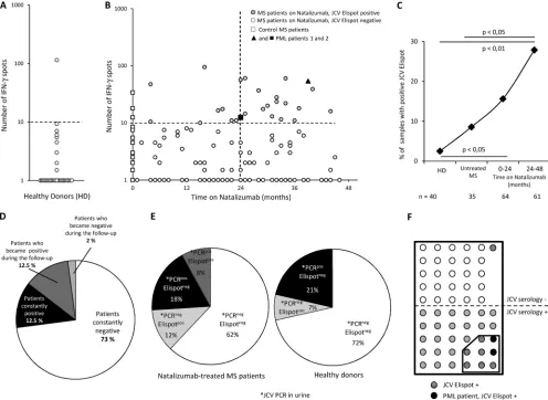
FIG 2Increased detection of JCV-specific TEM in patients on natalizumab. Panels A and B represent the number of IFN-␥spots per 0.25⫻106PBMC after
overnight activation with purified JC virus in 40 healthy donors (A) and in 99 MS patients according to the time on natalizumab (B). In panel B, the 2 patients with PML are indicated by a closed triangle and a closed square. The dashed line represents the positivity cutoff (see the text). Panel C represents the percentage of JCV-ELISPOT-positive samples in healthy donors (HD), control patients (Untreated MS), and patients treated with natalizumab for up to 2 years (0 –24 months) or more (24 – 48 months). Some patients treated with natalizumab were tested at 2 or 3 time points. The number of blood samples analyzed is indicated below. Statistical analysis used Fisher’s test. Panel D shows JCV ELISPOT results for patients who were tested at 2 or 3 time points. Panel E shows the results of urinary JCV PCR and JCV ELISPOT in 50 MS patients treated with natalizumab and in 14 healthy donors tested with both assays. Panel F shows results of JCV serology and JCV ELISPOT in 56 MS patients on natalizumab tested with both assays.
JC Virus Effector Memory T Cells and Natalizumab
on November 7, 2019 by guest
http://jvi.asm.org/
[image:3.585.43.539.286.648.2]matched for age and sex with MS patients. Characteristics of the MS patients are shown inTable 1. The patients and healthy donors gave their written consent to participate in the study, which was approved by the ethics committee of Pitié-Salpêtrière Hospital, Paris. An ELISPOT response was considered positive if the cor-rected number of spots (number of spots in activated wells minus number of spots in untreated wells) was at least 10, with fewer than 10 spots in untreated wells.
The proportion of positive samples was 2.5% in healthy donors
(Fig. 2A). In the MS group, the percentage of positive patients rose
with the time on natalizumab (Fig. 2BandC), from 8.8% in un-treated MS patients to 15.9% among patients un-treated with natali-zumab for less than 24 months and 27% among patients treated for more than 24 months (Fig. 2C) (significant for patients treated for more than 24 months versus untreated MS [Fisher’s exact test, P⬍0.05] and versus control subjects [P⬍0.01]). This increase in the frequency of detection of JCV TEM responses suggested that prolonged natalizumab treatment favored JCV reactivation.
A subset of natalizumab-treated patients were sampled at least twice (seeTable 1). As shown inFig. 2D, most of these patients either became or remained positive during the sampling period.
The kidney is a major reservoir for JCV. A significant per-centage of the general population excretes the virus in their urine but has no evidence of JCV in their blood (13–15). In addition, the presence of JCV DNA in the brain appears to be independent of its presence in the kidney (16). As shown inFig. 2E, 60% of MS patients with detectable JCV TEM in blood had no detectable JCV in their urine, and 69% of patients with positive JCV PCR in urine had no detectable JCV TEM. JCV urinary excretion without detectable JCV TEM was also ob-served in healthy donors (Fig. 2E). This suggests that the pres-ence of JCV replication in kidney does not necessarily lead to detectable JCV TEM in blood. Conversely, this raises the pos-sibility that JCV TEM detection in blood may reflect ongoing extrarenal JCV replication.
In a study using a novel JCV enzyme-linked immunosorbent assay (ELISA) method (17), the estimated seroprevalence of JCV in MS patients treated with natalizumab was 53.6% (17). Seropos-itivity does not necessarily correlate with the presence of TEM responses, which is related to virus reactivation. Long-lived plasma cells may persist in bone marrow for long time periods, perhaps for decades, maintaining sustained serum antibody titers without active pathogen replication (18,19). We used the same ELISA method to test 56 of our natalizumab-treated MS patients who had been tested by JCV ELISPOT, including the 2 PML pa-tients. As shown inFig. 2F, 53.6% of the patients were seropositive and 16.1% were ELISPOT positive. Twenty-seven percent of JCV-seropositive patients had detectable JCV TEM in their blood. Only one patient who was JCV ELISPOT positive was seronegative. Ac-cording to Gorelik et al., the false-negative rate of the JCV sero-logic assay is 2.5% (17).
Our results show that the frequency of detection of JCV TEM responses in MS patients increases with the time on natalizumab. A previous study also showed an increase in T cell responses to JCV after 12 months of natalizumab therapy, but this study was limited to 18 months of treatment (20), whereas we found that the strongest increase in effector T cell responses occurred after 24 months.
Our results thus point to JCV reactivation during natalizumab therapy, including in extrarenal sites. JCV has been detected in the
brain of HIV-seronegative individuals without PML (21). The di-minished CNS immunosurveillance induced by natalizumab might favor local virus reactivation that may start as asymptom-atic intermittent virus replication but could evolve toward sus-tained virus replication and then to PML. Virus reactivation could activate peripheral specific T lymphocytes, via antigen-presenting cells, in locations such as brain-draining cervical lymph nodes (22–24). As natalizumab targets the VLA-4 receptor, these specific cells cannot efficiently cross the blood-brain barrier and might thus accumulate in blood, facilitating their detection. PML is also a complication of late-stage HIV infection. Whether detection of JCV-specific effector CD4 and CD8 T cells may precede AIDS-associated PML is a point of interest that deserves further investi-gation. However, AIDS-associated PML is clearly different from natalizumab-induced PML. Most HIV-infected patients who de-velop PML have profound CD4 lymphopenia, which also alters CD8 T cell functionality through the lack of CD4 help (25). More-over, unlike patients on natalizumab, there is no blockade of T cell trafficking through the blood-brain barrier, and this may prevent JCV-specific TEM accumulation in blood.
Together, our results suggest that functional assays capable of detecting JCV-specific TEM responses might help to identify pa-tients at risk of developing PML during treatment with antibodies that inhibit lymphocyte trafficking through the blood-brain bar-rier.
REFERENCES
1.Brew BJ, Davies NW, Cinque P, Clifford DB, Nath A.2010. Progressive multifocal leukoencephalopathy and other forms of JC virus disease. Nat. Rev. Neurol.6:667– 679.
2.Gasnault J, Taoufik Y.2006. New trends in progressive multifocal leuko-encephalopathy. Rev. Neurol. (Paris)162:43–56.
3.Clifford DB, De Luca A, Simpson DM, Arendt G, Giovannoni G, Nath A.2010. Natalizumab-associated progressive multifocal leukoencepha-lopathy in patients with multiple sclerosis: lessons from 28 cases. Lancet Neurol.9:438 – 446.
4.Kleinschmidt-DeMasters BK, Tyler KL. 2005. Progressive multifocal leukoencephalopathy complicating treatment with natalizumab and in-terferon beta-1a for multiple sclerosis. N. Engl. J. Med.353:369 –374. 5.Langer-Gould A, Atlas SW, Green AJ, Bollen AW, Pelletier D.2005.
Progressive multifocal leukoencephalopathy in a patient treated with na-talizumab. N. Engl. J. Med.353:375–381.
6.Major EO.2010. Progressive multifocal leukoencephalopathy in patients on immunomodulatory therapies. Annu. Rev. Med.61:35– 47. 7.Van Assche G, Van Ranst M, Sciot R, Dubois B, Vermeire S, Noman M,
Verbeeck J, Geboes K, Robberecht W, Rutgeerts P.2005. Progressive multifocal leukoencephalopathy after natalizumab therapy for Crohn’s disease. N. Engl. J. Med.353:362–368.
8.Keating SM, Bejon P, Berthoud T, Vuola JM, Todryk S, Webster DP, Dunachie SJ, Moorthy VS, McConkey SJ, Gilbert SC, Hill AV.2005. Durable human memory T cells quantifiable by cultured enzyme-linked immunospot assays are induced by heterologous prime boost immuniza-tion and correlate with protecimmuniza-tion against malaria. J. Immunol.175:5675– 5680.
9.Reece WH, Pinder M, Gothard PK, Milligan P, Bojang K, Doherty T, Plebanski M, Akinwunmi P, Everaere S, Watkins KR, Voss G, Torn-ieporth N, Alloueche A, Greenwood BM, Kester KE, McAdam KP, Cohen J, Hill AV.2004. A CD4(⫹) T-cell immune response to a con-served epitope in the circumsporozoite protein correlates with protection from natural Plasmodium falciparum infection and disease. Nat. Med.
10:406 – 410.
10. Todryk SM, Pathan AA, Keating S, Porter DW, Berthoud T, Thompson F, Klenerman P, Hill AV.2009. The relationship between human effector and memory T cells measured byex vivoand cultured ELISPOT following recent and distal priming. Immunology128:83–91.
11. Chen Y, Bord E, Tompkins T, Miller J, Tan CS, Kinkel RP, Stein MC, Viscidi RP, Ngo LH, Koralnik IJ.2009. Asymptomatic reactivation of JC
on November 7, 2019 by guest
http://jvi.asm.org/
virus in patients treated with natalizumab. N. Engl. J. Med.361:1067– 1074.
12. Sallusto F, Lenig D, Forster R, Lipp M, Lanzavecchia A.1999. Two subsets of memory T lymphocytes with distinct homing potentials and effector functions. Nature401:708 –712.
13. Kitamura T, Aso Y, Kuniyoshi N, Hara K, Yogo Y.1990. High incidence of urinary JC virus excretion in nonimmunosuppressed older patients. J. Infect. Dis.161:1128 –1133.
14. Markowitz RB, Thompson HC, Mueller JF, Cohen JA, Dynan WS.
1993. Incidence of BK virus and JC virus viruria in human immunodefi-ciency virus-infected and -uninfected subjects. J. Infect. Dis.167:13–20. 15. Rossi A, Delbue S, Mazziotti R, Valli M, Borghi E, Mancuso R, Calvo
MG, Ferrante P.2007. Presence, quantitation and characterization of JC virus in the urine of Italian immunocompetent subjects. J. Med. Virol.
79:408 – 412.
16. Bayliss J, Karasoulos T, Bowden S, Glogowski I, McLean CA.2011. Immunosuppression increases latent infection of brain by JC polyomavi-rus. Pathology43:362–367.
17. Gorelik L, Lerner M, Bixler S, Crossman M, Schlain B, Simon K, Pace A, Cheung A, Chen LL, Berman M, Zein F, Wilson E, Yednock T, Sandrock A, Goelz SE, Subramanyam M.2010. Anti-JC virus antibodies: implications for PML risk stratification. Ann. Neurol.68:295–303. 18. Amanna IJ, Carlson NE, Slifka MK.2007. Duration of humoral
immu-nity to common viral and vaccine antigens. N. Engl. J. Med.357:1903– 1915.
19. Manz RA, Hauser AE, Hiepe F, Radbruch A. 2005. Maintenance of serum antibody levels. Annu. Rev. Immunol.23:367–386.
20. Jilek S, Jaquiery E, Hirsch HH, Lysandropoulos A, Canales M, Guig-nard L, Schluep M, Pantaleo G, Du Pasquier RA.2010. Immune re-sponses to JC virus in patients with multiple sclerosis treated with natali-zumab: a cross-sectional and longitudinal study. Lancet Neurol.9:264 – 272.
21. Tan CS, Ellis LC, Wuthrich C, Ngo L, Broge TA, Jr, Saint-Aubyn J, Miller JS, Koralnik IJ.2010. JC virus latency in the brain and extraneural organs of patients with and without progressive multifocal leukoenceph-alopathy. J. Virol.84:9200 –9209.
22. de Vos AF, van Meurs M, Brok HP, Boven LA, Hintzen RQ, van der Valk P, Ravid R, Rensing S, Boon L, BAt Hart Laman JD.2002. Transfer of central nervous system autoantigens and presentation in secondary lymphoid organs. J. Immunol.169:5415–5423.
23. Karman J, Ling C, Sandor M, Fabry Z.2004. Initiation of immune responses in brain is promoted by local dendritic cells. J. Immunol.173: 2353–2361.
24. Walter L, Albert ML.2007. Cutting edge: cross-presented intracranial antigen primes CD8⫹T cells. J. Immunol.178:6038 – 6042.
25. Gasnault J, Costagliola D, Hendel-Chavez H, Dulioust A, Pakianather S, Mazet AA, de Goer de Herve MG, Lancar R, Lascaux AS, Porte L, Delfraissy JF, Taoufik Y.2011. Improved survival of HIV-1-infected patients with progressive multifocal leukoencephalopathy receiving early 5-drug combination antiretroviral therapy. PLoS One6:e20967. doi:10 .1371/journal.pone.0020967.
JC Virus Effector Memory T Cells and Natalizumab