ESTIMATION OF RADIOLOGICAL AND PATHOLOGICAL PATTERN IN A CASE OF MASTALGIA
DISSERTATION SUBMITTED TO
In partial fulfillment of the requirement for the degree of M. S. (GENERAL SURGERY)
Branch I of
THE TAMIL NADU DR. M. G. R MEDICAL UNIVERSITY CHENNAI- 600032
DEPARTMENT OF GENERAL SURGERY TIRUNELVELI MEDICAL COLLEGE
CERTIFICATE BY THE GUIDE
This is to certify that the dissertation entitled “ESTIMATION OF RADIOLOGICAL AND PATHOLOGICAL PATTERN IN A CASE OF MASTALGIA” is a bonafide research work submitted by
DR.A.RAGHURAMAN, Postgraduate student in Department of General Surgery, Tirunelveli Medical College & Hospital, Tirunelveli to the Tamilnadu
Dr MGR Medical University, Chennai, in partial fulfillment of the requirement
for M.S. Degree (Branch - I) in General Surgery.
Date:
Place: Tirunelveli Dr. B.M.PABITHA DEVI, M.S.,Associate Professor Department of General Surgery
CERTIFICATE BY THE HEAD OF THE DEPARTMENT
This is to certify that the dissertation entitled “ESTIMATION OF RADIOLOGICAL AND PATHOLOGICAL PATTERN IN A CASE OF MASTALGIA” is bonafide and genuine research work carried out by
DR.A.RAGHURAMAN Post Graduate M.S student in Department of General Surgery, Tirunelveli medical college & Hospital, Tirunelveli under the guidance
of Dr. B.M.PABITHA DEVI, M.S., Associate Professor, Department of General Surgery, Tirunelveli Medical College Tirunelveli in partial fulfilment of
the requirements for the degree of M.S in GENERAL SURGERY.
Date:
Place: Tirunelveli Prof. Dr. VARADARAJAN M.S.,Professor and HOD, Department of General Surgery, Tirunelveli medical college & Hospital,
CERTIFICATE BY THE HEAD OF INSTITUTION
This is to certify that the Dissertation “ESTIMATION OF RADIOLOGICAL AND PATHOLOGICAL PATTERN IN A CASE OF MASTALGIA” presented here in by Dr.A.RAGHURAMAN is an original work done in the Department of General Surgery, Tirunelveli Medical College
Hospital, Tirunelveli for the award of Degree of M.S. (Branch I) General Surgery
under my guidance and supervision during the academic period of 2016 -2019.
Date:
Place: Tirunelveli Dr. S. M. KANNAN M.S., M.Ch.,The DEAN
DECLARATION
I solemnly declare that the dissertation titled “ESTIMATION OF RADIOLOGICAL AND PATHOLOGICAL PATTERN IN A CASE OF MASTALGIA” is done by me at Tirunelveli Medical College hospital, Tirunelveli. I also declare that this bonafide work or a part of this work was not
submitted by me or any others for any award, degree, or diploma to any other
University, Board, either in or abroad.
The dissertation is submitted to The Tamilnadu Dr. M.G.R.Medical
University towards the partial fulfilment of requirements for the award of M.S.
Degree (Branch I) in General Surgery.
Place: Tirunelveli
Date: Dr.A.RAGHURAMANPostgraduate Student,
M.S General Surgery, Department of General Surgery,
ACKNOWLEDGEMENT
I express my deep sense of gratitude and indebtedness to my respected
teacher and guide Dr. B.M. Pabitha Devi, M.S. Associate Professor, Department
of General Surgery, Tirunelveli Medical College, Tirunelveli, whose valuable
guidance and constant help have gone a long way in the preparation of this
dissertation.
I am also thankful to Assistant Professors Dr.S.Bethsy Priscilla, M.S.,
Dr. C. Rajmohan M.S., for their help.
I express my thanks to all of the staff members of the Department of
General Surgery and all my Postgraduates colleagues and friends for their help
during my study and preparation of this dissertation and also for their
co-operation.
I always remember my family members for their everlasting blessings and
encouragement.
Lastly, I express my thanks to my patients without whom this study would
CERTIFICATE – II
This is certify that this dissertation work title “ESTIMATION OF RADIOLOGICAL AND PATHOLOGICAL PATTERN IN A CASE OF MASTALGIA” of the candidate Dr.A.RAGHURAMAN with registration Number 221611358 for the award ofM.S. Degree in the branch of GENEARL SURGERY (I).I personally verified the urkund.com website for the purpose of plagiarism check. I found that the uploaded thesis file contains from introduction
to conclusion page and result shows 1 percentage of plagiarism in the dissertation.
CONTENTS
SNO TITLE PAGENO
1 INTRODUCTION 1
2 AIMS AND OBJECTIVES 2
3 REVIEW OF LITERATURE 5
4 RESULTS 56
5 DISCUSSION 70
6 CONCLUSION 79
7 BIBLIOGRAPHY
8 ANNEXURE
9 PROFORMA
LIST OF TABLES
SNO TITLE PAGENO
1 TABLE -1 (ANDI CLASSIFICATION OF BENIGN BREAST DISORDERS) 35 2 TABLE-2 ( RELATIVE CANCER RISK ASSOCIATED WITH BENIGN BREAST
DISORDERS AND IN SITU CARCINOMA OF THE BREAST) 38 3 TABLE-3 (CLASSIFICATION OF BENIGN BREAST DISORDERS) 39 4 TABLE-4 ADVANTAGES AND LIMITATIONS OF MAMMOGRAPHY 48 5 TABLE-5 ADVANTAGES AND LIMITATIONS OF USG 51
6 TABLE-6 AGE DISTRIBUTION 56
7 TABLE-7 AGE GROUP CORRELATIONS: 57
8 TABLE-8 LUMP AND MASTALGIA 58
9 TABLE-9 CYCLICITY OF MASTALGIA 59 10 TABLE-10 MENSTURAL CYCLE AND MASTALGIA 61
11 TABLE-11 FNAC CORRELATION 68
12 TALBE-12 EXCISIONAL BIOPSY REULTS 69 13 TABLE-13 SPLIT UP OF LESIONS IN RELATION TO CYCLICITY 72
14 TABLE-14 SPLIT UP OF LESIONS IN INDUVIDUALS BASED ON
MENSTRUAL HISTORY 74
15 TABLE-15 PREVIOUS HISTORY OF BENIGN BREAST DISEASES 75
16 TABLE-16 SPLIT UP OF LESIONS IN INDUVIDUALS WITH PREVIOUS
[image:11.595.75.537.135.663.2]LIST OF PICTURES
SNO TITLE PAGENO
1 FIG 1. MILK LINES 10
2 FIG 2. FOUR ANATOMICAL QUADRANTS OF BREAST 13 3 FIG 3. BREAST ANATOMY-SAGITTAL SECTION 17 4 FIG 4. BLOOD SUPPLY OF BREAST 19
5 FIG.5 AXILLARY LYMPH NODES: 22
6 FIG 6. LYMPHATICS OF BREAST 24
7 FIG 7. NEUROEDOCRINE CONTROL OF MILK REFLUX 28
8 FIG:8 MAMMOGRAPHY OF BREAST SHOWING DIFFERENT DENSITIES: 49
9
FIG:9 COMPOSITION A HAS LOW RISK, WHILE B &C HAVE MEDIUM RISK AND COMPOSITION D HAVE HIGH RISK FOR DEVELOPING
BREAST CANCER 49
10 FIG:10 USG OF A BREAST CYST: 51
11 FIG:11 USG DIFFERENTIATION BETWEEN BENIGN AND MALIGNANTLESIONS: 52
12 FIG:12 FNAC OF BREAST: 54
13 FIG:13 FIBROADENOMA (FNAC) 55
[image:12.595.73.527.111.681.2]LIST OF CHARTS
SNO TITLE PAGENO
1 CHART-1 SPLIT UP BASED ON CLINICAL EXAMINATIONS 56 2 CHART-2 SPLIT UP BASED ON USG AND FNAC RESULTS 57
3 CHART-3 LUMP AND MASTALGIA 58
4 CHART-4 CYCLICITY OF MASTALGIA 59
5 CHART-5CORRELATION BETWEEN CYCLICITY AND CLINICALEXAMINATION FINDINGS 60 6 CHART-6 CORRELATION BETWEEN CYCLICITY AND USG&FNAC FINDINGS 60 7 CHART-7 MENSTURAL CYCLE AND MASTALGIA 61
8 CHART-8 CORRELATION BETWEEN MENSTURAL CYCLE AND CLINICALEXAMINATION FINDINGS 62
9 CHART-9 CORRELATION BETWEEN MENSTURAL CYCLE AND USG&FNACFINDINGS 62
10 CHART-10 PREVIOUS BENIGN BREAST DISEASES 63
11 CHART-11 CLINICAL EXAMINATION FINDINGS IN INDUVIDUALS WITHPREVIOUS HISTORY OF BENIGN BREAST DISEASES 63
12 CHART-12 USG AND FNAC FINDINGS IN INDUVIDUALS WITH PREVIOUSHISTORY OF BENIGN BREAST DISEASES 64
13 CHART-17 BREASTFEEDING AND MASTALGIA 65 14 CHART-14 CLINICAL EXAMINATION FINDINGS 66
15 CHART-15 USG CORRELATION 67
16 CHART-16 FNAC CORRELATION 68
LIST OF ABBREVIATIONS
FAD
Fibroadenoma of the Breast
FC
Fibrocystic Disease of the Breast
NAD
No Abnormality Detected
Reg
Regular menstrual cycles
Ireg
Irregular menstrual cycles
USG
Ultrasonography
FNAC
Fine Needle Aspiration Cytology
DCIS
Ductal Carcinoma In Situ
1
INTRODUCTION
Mastalgia is the most common breast symptom in patients attending any breast clinic1. About 60 to 70 % of women experience
some degree of mastalgia, among them 10 to 20 % of cases present with severe pain 2, 3. The common concerns of patients coming with mastalgia in OPD are: the fear of cancer and the severe degree of pain affecting day to day activity. The majority of patients with mastalgia can be managed with either reassurance or simple drugs. Mastalgia in significant proportion of patients is associated with breast nodularity that may be tender or without a clinically palpable lump. Some degree of mastalgia and breast nodularity are found in normal population 3, 4. The most important responsibility of the clinician is to rule out benign breast disease or cancer and to reassure the patient.
2
AIMS AND OBJECTIVES OF THE STUDY:
To study the natural history and different modes of clinical presentation
of mastalgia.
To study the various radiological and pathological patterns of mastalgia.
To correlate clinical diagnosis with the histopathological diagnosis in
order to refine our diagnostic skills
SOURCE OF THE DATA:
Patients attending the surgery department of Tirunelveli medical college hospital, with complaints of mastalgia during December 2016 to September 2018.
STUDY DESIGN:
A prospective cohort study of a sample size of 200 patients fulfilling the inclusion criteria will be part of this study.
INCLUSION CRITERIA:
Patients above 13years of age who present with complaints of
mastalgia with or without lump or nipple discharge.
Lactating women.
3
EXCLUSION CRITERIA:
Patients with signs and symptoms suggestive of carcinoma breast.
Pregnant patients.
Patients who are not willing for follow up.
METHOD OF COLLECTION OF DATA:
All the patients coming to TVMCH surgery dept. with the features
suggestive of mastalgia will be subjected to detailed history and clinical examination.
All the patients will be subjected to ultra sonography, mammography
(only in patients more than 40yrs of age), FNAC (as and when required) besides routine investigations.
The patients who has lump and in whom surgery is contemplated will
undergo excisional biopsy and histopathological examination of excised lump.
INVESTIGATION AND INTERVENTION:
Routine blood investigations-HB%, TC, DC, ESR, RBS,
Urea, Creatinine.
4
Mammography as and when required (Mammography for
patients above the age of 40yrs).
FNAC and Excisional biopsy as and when required.
5
REVIEW OF LITERATURE
CLASSIFICATION OF BREAST PAIN
CYCLICAL MASTALGIA
Cyclical breast pain tends to occur 1 to 2 weeks before menses. The pain is diffuse and bilateral, at times radiating to the upper arm and axilla. Some experience more severe pain in one breast than the other. It is usually relieved by the onset of menstrual flow. Cyclical mastalgia affects age group between 30 and 40. Cyclical mastalgia may have spontaneous resolution in 22 % of patients and persists in up to 65 % of patients even after treatment 4. However, it can resolve with a pregnancy or menopause,
and because of this, it is postulated that cyclical mastalgia may occur due to hormonal stimulation of breast parenchyma at the end of the luteal phase of the menstrual cycle 5. For many, it may be a lifelong suffering till they
attain menopause, if left untreated 4.
NONCYCLICAL MASTALGIA
6
In up to 50 % of cases it can resolve without treatment but in others it’s more difficult to treat 3.
Chest wall pain can mimic mastalgia. Common causes of chest wall pain are costochondritis (Teitze’s disease), herpes zoster and referred nerve root pain as in cervical spondylitis. Non-chest wall pain can be due to ischemic heart disease, peptic ulcer and biliary pain. 5, 6.
ETIOLOGY OF MASTALGIA
ENDOCRINE ABNORMALITIES
Three main theories postulated:
1. Increased estrogen secretion from the ovary 2. Deficient progesterone production
3. Hyperprolactinemia
The serum hormone levels studies do not support the first two postulates, as hormonal levels are similar in both patient group and control group7 however, a recent study published showed a significantly depressed level of luteal progesterone, thus supporting the second theory
7
It is found that the patients with mastalgia had a significantly greater rise in prolactin levels when compared to the control groups . A study conducted at Cardiff Mastalgia Clinic also arrived at a result of rise in stimulated prolactin level in women presenting with mastalgia11 .
WATER RETENTION
In the study conducted at Cardiff Mastalgia Clinic, the total amount of body water was measured using radioactive water (D2O) in mastalgia patients and control groups women. It was observed that there was no significant differences in water gain in mastalgia patients compared to control group in between the 5th and 25th day of menstrual cycle13. So the
study concluded that simple retention of body water is not associated with breast pain 13.
PSYCHONEUROSIS
8
CAFFEINE AND METHYLXANTHINE
It is suggested that breast cells overstimulation may occur due to interference with adenosine triphosphate degradation pathway by methylxanthine. Minton et al. observed that caffeine restriction produced improvement in symptoms; however, the study was uncontrolled15 . Subsequent randomized control trials have failed to demonstrate the benefit of caffeine restriction in relieving mastalgia 16.
MISCELLANEOUS FACTORS
9
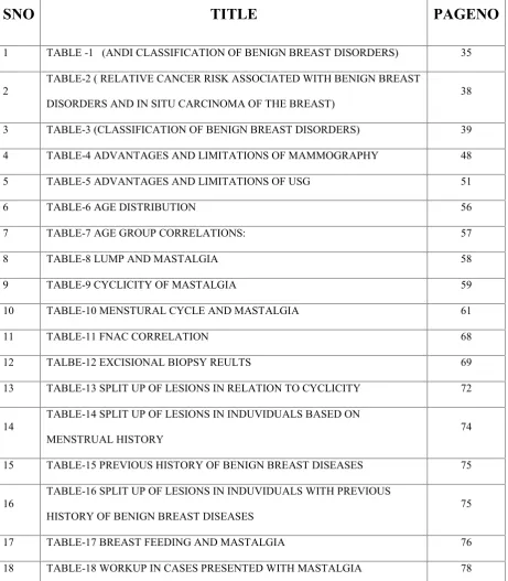
EMBRYOLOGY OF BREAST
Breasts develop from mammary ridges which extend from the axilla to inguinal region by the end of 6th week of gestation. This ridges or the
milk lines wont persist in later days and disappears. The part of the pectoral region is retained giving raise to breasts.
10
11
FUNCTIONAL ANATOMY OF BREAST
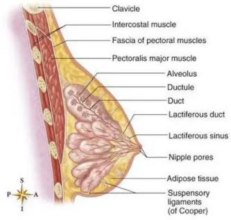
The breasts consist of mammary glands and associated skin and connective tissues. The mammary glands are modified sweat glands in the superficial fascia anterior to the pectoral muscles and the anterior thoracic wall. The base, or attached surface, of each breast extends vertically from ribs II to VI, and transversely from the lateral border of sternum to as far laterally as the mid-axillary line. The upper lateral region of the breast can project around the lateral margin of the pectoralis major muscle and into the axilla. This axillary process (axillary tail) may perforate deep fascia and extend as far superiorly as the apex of the axilla.
12
The tips of the nipples are fissured with the lactiferous ducts opening into them. The nipples are composed mostly of circularly arranged smooth muscle fibres that compress the lactiferous ducts during lactation and erect the nipples in response to stimulation, as when a baby begins to suckle. A well-developed, connective tissue stroma surrounds the ducts and lobules of the mammary gland. In certain regions, this condenses to form well-defined ligaments, the suspensory ligaments of breast, which are continuous with the dermis of the skin and support the breast.
The breast lies on deep fascia related to the pectoralis major, serratus anterior and the aponeurosis of external oblique muscle. The deep pectoral fascia envelops the pectoralis major muscle and is continuous with the deep abdominal fascia below. It attaches to the sternum medially and to the clavicle and axillary fascia above and laterally. Along the lateral border of the pectoralis major muscle, the anterior lamina of the deep pectoral fascia unites with the fascia of the pectoralis minor muscle and, more inferiorly, with the fascia of the serratus anterior. A posterior extension of this fascia is continuous with the fascia of the latissimus dorsi and forms the suspensory ligament of the axilla. A layer of loose connective tissue (the retromammary space) separates the breast from the deep fascia and provides some degree of movement over underlying structures.
13
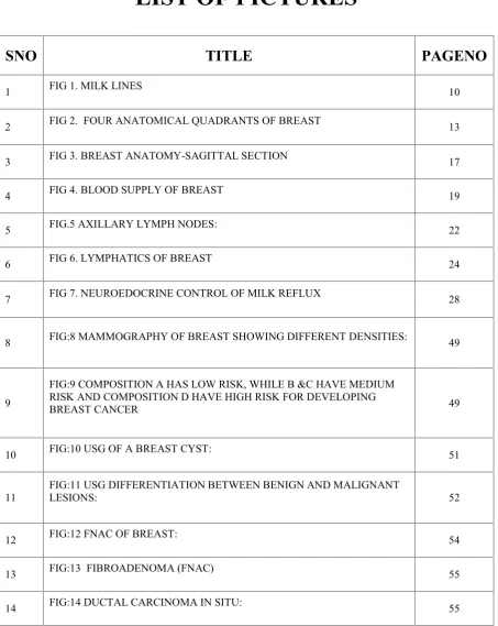
[image:27.595.105.512.252.681.2]For the anatomical location and description of tumors and cysts, the surface of the breast is divided into four quadrants (fig. 2). The upper outer quadrant of the breast contains a greater volume of tissue than do the other quadrants.
14
NIPPLE-AREOLA COMPLEX
The epidermis of the nipple-areola complex is pigmented very much when compared to the surrounding. At puberty, this pigment becomes darker and the nipple assumes an elevated position. Then in pregnancy, the areolar region enlarges and pigmentation is further increased in intensity. The areola also contains sweat glands, sebaceous glands, and Montgomery's tubercles (accessory glands, which produce small elevations on the surface of the areola). Smooth muscle bundle fibres, lie in a circumferential fashion inside the dense connective tissue and longitudinally inturn along the course of major ducts. They were responsible for the nipple erection in response to sensory stimuli. The dermal papilla which is located at the tip of the nipple has been provided with numerous sensory nerve endings and Meissner's corpuscles. This rich sensory innervation of the breast is of physiological importance, because the sucking of the child evokes a neurohormonal response that results in milk ejection.
INACTIVE AND ACTIVE BREAST:
15
The lactiferous ducts then opens through a narrowed orifice (0.2 to 0.35 mm in radius) into the ampulla of the nipple areola complex. Lactiferous sinus is the distal dialatedportion of the lactiferous duct that lies below the nipple areola complex. It has the lining of stratified squamous epithelium. Lactiferous ducts / major ducts are lined by 2 layers of cuboidal cells. The minor ducts are in contrast lined by a single layer of columnar or cuboidal cells.
The Myoepithelial cells arise from ectoderm and they reside between the epithelial cells in the basal lamina. Myoepithelial cells contain myofibrils which inturn are responsible for milk ejection. Inactive breast, the epithelium is sparse and less. In the early phases of the menstrual cycle in women, minor ducts are cord-like structures with small lumina. The estrogen stimulation occuring at the time of ovulation, causes the alveolar epithelium to increase in height and the duct lumen becomes more prominent with accumulation of some secretions. As the hormonal stimulation stops at the end, the alveolar epithelium regresses in height.
16
Development of the alveoli is asymmetric, and there is a considerable degree of variation in the development within a single lobule. Following the delivery of child, the breasts gains additional volume by means of hypertrophy. Then secretory products accumulate in the lumina of the minor ducts.
Alveolar epithelium contains abundant of some cell organelles like mitochondria, Golgi complexes and dense lysosomes.
Function of the alveolar epithelium in the production of milk:
(a) Endoplasmic reticulum produces the protein component of the milk (b) Lipid component of milk, are produced as free lipid droplets in the cytoplasm.
17
18
BLOOD SUPPLY OF BREAST:
The breast receives its principal arterial blood supply from
(a) perforating branches of the internal thoracic (or internal mammary) artery, a branch of the subclavian artery; it courses parallel with the lateral border of the sternum behind the transversus thoracis muscles. From the internal thoracic artery, perforating branches pass through the intercostal muscles of the first six interspaces and the pectoralis major muscle to supply the medial half of the breast and surrounding skin. Typically these arteries descend laterally toward the nipple-areolar complex so that most of the arterial supply arises above the level of the nipple. Therefore, radial incisions in the upper half of the breast are less likely to injure the major arterial supply than transverse incisions. The inferior parts of the breast below the level of the nipple are almost free of major vessels.
(b) lateral branches of the posterior intercostal arteries; and
(c) branches from the axillary artery, including the highest thoracic, lateral thoracic, pectoral branches of the thoracoacromial artery and unnamed mammary branches. The lateral thoracic artery is the most important of these vessels.
19
The veins of the breast and chest wall follow the course of the arteries, with venous drainage being toward the axilla. The three principal groups of veins are (a) perforating branches of the internal thoracic vein - The perforating tributaries from the medial half of the breast carry the greater part of the venous drainage. They enter the internal thoracic vein, which joins the brachiocephalic vein.
(b) perforating branches of the posterior intercostal veins, and (c) tributaries of the axillary vein.
[image:33.595.91.452.460.744.2]Batson's vertebral venous plexus, which invests the vertebrae and extends from the base of the skull to the sacrum, may provide a route for breast cancer metastases to the vertebrae, skull, pelvic bones, and central nervous system.
FIG 4. BLOOD SUPPLY OF BREAST
20
NERVE SUPPLY:
Lateral cutaneous branches of the third through sixth intercostal nerves provide sensory innervation of the breast (lateral mammary branches) and of the anterolateral chest wall. Cutaneous branches that arise from the cervical plexus, specifically the anterior branches of the supraclavicular nerve, supply a limited area of skin over the upper portion of the breast. The intercostobrachial nerve is the lateral cutaneous branch of the second intercostal nerve and may be visualized during surgical dissection of the axilla. Resection of the intercostobrachial nerve causes loss of sensation over the medial aspect of the upper arm.
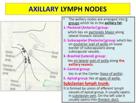
LYMPHATICDRAINAGE OF BREAST:
21
From this plexus most lymph (> 75%), especially from the lateral breast quadrants, drains to the axillary lymph nodes, initially to the anterior or pectoral nodes for the most part. However, some lymph may drain directly to other axillary nodes or even to interpectoral, deltopectoral, supraclavicular, or inferior deep cervical nodes.
Axillary nodes are 20–40 nodes, grouped artificially as pectoral (anterior), subscapular (posterior), central and apical. Surgically, the nodes are described in relation to pectoralis minor. Those lying below pectoralis minor are the low nodes (level 1), those behind the muscle are the middle group (level 2), while the nodes between the upper border of pectoralis minor and the lower border of the clavicle are the upper or apical nodes (level 3).There are six axillary lymph node groups recognized by surgeons
(a) the axillary vein group (lateral);
(b) the external mammary group (anterior or pectoral group), (c) the scapular group (posterior or subscapular),
(d) the central group,
22
FIG.5 AXILLARY LYMPH NODES:
23
Lymphatic vessels occasionally follow lateral cutaneous branches of the posterior intercostal arteries to the intercostal nodes.
24
[image:38.595.75.525.231.645.2]However, in many cases, the trunks open independently into the junction of the internal jugular and subclavian vein, the venous angle, to form the brachiocephalic veins. In some cases, they open into both of these veins.
25
PHYSIOLOGY OF THE BREAST
BREAST DEVELOPMENT AND FUNCTION
Breast development and function are initiated by a variety of hormonal stimuli, which includes the estrogen, progesterone, prolactin, oxytocin, thyroid hormone, cortisol, and growth hormone. Estrogen, progesterone, and prolactin especially have trophic effects that are essential to breast development and function. Estrogen is responsible for ductal development, whereas progesterone is responsible for differentiation of epithelium and lobular development. Prolactin is the hormonal stimulus for lactogenesis in pregnancy and the postpartum period. It up regulates hormone receptors and also stimulates epithelial development. Figure 7 depicts the secretion of neurotrophic hormones from hypothalamus, which inturn is responsible for regulation of the secretion of the hormones that affect the breast tissues.
The luteinizing hormone (LH) and follicle - stimulating hormone (FSH) regulates the release of estrogen and progesterone from the ovaries. The release of LH and FSH from the basophilic cells of the anterior pituitary is regulated by the secretion of gonadotropin-releasing hormone (GnRH) from hypothalamus.
26
In female neonate, the circulating estrogen and progesterone levels decrease after birth and remain low throughout childhood period because of the sensitivity of the hypothalamic-pituitary axis to the negative feedback mechanisms from these hormones. With the beginning of puberty, there is a decrease in the sensitivity of the hypothalamic-pituitary axis to negative feedback mechanisms and an increase in its sensitivity to positive feedback mechanism to estrogen. These physiologic events causes an increase in GnRH, FSH, and LH secretion and resultant increase in estrogen and progesterone secretion by the ovaries, which inturn leads to establishment of the menstrual cycle. At the beginning of the menstrual cycle, the breast increase in the size and density. With the onset of menstruation, the breast engorgement and epithelial proliferation decreases.
PREGNANCY, LACTATION, AND SENESCENCE
A dramatic increase in circulating ovarian and placental estrogens and progestins are evident during pregnancy, which causes alterations in the form and substance of the breast. The breast increases in size and density as the ductal and lobular epithelium proliferates.
The areolar skin darkens, and the accessory areolar glands (Montgomery's glands) become more prominent. In the 1st and 2nd
27
During the 3rd trimester, fat droplets accumulates in the alveolar epithelium and colostrum fills the alveolar and ductal spaces. In late pregnancy, prolactin initiate the synthesis of milk fats and protein.
After delivery of the placenta, circulating progesterone and estrogen levels falls, prolactin expresses its lactogenic action. Milk production and secretion are controlled by neural reflex arcs that begin in nerve endings of the nipple-areola complex. Maintenance of lactation needs regular stimulation of these neural reflexes, which stimulates prolactin secretion and milk letdown.
Oxytocin release is initiated by the auditory, visual, and olfactory stimuli associated with the process of nursing. Oxytocin results in contraction of the myoepithelial cell and expulsion of milk. After weaning of the infant, prolactin and oxytocin levels decreases. Dormant milk results in increased pressure within the ducts and alveoli, which causes atrophy of the epithelium.
28
FIG 7. NEUROEDOCRINE CONTROL OF MILK
29
INFECTIOUS AND INFLAMMATORY DISORDERS:
Except during the postpartum period, infections of the breast are rare. They are classified as:
Intrinsic (secondary to abnormalities in the breast)
Extrinsic (secondary to an infection in the adjacent structure, e.g., skin, thoracic cavity).
BACTERIAL INFECTION
Staphylococcus aureus and Streptococcus are the most frequent
organisms found in nipple discharge from an infected breast. Breast abscesses (typically staphylococcal infections) presents with tenderness, erythema, and hyperthermia. These abscesses have positive correlation to lactation and occur within the few weeks of breastfeeding. The staphylococcal infection progress and results in subcutaneous, subareolar, interlobular and retromammary abscesses, which needs to be drained. Before drainage ultrasonography is effective in delineating the required extent of the drainage area. Drainage is done with a circumareolar incisions or incisions paralleling Langer's lines.
30
They are treated with local wound care, including application of warm compresses, and with administration of IV antibiotics (penicillins or cephalosporins). Breast infections may be chronic, with recurrent abscess formation. Cultures are performed to identify acid-fast bacilli, anaerobic and aerobic bacteria, and fungi. Biopsy of the abscess cavity wall is recommended at the time of drainage to rule out underlying breast cancer with necrotic tumor. Hospital-acquired puerperal infections are less common nowadays, but lactating women who presents with milk stasis or non infectious inflammation may still develop this problem. Epidemic puerperal mastitis is initiated by virulent strains of methicillin-resistant S.
Aureus(MRSA) that are transmitted by the suckling neonate and may result
in considerable morbidity and occasional mortality.
31
Zuska's disease (recurrent periductal mastitis), is the recurrent retroareolar infections, abscesses and duct fistulas located near nipple.This syndrome is managed symptomatically, by IV antibiotics incision and drainage as and when required. Long-term control can be obtained by wide débridement of chronically infected tissue and/or terminal duct resection. But postoperative infections are worrisome. Smoking has been a risk factor for this condition.
MYCOTIC INFECTIONS:
Fungal infections of the breast are uncommon and usually caused by blastomycosis or sporotrichosis. Intraoral fungi are inoculated into the breast tissue by the suckling neonate. These present as mammary abscesses in close proximity to the nipple-areola complex. Pus mixed with blood may be expressed from sinus tracts. Antifungal agents are administered for the treatment of systemic (noncutaneous) infections. This therapy generally eliminates the need for surgical intervention, but some may benefit from drainage of an abscess, or even partial mastectomy, to eradicate the persistent fungal infection. Candida albicans of the breast skin presents as erythematous, scaly lesions of the inframammary or axillary folds. Scrapings from the lesions can be used to demonstrate fungal elements.
32
HIDRADENITIS SUPPURATIVA
Hidradenitis suppurativa of the nipple-areola complex or axilla is a chronic inflammatory condition that originates in the accessory areolar glands of Montgomery or within the axillary sebaceous glands. Chronic acne is a predisposing factor for developing hidradenitis. When located in and around the nipple - areola complex, this disease may be difficult to distinguish from other chronic inflammatory conditions, Paget's disease of the nipple, or invasive breast cancer. Involvement of the axillary skin is often contiguous and multifocal. Treatment involves Antibiotic therapy with incision and drainage of fluctuant areas. Sometimes excision of the involved areas may be required. Large areas of skin loss may require coverage with advancement flaps or split-thickness skin grafts.
MONDOR'S DISEASE:
Mondor's disease (variant of thrombophlebitis) is a rare condition that involves inflammation of the superficial veins of the anterior chest wall and breast.
33
A tender, firm cord like structure is found to follow the distribution of one of the major superficial veins. Rarely, bilateral presentation may be seen, and there may be no evidence of thrombophlebitis in other anatomic sites. This benign, disorder is not indicative of a cancer. When the diagnosis is doubtful, or when a mass like lesion is present near the tender cord, biopsy is indicated.
Treatment for Mondor's disease includes anti-inflammatory medications and application of warm fomentation along the symptomatic vein. Other measures include brassiere support as well as restriction of motion of the ipsilateral extremity and shoulder. The process usually resolve in 4 to 6 weeks. If they are refractory to therapy, excision of the involved vein segment is indicated.
BENIGN DISORDERS AND DISEASES OF THE BREAST:
Benign breast disorders and diseases encompass a wide spectrum of clinical and pathologic entities.
34
ABERRATIONS OF NORMAL DEVELOPMENT AND
INVOLUTION:
The principles guiding the classification of the aberrations of normal development and involution (ANDI) of benign breast conditions are as follows:
(a) Benign breast disorders and diseases are related to the normal processes of reproductive life and to involution.
(b) There is a spectrum of breast conditions that ranges from normal to disorder to disease.
(c) The ANDI classification encompasses all aspects of the breast condition, including pathogenesis and the degree of abnormality.
35
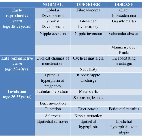
TABLE -1 (ANDI CLASSIFICATION OF BENIGN BREAST
DISORDERS)
NORMAL DISORDER DISEASE
Early reproductive years (age 15-25years) Lobular Development
Fibroadenoma Giant Fibroadenoma Stromal Development Adolescent hypertrophy Gigantomastia
Nipple eversion Nipple inversion Subareolar abscess
Mammary duct fistula
Late reproductive years
(age 25-40yrs)
Cyclical changes of menstruation
Cyclical mastalgia Incapacitating mastalgia Nodularity Epithelial hyperplasia of pregnancy Bloody nipple discharge Involution (age 35-55years)
Lobular involution Macrocysts Sclerosing lesions Duct involution
Dilatation Duct ectasia Periductal mastitis Sclerosis Nipple retraction
Epithelial turnover Epithelial hyperplasia
Epithelial hyperplasia with
[image:49.595.57.542.139.605.2]36
EARLY REPRODUCTIVE YEARS
Fibroadenomas are commonly seen younger women aged between 15 to 25 years. Fibroadenomas usually grow to 1 or 2 cm in diameter and then remain stable but may grow to a larger size. Small fibroadenomas (1 cm in size) are considered normal, whereas larger fibroadenomas (3 cm) are considered a disorder. Giant fibroadenomas (>3 cm) are disease. Similarly, multiple fibroadenomas (with more than 5 lesions in one breast) are very uncommon and are considered disease. The precise etiology of adolescent breast hypertrophy is not known. A spectrum of changes from limited to massive stromal hyperplasia (gigantomastia) is seen. Nipple inversion is a developmental disorder of the major ducts, which prevents normal protrusion of the nipple. Mammary duct fistulas occurs when nipple inversion predisposes to major duct obstruction, inturn leading to recurrent subareolar abscess.
LATER REPRODUCTIVE YEARS
37
Painful nodularity persisting for more than a week of the menstrual cycle is considered a disorder.
INVOLUTION
38
TABLE-2 ( RELATIVE CANCER RISK ASSOCIATED WITH
BENIGN BREAST DISORDERS AND IN SITU CARCINOMA
[image:52.595.88.502.162.651.2]39
TABLE-3 (CLASSIFICATION OF BENIGN BREAST
[image:53.595.89.504.157.683.2]40
PATHOLOGY OF NONPROLIFERATIVE DISORDERS
The histologic differentiation of benign, atypical, and malignant changes is of paramount importance for management of benign
breast diseases. Determining the clinical significance of these changes is a problem that is compounded by inconsistent nomenclature.
The classification system was originally developed by Page26, which separates
the various types of benign breast disorders and diseases into three clinical groups:
1. Nonproliferative disorders
2. Proliferative disorders without atypia 3. Proliferative disorders with atypia .
Nonproliferative disorders of the breast account for about 70% of benign breast conditions and carry no increased risk of breast cancer. This category includes cysts, fibroadenomas, duct ectasia, periductal mastitis, calcifications and related disorders.
41
Haagensen regarded duct ectasia as a primary event which leads to the stagnation of secretions, epithelial ulceration and resultant leakage of duct secretions (containing chemically irritating fatty acids) into periductal tissue.
This sequence produces a local inflammatory process with periductal fibrosis which results in nipple retraction. Another theory considers periductal mastitis as a primary process, which leads to weakening and secondary dilatation of ducts. It is possible that both processes occur together which explains the wide spectrum of problems seen, including nipple discharge, nipple retraction, inflammatory masses, and abscesses.
Calcium deposits are frequently seen in the breast. Most are benign and are due to cellular secretions and debris or may be due to trauma and inflammation. Calcifications that are strongly associated with cancer include microcalcifications, which vary in shape and density (<0.5 mm in size) and fine, linear calcifications, which may show branching. Fibroadenomas tend to have abundant stroma with histologically normal cellular elements. They are hormonal dependent similar to that of normal breast lobule.They lactate during pregnancy and involute in the postmenopausal period.
42
They may be divided into tubular adenomas and lactating adenomas. Tubular adenomas occurs in young non pregnant women,
Whereas lactating adenomas occurs during pregnancy or during the postpartum period. Hamartomas are discrete breast tumors which are usually 2 to 4 cm in diameter, firm in consistency and sharply circumscribed. Adenolipomas are sharply circumscribed nodules of fatty tissue that has elements of normal breast lobules and duct.
FIBROCYSTIC DISEASE
The term fibrocystic disease is nonspecific. Frequently, it is used to describe symptoms, to rationalize the need for breast biopsy, and to explain breast biopsy results. Synonyms include chronic cystic disease, chronic cystic mastitis, fibrocystic changes, cystic mastopathy, Schimmelbusch's disease, mazoplasia, fibroadenomatosis, Reclus' disease, and Cooper's disease26.
43
PATHOLOGY OF PROLIFERATIVE DISORDERS WITHOUT
CELLULAR ATYPIA:
Proliferative breast disorders without atypia include ductal epithelial hyperplasia, sclerosing adenosis, radial scars, complex sclerosing lesions and intraductal papillomas. Sclerosing adenosis is commonly seen during the reproductive and perimenopausal years and has no malignant potential.
Histological changes consist of ductal proliferation (proliferative), epithelial regression & stromal fibrosis (involutional). Sclerosing adenosis is causes distortion of breast lobular architecture and multiple microcysts predispose to this condition. It can sometimes present as a palpable mass. Benign calcifications are seen. Central sclerosis, apocrine metaplasia, epithelial proliferation and papilloma formation leads to the formation of radial scars to complex sclerosing lesions of the breast.
Radial scars are lesions less than 1cm in diameter, whereas larger ones are called complex sclerosing lesions. Radial scars are formed at sites of terminal duct branching (histological changes found to be radiating from a central area of fibrosis).
44
Mild ductal hyperplasia is characterized by the presence of three or four cell layers above the basement membrane. Moderate ductal hyperplasia is characterized by the presence of five or more cell layers above the basement membrane. Florid ductal epithelial hyperplasia occupies at least 70% of a minor duct lumen. It is found in >20% of breast tissue specimens, is either solid or papillary, and is associated with an increased cancer risk (see Table-2).
45
PATHOLOGY OF ATYPICAL PROLIFERATIVE DISEASES
The atypical proliferative diseases have some of the features of carcinoma in situ but lack a major defining feature of carcinoma. In 1978, Haagensen and colleagues described lobular neoplasam as a spectrum of disorders ranging from atypical lobular hyperplasia to lobular carcinoma in situ (LCIS).
CLINICAL ASSESSMENT AND INVESTIGATIONS
A detailed history and clinical examination will point to the diagnosis in most cases.
INVESTIGATIONS
Ultrasound examination is done in all patients presenting with mastalgia.
Mammogram is done in women aged 40 years and above.
FNAC is done in patients with clinically palpable lump or nodularity
and in patients with ultrasound findings of abnormalities.
Patients with FNAC reports of fibroadenoma with lump size >3cm went
46
BREAST IMAGING MODALITIES
MAMMOGRAPHY:
Screening:
Mammography, issuitable for screening especially in women above 40yrs of age.
Diagnostic:
Mammography is indicated as a diagnostic method in determining whether a lesion has a malignant potential or benign. It also screens for occult disease found in the surrounding tissue.
CONVENTIONAL MAMMOGRAPHY
Mammography is practically a high-resolution x-ray imaging of compressed breast. This process involves radiation transmission through the breast tissue and the eventual projection of anatomical structures on a film screen/image sensor.
Two views of each breast,
Craniocaudal (CC) view
Mediolateral oblique (MLO - 450) view
are obtained by mammography.
47
Misconceptions regarding the risk of radiation from mammography persist, despite the fact that no women has shown to develop breast cancer as a result of mammography. Studies show even after multiple examinations at doses much higher than the current dose of 3 to 4 mGy (0.3– 0.40 rad) has no positive correlation with development of breast cancer. The adverse effects of screening include pain and discomfort (breast compression), patient recall to do additional imaging and false-positive biopsies. The risk benefit ratio from screening, are in favour of detecting early lesions21.
The sensitivity of mammography depends on the patient factor like composition and density of the tissue. It is difficult to look for small lesions in denser tissue, even though some small foci of calcification can be seen. Initially, mammogram is used to detect any mass or calcifications followed by architectural distortion.
A mass by definition is a space occupying lesion imaged in two different views in mammography. It is further categorized by its shape, margins, density, size, orientation and calcifications.
48
Benign micro calcifications are usually smooth and sharply defined and have high uniform density. Malignant micro calcifications appear in irregular shape and are variably distributed through breast tissue23, 24.
[image:62.612.130.530.286.700.2]In the official BI-RADS publication, the calcifications have been described by its appearance and distribution in the breast tissue23, 24, 25.
TABLE-4 ADVANTAGES AND LIMITATIONS OF
49
FIG:8 MAMMOGRAPHY OF BREAST SHOWING DIFFERENT
DENSITIES:
FIG:9 COMPOSITION A HAS LOW RISK, WHILE B &C HAVE MEDIUM
RISK AND COMPOSITION D HAVE HIGH RISK FOR DEVELOPING
BREAST CANCER
[image:63.612.177.391.411.686.2]50
ULTRASONOGRAPHY OF BREAST:
USG is mainly used as a cheap and efficient tool to differentiate cystic lesion from solid breast masses. USG can be safely performed in young or pregnant patients as it do not produce any form of ionizing radiation. USG is useful in detecting breast masses which are hidden on mammogram and to evaluate breast lesions in younger women especially less than 30yrs of age. Abnormalities demonstrated on the mammogram can further be followed up using USG. USG is also a useful tool in the guidance of therapeutic procedures and biopsies.
Women younger than 30years have dense glandular breast tissue which reduces the diagnostic sensitivity of mammogram, but USG is especially useful in these cases. Evaluation of breast abscesses is better done with USG. Fine needle aspiration cytology (FNAC) of the lesion can be done under ultrasound guidance.
51
TABLE-5 ADVANTAGES AND LIMITATIONS OF USG:
[image:65.595.138.460.544.772.2]52
FIG:11 USG DIFFERENTIATION BETWEEN
BENIGN AND MALIGNANT LESIONS:
FNAC:
53
PROCEDURE:
54
55
FIG:13 FIBROADENOMA (FNAC)
[image:69.595.75.558.441.634.2]
56
RESULTS
[image:70.595.72.466.162.438.2]AGE AND MASTALGIA:
TABLE-6 AGE DISTRIBUTION
AGE NUMBER OF
PATIENTS
PERCENTAGE
15-20 44 22%
21-25 78 39%
26-30 42 21%
31-35 22 11%
36-40 10 5%
41-45 4 2%
CHART-1 SPLIT UP BASED ON CLINICAL EXAMINATIONS:
0 10 20 30 40 50 60 70 80 90
15-20 21-25 26-30 31-35 36-40 41-45
57
TABLE-7 AGE GROUP CORRELATIONS:
AGE GROUP
CLINICAL EXAMINATION USG AND FNAC
NAD N O D U L A R ITY L U MP A B C E SS
NAD FC FA
D A B C E SS
15-20(44pts) 26 8 10 - 24 10 10 -
21-25(78pts) 46 18 10 4 44 20 10 4
26-30(42pts) 30 10 2 - 30 10 2 -
31-35(22pts) 7 10 5 - 7 10 5 -
36-40(10pts) 3 2 5 - 3 2 5 -
41-45(4pts) 4 0 0 - 4 0 0 -
TOTAL(200pts) 116 48 32 4 112 52 32 4
CHART-2 SPLIT UP BASED ON USG AND FNAC RESULTS:
0 10 20 30 40 50 60 70 80 90
15-20 21-25 26-30 31-35 36-40 41-45
58
[image:72.595.71.498.135.327.2]LUMP AND MASTALGIA:
TABLE-8 LUMP AND MASTALGIA
LUMP NUMBER OF
PATIENTS
(n=200)
PERCENTAGE
Present 84 42%
Absent 116 58%
CHART-3 LUMP AND MASTALGIA
Present
42%
Absent
58%
59
[image:73.595.108.489.236.608.2]CYCLICITY OF MASTALGIA:
TABLE-9 CYCLICITY OF MASTALGIA:
TYPE OF
MASTALGIA
NUMBER OF
PATIENTS
(n=200)
PERCENTAGE
Cyclical 88 44%
Non Cyclical 112 56%
CHART-4 CYCLICITY OF MASTALGIA:
Cyclical
44%
Non
Cyclical
56%
60
CHART-5 CORRELATION BETWEEN CYCLICITY AND CLINICAL
EXAMINATION FINDINGS:
CHART-6 CORRELATION BETWEEN CYCLICITY AND USG&FNAC
FINDINGS: 0 20 40 60 80 100 120
CYCLICAL(88pts) NON CYCLICAL(112pts)
CYCLICITY OF MASTALGIA CLINICAL EXAMINATION FINDINGS
ABCESS LUMP NODULARITY NAD 0 20 40 60 80 100 120
CYCLICAL(88pts) NON CYCLICAL(112pts)
CYCLICITY OF MASTALGIA USG FINDINGS
ABCESS
FAD
FC
61
[image:75.595.115.521.406.623.2]MENSTURAL CYCLE AND MASTALGIA:
TABLE-10 MENSTURAL CYCLE AND MASTALGIA
MENSTURAL
CYCLES
NUMBER OF
PATIENTS
(n=200)
PERCENTAGE
REGULAR 142 71%
IRREGULAR 58 29%
CHART-7 MENSTURAL CYCLE AND MASTALGIA:
142
58
MENSURATION AND
MASTALGIA
62
CHART-8 CORRELATION BETWEEN MENSTURAL CYCLE AND
CLINICAL EXAMINATION FINDINGS:
CHART-9 CORRELATION BETWEEN MENSTURAL CYCLE AND
USG&FNAC FINDINGS: 0 20 40 60 80 100 120 140 160
REGULAR (142pts) IRREGULAR (58pts)
MENSTURAL CYCLE & MASTALGIA CLINICAL EXAMINATION FINDINGS
ABCESS LUMP NODULARITY NAD 0 20 40 60 80 100 120 140 160 REGULAR(142PTS) IRREGULAR(58PTS)
MENSTURAL CYCLE & MASTALGIA USG FINDINGS
ABCESS
FAD
FC
63
PREVIOUS BENIGN BREAST DISEASES:
CHART-10 PREVIOUS BENIGN BREAST DISEASES:
CHART-11 CLINICAL EXAMINATION FINDINGS IN INDUVIDUALS
WITH PREVIOUS HISTORY OF BENIGN BREAST DISEASES:
37%
63%
Previous Benign Breast Diseases
Present
Absent
0 20 40 60 80 100 120 140
PRESENT(74pts) ABSENT(126pts)
PREVIOUS HISTORY OF BENIGN BREAST DISEASES CLINICAL EXAMINATION FINDINGS
ABCESS
LUMP
NODULARITY
64
CHART-12 USG AND FNAC FINDINGS IN INDUVIDUALS WITH
PREVIOUS HISTORY OF BENIGN BREAST DISEASES:
Among the 74pts presented with previous history of benign breast diseases
46pts found to have pathological and radiological findings (37pts with fibrocystic disease, 5pts with fibroadenoma and 4pts with abscess).
Among the 126pts presented with previous history of benign breast
diseases 42pts found to have pathological and radiological findings (15pts with fibrocystic disease and 27pts with fibroadenoma).
0 20 40 60 80 100 120 140
CYCLICAL(88pts) NON CYCLICAL(112pts)
PREVIOUS HISTORY OF BENIGN BREAST DISEASES USG FINDINGS
ABCESS
FAD
FC
65
BREAST FEEDING AND MASTALGIA:
Breast feeding history is not applicable in 50% of unmarried
patients(100pts).
Out of the remaining 100 patients, 94(47%) gave positive breastfeeding
history
Only 6 (3%)patients didn’t breastfeed.
CHART-13 BREASTFEEDING AND MASTALGIA:
47%
3%
50%
Breast Feeding
Given
Not given
66
CLINICAL EXAMINATION IN PATIENTS WHO PRESENTED WITH
COMPLAINTS OF LUMP BREAST:
97pts (48%) presented with complaints of breast lump/lumpiness.
84pts (42%) were found to have lump/nodularity/abcess on clinical
examination.
48pts (24%) had nodularity on clinical examination.
32pts (16%) had lump breast on clinical examination.
4pts (2%) had abscess on clinical examination.
CHART-14 CLINICAL EXAMINATION FINDINGS:
58%
16%
24%
2%
CLINICAL EXAMINATION
NAD LUMP
67
USG CORRELATION:
97pts (48%) presented with complaints of breast lump/lumpiness.
84pts (42%) were found to have lump/nodularity/abscess on clinical
examination.
88pts (44%) were found to have lump/nodularity/abscess in USG.
52pts (26%) had fibrocystic disease on USG.
32pts (16%) had fibroadenoma on USG.
4pts (2%) had abscess on USG.
CHART-15 USG CORRELATION:
56%
26%
16%
2%
USG FINDINGS
NAD
FC
FAD
68
FNAC CORRELATION:
88pts (44%) were found to have lump/nodularity/abscess in USG.
Among them 84pts (42%) went in for FNAC (except the 4pts with abscess).
52pts (26%) had fibrocystic disease on FNAC.
[image:82.595.117.481.503.696.2] 32pts (16%) had fibroadenoma on FNAC.
TABLE-11 FNAC CORRELATION:
FNAC
FINDINGS
NUMBER OF
PATIENTS
PERCENTAGE
NOT APPLICABLE 116 58%
FC 52 26%
FAD 32 16%
CHART-16 FNAC CORRELATION
116 52
32
NA
FC
FAD
0 50 100 150
FNAC FINDINGS
69
EXCISION BIPOPSYCORRELATION:
32pts who had fibroadenoma in USG and FNAC went in for excision
biopsy.
28pts had fibroadenoma.
2pts had fibrocystic changes.
2pts had Ductal Carcinoma In Situ (DCIS).
TALBE-12 EXCISIONAL BIOPSY REULTS
EXISIONAL
BIOPSY RESULTS
NUMBER OF
PATIENTS
PERCENTAGE
NOT APPLICABLE 168 84%
FC 2 1%
FAD 28 14%
DCIS 2 1%
CHART-17 EXISIONAL BIOPSY RESULTS:
168
2
28
2
NA
FC
FAD
DCIS
0 50 100 150 200
BIOPSY FINDINGS
70
DISCUSSION
AGE AND MASTALGIA:
Mastalgia is more prevalent among women of reproductive age.
The range of ages involved that are included in this study is between 15and
45 years.(TABLE-6) .
The average age of patients presenting in our series is 25.7 years.The
median age is 24 years.
In Cardiff breast clinic study also corresponds to similar observations that
mastalgia is more a disease of women of reproductive age group.
The median age of their age group was 36 years and their study population
included 212 women (age ranging between 12 and 51 years)11.
The results shows 122 patients were in the age group of 25 or less.
These results highlight the fact that 4 cases (2%) was diagnosed by USG
71
CYCLICITY AND MASTALGIA:
Breast pain can be classified as cyclical and non-cyclical in relation to their occurrence with menstrual cycle.
Cyclical mastalgia occurs 1-2 weeks prior to mensus and often bilateral, diffusely involves the whole of the breast with occasional radiation to the upper arm and axilla.
Cyclical mastalgia is usually seen among women 30-40 years of age. It is found to be relieved spontaneously with onset of mensus in nearly 22%
of patients and being persistent in 65% of patients despite treatment. Among the 88pts presented with cyclical pain 31pts found to have
pathological and radiological findings (27pts with fibrocystic disease and 4pts with abscess).
72
TABLE-13 SPLIT UP OF LESIONS IN RELATION TO CYCLICITY:
CYCLICITY OF MASTALGIA
CLINICAL EXAMINATION
USG AND FNAC
NAD NODU L AR IT Y L UM P ABCES S
NAD FC FA
D
ABCES
S
CYCLICAL (88pts)
59 25 0 4 57 27 0 4
NON CYCLICAL (112pts)
57 23 32 0 55 25 32 0
TOTAL (200pts)
116 48 32 4 112 52 32 4
In a study conducted by srivastav et al non-cyclical mastalgia is slightly more prevalent than cyclical mastalgia in women of north india.
73
MENSTURAL CYCLE AND MASTALGIA:
It is popularly believed that when breast pain due to altered hormonal status occurs in women due to engorgement and ductal dilatation it is logical to think that uterus function in relation to this hormonal disarray should also be altered which can manifests as irregular menstrual cycles.
But this is not supported by the results observed as in our study. Most of the patients have regular menstrual cycles (71%).
Among the 142pts presented with regular cycles 79pts found to have pathological and radiological findings (45pts with fibrocystic disease, 30pts with fibroadenoma and 4pts with abscess).
74
TABLE-14 SPLIT UP OF LESIONS IN INDUVIDUALS BASED ON
MENSTRUAL HISTORY:
MENSTURAL CYCLE CLINICAL EXAMINATION USG AND FNAC
NAD N O D U L A R ITY L U MP A B C E SS
NAD FC FA
D A B C E SS
REGULAR (142pts) 66 42 30 4 63 45 30 4
IRREGULAR (58pts) 50 6 2 0 49 7 2 0
TOTAL
(200pts)
116 48 32 4 112 52 32 4
Out of the 200 patients who underwent the study 142 patients (71%) had
regular cycles.
58patients (29%) had irregular cycles on eliciting history.
Srivastav et al conducted a similar study in north Indian women and
75
[image:89.595.74.524.497.781.2]PREVIOUS HISTORY AND MASTALGIA:
TABLE-15 PREVIOUS HISTORY OF BENIGN BREAST DISEASES:
HISTORY NUMBER OF
PATIENTS
PERCENTAGE
Present 74 37%
Absent 126 63%
Previous history of benign breast symptoms is found to have a frequent
association with the patients currently presenting with mastalgia.
History of previous benign diseases for which the patient had received
medical or surgical treatment is elicited in 37% of individuals in our study.
TABLE-16 SPLIT UP OF LESIONS IN INDUVIDUALS WITH
PREVIOUS HISTORY OF BENIGN BREAST DISEASES:
PREVIOUS HISTORY
CLINICAL EXAMINATION USG AND FNAC
NAD NODU L AR IT Y L UM P ABCES S
NAD FC FA
D
ABCES
S
PRESENT
(74pts) 31 34 5 4 28 37 5 4
ABSENT
(126pts) 85 14 27 0 84 15 27 0
76
Out of the 200 patients who underwent this study 74 (37%) patients had
positive history of previous benign breast diseases.
126 (63%) patients had no such history.
This is a significant positive correlation.
BREASTFEEDING AND MASTALGIA:
In an epidemiological study by srivastav et al from AIIMS observed that
there is no significant relation between breastfeeding and mastalgia.
In this study also there was no significant correlation found between
breastfeeding and mastalgia.
In our part of the world breastfeeding practices are strong and most of the mothers who participated in the study gave history for breastfeeding.
TABLE-17 BREAST FEEDING AND MASTALGIA:
BREAST FEEDING NO OF WOMEN PERCENTAGE
Given 94 47%
Not given 6 3%
Not Applicable 100 50%
Only 3% of women have not given breast milk.
And this seemingly increased incidence of breast pain among breastfed
77
MASTALGIA AND LUMP:
Mastalgia as a clinical condition is far more prevalent then lump breast.
But lump breast draws the attention of the patient to the previously
prevalent breast pain and make them to approach a doctor for medical advice.
In a clinical study published by Kelley et al breast lump is found to be
associated with breast pain presented in 29% of patients(n= 350 patient).
97pts (48%) presented with complaints of breast lump/lumpiness.
84pts (42%) were found to have lump/nodularity/abscess on clinical
examination.
88pts (44%) were found to have lump/nodularity/abscess in USG.
Among them 84pts (42%) went in for FNAC (except the 4pts with
abscess).
32pts who had fibroadenoma in USG and FNAC went in for excision
78
TABLE-18 WORKUP IN CASES PRESENTED WITH MASTALGIA
Patient complained of lumpiness Patient found to have Lump/ Nodularity/ Abscess on clinical examination
Patient found to have Fibroadenoma/ Fibrocystic disease/ Abscess on USG Patient with Fibroadenoma/ Fibrocystic disease in USG
underwent FNAC Patients with FNAC reports of Fibroadenoma (lump >3cm) went for Excision Biopsy Present in 97pts (48%) Present in 84pts (42%)
Present in 88pts (44%)
84pts went in for FNAC
32pts went in for Excision
Biopsy
Nodularity in 48pts
FC in 52pts FC in 52pts 28pts had FAD
Lump in 32pts
FAD in 32pts FAD in 32pts 2pts had FC
Abscess in 4 pts
Abscess in 4 pts - 2pts had DCIS
Absent in 103pts (52%) Absent in 116pts (58%) Absent in 112pts (56%) - -
4pts (2%) with nodularity were missed in clinical examination especially
in age group below 25yrs. These were picked up by USG.
There seems to be a near 100% correlation between USG and FNAC
establishing the USG superiority in breast lesions.
The 32pts with USG and FNAC results as fibroadenoma and lesion more
than3cm in size underwent excision biopsy and in those:
2pts (1%) were found to have fibrocystic changes.
79
CONCLUSION:
1. Non-cyclical mastalgia is more prevalent than cyclical mastalgia among women in south India which needs more evaluation.
2. Lumpiness of one or both breasts is a frequently associated complaint with mastalgia which needed detailed history, clinical examination and investigations to differentiate benign from malignant lesions.
3. Presence of previous history of benign breast diseases or treatment is a risk factor for mastalgia which may be due to improper diagnosis and management.
4. USG is the best diagnostic tool in young females as they have dense breasts. 5. Triple assessment for any breast lump or mastalgia forms gold standard in
BIBLIOGRAPHY
1) Barton MB, Elmore JG, Fletcher SW (1999) breast symptom among women enrolled
in a health maintenance organization: frequency, evaluation and outcome. Ann Intern
Med 130:651–657
2) Ader DN, South-Paul J, Adera T, Deuster PA (2001) Cyclical mastalgia: prevalence
and associated health behavioral factors. J Psychosom Obstet Gynaecol 22:71–76
3) Mansel RE, Webster DJT, Sweetland HM (2009) Breast pain and nodularity. In: Mansel
RE, Webster DJT, Sweetland HM (eds) Benign disorders and disease of the breast, 3rd
edn. Saunders Elsevier, Philadelphia, pp 107–138
4) Kumar S, Rai R, Das V, Dwivedi V, Kumar S, GG A (2010) Visual analogue scale for
assessing breast nodularity in non discrete lumpy breasts: the Lucknow Cardiff breast
nodularity scale. The Breast 19:238–242
5) Wisbey JR, Kumar S, Mansel RE, Preece PE, Pye JK, Hughes LE (1983) Natural
history of breast pain. Lancet ii:672–674
6) Smith RL, Pruthi S, Fitzpatrick LA (2004) Evaluation and man-agement of breast pain.
Mayo Clin Proc 79(3):353–372
7) Malarkey WB, Schrooeder LL, Stevens VC, James AG, Lanese RR (1977) Twenty four
hour preoperative endocrine profiles in women with benign and malignant breast
disease. Cancer Res 37:4655–4659
8) Sitruk-Ware R, Sterkers N, Mauvais-Jarvis P (1979) Benign breast disease.1: hormonal
investigation. Obstet Gynecol 53:457–460
9) Peters F, Pickcardt CR, Zimmerman G (1981) PRL, TSH and thyroid hormones in
10) Kumar S, Mansel RE, Scanlon MF, Hughes LE (1984) Altered res