THE ROLE OF LOW LYING PUBIC TUBERCLE IN THE
DEVELOPMENT OF INGUINAL HERNIA – A CASE
CONTROL STUDY
DISSERTATON SUBMITTED FOR M.S DEGREE EXAMINATON
BRANCH I
(GENERAL SURGERY)
K.A.P.V GOVERNMENT MEDICAL COLLEGE AND MAHATMA
GANDHI MEMORIAL HOSPITAL, TIRUCHIRAPALLI
THE TAMILNADU DR.M.G.R. MEDICAL UNIVERSITY
CHENNAI
DECLARATON BY CANDIDATE
I hereby declare that this dissertation entitled “THE ROLE OF
LOW LYING PUBIC TUBERCLE IN THE DEVELOPMENT OF
INGUINAL HERNIA –A CASE CONTROL STUDY” is the bonafide
and genuine research work carried out by me under the guidance of
Dr. P. RAJAGOPAL M.S., Associate Professor, Department of General
Surgery, KAPV Government Medical College and Mahatma Gandhi Memorial Hospital, Tiruchirapalli.
KAPV GOVERNMENT MEDICAL COLLEGE AND
MAHATMA GANDHI HOSPITAL
CERTIFICATE BY GUIDE
This to certify that this dissertation entitled “THE ROLE OF LOW LYING PUBIC TUBERCLE IN THE DEVELOPMENT OF
INGUINAL HERNIA –A CASE CONTROL STUDY” is a bonafide
work of research done by Dr.S.S.ABHINAND in partial fulfillment of requirement for the degree of MS in GENERAL SURGERY.
Date Signature of the Guide
ENDORSEMENT BY HEAD OF THE DEPARTMENT
AND HEAD OF THE INSTITUTION
This is to certify that this dissertation entitled “THE ROLE OF
LOW LYING PUBIC TUBERCLE IN THE DEVELOPMENT OF
INGUINAL HERNIA –A CASE CONTROL STUDY” is the bonafide
research work done by Dr. S.S.ABHINAND under the guidance of
Dr. P. RAJAGOPAL M.S., General Surgery, Associate Professor,
Department of General Surgery, KAPV Government Medical College and Mahatma Gandhi Memorial Hospital, Tiruchirapalli.
Prof. Dr.S.LILY MARY M.D., Dr. A.THULASI M.S., D.G.O.,
Dean, Professor and Head of Department,
K.A.P.V. Govt. Medical College, Department of Surgery
Trichy. K.A.P.V. Govt. Medical College,
Trichy
Place: Trichy
COPYRIGHT DECLARATION
I hereby declare that the KAPV Medical College and Mahatma Gandhi Memorial Government Hospital, Tiruchirapalli, shall have the rights to preserve, use and disseminate this dissertation in print or electronic format for academic/research purpose.
Date: Dr.S.S.ABHINAND M.B.B.S.,
ACKNOWLEDGEMENT
Success and final outcome of this study required a lot of guidance and assistance from many people and I am grateful to have got all this assistance all along till the completion of my study.
I express my gratitude to Dr. S. LILLY MARY M.D., Dean, KAPV Government Medical College and Mahatma Gandhi Memorial Hospital, Tiruchirapalli, for her invaluable support in conducting this study.
I acknowledge my sincere thanks to Dr.A.THULASI M.S., D.G.O., Professor and Head of Department of General Surgery for his valuable advice and encouraging the study.
I wish to express my heartfelt respect and gratitude to my guide and unit chief, Dr.P.RAJAGOPAL, M.S., Associate Professor, Department of General Surgery for his constant advise, invaluable suggestions and words of encouragement
I am extremely thankful to my unit Assistant Professors
Dr. S..SENTHILVEL M.S., Dr.V.SUJATHA M.S., and
Dr. V. VIMAL M.S., for their valuable guidance throughout my
I also wish to thank Dr. R.MOHAN, M,S., M.Ch., Chairman of the Ethical Committee of KAPV Medical College and Mahatma Gandhi Memorial Hospital, for granting me permission to conduct this study.
TABLE OF CONTENTS
S.NO PARTICULARS PAGE NO.
1 INTRODUCTION 1
2 AIM AND OBJECTIVE 4
3 REVIEW OF LITERATURE 6
4 STUDY DESIGN AND METHODOLOGY 61
5 OBSERVATIONS AND RESULTS 65
6 DISCUSSION 85
7 SUMMARY 89
8 CONCLUSION 93
BIBLIOGRAPHY 94
ANNEXURES
i. Proforma
ii. Consent Form
iii. Ethics Committee Clearance
iv. Plagiarism Receipt
v. Master Chart
LIST OF ABBREVIATIONS
EHS- European Hernia Society. USG- Ultrasonogram.
CT- Computerised Tomogram.
GPRVS- Giant Prosthetic Reinforcement of Visceral sac. TAPP- Transabdominal Preperitonial.
TEP- Totally Extra-peritoneal. BMI- Body Mass Index.
SS Line- Distance between two anterior superior iliac spine ST Line- Distance between pubic tubercle to SS line
1
2
DEFINITION
Hernia is the abnormal protrusion of a part or whole of the viscus through a normal or abnormal opening in the cavity that contains it.1The inguinal hernia based on anatomical characteristic divided into two types. The most common type is indirect inguinal henia, in which hernia sac emerge lateral to inferior epigastric artery.2 It occur due to the persistence of processus vaginalis. Direct inguinal hernia occur medial to the inferior epigastric vessels when abdominal contents protrudes along a weak spot in the fascia transversalis which forms the posterior wall of the inguinal canal. Inguinal canal is 3.75cm in length 3extends from deep to superficial inguinal ring. There are various defensive mechanisms of the inguinal canal to prevent the formation of hernia which are based on anatomical factors.
3
individual to suffer from an inguinal hernia is the location of the pubic tubercle.6
Even though inguinal hernia is the most common type of hernia, the
4
AIMS AND
5
AIMS AND OBJECTIVES
1. The aim is to find out the relationship of pubospinal distance between cases
and control
2. 2.To study the clinical profile of inguinal hernia
3. To study the prevalence of hernia in various age group
6
REVIEW OF LITERATURE
7
REVIEW OF LITERATURE
Hernia is one of the commonest surgical problem for which a general surgeon is called for. Hernia usually occur following disruption of the fibro muscular wall. The most common site is inguinal region in both sexes. The content can be anything which passes through the defect. The word ―hernia‖ is derived from a Latin term meaning ―a rupture7.‖ The earliest reports of abdominal wall hernias date back to 1500 BC. During this early period, abdominal wall hernia were treated with trusses or bandage dressings. The first report of operative repair of a groin hernia dates back to the first century AD.
The groin hernia classification based on the anatomy of the defect (i.e., inguinal versus femoral) dates back to the 14th century. The hernia is classified into direct and indirect based on anatomy first reported way back on 1559.
Incidence
8
The femoral hernia even though quite rare as compared to inguinal hernia, it is seen more common in women. This is true in umbilical hernia also. The male to female ratio is 25:1 for femoral hernia and 2:1 for umbilical hernia. Indirect inguinal hernia is most common type of hernia irrespective of age and sex. One among two patients suffering from femoral hernia will develop inguinal hernia in long term follow up. Both indirect inguinal and femoral hernias occur more commonly on the right side11. This is attributed to a delay in atrophy of the processus vaginalis after the normal slower descent of the right testis to the scrotum during foetal development. The predominance of right-sided femoral hernias is thought to be due to the compressing effect of the sigmoid colon on the left femoral canal.
9
ANATOMY OF GROIN REGION AND ABDOMINAL WALL
Anatomy of anterior abdominal wall and groin is essential for surgical repair and understanding the pathology of hernia.
Structure of the Anterior Abdominal Wall
The anterior abdominal wall is made up of skin, superficial fascia, deep fascia, muscles, extraperitoneal fascia, and parietal peritoneum.
Skin
The skin is loosely attached to the underlying structures except at the umbilicus, where it is adherent to the scar tissue.
Nerve Supply
The cutaneous nerve supply to the anterior abdominal wall is derived from the anterior rami of the lower six thoracic and the 1st lumbar nerves. The thoracic nerves are the lower five intercostal and the subcostal nerves. The 1st lumbar nerve is represented by the iliohypogastric and the ilioinguinal nerves.
Blood Supply
Arteries
10
epigastric, the superficial circumflex iliac, and the superficial external pudendal arteries, branch of femoral artery.
Veins
The venous drainage above umbilicus mainly drains into the axillary vein via the lateral thoracic vein and below umbilicus into the femoral vein via the superficial epigastric and the great saphenous veins.
Superficial Fascia
The superficial fascia is divided into a superficial fatty layer (fascia of Camper) and a deep membranous layer(Scarpa‘s fascia)
Deep Fascia
The deep fascia in the anterior abdominal wall is merely a thin layer of connective tissue covering the muscles; it lies immediately deep to the membranous layer of superficial fascia.
Muscles of the Anterior Abdominal Wall
11
The lower part of the rectus sheath may contain a small muscle called the pyramidalis.
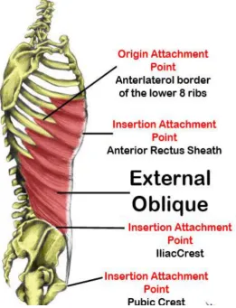
External Oblique13
[image:23.595.169.431.366.707.2]The external oblique muscle is a broad, thin, muscular sheet that arises from the outer surfaces of the lower eight ribs and fans out to be inserted into the xiphoid process, the linea alba, the pubic crest, the pubic tubercle, and the anterior half of the iliac crest . Most of the fibers are inserted by means of aponeurosis.
12
A triangular-shaped defect lies in the external oblique aponeurosis lies above and medial to the pubic tubercle. This is known as the
superficial inguinal ring14. The spermatic cord (or round ligament of the uterus) passes through this opening and carries the external spermatic fascia (or the external covering of the round ligament of the uterus) from the margins of the ring.
Between the anterior superior iliac spine and the pubic tubercle, the lower border of the aponeurosis is folded backward on itself, forming the
inguinal ligament. From the medial end of the ligament, the lacunar ligament extends backward and upward to the pectineal line on the superior ramus of the pubis. Its sharp, free crescentic edge forms the medial margin of the femoral ring. On reaching the pectineal line, the lacunar ligament becomes continuous with a thickening of the periosteum called the pectineal ligament15.
13
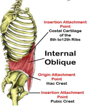
Internal Oblique16
[image:25.595.150.464.331.708.2]The internal oblique muscle is also a broad, thin, muscular sheet that lies deep to the external oblique; most of its fibers run at right angles to those of the external oblique. It arises from the lumbar fascia, the anterior two thirds of the iliac crest, and the lateral two thirds of the inguinal ligament. The muscle runs upward and forward. The muscle is inserted into the lower borders of the lower three ribs and their costal cartilages, the xiphoid process, the linea alba, and the symphysis pubis.
14
The internal oblique has a lower free border that arches over the spermatic cord (or round ligament of the uterus) and then descends behind it to be attached to the pubic crest and the pectineal line. Near their insertion, the lowest tendinous fibers are joined by similar fibers from the transversus abdominis to form the conjoint tendon. The conjoint tendon is attached medially to the linea alba, but it has a lateral free border.
As the spermatic cord (or round ligament of the uterus) passes under the lower border of the internal oblique, it carries with it some of the muscle fibers that are called the cremaster muscle17. The
cremasteric fascia is the term used to describe the cremaster muscle and
its fascia.
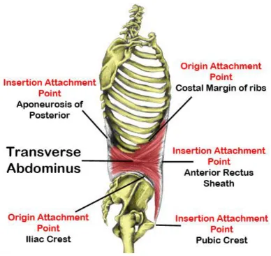
Transversus abdominis
15
Fig 3. Anatomy of Transverse Abdomius
It is inserted into the xiphoid process, the linea alba, and the symphysis pubis. The lowest tendinous fibers join similar fibers from the internal oblique to form the conjoint tendon, which forms roof of inguinal canal .
Rectus Abdominis18
16
[image:28.595.117.467.244.572.2]symphysis pubis and from the pubic crest. It is inserted into the 5th, 6th, and 7thcostal cartilages and the xiphoid process . When it contracts, its lateral margin forms a curved ridge that can be palpated and often seen and is termed the linea semilunaris. This extends from the tip of the ninth costal cartilage to the pubic tubercle.
Fig 4. Anatomy of Rectus Abdomius
The rectus abdominis muscle is divided into distinct segments by three transverse tendinous intersections: one at the level of the xiphoid process, one at the level of the umbilicus, and one halfway between these two . These intersections are strongly attached to the anterior wall of the
17
of the external oblique, internal oblique, and transversus, which form the rectus sheath.
Pyramidalis
The pyramidalis muscle is often absent. It arises by its base from the anterior surface of the pubis and is inserted into the linea alba . It lies in front of the lower part of the rectus abdominis.
Rectus Sheath 19
The rectus sheath is a long fibrous sheath that encloses the rectus abdominis muscle and pyramidalis muscle and contains the anterior rami of the lower six thoracic nerves, the superior and inferior epigastric vessels and lymph nodes. It is formed mainly by the aponeuroses of the three lateral abdominal muscles.
For ease of understanding, the rectus sheath is considered at three levels. Above the costal margin, the anterior wall is formed by the aponeurosis of the external oblique. The posterior wall is formed by the thoracic wall—that is, the 5th, 6th and 7th costal cartilages and the intercostal spaces.
18
front of the muscle, and the transversus aponeurosis is directed behind the muscle.
Between the level of the anteriosuperior iliac spine and the pubis, the aponeuroses of all three muscles forms the anterior wall. The posterior wall is absent, and the rectus muscle lies in contact with the fascia transversalis.
Inguinal Canal20
The inguinal canal is an oblique passage through the lower part of the anterior abdominal wall. In the males, it allows passage of structure from the testis to the abdomen. In females, it gives the pathway for round ligament of the uterus to pass from the uterus to the labium majora. That is about 4cm long in the adult and extends from the deep inguinal ring, a opening in the fascia transversalis downward and medially to the superficial inguinal ring, a hole in the aponeurosis of the external oblique muscle. It lies parallel to and above the inguinal ligament. In the newborn child, the deep ring directly posterior to the superficial ring so that the canal is shorter in children21.
19
margins of the ring give attachment to the internal spermatic fascia (or the internal covering of the round ligament of the uterus)22.
The superficial inguinal ring is a triangular-shaped opening in the aponeurosis of the external oblique muscle and situated above and medial to the pubic tubercle. The margins of the ring, sometimes called the crura, give attachment to the external spermatic fascia23.
[image:31.595.96.535.297.605.2]Walls of the Inguinal Canal
Fig 4. Anatomy of Inguinal Canal
20
Posterior wall. It is also a strong boundary. It completely covers the superficial inguinal ring. It is formed by fascia transverslis with contribution from conjoint muscle medially.
Roof or superior wall. From the insertion of internal oblique some muscle fibre contribute along with the transversalis abdominis to form the conjoint muscle.
Floor or inferior wall. The medial part of inguinal ligament is called lacunar ligament. This along with lower free border inguinal ligament forms the floor.
Mechanics of the Inguinal Canal24
The inguinal canal is an oblique passage through the lower part of the anterior abdominal wall. In the males, It allow passage of structure from the testis to the abdomen. In females, it gives the pathway for round ligament of the uterus to pass from the uterus to the labium majora.
The inguinal canal which lies in the lower anterior abdominal wall is regarded as a site of potential weakness in male and females. But nature has its own mechanism to tide over this weakness.
1. The oblique nature of the canal doesn‘t allow the weakest part the lower abdomen internal ring and superficial ring to lie opposite to each other.
21
3. The posterior wall with its counterpart conjoint muscle protects the superficial ring.
4. During intra abdominal pressure increases, the roof of canal that is the conjoint muscle collapses and become a flat structure that compress the inguinal canal. The compressed inguinal canal in turn prevents the intra abdominal content to enter into the canal.
5. When we do the squatting position during excessive straining like parturition the hip joints lies in a flexed position with thighs lying against the anterior abdominal wall. This manoeuvour thus protect weakened lower abdomen .
Femoral Canal25
22
Fig 5. Anatomy of Femoral Canal
• Fruchaud's Myopectineal Orifice 26
Traditionally the hernias of the groin have been defined as separate entities, which create confusion. Fruchaud's concept of the anatomy of hernias of the groin is important. Rather than dividing inguinal hernia as direct, indirect, femoral etc. Fruchaud believes that all lower abdominal hernia orginate from this weak area that he coined as the myopectineal orifice.
23
Fig 6. Anatomy of Fruchaud's Myopectineal Orifice
This bony muscular structure will be bridged and bisected by the inguinal ligament, pathway for spermatic cord and femoral vessels and safeguarded posteriorily by transversalis fascia. Therefore the rectitude of the myopectineal orifice is hang on the transversalis fascia. A groin hernia when there is a breach in the transversalis fascia spanning the myopectineal orifice allowing the peritoneal sac to come out. Thus weakness in the transversalis fascia is the main pathology of lower abdominal hernias.
Nerves in the groin area27
24
groin area. The genital branch supply the lateral aspect of scrotum and cremaster muscle. The labia is also innervated by genital branch of genitofemoral nerve. This nerve along with cremaster vessels at illio pubic tract forms a neurovascular bundle.
Risk Factors
There are several known risk factors, which can leads an individual developing an inguinal hernia. A non-exhaustive list is presented below:
Male gender: Males are far more prone to developing an inguinal
hernia than females due to anatomical features.
Family history: The risk of developing an inguinal hernia
increases if an individual had a first degree relative (parents/siblings) with the same condition .
Obesity: Moderate or severe obesity can result in constant increase pressure in the abdomen, which can contribute to the development of a hernia .
Pregnancy: This can leads to weakening of the abdominal muscles
and can also result in increased pressure inside the abdomen .
Comorbidities: Having a condition such as cystic fibrosis, chronic
25
abdomen, can result in an inguinal hernia due to repetitive excess pressure in the abdominal wall.
Chronic cough: A chronic cough due to a medical condition
(usually chronic), or due to smoking, increases risk of developing an inguinal hernia due to repetitive straining and pressure in the abdominal cavity .
Smoking: Studies of connective tissue which has been obtained from inguinal hernia patients have shown that smoking induces hernial formation as a result of defect in the connective tissue metabolism. It has also been reported that smoking is a significant risk factor for recurrence of inguinal hernias, also probably due to defective connective tissue metabolism seen in smokers.
Chronic constipation: This condition can lead to straining during
bowel movements and this can lead to the formation of an inguinal hernia. Straining during urination can also result in inguinal hernia..
Previous abdominal surgeries: Well known risk factor due to
injury to illeo inguinal nerve and subsequent muscle weakness.
26 Classification systems of groin hernias
The hernia surgery the recurrence depends upon the pathology. In a direct hernia if the defect is large the chance of recurrence compared to a small indirect hernia is 5:1. So groin hernia must be classified in a uniformily accepted manner. It should able to explain the underlying pathology and clinical features.
The classification must localise the defect , it size and should clearly define the strength of the posterior inguinal wall.
The Following are some of the commonest classification systems around:
1. STOPPA’S CLASSIFICATION28
Type I
Indirect hernia with a normal internal Ring measuring less than 2 cm. Inguinal floor is normal
Type II
Indirect hernia with deep ring > 2cm inguinal floor is normal
Type III
Indirect hernia/ direct hernia/ femoral hernia with a weak inguinal floor
Type IV
27
2. NYHUS CLASSIFICATION 29
Type I
Indirect inguinal hernia— here the deep ring is normal (e.g. hernia in pediatric age group)
Type II
Indirect inguinal hernia— here the deep ring is found to be dilated but posterior wall of inguinal canal is normal. The inferior epigastric vessel remains intact.
Type III
Pathology lies on posterior wall
A Defect can be at hesselbacks triangle that is direct hernia
B Pantaloon hernia
C Femoral henia
Type IV
This type is for recurrent hernias
A for Direct hernia B for indirect hernia C for Femoral hernia
28
3. GILBERT CLASSIFICATION30
Type I
Hernia through a relaxed internal ring through which peritoneal content travel out as indirect hernia.
Type II
Through a dilated internal ring which admits one finger but not two finger. Hernia appear only on raised intra abdominal pressure after reduction.
TypeIII
Hernia passes through dilated internal ring which permits more than two finger breadth. Hernia reappear soon after reduction without an impulse.
Type IV
Its for Direct hernia as a result of blow out defect in the posterior boundary of inguinal canal. Deep ring is normal.
Type V
Direct hernia through punched out anomally in the transversalis fascia. Deep ring is normal.
Type VI
29 Type VII
Femoral hernia
4. EUROPEAN HERNIA SOCIETY (EHS) CLASSIFICATION 31
EHS groin hernia classification
P/ R
0 1 2 3
L
M
F
As shown in the table above, 1, 2 and 3 indicate the diameter of hernia orifice. 1 means it admits 1 finger, 2 means it admits more than 1 finger and less than 3 finger and 3 means it admit 3 or more fingers. Thus a hernia orifice of 1.5 cm is regarde as a size 2 hernia. Based on relation with inferior epigastric vessel L means direct hernia and M means indirect inguinal hernia. F is abbreviated to femoral hernia. If the hernia is combined it is depicted by ticking the appropriate box. In this classification P represent primary hernia and R represent recurrent hernia.
5. ANATOMICAL CLASSIFICATION OF INGUINAL HERNIA
30
Type II Hernia passes through posterior wall of inguinal canal hesselbachs triangle, which lies medial to inferior epigastric artery.
6. CLASSIFICATION ACCORDING TO EXTENT
Type I Bubonocele. Here sac is confined to inguinal canal.
Type II Funicular. Here sac crosses the superficial inguinal ring but after entering the scrotum it doesn‘t reach the base.
Type III Complete .Here sac travels upto the base of the scrotum.
COMPOSITION OF A HERNIA 32
Ingeneral hernia made up of 3 anatomical part.
1) The sac. Its a peritoneal fold, it has 3 parts: the neck , the fundus and the body. The direct hernia and incisional hernia has no neck, in indirect hernia diameter of neck determines the risk of strangulation. If the hernial contents undergo strangulation it‘s better to open the fundus of sac, to avoid infectious fluid to spell into peritoneal cavity. The body of the sac has high variation. In long-standing hernia the wall of the sac will be indurated.
2) The coverings: They are derived from structures around the sac, in inguinal hernia its formed by anterior abdominal wall layers. 3) The Contents: These could be any of the following:
31
Intestine = enterocele. If it is appendix named as amaydles hernia. Usually it contain small intestine.
A portion of the circumference of the intestine = Richter‘s hernia;
Some times the bladder with or without sigmoid colon will form the contents of a inguinal hernia, which is called sliding hernia Ovary with or without fallopian tube.
A Meckel‘s diverticulum sometimes seen in hernia sac and is called Littre‘s hernia
1.8 Presentation of Inguinal hernias
Patients with lower abdominal hernia present in a innumerable ways, from the asymptomatic swelling which increases in size on straining and disappear when lies down will be the common presentation which is seen in about 30% of patients. Dull aching pain along with swelling which is worst during exertion or at night time accounts for 2/3 rd of patients. Severe pain in groin hernia is a clue to its complication like strangulation/ irreducibility with obstruction.
32
increase the production of lactic acid with local vasodilator property to tide over the acute situation. Sometimes this mechanism doesn‘t be enough and henial content undergoes strangulation.
There is a direct relation with painful presentation with duration of disease. The patients who has a hernia for 10 years 90% will be complaining of pain. In indirect inguinal hernia as content passes through two rigid rings that is superficial and deep ring they are more common for complication and pain as compared to direct were neck is wide and around 6% of patients will have features of obstruction at the time of presentation.
For those patients presenting with an acute hernia the symptoms vary according to the pathology within the hernia and the presentation could be as follows.
33
2. Obstructed hernia: Here the patient presented with features of intestinal obstruction. On groin one can very well see an irreducible inguinal hernia with area of tenderness. Usually its an enterocoele which is obstructed by the narrow neck or overcrowding within the sac. The patient will have colicky pain, vomiting and abdominal distension. If the pain become severe and tenderness increases one should suspect strangulation. The treatment of obstructed hernia is emergency surgical correction after stabilising the patient.
Incarcerated hernia is medical term used to describe when a portion of the colon is obstructed by hard stools. Here the indentation test is positive.
3. Strangulated hernia: a strangulated inguinal hernia is a medical emergency. It usually occurs after the irreducibility and obstruction sets in. Here the narrow rigid neck compresses the venous channel resulting in congestion and oedema of the bowel. There will accumulation of exudates in the sac which compromise further by impending the arterial supply as well.
34
The patient will be presented with features of intestinal obstruction along with severe pain in the groin not respond to analgesics. On examination patient may be febrile, there will be tachycardia and tachypnoea. The skin over the groin will show discoloration with severe tenderness. Here cough impulse will be absent.
Hence, all patients presenting with bowel obstruction require a thorough physical examination of the groin region for inguinal and femoral hernias. If there is no bowel in the hernia sac, an incarcerated groin hernia may alternatively present as a hard, painful mass that is tender to palpation. The physical exam differs between an incarcerated and a strangulated hernia. The incarcerated hernia may be mildly tender due to venous congestion from the tight defect. The strangulated hernia will be tender and warm and may have surrounding skin erythema secondary to the inflammatory reaction from the ischemic bowel. The patient with the strangulated hernia may have a fever, hypotension from early bacteraemia, and a leuckocytosis.
35
hernia clearly requires emergency operation immediately following diagnosis.
Diagnosis
History and Clinical Examination:
The diagnosis of groin hernia is essentially clinical. The patient should be thoroughly examined both in standing and supine position. Any risk factor for hernia such as constipation, micturition difficulty and chronic cough should be elicited. The abdominal condition like previous surgery especially for appendix, abdominal tumour, ascitis should be included in history taking.
It is very important to inspect groin area in all patient presented with features of intestinal obstruction or peritonitis. If the inguinal region appears normal, ask the patient to cough or strain. If there is hernia there will be impulse on coughing.
use-36
full are Ziemans test and finger invagination test. This division is not important, because hernia is repaired through same incision and steps of hernia surgery are almost similar.
If the hernia is complete in nature, it must be differentiate from hydrocele. In inguinal hernia the cord structure is palpable at the root of the scrotum. The testis will be also palpable on either side in case of hernia. In hernia there will be impulse on coughing. Less reliable test is transilumination test; here a torch light is placed over the swelling in the dark room. If the swelling allow passage of light the test is positive. Usually hydrocele is brilliantly transluminent swelling.
37 Investigations
The inguinal hernia is clinical diagnosis. USG can be used to differentiate inguinal hernia from the femoral hernia or other swelling in the groin area. If the diagnosis is confirmed, USG can diagnosis type of hernia indirect or direct though exact specification is seldom needed. Herniography is no longer justified due to its invasiveness.
The situation may arise when inguinal hernia may present as an irreducible mass lower abdomen. In this situation the cough impulse test will be negative. So to get a correct diagnose it may be essential to do USG and CT scan abdomen.
Differential diagnosis33
The differential diagnosis if an groin hernia includes the following: 1. Femoral hernia
2. Hydrocele
3. Undesecended testicle 4. Lymph node
5. Lipoma
6. Femoral artery aneurysm 7. Saphena varix.
Prosthetic Material for Hernioplasty
38
The use of mesh in hernia repair has become the standard repair world wide. This was based on the definite reduction in recurrence rates as well as post operative pain scores in these patients. Early in the 20th century Billroth said ‗ if an adequate tissue replacement is identified, the problem of hernia would no longer exist‘ this triggered a search for an ideal tissue replacement, and since then several materials have emerged as suitable for routine use in hernia surgery, as they fulfill the characteristics of an ideal prosthesis
1. Not modified physically by tissue fluid. 2. Chemically inert.
3. Does not leads to an inflammatory or foreign body reaction. 4. Does not cause carcinogenesis.
5. Does not cause allergic or hypersensitivity responses. 6. Resistant to mechanical strain.
7. Conformable. 8. Sterilizable.
Meshes are broadly classified into two type
Biological mesh and Synthetic mesh
Biological mesh: They are sterile, decellularised, non immunogenic
39
neovascularisation and collagen deposition before they are broken down by biological enzymes.
Synthetic mesh: They are made up of polypropylene, polyester or
polytetrafluroethylene. The polypropylene is monofilament and hydrophobic which prevent growth of microorganism but cause influx of fibroblast resulting in collagen deposition and form a strong barrier. Polyester mesh is braided filament mesh which is hydrophilic and allow bacteria to grow, but this property also aid rapid growth of fibroblast and neovascularisation holding the infection. PTFE is a flat sheet which does not cause any tissue in growth.
Tissue separating mesh. They are mesh with one side coated with
polycellulose, collagen and PTFE which prevent adhesion by forming sticky surface. They are used as intraperitoneal mesh.
Position of mesh
Onlay: just outside the muscle in subcutaneous plane Inlay: mesh is kept within the defect
Sublay: mesh is kept between the fascial plane or kept against the mesh or fascia extraperitoneally.
Treatment of Inguinal Hernia
40
patient harbouring non complicated direct hernia can be wait and watch. Since in modern days local anaesthesia is enough to perform this type of sugeries, surgical repair should be the first treatment option. Morbidity and mortality associated with elective groin hernia repair is much less than that with complicated one. If the hernia undergoes strangulation, the mortality rate approaches 5 %.
Non operative Management
There is no role for non operative management. The truss which is used in the past only has a historical significance. As already mentioned the direct hernia in the elderly male can be watch and wait.
Operative Management
General considerations
41
to open method. It‘s not suitable in patient who are not able to tolerate general anaesthesia.
The major indication for a surgeon to choose any one inguinal hernia repair over another is personal experience with a particular operation. Thus, in theory, any patient can be considered a candidate for any of these procedures. Some general guidelines are useful. The overriding consideration should be the need to tailor the operation to the patient's particular hernia. For example, a simple Marcy repair would be completely adequate for a paediatric patient with a Nyhus type 1 hernia but not for an elderly patient who has an indirect hernia in conjunction with extensive destruction of the inguinal floor. The giant prosthetic reinforcement of the visceral sac (GPRVS), is a hernia surgery which is done when there is post abdominal wall weakness and bilateral huge hernia.
The contraindication for surgery is a few, since hernia repair can be done under local anaesthesia. The patient refusal and uncontrolled coagulopathy are two main contraindications. There are two approach for hernia surgery the anterior and posterior.
ANTERIOR APPROACH
Anaesthesia
42
as they will not tolerate pain. In anxious, noncooperative patient better to give regional or general anaesthesia. The local anaesthesia is preferred method in patient who have systemically compromised such as having cardiovascular disease, copd , uncontrolled hypertension etc.
The post operative complications are less with local anaesthesia. They can be early mobilised, less urinary retention and coast is also considerable. It can be done as a day care surgery. The hernia clinics in most of the western countries prefer local anaesthesia. A combined long acting and short acting drug is given. First skin and subcutaneous tissue is infiltrated with local anaesthetic, after visualising the external oblique deeper structure is also infiltrated.
Operative technique
The operative procedure in hernia is eventhough innumerable, each technique follow some basic steps. Whether it use any prosthetic material or not, type of anaesthesia, irrespective of age and sex. Careful handling of cord reduces post operative oedema and discomfort to the patient. By careful dissection and safe guarding the major nerves can prevent the post operative pain and chronic pain.
43
incised and external oblique aponeurosis is identified by white glistening structure.
The external oblique aponeurosis is opened by creating a small incision using 11 bade. The incision is then extend on either side both medially and laterally. Laterally it should cross the deep inguinal ring and medially it should reach up to superficial inguinal ring. Then upper and lower flaps are raised. The upper flap is raised underneath the rectus sheath medially and internal oblique muscle laterally. The lower flap was raised upto the inguinal ligament.
The illiohypogastric and illioinguinal nerve can be identified beneath the external oblique at this time. It is better to preserve both this nerve rather than cutting it or taking bite over it. It is usually separated by blunt dissection and safe guarded from the operative field.
After safe guarding the nerve the cremaster muscle is identified. laterally it is attached to inguinal ligament , but medially it is free. The cremaster muscle is opened longitudinally using electro cautery. The cremaster opened in the whole length. After opening cremaster and attaining haemostasis the cord structure is hooked out by passing index finger of right hand at superficial inguinal ring meets with other finger from opposite hand.
44
usually pearly white in colour. The sac is completely freed from cord structure. The cord structures are lateralised. The sac is opened and content are reduced. The sac is ligated and excess sac is excised. Some surgeon doesn‘t believe in excision of sac as they can cause post operative pain. They believe in reduction of the sac.
After dealing with sac, the operation is preceded depending upon the type of surgery. The superficial ring should be dilated enough to prevent the strangulation of the cord structure. After that scarpa fascia is closed followed by skin.
Details of Specific Repairs
1. Marcy repair34
The Marcy repair is the non prosthetic repair done for paediatric hernias. Here a sleeve like incision is made along the internal spermatic fascia. The sac is dissected out. The spermatic fascia closed using non absorbable material. The dilated internal ring is narrowed using the same suture material. One of the major indications for this type of repair is nyhus type 1 inguinal hernia.
2. Bassini repair35
45
suture material. The first bite is taken from the pubic tubercle which he called as the key bite.
At the time of Bassini this was a revolutionary surgery with acceptable recurrence rate. The key to surgery is the use of non absorbable suture material. If absorbable suture material is used its tensile strength will be lost in days to weeks. After strengthening the posterior wall the external oblique , scarpa and skin layer. Long term result is excellent in good hands.
3. McVay (Cooper’s ligament) repair 36
A relaxing incision is made over the rectus sheath above pubic tubercle upto 4 cm after completion of this procedure. It‘s a pure non prosthetic repair were non absorbable suture material used to oppose the transversalis fascia to coppers ligament still it reaches the femoral ring. Lateral to femoral ring the transversalis fascia is approximated to illio pubic tract upto the entrance of cord. It helps to repair all 3 hernias direct, indirect and femoral .
4. Maloney darn 37
46 5.Shouldice repair 38
It‘s a multi layer repair. It can be done under local anaesthesia. After doing herniotomy, the transversalis fascia is incised from deep ring to pubic tubercle. The lower flap is sutured to post part of upper flap. The upper flap is sutured to inguinal ligament, followed by conjoint tendon approximated to inguinal ligament in two layers. As a last step the external oblique aponeurosis closed in two layers in front of the cord. Hence its a six layer repair. The suture material used is polypropylene or polyethylene. The recurrence rate is only less than 1%.
6. Lichtenstein repair39
The tension-free mesh repair first introduced in 1984 by Lichtenstein and colleagues. The operation is a prosthetic mesh repair by strengthening the posterior abdominal wall. It usually uses prolene mesh. Long term outcome of this surgery is found to less recurrent rate, for patient view it is associated with less chronic pain and early return to normal activity.
47
Followed by this a prolene mesh is placed over the inguinal floor. Usual length of mesh used is 6x8 cm. Mesh should cross the deep inguinal and 1cm medial to pubic tubercle. The mesh is transfixed to posterior abdominal wall using 1-0 prolene. First bite is taken at the pubic tubercle , followed by continuous suture into inguinal ligament. The prolene mesh is transfixed to fascia transversalis.
A fish tail is created at the lateral end, dividing the mesh into upper 2/3rd and lower 1/3rd creating a shutter valve. This step is essential for preventing the recurrences. After creating the fish tail the mesh is sutured to inguinal ligament using single interrupted suture. The mesh should be loose over the inguinal region to ensure that there is no tension over the mesh even when patient stands upright.
The illioinguinal nerve and illiohypogastric runs below the mesh and care should be taken to avoid stitches around that vicinity to prevent chronic pain and neuralgia. Some surgeons avoid bite directly over bone to avoid pubic osteitis, they take first bite from the lateral end of rectus muscle.
After placing the mesh the external oblique is closed in a routine continuous absorbable sutures, followed by approximation of Scarpas membrane and skin.
48
hernias; other complications occurred only rarely. Other authors who used this technique outside the Lichtenstein clinic have reported low recurrence rates, indicating that good results are reproducible outside specialist units
7. Plug and Patch repair 40
This technique was first introduced by Gilbert. Here incision is made similar to other techniques. The sac is identified if there is indirect hernia. The content of the sac reduced. The excess sac ligated and excised. After that a sheet of polypropylene mesh is mould up like a cigar, tied, inserted into the defect, and fixed with interrupted sutures to either the deep ring (for an indirect hernia) or the neck of the defect (for a direct hernia).
49 2. POSTERIOR APPROACH
Preperitoneal Approach (Stoppa-Rignault-Wantz Repair - Giant
Prosthetic Reinforcement of Visceral Sac GPRVS) 41
The whole concept GPRVS is derived from Fruchaud, a scientist who first explained Fruchaud myopectineal orifice. He believe that all groin hernia occur due to weakness in this myopectineal orifice or in other words inability of the transversalis fascia to hold the peritoneal content. Stoppa was student of fruchaud, this lead him to develop this pre peritoneal repair.
Here a large prosthetic mesh is placed in the pre peritoneal layer over the fruchaud myopectineal orifice. The mesh is kept in such a way that it entirely replaces the weak transversalis fascia at this site. In this technique the anatomical type of hernia present is unimportant. Thus the preperitoneal layer is strengthened, the preperitoneal layer lies between the fascia transversalis and peritoneum. The transversus abdominis muscle and its aponeurosis and fascial coverings are probably the most important layer in the groin. The aim of hernia repairs should be to return this layer to normal. By strengthening the preperitoneal area, this goal can be achieved.
50
incision. The peritoneum can be dissected far from the undersurface of the fascia transversalis to expose the defect through which the hernia protrudes. Alternatively, a lateral rectus approach via a transverse incision can be used to expose the defect. A few inches above the pubic tubercle, the rectus sheath can be incised and the rectus muscle is retracted medially.
Access is then gained to the preperitoneal space, through which the repair is performed using a large prosthesis that cross far beyond the margins of the myopectineal orifice and covers the visceral sac. The mesh is held intact by intra-abdominal pressure, which pushes outward toward the undersurface of the transversalis fascia. Later, as a consequence of connective tissue in-growth, the mesh becomes incorporated in the body tissues, which further strengthens this layer. The mesh also adheres to the peritoneum, so that the peritoneum cannot protrude through the parietal defect.
51
complications involving these structures that occur with other hernia repairs are very rare with the preperitoneal repair.
The mesh is made up of multifilament fibres of Dacron, which is soft, elastic, flexible, and rapidly integrated into tissue. Other meshes are not suitable because they are semi rigid and buckle when bent in two directions. In a bilateral repair, the chevron-shaped mesh is used which measure transversely 2cm less that two anterior superior iliac spine and vertically it corresponds to length from the umbilicus and the symphysis pubis. The mesh is placed in the preperitoneal space so that it underlies the rectus muscle for a width of about 2 to 3 cm and extends this same distance above the level of the myopectineal orifice in all directions. The pre peritoneal or posterior method for the repair of inguinal hernias is particularly useful with very large or recurrent hernias. Laparoscopic hernioplasty is an extension of the preperitoneal concept. In many of the laparoscopic repairs, the mesh is placed in the preperitoneal space.
3. LAPAROSCOPIC APPROACH42
52
infraumbilicus rather than through a routine inguinal incision. This is followed by dissection of the preperitoneal space with placement of a mesh which is similar to the routine preperitoneal operation.
Transabdominal preperitoneal repair (TAPP)
The procedure is begun with a thorough diagnostic laparoscopy to rule out unrelated pathology and carefully inspect both myopectineal orifices. Two additional trochar are placed just lateral to the rectus muscle at the level of umbilicus. For a unilateral hernia, a transverse incision is begun at the lateral side of the medial umbilical ligament and extended to open its lateral leaf to the anterior superior iliac spine. If the medial umbilical ligament appears to compromise exposure, it can be divided. Electrocautery is used to minimize bleeding from the remnants of the embryologic umbilical artery. A complete dissection in the preperitoneal space is essential. It is usually performed with blunt dissection and liberal use of electrocautery, as bleeding in this area is particularly troublesome if it interferes with illumination. The ipsilateral and contra-lateral pubic bone with its tubercle, the inferior epigastric vessels, Mcvarys ligament, and the iliopubic tract are identified.
53
prosthesis to lie flat in this area. If small, an indirect sac is freed away from the cord structures and reduced. If large, the sac is divided at a convenient point distal to the deep ring and only the proximal portion is mobilized. A direct sac readily reduces during the preperitoneal dissection. An easily visible layer of fatty tissue separates the thinned out transversalis fascia lining the defect and the peritoneum.
A large piece of polypropylene mesh (at least 15 × 10 cm) is stapled in place, starting from the contra-lateral pubic tubercle medially and followed by anterior abdominal wall superiorly at least 2 cm above the hernia defect, to the anterior superior iliac spine laterally, and to Cooper's ligament inferiorly. Most surgeons prefer to fasten the prosthesis with staples or tacks. Some surgeons feel fixation is not necessary at all when a large prosthesis is used that covers the entire myopectineal orifice. Staples tacks are avoided below the iliopubic tract when lateral to the internal spermatic vessel because of the danger of damage to the important nerves in this area.
54
vertically. This is to avoid damage to lateral cutaneous nerve of thigh and femoral branch of genitofemoral nerve.
For bilateral inguinal hernias, the similar peritoneal incision and preperitoneal dissections are used. The symphysis pubis is visualised completely for proper communication with each other. This technique allows the placement of one large mesh (at least 25 ×7.5 cm) that essentially covers the entire lower abdomen.
B. Totally extraperitoneal repair (TEP)
55
Popular alternatives are to use a water- or air-filled balloon dissector to perform the preperitoneal dissection and to place the two accessory cannulae on either side of the umbilicus, as in the TAPP procedure, instead of in the midline. The presumed advantages of the TEP procedure are that the inherent complications of entering the peritoneal cavity, such as intra-abdominal organ injury or postoperative bowel obstruction secondary to adhesions or trocar site herniation, are avoided. However, the operative space is limited, and considerable experience is required to become familiar with the anatomy from this perspective.
Complications 43
Since the hernia surgery is most frequently performed surgery now days the complication of this surgery outnumber the rest. Most of this complication is unpreventable. But some complication can be avoided by careful dissection..Thorough knowledge and understanding of this problem is essential, those who complaints high pain in the immediate post operative period go on to develop chronic pain later on. Thromboembolic complications are infrequent, with use of local anaesthesia and early mobilisation.
Some of the commonest complications are listed below: 1) Chronic pain
56
3) Numbness
Numbness is reported in 9% of patients undergoing open hernia repair
4) Ischaemic orchitis and testicular atrophy
The clinical feature starts with testicular pain, with signs of inflammation like fever and leuckocytosis. Its usually starts two days after the surgery. It usually spontaneously resolves in 60% of cases. In a few patient it persist resulting in testicular atrophy. This most dreaded complication occurs in 0.03 to 0.5% and 0.8 to 5% of patients after the repair of primary and recurrent hernias, respectively.
5) Wound Complications
Haematoma
Post herniorrhaphy bleeding and hematoma usually seen with disruption of cremasteric artery, inferior epigastric vessels or cremasteric vessels. Injuries to the deep circumflex artery or the external iliac vessels may result in a large retroperitoneal haematoma.
57
Seroma
Seromas occurs more commonly after mesh repair. It usually resolves by conservative management, if it is not responding then aspiration under aseptic precaution with antibiotic coverage is essential.
6) Infection
In generally nearly 5% of the patient develops post operative infection. Local treatment is directed at irrigating purulent material, lysing cellular, fibrous, and fibrinous debris, and destroying the infectious agent. Saline solution, granulated sugar, and topical antimicrobial substances are all useful. Complete incorporation can be expected with meshes of both polypropylene and polyester (but not with expanded polytetrafluoroethylene) in 3 to 4 weeks, providing the mesh firmly contacts tissue and is not floating free. Systemic antibiotics, of course, are essential.
58
implantation and infection may be months or years. In these cases and in all infected prosthetic wounds that have healed with a sinus, it is rarely possible for the prosthesis to become reintegrated, excision of the sequestered mesh is necessary. Only the sequestered mesh must be removed; the integrated mesh can remain. The use of prophylactic antibiotics is controversial.
7) Osteitis Pubis
It‘s an osteomyelitis of pubic as aresult of suturing and transfixation of mesh into periosteum. Now days its incidence decreased due to better surgical technique and avoidance of bite.
8) Prosthesis related complications
Tissue response, to foreign object is variable. It can cause contraction, erosion and trigger infection. When it erodes it can reach the abdominal cavity and cause fistula formation.
9) Complications Related to Laparoscopy
Vascular Injury
59
common iliac arteries and veins, and inferior vena cava. Injuries to the renal vessels have also been reported. These vessels are fixed and may be penetrated even if the safety mechanisms of the needle or trocar are working properly. The mesenteric and omental vessels are also at risk, especially in the presence of adhesions. The epigastric arteries may be injured with secondary cannula placement.
Visceral Injury
60
Port-site hernias
This is a complication seen after the port placement. The wound created by placing port should be closed in layers otherwise hernia can occur from this site resulting in small bowel obstruction.
10) Recurrence
After the hernia surgery weakness in the fascia transversalis can occur resulting in recurrence. The recurrence largely depends upon the type of technique used, the profession of the patient, patient risk factor and surgeon skill.
61
62
MATERIALS AND METHODS
SOURCE OF DATA: The study will be conducted on patients
attending out-patient and inpatient of Mahatma Gandhi memorial general hospital Trichy which is attached to government KAPV medical college from 2015 January to march 2016.
INCLUSION CRITERIA
Patients with inguinal hernia irrespective of sex and occupation were included in the study
EXCLUSION CRITERIA
Patients with obvious risk factors like obstructive uropathy, intra abdominal malignancies.
Patient with age less than 16 years as exact position of pubic tubercle cannot be forecasted due to the growth of skeletal system.
Patient with congenital and acquired pelvic anomalies
63
the SS Line was measured in centimeters. This line was designated as ST line. The midpoint between the anterior superior iliac spine and the pubic symphysis was marked as the midinguinal point and the distance from it to the centre of the superficial inguinal ring was measured, the inguinal ligament length was measured as well. All these measurement thus obtained were tabulated and analyzed using Chi-square test and students ‗t' test. Similar measurement was done on control as well.
64
65
OBSERVATION AND
66
OBSERVATION
The study was conducted at MGM General Hospital which is attached to KAPV Government medical college Tiruchirapalli. Total 150 patients who are admitted at my hospital are chosen based on prefixed criteria. The controls are selected from the out patient department which matches with patient with regard to age , sex and BMI.
AGE
The patient with age more than 16 years are choosen. The lowest age was 17 years and highest age was 83 years. The distribution of cases is shown in the table below, the highest incidence was noted in 50-60 age group with 28%. The lowest incidence 20-30 age group with 5.3%
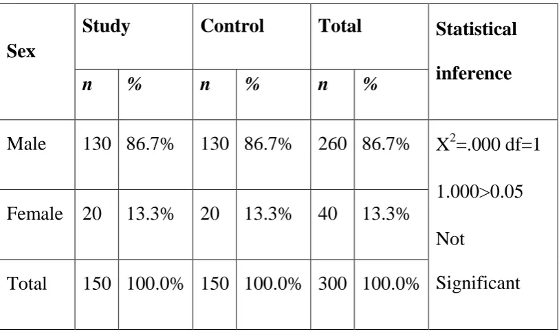
GENDER
CHART 1: AGE WISE DISTRIBUTION OF CASE
0 5 10 15 20 25 30 35 40 45
67
The male show dominance among patient with incidence of 132 among 150, female form minority with rest 18 patients.
CHART 2: GENDER DISTRIBUTION OF CASES
BASED ON ANATOMY OF HERNIA
Based on the anatomy which was confirmed intraoperatively the hernia is divided into indirect, direct and pantaloon type with both components. Of this 150 hernia 19 hernia are bilateral , of bilateral type 12 has both component as direct and 7 both component indirect. In rest 40 patients have direct hernia and 86 have indirect hernia.5 patients had pantaloon type.
male 87% female
68
CHART 3: CLINICAL DISTRIBUTION OF CASES
Based on clinical presentation
The most common clinical presentation is painless swelling in the inguinal region which forms main complaint in 89 patients; pain is the second prominent complaint which forms 37 out of 150 patients. In rest 24 patients pain and swelling both were presenting complaint.
0 10 20 30 40 50 60 70 80 90
Indirect Direct Bilateral Pantaloon
Indirect
69
CHART 4: DISTRIBUTION BASED ON CLINICAL
PRESENTATION
Based on complication
Complications which I came across in my studys were recurrence, obstruction, irreducibility and strangulation. Irreducibility was present in 27 patient, of this 27 patient 14 patient had features of obstruction like vomiting, constipation and abdominal distention. The features of strangulation present in 4 patient with severe pain, tenderness and redness in the skin over inguinal region. Recurrence was found in only 3 patients
0 10 20 30 40 50 60 70 80 90 100
70
CHART 5: BASED ON COMPLICATION
Duration of disease
Of the 150 patients, 73 patients presented within one year, 37 patients between one and two year. 28 patient between 2nd and 3rd year . In rest the symptoms are present for more than 3 years.
0 5 10 15 20 25 30
71
CHART 6: BASED ON DURATION OF DISEASE
Associated systemic disease
Most common systemic disease which was present in my patient was hypertension in 32 patients, followed by diabetic in 21 patients. Both diabetic and hypertension were present in 7 patients. The other diseases like cad, cva, ckd etc were present in 9 patients The patients with other chronic diseases like connective tissue disorder, copd, asthmatics, bph were excluded from the study.
0 10 20 30 40 50 60 70 80
<1 year 1-2 year 2-3 year >3 year
Series 1
Column1
72
CHART 7: BASED ON ASSOCIATED SYSTEMIC DISEASE
BASED ON PARITY
Of the 18 females included in our study all of them were parous women.
BASED ON THE SITE OF LESION
Of the 150 patients 99 patient pathology was on right side and on 32 patients the hernia was on left side. In rest that is on 19 patients the lesion was bilateral.
hypertension
diabetic
hpertension and diabetic
73
CHART 8: BASED ON SITE OF LESION
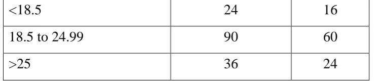
BASED ON BMI
Based on BMI the patient is divided into three class, first class is <18.5, second class between 18.5-24.99 and last class with BMI more than 25. The results I have shown in the graph below.
right side
left side
74
CHART 9: BASED ON BMI
0 10 20 30 40 50 60 70 80 90
75
RESULT
The following tables depict the various results
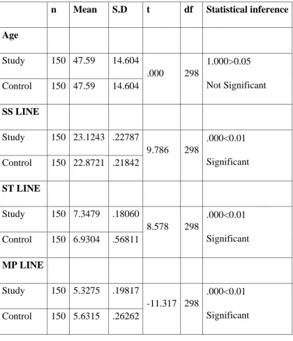
TABLE 1: COMPARING VALUES OF SS, ST AND MP WITH
CONTROL
n Mean S.D t df Statistical inference
Age
Study 150 47.59 14.604
.000 298
1.000>0.05 Not Significant Control 150 47.59 14.604
SS LINE
Study 150 23.1243 .22787
9.786 298
.000<0.01 Significant Control 150 22.8721 .21842
ST LINE
Study 150 7.3479 .18060
8.578 298
.000<0.01 Significant Control 150 6.9304 .56811
MP LINE
Study 150 5.3275 .19817
-11.317 298
[image:87.595.87.508.235.724.2]76
CHART 10: GRAPHICAL REPRESENTATION OF CASES AND
CONTROL BY SS, ST AND MP VALUUES
In my study there was 150 cases and 150 controls. Among them the average SS value for case was 23.12 which was much above the average in control group which was 22.87. The t value was also significant (9.786). When it come to ST value the mean was 7.34 in the study group and mean was only 6.93 in the control group. The statistical significance was proved with t value 8.57. The average MP distance was 5.63 in control group which was much higher than study group with distance of 5.327.
0 5 10 15 20 25
study
control
SS LINE
ST LINE
77
TABLE 2: DISTRIBUTION OF CASES AND CONTROL BASED
ON GENDER
Sex
Study Control Total Statistical
inference
n % n % n %
Male 130 86.7% 130 86.7% 260 86.7% X2=.000 df=1 1.000>0.05 Not
Significant Female 20 13.3% 20 13.3% 40 13.3%
Total 150 100.0% 150 100.0% 300 100.0%
In my study the male cases were predominant with percentage of 86.7%, the females was only minority with 13.3% . The control was choosen with similar age and sex , hence chi square test was not significant.
TABLE 3: CLINICAL PROFILR OF CASES
Particulars No.of respondents
(n=150)
Percentage
(100%)
Duration
<1yr 73 48.7
1 to 2yrs 37 24.7
2 to 3yrs 28 18.7
[image:89.595.95.502.134.378.2]78 Irreducible
No 123 82.0
Yes 27 18.0
Obstructive Symptoms
No 136 90.7
Yes 14 9.3
Strangulation Features
No 146 97.3
Yes 4 2.7
Recurrence
No 146 97.3
Yes 4 2.7
Hypertension
YES 32 21.3
NO 118 78.7
Diabetes mellitus
No 129 86.0
Yes 21 14.0
Any other chronic disease
No 141 94.0
Yes 9 6.0
site of lesion
RT 99 66.0
LT 32 21.3
Bilateral 19 12.7