Boar spermatozoa develop ability to bind to oviduct epithelium during passage through the epididymis
141
0
0
Full text
(2) ii STATEMENT OF ACCESS I, the undersigned, author of this work, understand that James Cook University will make this thesis available for use within the University Library and, via the Australian Digital Theses Network, for use elsewhere. I understand that, as an unpublished work, a thesis has significant protection under the Copyright Act and; I do not wish to place any further restriction on access to this work. ________________. _______________. Santiago T. Peña, Jr.. Date. STATEMENT OF SOURCES DECLARATION. I declare that this thesis is my own work and has not been submitted in any form for another degree or diploma at any university or other institution for tertiary education. Information derived from published or unpublished work of others has been acknowledged in the text and a list of references is given.. __________________. ________________. Santiago T. Peña, Jr.. Date.
(3) iii DECLARATION OF ETHICS The research presented and reported in this thesis was conducted within the guidelines for research ethics outlined in the Joint NHMRC/AVCC Statement and Guidelines on Research Practice (1997), the James Cook University Policy on Experimentation Ethics, Standard Practices and Guidelines (2001), and the James Cook University Statement and Guidelines on Research Practice (2001). The research methodology received clearance from the James Cook University Experimentation Ethics Review Committee (Animal Ethics approval number A1007).. ___________________. ________________. Santiago T. Peña, Jr.. Date.
(4) iv STATEMENT ON THE CONTRIBUTION OF OTHERS Professor Phillip Summers supervised the research reported in this thesis, carried out the surgical castrations of the boars, provided advice and assistance with the preparation of the thesis and was a co-author on all papers resulting from this thesis. A stipend was provided by the Australian Agency for International Development (AusAID) for the duration of the research candidature. Project costs were met from IRA and Reproduction Service accounts held by Professor Summers.. ___________________. __________________. Santiago T. Peña, Jr.. Date.
(5) v ACKNOWLEDGEMENT The LORD bless thee, and keep thee: The LORD make his face shine upon thee, and be gracious unto thee: The LORD lift up his countenance upon thee, and give thee peace. Numbers 6: 24-26 (Holy Bible, KJV). I thank God first of all for this rare opportunity he has given me to continue my professional upliftment in this prestigious school, the School of Veterinary and Biomedical Sciences, James Cook University, Townsville, North Queensland, Australia. I thank Him for the great chance to enjoy bountiful blessings he has bestowed upon me in this country and in advancing my knowledge of my professional career along the course of my study. I thank my scholarship sponsor, the Australian Agency for International Development (AusAID) in providing me financial assistance for without it, I couldn’t surely have what I am having now. I also thank and highly acknowledge the exemplary performance of my supervisor, Prof Phillip Summers, for his diligence and ever ready support in the final accomplishment of this work. Not to forget are the helping hands of Chris Coleman and Jeffrey Palpratt during the laboratory works and travels of this research. I thank my mother, family, pastors and friends for prayers and encouragement while I am away from home. Most importantly, I thank God for my wife Angelica, my ever faithful helpmeet. To all of you, I tender this humble piece of work..
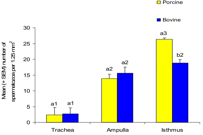
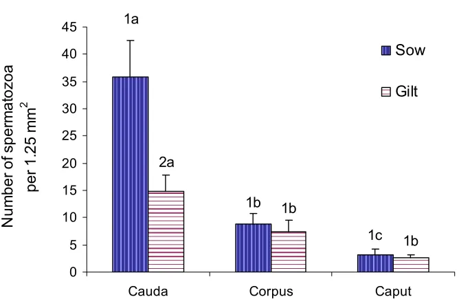
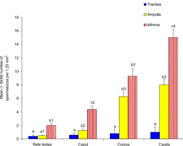
(6) vi ABSTRACT The aim of this study was to investigate the relationship of maturation of spermatozoa in the epididymis and the ability to bind to oviduct epithelium. It was hypothesized that testicular spermatozoa need to pass through the regions of the epididymis in order to acquire the ability to bind to the oviduct. Spermatozoa were collected from the rete testis and the caput, corpus and cauda epididymides from 10-14 month-old Large White or Large White x Landrace boars. Boars were first unilaterally castrated and then slaughtered four to five weeks later to obtain the second testicle, epididymis and seminal vesicles. Oviducts were obtained from slaughtered pre-pubertal gilts and explants from the isthmus and ampulla prepared. Spermatozoa were suspended in modified Androhep medium, added to oviduct explants and incubated at 390 C in a humidified atmosphere containing 5% CO2 in air for 15 minutes. The number of spermatozoa attached to 1.25 mm2 of explant was counted after fixation and staining of explants. The possibility of using oviducts from slaughtered cows rather than porcine oviducts was examined using ejaculated spermatozoa. Significantly more ejaculated spermatozoa bound to the isthmus of gilts than cows hence, porcine oviducts were used in the succeeding experiments. There was a sequential increase in the number of spermatozoa that bound to the oviductal epithelium from the rete testis to the cauda epididymidis (2 ±0.30, 4.36 ±0.53, 9.3 ±1.60 and 15±1.22 for rete testis, caput corpus and caudal spermatozoa on isthmic explants, respectively). Significantly more (P ≤0.05) spermatozoa, either ejaculated or epididymal, bound to isthmus than ampulla explants (26.33±2.27 and 13.55±1.42 ejaculated spermatozoa on isthmic and ampullary explants, respectively). Incubation in medium containing albumin and asialofetuin which are known to contain mannose and lactose respectively inhibited the binding of epididymal spermatozoa to oviduct explants (3.42±0.56 caudal spermatozoa on isthmic explants in medium with albumin and 14.75±2.02 caudal spermatozoa on isthmic explants in modified Androhep medium)..
(7) vii. The number of spermatozoa from the caput and corpus that bound to oviduct explants significantly (P ≤0.05) increased after incubation in caudal fluid for 30 minutes (7.52±1.10 and 12.78±1.64 corpus spermatozoa on isthmic explants for modified Androhep and caudal fluid, respectively). This result suggests that caudal fluid has distinct features that directly or indirectly influence the attachment of spermatozoa to oviduct epithelium. The motility of spermatozoa also decreased after incubation in caudal fluid for 30 minutes. This result is not surprising because in vivo, spermatozoa remain in a quiescent state during storage in the cauda epididymidis. Exposure of epididymal spermatozoa to seminal plasma for 30 minutes significantly reduced the number of spermatozoa that bound to oviduct explants while exposure for one minute significantly increased the number of bound caput spermatozoa. Incubation in seminal plasma also caused capacitation of spermatozoa and this is the likely reason for the reduction in the number of bound spermatozoa. There is also the possibility that some components in the seminal plasma may form a coating over the plasma membrane of spermatozoa sufficient to inhibit the expression of pre-existing binding molecules. On the other hand, the seminal plasma may also be a source of binding molecules for immature caput spermatozoa. There was an increase in the motility of spermatozoa after exposure to seminal plasma. In conclusion, this study found that as spermatozoa pass down the epididymis, there is an increase in the number of spermatozoa that bind to oviduct explants. This result was interpreted to mean that the maturation of spermatozoa in the epididymis involves the acquisition of the ability to bind to oviduct epithelium..
(8) viii TABLE OF CONTENTS Page No. Statement of Access. ii. Statement of Sources. ii. Declaration of Ethics. iii. Statement of the Contribution of Others. iv. Acknowledgement. v. Abstract. vi. Table of Contents. viii. List of Tables. xii. List of Figures. xiii. List of Abbreviations. xvi. CHAPTER 1 .............................................................................................................. 1 INTRODUCTION..................................................................................................... 1 1.1. Introduction ................................................................................................. 1. 1.2. Working Hypothesis.................................................................................... 4. 1.3. Objectives.................................................................................................... 4. CHAPTER 2 .............................................................................................................. 6 REVIEW OF LITERATURE .................................................................................. 6 2.1. Introduction ................................................................................................. 6. 2.2. A General Perspective to the Role of the Epididymis................................. 6. 2.3. The Fertilising Ability of Epididymal Spermatozoa ................................... 7. 2.4. Comparative Morphology of Spermatozoa from the Caput, Corpus and Cauda Epididymidis .................................................................................... 9. 2.5. Histological Features of the Boar Epididymis .......................................... 10. 2.6. Ultrastructural Characteristics of the Boar Epididymis ............................ 12. 2.7. Physiological Correlations ........................................................................ 14. 2.8. The Epididymal Plasma ............................................................................ 15. 2.8.1. Epididymal secretory proteins........................................................... 16. 2.8.2. Epididymal secretory proteins of the boar and related compounds .. 17. 2.8.3. Secretory enzymes in the epididymal fluid of the boar..................... 21.
(9) ix 2.9. Maturation of Boar Spermatozoa in the Epididymis................................. 22. 2.9.1. Modifications of the DNA-protein complex ..................................... 23. 2.9.2. Modifications of the plasma membrane ............................................ 23. 2.9.3. Concentration of spermatozoa during the epididymal transit ........... 27. 2.10. Protection of spermatozoa during storage in the cauda epididymidis....... 27. 2.11. The Oviduct............................................................................................... 28. 2.11.1. Structural anatomy and histology of the oviduct .............................. 28. 2.11.2. Blood supply to the oviduct .............................................................. 30. 2.12. The Oviduct Luminal Fluid....................................................................... 30. 2.13. Binding of Spermatozoa to the Oviduct.................................................... 33. 2.13.1 2.14. The role of the sperm reservoir ......................................................... 34. Mechanisms Involved in the Formation of the Sperm Reservoir.............. 37. 2.14.1. Physical aspects................................................................................. 38. 2.14.2. Carbohydrate recognition.................................................................. 39. 2.15. Features of the Binding of Spermatozoa to Oviductal Explants ............... 41. 2.16. Sperm-to-Oviduct Binding in Relation to the Region of the Oviduct, the Stage of the Oestrous Cycle and the Reproductive Status of the Animal . 42. 2.17. Sperm-to-Oviduct Binding between Epididymal and Ejaculated Spermatozoa and between Capacitated and Uncapacitated Spermatozoa 43. 2.18. The Release of Spermatozoa from the Sperm Reservoir .......................... 43. 2.19. Sperm-Oviduct Binding and the Role of the Seminal Plasma .................. 44. 2.20. Conclusion................................................................................................. 46. CHAPTER 3 ............................................................................................................ 47 MATERIALS AND METHODS ........................................................................... 47 3.1. Boars ......................................................................................................... 47. 3.2. Preparation of oviductal explants for the binding assay ........................... 47. 3.3. Preparation of spermatozoa and determination of motility characteristics49. 3.3.1. CASA settings and definitions .......................................................... 49. 3.4. Co-incubation of spermatozoa and explants ............................................. 51. 3.5. Fixation and counting bound spermatozoa................................................ 51. 3.6. Comparison of the binding capacity of ejaculated boar spermatozoa to porcine and bovine oviducts...................................................................... 52. 3.7. The binding of boar epididymal spermatozoa to porcine oviducts ........... 52.
(10) x 3.7.1. Comparison of the binding capacity of epididymal boar spermatozoa to oviducts of sows and gilts ............................................................. 53. 3.8. The binding of epididymal spermatozoa after dilution with either albumin, asialufetuin and modified Androhep solutions.......................................... 54. 3.9. The binding of epididymal spermatozoa after incubation in caudal fluid. 55. 3.10. The binding of epididymal spermatozoa to oviductal epithelium after incubation with seminal plasma ................................................................ 55. 3.10.1. Determination of the percentage of live and dead spermatozoa ....... 57. 3.10.2. Capacitation status of spermatozoa ................................................... 57. 3.11. Data presentation and analyses ................................................................. 58. CHAPTER 4 ............................................................................................................ 59 BINDING OF BOAR EPIDIDYMAL SPERMATOZOA TO OVIDUCTAL EPITHELIUM......................................................................................................... 59 4.1. Introduction ............................................................................................... 59. 4.2. Binding of ejaculated boar spermatozoa to porcine and bovine oviductal explants...................................................................................................... 61. 4.3. Motility of epididymal spermatozoa ......................................................... 62. 4.4. Binding of boar epididymal spermatozoa to the oviductal epithelium of sows and gilts ............................................................................................ 64. 4.5. Developmental influence on the binding ability of boar spermatozoa from the rete testis and the caput, corpus and cauda epididymidis to porcine oviducts ..................................................................................................... 64. 4.6. Comparison of the binding capacity of epididymal spermatozoa between boars .......................................................................................................... 67. 4.7. Carbohydrate-binding molecules recognised by epididymal spermatozoa…………………………………………………………….. 67. CHAPTER 5 ............................................................................................................ 77 EXPOSURE TO CAUDAL EPIDIDYMAL FLUID INCREASES THE BINDING ABILITY OF SPERMATOZOA FROM THE CORPUS AND THE CAPUT EPIDIDYMIDIS……………………………………………………… 77 5.1. Introduction ............................................................................................... 77. 5.2. Motility of epididymal spermatozoa after incubation in caudal fluid....... 79.
(11) xi 5.3. Binding of epididymal spermatozoa after incubation in caudal fluid ....... 80. CHAPTER 6 ............................................................................................................ 85 INFLUENCE OF SEMINAL PLASMA ON THE BINDING ABILITY OF EPIDIDYMAL SPERMATOZOA……………………………………………… 85 6.1. Introduction …………………………………………………………….. 85. 6.2. Motility characteristics of epididymal spermatozoa before and after incubation with seminal plasma ................................................................ 87. 6.3. Capacitation status of spermatozoa before and after incubation in modified Androhep medium or seminal plasma....................................................... 90. 6.4. The viability of spermatozoa before and after incubation in modified Androhep medium or seminal plasma....................................................... 92. 6.5. The influence of seminal plasma on the binding capacity of epididymal spermatozoa to oviductal epithelium......................................................... 95. CHAPTER 7 .......................................................................................................... 101 GENERAL DISCUSSION.................................................................................... 101 7.1 Scope of research project…………………………………………………… ...101 7.2 Future research directions…………………………………………………… 106 7.3 Conclusion……………………………………………………………………..107 REFERENCES ...................................................................................................... 109.
(12) xii LIST OF TABLES Page No. 48. Table 3.1. Composition of Modified Tyrode’s Solution. Table 3.2. Composition of the Modified Androhep Solution. 51. Table 3.3. Composition of Gill’s Haematoxylin stain. 52. Table 4.1. Motility characteristics of epididymal spermatozoa immediately after collection 63. Table 5.1. Motility characteristics (mean ± SEM) of epididymal spermatozoa after incubation in either modified Androhep medium or caudal fluid. 80. Table 6.1. Motility characteristics (mean ± SEM) of spermatozoa before and after incubation for 30 minutes in either modified Androhep medium or seminal plasma. 90. Table 6.2. Motility characteristics (mean ± SEM) of spermatozoa before and after incubation for one minute in either modified Androhep medium or seminal plasma. 92.
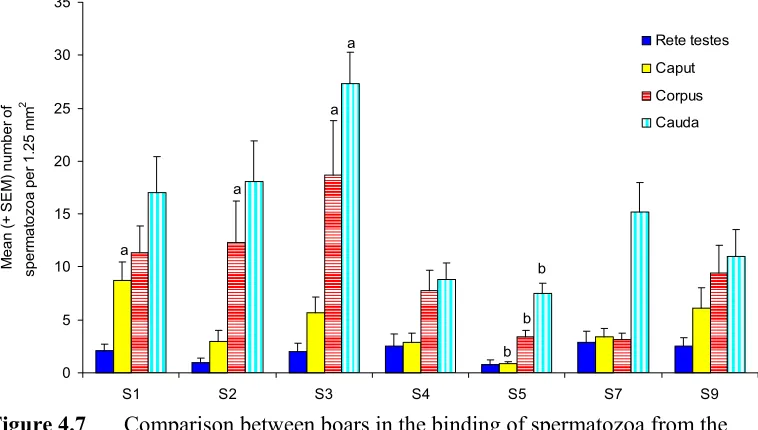
(13) xiii LIST OF FIGURES Page No. Figure 4.1. Binding of ejaculated porcine spermatozoa to porcine and bovine oviductal and tracheal explants.. 62. Figure 4.2. The mean (+ SEM) percentage of motile spermatozoa from the rete testis and epididymis. 63. Figure 4.3. Binding of epididymal spermatozoa to the isthmus.. 64. Figure 4.4. Binding of epididymal spermatozoa to the ampulla.. 65. Figure 4.5. Binding of boar spermatozoa from the rete testis and epididymis to the isthmus and ampulla. 66. Figure 4.6. Comparison of the binding of ejaculated and caudal spermatozoa to isthmic and ampullary explants. 66. Figure 4.7. Comparison between boars in the binding of spermatozoa from the rete testis and epididymis to isthmic explants. 68. Figure 4.8. Comparison between boars in the binding of spermatozoa from the rete testis and epididymis to ampullary explants. 68. Figure 4.9. Comparison between the left and the right testicles of boars in the binding capacity of spermatozoa to isthmic explants. 69. Figure 4.10. Comparison between the left and the right testicles of boars in the binding capacity of spermatozoa to ampullary explants. 69. Figure 4.11. The binding of epididymal spermatozoa to isthmic explants after incubation of spermatozoa in medium containing either albumin, asialufetuin or modified Androhep medium.. 70. Figure 4.12. The binding of epididymal spermatozoa to ampullary explants after incubation of spermatozoa in medium containing albumin, asialufetuin or modified Androhep medium. 70. Figure 5.1. The reduction in the motility of spermatozoa from the caput and the corpus after incubation in caudal fluid. 79.
(14) xiv Figure 5.2. The influence of incubation of epididymal spermatozoa in caudal fluid on the binding of spermatozoa to isthmic explants. 81. Figure 5.3. The influence of incubation of epididymal spermatozoa in caudal fluid on the binding of spermatozoa to ampullary explants. 81. Figure 6.1. The mean (+ SEM) percentage of motile spermatozoa from the rete testis and the epididymis of boars immediately after collection into either modified Androhep medium or seminal plasma. 88. Figure 6.2. The mean (+ SEM) percentage of motile spermatozoa from the rete testis and the epididymis of boars after incubation for 30 minutes in either modified Androhep medium or seminal plasma. 89. Figure 6.3. The mean (+ SEM) percentage of motile spermatozoa from the rete testis and the epididymis of boars before and after incubation for one minute in either modified Androhep medium or the seminal plasma. 91. Figure 6.4. The capacitation status of spermatozoa before incubation in modified Androhep medium (control) or after incubation for 30 minutes in either modified Androhep medium or seminal plasma. 93. Figure 6.5. The capacitation status of spermatozoa after incubation for one minute in either modified Androhep medium (control) or seminal plasma. 93. Figure 6.6. The percentage (+SEM) of live spermatozoa before incubation in modified Androhep medium or after incubation for 30 minutes in either modified Androhep medium or seminal plasma. 94. Figure 6.7. The percentage (+SEM) of live spermatozoa after incubation for one minute in either modified Androhep medium or seminal plasma. 94. Figure 6.8. Effect of incubation for 30 minutes in either modified Androhep medium or seminal plasma on the binding of spermatozoa from the rete testis and epididymis to isthmic explants. 95. Figure 6.9. Effect of incubation for 30 minutes in either modified Androhep medium or seminal plasma on the binding of spermatozoa from the rete testis and epididymis to ampullary explants. 96. Figure 6.10. Effect of incubation for one minute in either modified Androhep medium or seminal plasma on the binding of spermatozoa from the rete testis and epididymis to isthmic explants. 96.
(15) xv Figure 6.11. Effect of incubation for one minute in either modified Androhep or seminal plasma on the binding of spermatozoa from the rete testis and epididymis to ampullary explants. 97.
(16) xvi LIST OF ABBREVIATIONS. ANOVA. analysis of variance. ATP. adenosine triphosphate. BSP. bovine seminal plasma protein. cAMP. cyclic Adenosine Monophosphate. cAMP-PKA. cAMP-protein kinase A. CTC. chlortetracycline. CRISP. cysteine-rich secretory protein. CTP. cholesterol transferase protein. DNA. deoxyribonucleic acid. EAP. oestrous-associated glycoprotein. E-RABP. epididymal retinoic acid-binding protein. FITC-sZP. fluorescein-conjugated solubilized zona pellucida. GPX. glutathione peroxidase. g. g-force/g-load/gravitational acceleration. HBPs. heparin-binding proteins. HE. human epididymal protein. HPLC. high performance liquid chromatography. HTM-IVOS. Hamilton-Thorne Machine-integrated visual optical system. kDa. kilodalton. K+/Na+ ratio. potassium/sodium ratio. MAN2B. 135 kDa alpha-D-mannosidase. MMPs. matrix metalloproteinases. mRNA. messenger ribonucleic acid. OECM. oviduct epithelial cell monolayer. OGP. oestrogen-dependent protein. PAGE. polyacrylamide gel electrophoresis. PGDS. prostaglandin D2 synthase. pI. isoelectric point. pOGP. porcine oestrogen-dependent protein. SDS. sodium dodecyl sulfate. TALP. Tyrode’s albumin lactate phosphate.
(17) 1. CHAPTER 1 INTRODUCTION. 1.1. Introduction. Food animals are very important resources of proteins and other nutrients in the human diet. They provide a number of essential amino acids and vitamins not usually found in plants. Of the many animal resources available throughout the world, pigs as well as poultry are the major and more stable meat sources. The consumption of meat per animal species worldwide is shared between pork (43%), poultry (27%), beef/veal (26%) and 4% from others (Du, 2001). The worldwide demand for meat has been predicted to increase by more than four percent above the consumption of the past decade (FAS, 2004). This is largely due to the increasing demand and growing economies of developing nations in the Asian region in which by the year 2020, developing nations will utilise 60% of the world’s meat and 52% of the world’s milk production (ILRI, 2004). The demand for food has been projected to increase significantly between 3000 kcal/person/day in 2015 and above in 2030 (FAO, 2003). Comparative data of the dietary intake per capita of major meats in USA, Australia and The Philippines is 23%, 20.38% and 11.23 % for pork; 23%, 30.56% and 7.44% for chicken; and 29%, 35.16% and 2.81% for beef (LDC, 2004). These data indicate that pig meat plays a very important role in a country like The Philippines where pig farming is the largest livestock industry. In addition to being a major source of food, pig farming also provides business and employment opportunities for people. In 2002, the total production of pork reached as much as 1.67 M tonnes valued at about 86.72B pesos of which smallholder farmers contributed 76.8% of the total production (SwiIN, 2004). Fertility is an important aspect in animal production. Unfortunately often the fertility of males doesn’t receive as much attention as it should in the management of the herd. In pigs, boar fertility is usually related to the prolificacy of the sow as measured by the farrowing rate, litter size or the number of returns to service (Hughes and.
(18) 2 Varley, 1980; Colenbrander et al., 1993), but this system doesn’t really guarantee the highest culling standards for boars especially if the animal under consideration has an outstanding genetic merit. The quest for understanding the complex attributes of fertility in male is of paramount concern. Whether it is by natural or artificial insemination of spermatozoa into the female reproductive tract, the highest possible fertility of spermatozoa is always a must as it constitutes 50% in the actual success attainable in every conception. While much attention has been directed to understanding the complex cellular events in the production of spermatozoa and their effect on fertility, there has been a limited focus on aberrations in the function of the epididymis and their role in infertility of domestic animals. While much attention has been directed to understanding the complex cellular events in the production of spermatozoa and their effect on fertility, there has been a limited focus on aberrations in the function of the epididymis and their role in infertility of domestic animals. One of the organs whose anatomical and physiological attributes are almost unknown to the secular world is the epididymis. This is probably due to its relatively small size, not so conspicuous location and thought to be just an integral part of the testicle. The epididymis is an organ structure comprised of a strongly convoluted duct system with a length of about 60 metres in the boar, 40 in the bull and 20 in man (Mann and Lutwak-Mann, 1981). Upon leaving the testes and passing through the efferent ducts, spermatozoa enter the epididymis. The word epididymis was coined in 1830 by Sir Astley Cooper when he first described this organ (Mann and Lutwak-Mann, 1981): “Of the epididymis: This body may be considered as an appendix to the testis, and its name is derived from its being placed upon this organ, as the testes were anciently called didymi. It is of a crescenti form; its upper edge is rounded, its lower edge is thin. Its anterior and upper extremity is called its caput, the middle part its body and the lower part its cauda.” The epididymis is divided into three major compartments: the head (caput), the body (corpus) and the tail (cauda) (Mann and Lutwak-Mann, 1981; Briz et al., 1993). The epididymis has a significant role in the maturation of testicular spermatozoa (Hafez, 1993). Testicular spermatozoa are both immotile and infertile (Brooks, 1983) and.
(19) 3 despite the fact that the post-testicular mammalian spermatozoon is equipped with a modified nucleus of unique DNA content and a highly organized cytoplasm, it does not have all the capabilities which are essential both for survival in the female reproductive tract or the capacity to fertilise oocytes (Amann et al., 1993). The epididymis serves as an area where the full development and maturation of spermatozoa can continue leading to the acquisition of progressive motility and fertilising capacity. A comprehensive understanding of the biological and physiological functions of the epididymis is still limited. Although the epididymis has been part of biological investigations since early in the 19th century, it was only in the 1970’s when the anatomical features, biochemistry and functionality of the epididymis has been deeply considered. A complex array of proteins and secretory products in the epididymis has been identified (Syntin et al., 1996), but a detailed understanding of the cellular changes that occur to spermatozoa at different sites of the epididymis is still largely unknown. While the epididymis performs a crucial role in the fertility of the male, the oviducts also have a number of roles that are essential for successful reproduction. The mammalian oviduct is not just a passive conduit between the uterus and the ovaries but is the primary organ involved in sperm capacitation, fertilisation and early development of the embryo (Hunter, 1998; Rodriguez-Martinez, 2001). As with the epididymis, the functional significance of the oviduct was not immediately appreciated. Those spermatozoa that reach the oviduct in the pig are mostly retained at the uterotubal junction and attached to the caudal portion of the isthmus. These regions constitute the sperm reservoir which in pigs is established one to two hours after insemination (Hunter, 1981). The sperm reservoir is known to function in three major ways (Topfer-Petersen et al., 2002). Firstly, it prolongs sperm viability with respect to the onset of ovulation regardless of the time of insemination. Spermatozoa stored in the caudal isthmus retain their viability for 36 h or longer (Hunter, 1984). Secondly, it controls capacitation to coincide with the onset of ovulation. With the limited life of spermatozoa, capacitation must be controlled for it to happen at the.
(20) 4 right time. The oviduct regulates this complex sequence of cellular and membrane changes (Petrunkina et al., 2003). Thirdly, the sperm reservoir prevents polyspermy (Hunter, 1993). The mechanisms by which spermatozoa bind to the epithelium of the oviduct are still largely unclear. However, the physical environment of the oviduct and the involvement of a carbohydrate-mediated interaction between spermatozoa and oviductal epithelium appear to be the basis for this complex sperm-to-oviduct interaction (Pollard et al., 1991; Green et al., 2001). At oestrus, the lumen of the oviduct becomes smaller due to oedema of the lamina propria in response to high concentrations of circulating oestrogens. In the pig, the caudal isthmus of the oviduct secretes a characteristic visco-elastic and tenacious mucus just before ovulation (Hunter, 1998). After ovulation, the quantity and the viscosity of the luminal secretions decrease. On the other hand, a carbohydrate-mediated interaction has been widely observed in many animal species and with other cellular interactions such as between spermatozoa and Sertoli cells (Raychoudhury and Millette, 1997) and between spermatozoa and the zona pellucida (Oehninger et al., 1991). Oligosaccharides and lectins present in the plasma membrane and mucosal surface of spermatozoa and the oviduct, respectively, function as ligand and receptors which actively participate in sperm-oviduct interaction. Different sugar monomers, oligosaccharides or glycoproteins inhibit the binding of spermatozoa to oviductal epithelial cells or explants in a species specific manner (Suarez, 2001). However, it is not yet known how far these identified receptor-ligand systems influence this interaction between spermatozoa and the oviduct epithelium (Hunter, 2003). It has been found recently that proteins secreted by the seminal vesicles of the bull adhere to the plasma membrane of spermatozoa and functions as primary oviduct-binding proteins (Gwathney et al., 2003, 2006).. The relationship that exists between the epididymis and the oviducts is the fact that while spermatozoa progress through different regions of the epididymis, it would be expected that a number of maturational changes occur to spermatozoa which are.
(21) 5 necessary for the development of ligands or receptors essential for binding to the oviductal mucosa. Spermatozoa undergo maturation processes principally within the caput and corpus regions of the epididymis and are stored in the cauda. Further to this is the possible influence of certain components of the seminal plasma on the interaction of spermatozoa to the oviductal epithelium.. 1.2. Working Hypothesis. Testicular spermatozoa of the boar need to pass through the caput, corpus and cauda epididymidis to develop the full capacity for attachment to the epithelium of the oviduct.. 1.3. Objectives . Examine and compare the capacity of spermatozoa from the rete testes, caput, corpus and cauda epididymidis of the boar to attach to the isthmic and ampullary epithelium with the use of an in vitro binding assay.. . Determine whether the binding is species-specific by examining the attachment of porcine epididymal spermatozoa to the bovine oviduct.. . Investigate the influence of secretions of the seminal vesicle on the capacity of porcine epididymal spermatozoa to bind to the oviductal epithelium.. . Determine if incubation of spermatozoa from the caput and corpus with fluid from the cauda epididymidis will influence the binding capacity of caput and corpus spermatozoa..
(22) 6. CHAPTER 2 REVIEW OF LITERATURE. 2.1. Introduction. This literature review is divided into three major areas. Firstly, the developmental changes and maturational processes that occur to spermatozoa in the epididymis with special reference to the boar are discussed. These include the morphological and ultrastructural features of the epididymal epithelium and its characteristic secretory products which play significant roles in maturation of boar spermatozoa. Secondly, the structural anatomy and histology of the mammalian oviduct will be considered. The oviductal fluid content and composition is also discussed particularly with respect to oviductal regions and the stage of the oestrous cycle. Thirdly, a discussion will focus on the interaction of spermatozoa with the oviduct both in vitro and in situ. This includes the morphological features of the binding of spermatozoa to oviduct epithelium, its mechanism, significance and other factors which might be involved in the formation of the sperm reservoir.. 2.2. A General Perspective of the Role of the Epididymis. While research on testicular and ovarian physiology has been undertaken for many years, a detailed study of the epididymis and its crucial role in the maturation of spermatozoa has only occurred in the past few decades. The epididymis is important in the final development of the motility and fertilising capacity spermatozoa (Brooks, 1983; Amann et al., 1993; Hafez, 1993). It also enables the production of a population of spermatozoa with different potential capacities to fertilise oocytes (Amann et al., 1993) which is important to increase the likelihood of fertilized oocytes in a given time. This heterogeneity in the sperm population is essential with respect to the time of ovulation, especially in a species like the pig. Pigs ovulate about 40-42 hours from the beginning of oestrus and thus spermatozoa which are able to maintain full potential and remain viable for an extended period of time will be the ones most likely to fertilise oocytes..
(23) 7 The maturation processes principally occurs within the caput and corpus epididymidis, while the prime purpose of the cauda is to store spermatozoa. Interestingly, complete maturation of spermatozoa and the development of the full capacity to fertilise oocytes have never been obtained in vitro with testicular spermatozoa or those retrieved from the proximal epididymis (Moore and Akhondi, 1996; Dacheux et al., 2003). However, when epididymal spermatozoa were incubated with epididymal cell cultures, an increase in the motility had been observed and in some cases, the capacity to fertilise oocytes also improved although the binding to the zona pellucida did not improve (Moore and Akhondi, 1996). Early studies have reported that the electrophoretic profiles of epididymal fluid from different regions were different to other body fluids including blood plasma. The presence of hundreds of proteins present across all mammalian species (Dacheux et al., 2003) with some uniquely found and specific to the epididymal secretions suggests that such proteins were the molecular agents responsible for the many significant changes that occur to spermatozoa as they traverse the epididymal duct (Holland and Nixon, 1998). The epididymis also performs a lot of other functions, which are directly or indirectly essential to the successful penetration and fertilisation of the oocyte. These include the following: secretion of epididymal plasma; modification and development of the plasma membrane of spermatozoa; eradication of old and dead spermatozoa; concentration of spermatozoa; maintenance of the viability of spermatozoa during storage in the cauda epididymidis and sperm transport (Mann and Lutwak-Mann, 1981; Amann et al., 1993). Transport of spermatozoa along the epididymal duct is achieved by the periodic contractions of the smooth muscle fibers lining the epididymal duct of the caput and corpus and the low-frequency contractions in sections around the smooth muscle of the cauda (Robaire and Hermo, 1988; Turner, 1991). The cauda also produces short but forceful contractions mediated by its resident nerve cells (Jones, 1999).. 2.3. The Fertilising Ability of Epididymal Spermatozoa. Ejaculated spermatozoa and those directly taken from the cauda are comparable in terms of maturity status and fertility potential (Yanagimachi, 1994). There are some.
(24) 8 reports that state that that caudal spermatozoa of the rat (Shalgi et al., 1981) and pigs (Nagai et al., 1984) fertilise more oocytes in vitro than ejaculated spermatozoa. Spermatozoa from the cauda epididymidis of the boar penetrated more zona-intact oocytes than ejaculated spermatozoa that were subjected to similar pre-treatment conditions (Nagai et al., 1984). The same authors added that spermatozoa were incapable of penetrating oocytes when they were exposed to the seminal plasma. The fertilising ability of epididymal spermatozoa of the boar had been studied by Holtz and Smidt (1976). It was found that essential changes occur to spermatozoa as they move down the epididymis and these changes specifically involve the acquisition of fertilising capacity. The fertilisation rate of spermatozoa improved considerably from the caput to the cauda, although complete maturation was not achieved until spermatozoa reached the cauda. This is consistent with the general observation that maturation of spermatozoa is finalised only when spermatozoa have arrived in the proximal cauda epididymidis (Turner, 1995). Furthermore, while spermatozoa from the upper corpus epididymidis of the boar demonstrate penetration and activation of some oocytes from ipsilateral oviduct, spermatozoa from the cauda epididymidis did not from oocytes recovered from the contralateral oviduct (Hunter et al., 1976). It was suggested that cauda spermatozoa were possibly invested with inhibitory compounds which stabilized the plasma membrane during the epididymal transit (Hunter et al., 1976). Exposure to those factors could delay expression of capacitation unlike those spermatozoa liberated from the proximal portion of the epididymal duct. Substances known to inhibit capacitation are present between the proximal and distal portions of the epididymal duct (Hunter et al., 1976). High concentrations of enzyme inhibitors (i.e. trypsin inhibitor) are present in boar rete testis fluid and epididymal plasma (Suominen and Setchell, 1972). Because of this, the ability of spermatozoa to penetrate oocytes would be likely to be blocked since the expression of the necessary enzymes is inhibited. Further studies on biochemical and physiological aspects relating to these limitations in understanding are needed. In the hamster and mouse, acquisition of the ability to penetrate oocytes is normally achieved when spermatozoa have reached the cauda (Mann and Lutwak-Mann, 1981). While penetration of oocytes is possible by rabbit spermatozoa isolated from the corpus, the fertilisation rate was still minimal..
(25) 9 2.4. Comparative Morphology of Spermatozoa from the Caput, Corpus and Cauda Epididymides. The quality of spermatozoa irrespective of the animal species is greatly influenced by a number of factors which can be either environmental (i.e., photoperiod, relative humidity, temperature and handling) and/or within the animal itself including but not limited to the breed, age, viral or bacterial infection (Swierstra, 1973; Colenbrander et al., 1993). In intensive swine production where artificial insemination is used, the frequency of semen collection can substantially affect the concentration as well as motility characteristics and morphology of spermatozoa (Bonet et al., 1991). A number of important factors which determine the maximum frequency of semen collection without severely affecting the performance of the boar include the rate of sperm production by the animal, the concentration of spermatozoa per ejaculate and the duration of the passage of spermatozoa in the epididymal tract. By using autoradiographic methods, Swierstra (1968) determined that the time required by boar spermatozoa to travel through the epididymis was three days for the caput, two days for the corpus, and four to nine days for the cauda. The duration of epididymal transit also varies between species and can be lowered by as much as 10-20 % when the frequency of collection/ejaculation is increased (Setchell, 1991). The comparative morphology of boar spermatozoa collected from different regions of the epididymis had been extensively discussed (Briz et al., 1993; 1995; 1996). The proportion of mature or immature spermatozoa with a proximal or distal cytoplasmic droplet differed significantly as spermatozoa pass down the epididymal tract. In the caput, 99% of spermatozoa have a proximal cytoplasmic droplet while only 1% of spermatozoa in the corpus have a proximal cytoplasmic droplet. On the other hand, the percentage of immature spermatozoa with a distal cytoplasmic droplet is 0.5% in the caput, 81% in the corpus and 18% in the cauda. Malformations in epididymal spermatozoa were also apparent and can be divided into three main types. These are a folded tail that begins at the Jensen’s ring, which develops in the cauda due to the failure of the expulsion of the distal cytoplasmic droplet; the coiling of the tail which originates in the corpus; and the fusion of tails which also develops in the corpus (Briz et al., 1996). Other malformations include bicephalic spermatozoa, anomalous cephalic form, macrocephalic spermatozoa and microcephalic spermatozoa..
(26) 10 Caput spermatozoa are also characterised by a mitochondrial sheath that is composed of larger mitochondria of unequal sizes and low electron-dense matrices. The opposite is true for those spermatozoa from the cauda. Caput spermatozoa are also characterised by the high level of acrosomal protuberance and the mid-piece being highly flexible. Spermatozoa with detached heads as well as the tendency to agglutinate also increased in the cauda.. 2.5. Histological Features of the Boar Epididymis. The interaction of spermatozoa with the epididymal milieu is an essential event in the life of spermatozoa as this leads to the over-all structural changes to spermatozoa which subsequently affect sperm functions. Thus the histological make-up of the epididymal epithelium is one important factor to consider in the characterisation of the physiological mechanisms within the epididymal duct. Moreover, a good understanding of epididymal histology would help direct a focus in research specific to regions where the epididymal epithelium brings specific changes to the spermatozoon and in this way facilitate an understanding of the molecular aspects of sperm maturation (Amann et al., 1993). Briz et al. (1993) and Stoffel and Friess (1994) have undertaken an intensive morphological study on the ductus epididymidis of the boar (Sus domesticus) hence, the following discussion is largely based on their work. As with other animal species, the epididymis of the boar is divided into three main regions: the head (caput); a narrower body (corpus or isthmus); and the tail (cauda). It can also be further subdivided into regions based on morphological characteristics: the caput comprising of the medial limb (initial segment); the bend (proximal caput) and lateral limb (distal caput); the corpus; and the proximal and distal cauda (Stoffel and Friess, 1994). In general, the whole epididymal duct of the boar appears to be histologically similar being lined with a pseudostratified epithelium with stereocilia and covered with a muscular connective sheath, although variations in the duct diameter, epithelial height, and length of stereocilia occur among the regions (Briz et al., 1993). A similar description was given by Bassols et al., (2004) for epididymal epithelial cells.
(27) 11 cultured in vitro. Cultured epididymal epithelial cells first appeared as a columnar or cuboidal shape but gradually changed to a flattened, polygonal shape (Bassols et al., 2004). The epithelium in the caput region of the epididymis has the greatest height and becomes progressively smaller in the corpus and cauda. The total membrane surface of the stereocilia is also greater in the caput as well as in the corpus while stereocilia in the caudal region become less abundant and are rather short. The cell height within the initial segment ranges from 40 µm and 60 µm, which gives the lumen a characteristic stellate appearance. The lumen of the proximal caput through to the distal cauda is spherical, and the epithelial height decreases from approximately 85 µm in the proximal caput and about 35 µm in the distal cauda. The distance from one basement membrane to another varies in between regions. The initial segment is between 450 µm and 550 µm, the proximal caput is from 425-460 µm and the distal caput is about 350 µm increasing rapidly from 500 µm in the distal corpus to 700 µm and over 1mm in the proximal and distal cauda, respectively (Stoffel and Friess, 1994). The blood to the caput and corpus epididymidis is supplied by two branches arising from the testicular artery while the majority of the cauda receives blood from the deferential artery (Stoffel and Friess, 1994). After encircling the testis, the testicular artery joins the epididymal branch commencing at the caput epididymidis. The close association of the epididymal branches with the pampiniform plexus is also worth noting here. Anastomoses can be observed at the level of the vascular cone and drainage is accomplished by the epididymal veins (Stoffel and Friess, 1994). The peritubular smooth muscle is comprised of seven to ten concentric layers, which extend to the distal corpus. In the proximal cauda, a considerable gradual increase (two to three fold) in the thickness of the periductal muscle can be observed (Jones, 1999). An abrupt increase by the same magnitude is also found in the distal cauda. The boar epididymal tubule is also surrounded and supported by a larger amount of connective tissue than the efferent ductules (Bassols et al., 2004). This is an important point to consider when obtaining epididymal epithelium for in vitro culture..
(28) 12 Vascularization is more intense in the caput and corpus while the cauda contains a much thicker muscular-connective tissue sheath. The cavity of the cauda is also noted for the more frequent appearance of body cells, described as spherical in shape with a single nucleus, measuring about 6 µm in diameter and with scarce, granular cytoplasm (Briz et al., 1993).. 2.6. Ultrastructural Characteristics of the Boar Epididymis. There are five major types of epithelial cells that are found throughout the three regions of the epididymal duct: principal, basal, clear, narrow, and basophilic cells (Briz et al., 1993). The boar epididymis also has another group called mitochondriarich cells (Stoffel and Friess, 1994). Macrophages and wandering leucocytes are also present within the connective tissue that surrounds the epididymal duct (Setchell et al., 1994). The principal cells comprise the majority of the epithelial cells and occupy about 7090% of the epididymal epithelium in most species (Amann et al., 1993). They are columnar with a slightly basophilic cytoplasm, stereocilia, and a pale oval nucleus containing one or two prominent nucleoli. They extend from the basal membrane where they adhere by means of hemidesmosomes up to the free surface of the epithelium. The principal cells also contain abundant secretory granules as well as mitochondria and cisternae of endoplasmic reticulum lying in the supranuclear cytoplasm. The pyramidal basal cells with a diameter of about 6.5 µm and a maximum height of 5.5 µm are distributed along the length of the epididymal duct. They are attached by hemidesmosomes to the basal membrane. The nucleus is sub-spherical in shape with deep infoldings, and the cytoplasm contains scattered mitochondria and cisternae of rough endoplasmic reticulum. The clear cells are slightly vacuolated cells distributed amongst the principal cells, bearing basal outfoldings that contact the basal membrane. Their nucleus is medially located and it bears a deep infolding, which progressively define asymmetrical nuclear lobes. The perinuclear cytoplasm is highly vacuolated and contains abundant.
(29) 13 spherical mitochondria and some secretion granules which have a well defined electron-dense matrix. A small number of narrow cells are also present arranged along with the other epithelial cells. The nucleus is bilobed and elongated, enclosing a dense granular euchromatin and a cytoplasm, which is highly vacuolated. The spheroidal basophilic cells, which are located at different levels between principal cells, are characteristically unique exhibiting high nucleoplasmic difference in staining. They have a rounded nucleus and a cytoplasm containing sparse numbers of mitochondria, some cisternae of rough endoplasmic reticulum, and also some lysosomes of the primary and secondary type. The mitochondria-rich cells are sporadically located between principal cells in the initial segment and the proximal caput (Stoffel and Friess, 1994). They have a broad apex, which expands over the neighbouring cells and a basally projecting slender stem. As the name implies, the most peculiar characteristic of these cells is the presence of numerous mitochondria. The epididymal duct sheath is composed of muscular-connective tissue, consisting of two layers of smooth muscle tissue: the thick internal or longitudinal layer and a thinner external or circular layer. As the region of the epididymal canal becomes more distal, the intrinsic connective tissue sheath becomes more fibrous. In addition, the cauda region contains a much thicker muscular connective tissue sheath. Tight junctions are also commonly observed between the smooth muscle cells. The cauda is also characterised by bundles of collagen fibres intruding between myoblasts and thus indentations in the contour of the cells can be observed (Briz et al., 1993). In culture, the three regions of the epididymis show similar ultrastructural characteristics when examined by electron microscopy (Bassols et al., 2004). Moreover, cultured boar epididymal epithelial cells have a tight adherence to each other through interdigitations, desmosomes, and tight junctions located on their lateral and basal membranes, with some apparent intercellular spaces present. The nuclei are oval with prominent nucleoli. The cytoplasm contains numerous mitochondria, extensive rough endoplasmic reticulum and a well-developed Golgi.
(30) 14 apparatus. Moreover, multivesicular bodies, residual bodies, lipid droplets, dense granules and bundles of filaments (10 nm) are also present. The features of cultured boar epididymal epithelium are similar to the epithelium in situ (Bassols et al., 2004). These include the cell polarity, epithelial integrity, and absorptive and secretory activity. The numerous stereocilia frequently found on the apical surface of the cells is evidence of this characteristic polarity. The epithelial integrity is achieved and maintained by interdigitations and junctional complexes in the lateral and basal membranes. The absorptive and secretory activities of boar epididymal cells in vitro was also demonstrated by the presence of numerous pinocytic vesicles in the apical cytoplasm, multivesicular bodies, residual bodies, an extensive rough endoplasmic reticulum, a well-developed Golgi apparatus, abundant ribosomes and numerous other vesicles in perinuclear regions.. 2.7. Physiological Correlations. Much has yet to be understood about the specific functions of the cells comprising the epididymal epithelium. Briz et al. (1993) suggested that the basal, principal, clear, and narrow cells could be the four developmental stages of the same cell type. The proximal principal cells are known to specialize in fluid-phase and receptormediated endocytosis and secretory activity (Amann et al., 1993). Renewal of principal cells is the basic function of basal cells. As basal cells mature to columnar cells with apical secretory granules, they become clear cells capable of apocrine function and finally undergo regression to become narrow cells. Basophilic cells have been suggested to be leukocytes which have migrated from the underlying connective tissue to the epididymal duct cavity (Briz et al., 1993). It should be noted that the absorptive activity is particularly prominent in the proximal and distal caput as determined by ultrastructural examination. This is largely due to the presence of extensive cisternae of rough endoplasmic reticulum and the well-developed Golgi apparatus both in the distal caput and corpus regions which participate in active protein synthesis and secretion (Stoffel and Friess, 1994). The abundant vascularization of the caput and the corpus is correlated with their.
(31) 15 greater synthetic and secretory activity and possibly the transport of testicular androgens (Briz et al., 1993). The abundant muscular-connective tissue sheath of the cauda is related to the basic function of this region as a storage area and the intense contraction needed during ejaculation. The physiological mechanism correlated to this contraction is the presence of abundant collagen fibres and muscular components within the muscular sheath itself. More importantly, tight junctions present in large numbers between smooth muscle cells are responsible for facilitating the overall contraction of the caudal region of the epididymis (Briz et al., 1993) and thus providing a forceful expulsion of spermatozoa during ejaculation. A detailed study of the lymphatic drainage system of the epididymis has been done mainly in the rat with lymphatic sinusoids observed in intertubular spaces of the epididymal duct (reviewed by Setchell et al., 1994). Most of the vessels come out from the caput with only one or two from the corpus and one from the cauda. These vessels join to the main testicular lymphatic trunk and together with the spermatic artery end up in the para-aortic group of lymph nodes.. 2.8. The Epididymal Plasma. The epididymal plasma is composed of fluid coming from different sources. This includes the testes from the secretions of the Sertoli cells; the epididymal spermatozoa from the metabolism of phospholipids producing small amounts of epididymal glycerylphosphorylcholine; and the remainder as a direct result of the biosynthetic activity of the epididymal epithelium (Mann and Lutwak-Mann, 1981; Dacheux et al., 2003). The epididymal plasma has a particularly high K+/Na+ ratio and has unique biochemical features due to the presence of a wide range of organic constituents including glycerylphosphorylcholine and carnitine, sialomucoproteins, lipoproteins, and various enzymes (Mann and Lutwak-Mann, 1981). 2.8.1 Epididymal secretory proteins The epididymal duct provides suitable and appropriate secretory elements necessary for the maturation of the spermatozoa. Among the most important of these elements.
(32) 16 are the epididymal secretory proteins (Dacheux et al., 2003). They are secreted into the lumen apically, and make contact with, or are adsorbed onto the surface of spermatozoa (Kirchhoff, 1998). Other proteins including those secreted by Sertoli cells are reabsorbed in the caput. Epididymal proteins that are not secreted by the epididymal epithelium may come from the epididymal blood supply that transcytose to the epididymal lumen (Dacheux et al., 2003). Complete characterisation and identification of all the proteins secreted by the mammalian epididymal epithelium has not been fully accomplished. At present, only a small number have been named and identified and the number varies with species and the author (Dacheux et al., 2003). To date, fewer than 20 proteins have been named in 15 species and more than one species secretes several (about 15) of these proteins. Proteins are secreted in specific regions of the epididymis with the proximal region being the most active. In all the species studied, most of the epididymal proteins that have been identified include clusterin, glutathione peroxidase (GPX), lactoferrin, epididymal retinoic-acid-binding protein (E-RABP), osidases (hexosaminidase, mannosidase, and galactosidases), cholesterol transferase protein (CTP)/human epididymal proteins (HE1), and prostaglandin D2 synthase (PGDS) (Dacheux et al., 1998a). A similarity exists among these proteins with regard to changes in their luminal concentration. This includes a balance between secretion, reabsorption and degradation rates. It appears also that the similar components found in equivalent epididymal regions follow a pattern of secretion, which is similar in most animals (Dacheux et al., 1998b). Secretory proteins of the epididymis are characteristically polymorphic both in molecular mass and isoelectric point (pI) (Dacheux et al., 2003). 2.8.2 Epididymal secretory proteins of the boar and related compounds Proteins present in the epididymal fluid can be grouped into unregionalized and regionalized proteins (Syntin et al., 1996). Most of the unregionalized proteins are not secreted in the epididymal lumen but are probably secreted by the surrounding tubular tissues. There were no major differences observed in the extratubular.
(33) 17 medium. On the other hand, electrophoretic studies revealed that those proteins present within the epididymal lumen were highly regionalized. Only a small number of proteins are secreted by all regions of the epididymis (Syntin et al., 1996). The proximal part of the epididymis is the primary source of proteins. In the caput alone, at least a hundred protein compounds are secreted. In the boar, the secretory activity in the proximal region is about six to eight times more than the distal region (Dacheux et al., 2003). This high synthetic activity is largely related to the secretion of the two compounds, “train” A (25-41 kDa, pI 5-6.6) and “train” F/G (40 kDa) which represent 34.5% and 31.4%, respectively of the total proteins secreted by the epididymis (Syntin et al., 1996). The corpus secretes about 13 proteins, among which two trains have been identified, while the cauda secretes only two minor proteins comprising less than 0.25% of the total regionalized proteins (Syntin et al., 1996). All together, 83%, 16%, and 1% of the overall protein secretion is secreted in the caput, corpus and cauda, respectively. A total of 146 epididymal proteins have been identified as secreted by the boar epididymis (Syntin et al., 1996). There are at least five regions physiologically distinguishable throughout the epididymal duct with reference to the origin and intensity of protein secretion. These are the proximal caput, the middle caput, the proximal corpus, the distal corpus, and the cauda (Syntin et al., 1996). The regional differentiation in the secretory activity of the epididymal organ is progressively established as the animal matures (Syntin et al., 1999). A number of mechanisms are associated with the maintenance of this regionalization in the adult boar. Of the 48% of all proteins secreted, 33.6% are dependent upon androgen stimulation while 14.4% are dependent upon androgen repression. Forty-seven percent are controlled by some other factor and about 5% were unregulated. Moreover, the secretion of at least half of the specific proteins in the proximal caput alone is influenced by the factors that are coming from the testes (Syntin et al., 1999). Using N-terminal amino microsequencing, clusterin, glutathione peroxidase, retinolbinding protein, lactoferrin, beta-N-acetyl-hexosaminidase, alpha-mannosidase, and procathepsin L have been identified as major proteins secreted by the porcine epididymis (Syntin et al., 1999). About 70% of the secretory activity of the.
(34) 18 epididymis of the boar is particularly related to the production of clusterin, lactoferrin, glutathione peroxidase, and prostaglandin D2 synthase (Dacheux et al., 2003). Clusterin, a cystine- rich secretory protein (CRISP) is considered to be one of the most intensively secreted proteins consisting of about 25-30 % of the total secretion in several species (Dacheux et al., 2003). This is referred to as train F/G in the epididymis and T2 in the testis (Syntin et al., 1996). This protein is only found in the proximal epididymis of the boar, close to the secretion site. The proportions of other proteins seem to be species dependent. Glutathione peroxidase, which represents train B, is secreted in the caput region. It comprises about 1.5% of the total production of epididymal proteins and it is partially reabsorbed as it passes through the epididymis accounting for about 2.8% of the total proteins in the cauda (Syntin et al., 1996). However, the concentration of glutathione peroxidase in the porcine epididymis is considerably lower compared to the optimal concentration of the purified protein (Okamura et al., 1997). Thus, glutathione peroxidase is considered to be enzymatically quiescent in the boar epididymis in terms of its functional activity. Glutathione peroxidase protects spermatozoa from untimely acrosome reaction and helps to maintain the fertilising ability of spermatozoa in the epididymis (Okamura et al., 1997). Retinoic acid-binding protein (train N) is secreted by the corpus and throughout the posterior epididymis. The secreted isoforms however tend to become more basic in the cauda (Syntin et al., 1996). A porcine homolog of the major protein human epididymal protein (HE1) has been purified from cauda epididymal fluid, but the HE1 homolog mRNA has only been detected in the caput and corpus epididymidis (Okamura et al., 1999). The HE1 homolog is secreted into the epididymal fluid as a 19-kDa glycoprotein. Subsequently its sugar moiety is gradually processed to form a 16-kDa protein as it passes through the epididymis. The HE1 homolog is capable of binding cholesterol, suggesting a major involvement in the lipid composition of the sperm membrane during maturation in the epididymis (Okamura et al., 1999). Furthermore, this.
(35) 19 porcine homolog was related to the other human epididymal proteins HE3, HE4, HE5, and HE12 (Schafer et al., 2003). The occurrence of a 40kDa protein in the fluid of the distal caput epididymidis of the boar has been known for quite a long time. Okamura et al. (1995) identified this protein as procathepsin L. Northern blot analysis has demonstrated the abundance of this protein in the distal caput. Procathepsin activity was demonstrated only in the distal caput and it was absent in proximal and mid-caput. However, in the mid-caput preceding the site of procathepsin L secretion, the mRNA for prostaglandin D2 synthase was found to be very high. The mRNA of prostaglandin D2 synthase enhances cathepsin L expression in cultured cells thus suggesting a possible regulatory mechanism by prostaglandin D2 synthase in the locally restricted expression and secretion of cathepsin L in the epididymis (Okamura et al., 1995). Lactoferrin is secreted by the distal portion of the epididymis in various animal species. In boars, lactoferrin is synthesized from the distal caput to the caudal region of the epididymis and is first secreted as a 75 kDa protein which becomes a 70 kDa glycoprotein upon reaching the cauda (Jin et al., 1997). One of its primary actions is to remove free iron in fluids and inflamed areas, thus preventing free radicalmediated damage as well as transporting metals from invading microbial and neoplastic cells (reviewed by Dacheux et al., 2003). It is also known as an important component of the non-specific immune system by participating in various antibacterial, anti-mycotic, anti-viral, anti-neoplastic and anti-inflammatory activities. In addition, lactoferrin might be also involved in preventing tail-to-tail agglutination of mature spermatozoa (Jin et al., 1997). A number of glycoconjugates have been discovered in the boar epididymal fluid using lectins that specifically recognised sugar residues. Stereocilia, the cellular apex and Golgi areas of the principal cells were the main cellular structures stained by lectins. Also, N-and O-glycoproteins were closely associated with the principal cells and the cytoplasm of principal cells in the three regions of the epididymal duct contained a granular distribution with Galanthus nivalis, a lectin that binds alpha 1, 3-mannose. A subpopulation of Maackia amurensis-positive epithelial cells whose morphology is similar to the principal cells is present. These are well distributed.
(36) 20 throughout the whole epididymal duct and their function is thought to be related to the production of glycoproteins that contain the specific sequence neuraminic acid α2, 3 galactose ß1, 4-N-acetylglucosamine ß1 (Calvo et al., 2000). Furthermore, lectin staining was not only confined to the regions of the duct epithelium but also to spermatozoa along the epididymal duct (Calvo et al., 2000). In the boar, spermatozoa from the caput epididymidis were weakly stained by Helix pomatia and Glycine max (both with affinity for terminal alpha-acetylgalactosamine) whereas an increase in staining was found in spermatozoa from the corpus and cauda. Moreover, staining with Arachis hypogea (binds to galactose beta 1, 3-Nacetylgalactosamine alpha 1) was observed in spermatozoa from the caput epididymidis but not on spermatozoa from the corpus and cauda. These findings all suggest that while glycan molecules are present along the epididymis, the specificity of spermatozoa to bind certain carbohydrate molecules depends upon their location in the epididymis. Anti-agglutinin is one of the specific proteins secreted in the boar epididymis and inhibits sperm head-to-head agglutination. By using the biotin-streptavidin method, this protein was localized in the large Golgi apparatus of all the principal cells of the caput and corpus epididymidis and was closely associated with the luminal surfaces, particularly around and between stereocilia (Dacheux and Dacheux, 1988). The antiagglutinin protein has been purified from the cauda epididymal plasma using precipitation with ammonium sulfate, anion-exchange chromatography, and reversephase HPLC (Harayama et al., 1996). Characterization by electrophoretic and membrane blotting techniques have also confirmed that porcine anti-agglutinin contains sialic acid residues involved in the immunoreactivity and molecular heterogeneity of the protein (Harayama et al., 1996). 2.8.3 Secretory enzymes in the epididymal fluid of the boar The epididymal fluid contains several secretory enzymes that are involved in glycoprotein metabolism or protein activation. Two groups of enzymes which are secreted abundantly in different regions of the epididymis in many species are glycosidases and glycosyltransferases (Dacheux et al., 2003). The glycosyltransferases include galactosyltransferase, glucosaminyltransferase,.
(37) 21 fucosyltransferase, and sialyltransferase (Tulsiani et al., 1998). These enzymes operate in vivo at their optimum pH level (6.6-7) although competition may occur with other enzymes such as pyrophosphatase and phosphatases, which may result in the reduction of their function with the epididymal luminal fluid (Tang, 1998). The most frequently occurring glycosidases in different species are mannosidases, beta hexosaminidase, and beta gluroronidase. In boars, at least two kinds of alpha-Dmannosidase (lysosomal type enzyme and 135 kDa alpha-D-mannosidase (MAN2B) have been identified (Jin et al., 1999). The lysosomal type consists of 63 and 51 kDa subunits at equimolar amounts. It can cleave alpha1-2 linked mannosyl residues and less likely, 1-3 and alpha 1-6 linked mannosyl residues. The activity of the lysosomal type is greater than the MAN2B2 suggesting that the lysosomal type alpha-Dmannosidase is the predominant active enzyme in the porcine epididymis (Jin et al., 1999). Caput principal cells of the boar epididymis contain tubulovesicular structures characterised by sparsely granulated endoplasmic reticulum. These are poorly developed in the proximal caput but exist in large numbers in the apical cytoplasm of the distal caput principal cells. They have glucose-6-phosphatase activity but their main function has yet to be determined (Geissbuhler et al., 1998). An enzyme, betaD-galactosidase has been reported in the rat and is capable of digesting a glycoprotein in vitro (Tulsiani et al., 1998). This suggests that there are enzymes in the epididymal duct which could be involved in modifications of the sperm surface glycoproteins during epididymal maturation. Proteases and protease inhibitors are also present in the epididymal fluid and are involved in various physiological processes. In the boar, several proteases have been identified that are actively secreted by the epididymal epithelium, such as cathepsins (D, S and procathepsin L) and matrix metalloproteinases (MMP2 -3-9) (Dacheux et al., 2003). By using a monodimensional and bidimensional zymography, Metayer et al., (2002) demonstrated that the epididymal fluid of the boar contains gelatinases. Moreover, all gelatinases with molecular weights of greater than 54 kDa were considered to be metalloproteases. The cauda epididymidis of the boar specifically contains metalloprotease proteins of around 60 kDa (Metayer et al., 2002). However, the exact actions of these enzymes have not yet been completely elucidated. They.
(38) 22 might also be inhibited by a number of protease inhibitors which originate from the testes such as cystin C, alpha2 – macroglobulin (Peloille et al., 1997) and other tissue inhibitors (Metayer et al., 2002).. 2.9. Maturation of Boar Spermatozoa in the Epididymis. As spermatids differentiate to become mature spermatozoa, their capacity for biosynthetic activity decreases (reviewed by Amann et al., 1993). Spermatozoa are therefore largely dependent upon exogenous molecules and ions and modify or degrade preformed substances such as glycoproteins, carbohydrates and lipids to provide energy (Bedford and Hoskins, 1990). As spermatozoa are transported along the epididymis, induced changes occur in them, subject to the uniqueness of the luminal fluid, whose composition is controlled regionally by the secretions of the epididymal epithelium as well as the selective removal of water and some undesirable substances (Amann, 1987; Dacheux et al., 1991; Turner, 1995). The many changes which occur to the spermatozoa during the epididymal transit include: modifications of sperm dimensions; increase in their susceptibility to cold shock; increased surface negative charge; reduced whole-cell isoelectric point; increase in disulfide cross linking, changes in lipid, proteins and antigenic composition; modifications in enzyme activity and alterations in lectin binding to the cell surface (reviewed by Brooks, 1983). A number of structural changes also occur in spermatozoa during this maturation process, including the alteration and migration of the cytoplasmic droplet, the reduction in the amount of the cytoplasm, alterations in the size, shape, and contents of organelles such as the acrosome and sperm membranes (reviewed by Mann and Lutwak-Mann, 1981). The over-all maturation changes that occur to spermatozoa during their maturation in the epididymis can be divided largely into two areas. These are modification of the DNA-protein complex and structural changes in the plasma membrane. 2.9.1 Modifications of the DNA-protein complex Caput spermatozoa are incapable of pronuclear formation. A specific factor is activated or added to spermatozoa in the caput epididymidis to prevent this from occurring. Moreover, the nucleus of the spermatozoon becomes more resistant to.
(39) 23 dissolution by sodium dodecyl sulfate (SDS) or SDS plus dithiothreitol, as spermatozoa are transported along the epididymal tract (Bedford and Hoskins, 1990). This increase in resistance may be the result of increased disulfide binding which leads to the stabilization of the chromatin within the nucleus. Interestingly, the cytoplasm of the oocyte has the only biological medium necessary to break down this DNA-nucleoprotein complex by inducing chromatin decondensation at fertilisation (Amann et al., 1993). 2.9.2 Modifications of the plasma membrane The plasma membrane of the spermatozoon is uniquely subdivided into well-defined regional domains that differ in composition, organization, and the corresponding functions. The diversity in the nature of the surface of the spermatozoan has been revealed in studies of surface charge, lectin binding to specific sugar moieties, freezefracture patterns, intramembranous particle distribution, membrane fluidity, lipid composition, protein composition, and antibody labeling (reviewed by Eddy and O’Brien, 1994). This variability in the composition and organization of the sperm plasma membrane suggests that the sperm surface is a mosaic of restricted surface domains which perform specialized functions. Most sperm surface domains are likely to be established during spermiogenesis However, during maturation in the epididymis, spermatozoa undergo additional shape and surface changes acquiring the final form and composition of some surface domains. Domains on the middle piece and the acrosomal region are the ones mostly affected or influenced by epididymal maturation (Eddy and O’Brien, 1994). Remodelling of the plasma membrane is achieved by modification or realignment of the pre-existing membrane components, addition of new components to the membrane and removal or loss of membrane components (Jones, 1998). Lipids of the cell membrane of spermatozoa are characterized by the following 1) the occurrence of large amounts (30-40%) of plasmalogens; 2) a high content of polyunsaturated fatty acids; mostly 20:4, 2:5; and 3) a relatively low cholesterol/ phospholipid ratio of about 0.26 to 0.45, depending upon the species (Jones, 1998). In porcine spermatozoa, phospholipids make up 70% of the total plasma membrane lipid followed by steroids with a cholesterol/phospholipid molar ratio of 0.12.
Figure


+7


Outline
Epididymal secretory proteins of the boar and related compounds
Maturation of Boar Spermatozoa in the Epididymis
The Oviduct
The Oviduct Luminal Fluid
Binding of Spermatozoa to the Oviduct
Mechanisms Involved in the Formation of the Sperm Reservoir
The binding of epididymal spermatozoa after incubation in caudal fluid
Scope of research project
Future research directions
Related documents
Our results suggest that a shorter duration of colitis, a low dose of total PSL, a high dose of pre-operative PSL, severe disease, and the RST are risk factors for urgent/
Available to that user in IntelliSpace PACS Radiology and IntelliSpace PACS Enterprise with the proper permissions (see the PACS Admin Tool > Security > Policy and Rights
Citing a document produced at a plenary by Intersindical, on the 30th of November, 1974, another point of view is presented, also in the form of a confession. It associates the
This study found elevated expression of Foxp3 genes in BCL patients, further supporting the role of Treg cells in the occurrence and progres- sion of BCL, in addition to the
The population were well aware about schizophrenia disease regarding to the prevalence of schizophrenia among gender, onset of the disease, other associated disease or
NT-proBNP is a valuable biomarker in differential diagnosis of pulmonary and cardiac dyspnea among elderly patients due to the high sensitivity of the testing method and the
Parameters associated with the first principal component, that is climatic water balance (CWB), average number of days of active plant growth, number of days of the
How can be these alternative uses of entrepreneurial capacity – and their different conse- quences in terms of economic growth- be integrated in a single model without assuming
