ACCURACY OF FIVE INDIRECT BONDING TECHNIQUES AN IN VITRO STUDY
A Dissertation submitted
in partial fulfilment of the requirements
for the degree of
MASTER OF DENTAL SURGERY
BRANCH V
ORTHODONTICS & DENTOFACIAL ORTHOPEDICS
THE TAMILNADU DR.M.G.R. MEDICAL UNIVERSITY
CHENNAI - 600032
MELMARUVATHUR – 603319.
ORTHODONTICS & DENTOFACIAL ORTHOPEDICS
CERTIFICATE
This is to certify that DR S.SUGANYA, Post graduate student (2014-2017) in the Department of Orthodontics and Dentofacial Orthopedics, Adhiparasakthi Dental College and Hospital,
Melmaruvathur – 603319, has done this dissertation titled
“MEASUREMENT AND COMPARISION OF BRACKET
TRANSFER ACCURACY OF FIVE INDIRECT BONDING
TECHNIQUES – AN IN VITRO STUDY” under our direct guidance and supervision in partial fulfillment of the regulations laid down by The Tamilnadu Dr .M.G.R Medical University, Chennai – 600032 for MDS; (Branch V) Department of Orthodontics& dentofacial Orthopedics Degree Examination.
Co-Guide Guide
Dr. R. RAMYA. , MDS Dr. V.SUDHAKAR , MDS
Reader Professor and HOD
Dr. S.THILLAINAYAGAM
I thank the ALMIGHTY for answering my prayers and making me what I am today.
I thank our Correspondent Dr. T. Ramesh, MD. , for his vital encouragement and support .
I am thankful to Dr. S. Thillainayagam M.D.S. , our beloved principal, Adhiparasakthi Dental College and Hospital , Melmaruvathur for providing me with the opportunity to utilize the facilities of the college. I avail this opportunity to express my gratitude and reverence to my b eloved teacher Dr.V.Sudhakar MDS., Professor and Head, Department of Orthodontics and Dentofacial Orthopedics, Adhiparasakthi Dental College and Hospital , Melmaruvathur. His pursuit for perfection and immense support were a source of constant inspiration t o me and without which such an endeavour would never have materialized.
It is my duty to express my thanks to my Co -Guide Dr.R.Ramya MDS., Reader for her valuable suggestions and encouragement throughout the completion of my Main dissertation. I am thank ful and express my gratitude to my teachers Dr.P.Preethi MDS. , Senior lecturer, Dr.Niveditha Kini MDS., Senior lecturer.
I am thankful and I express my gratitude to my previous teachers
Dr. K.Vijayalakshmi MDS.Professor and HOD.
I warmly acknowledge my juniors Dr.Bala Vignesh and Dr. Karikalan and also my Sub Juniors.
favours rendered.
I extend my thanks to Mr. Babu, and all other staff at Netwayfor their dedication and marvellous job in shaping this dissertation to its finest form.
I owe my gratitude to my Parents who stood beside me during my hard time and sacrificed so much to make me what I am today.
Dr. S.SUGANYA
TITLE OF THE DISSERTATION “Measurement and comparision of bracket transfer accuracy of five indirect bonding techniques” -An in vitro study
PLACE OF THE STUDY Adhiparasakthi Dental College and Hospital, Melmaruvathur – 603319
DURATION OF THE COURSE 3 years
NAME OF THE GUIDE Dr. V.Sudhakar., MDS. NAME OF CO -GUIDE Dr. R.Ramya., MDS.
I hereby declare that no part of the dissertation will be utilized for gaining financial assistance or any promotion without obtaining prior permission of the Principal, Adhiparasakthi Dental College and Hospital, Melmaruvathur – 603319. In addition, I dec lare that no part of this work will be published either in print or in electronic media without the guides who has been actively involved in dissertation. The author has the right to reserve for publish work solely with the permission of the Principal, Adh iparasakthi Dental College and Hospital, Melmaruvathur – 603319
Co-Guide
Dr. R. RAMYA. , MDS
Reader
Guide & Head of the Department
Dr. V.SUDHAKAR , MDS
Professor and HOD
Signature of candidate
AIM:
To measure and compare bracket transfer accuracy of five indirect bonding (IDB) techniques.
MATERIALS AND METHODS:
Five groups were studied. In each group 10 working models and 10 study models are taken.
Group I – PolyVinyl Siloxane (PVS – Putty) Group II – Single Vacuum Form (Single -VF)
Group III – Clear Polyvinyl Siloxane (PVS -Clear) Group IV - Double Vacuum Form - Double VF
Group V – Polyvinyl Siloxane (Clear) Vacuum form (PVS -VF)
Brackets were bonded on 50 identical stone working models (10 per technique) . IDB trays were fabricated to transfer brackets to another 50 identical stone study models (10 per technique). The MesioDistal (M -D: x-axis), OcclusoGingival (O -G: y-axis), and BuccoLingual (B -L: z-axis) positions of each bracket are measured using the Photographic and Caliper measurements.
RESULTS:
When the techniques were compared, bracket transfer accuracy was similar for PVS VF, PVS Clear, and PVS putty, whereas Double -VF showed significantly less accuracy in the O -G direction. Single -VF was less accurate in all three directions (M -D, O-G & B-L).
CONCLUSION:
S.
No Title
Page
No
1. INTRODUCTION 1
2. AIMS AND OBJECTIVES 4
3. REVIEW OF LITERATURE 5
4. MATERIALS AND METHODS 13
5. RESULTS 31
6. DISCUSSION 42
7. SUMMARY AND CONCLUSION 47
Figure No Title
Page
No
Figure 1
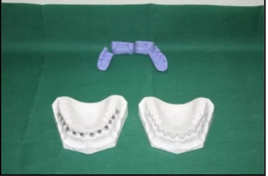
Five groups of IDB technique Working & Patient models
16
Figure 2 Armamentarium Materials 17
Figure 3 Working & Patient model 18
Figure 4 Bonding adhesive kit 19
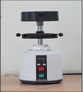
Figure 5 Vacuum form machine 20
Figure 6
Jig with positioning blocks for photographic measurements
21
Figure 7 Vernier caliper measurements 22
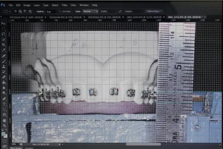
Figure 8 Grid line measurements 23
Figure 9 PVS Putty 24
Figure 10 Single - VF 25
Figure 11 PVS Clear 26
Figure 12 Double VF 27
TABLE
NO TITLE PAGE NO
Table 1 Tray descriptions for indirect bonding
S.NO TITLE PAGE NO
Chart 1 Mean Difference in M -D dimension – X axis – Point A 38
Chart 2 Mean Difference in M -D dimension – X axis – Point B 38
Chart 3 Mean Difference O -G dimension Y -axis – Point A 39
Chart 4 Mean Difference in O -G dimension – Y axis – Point B 39
Chart 5 Mean Difference in B -L dimension – Z axis – Point A 40
B-L : Bucco-Lingual
DVF : Double Vacuum Form
IDB : Indirect Bonding Technique
M-D : Mesio-Distal
O-G : Occluso-Gingival
PVS-VF : Poly Vinyl Siloxane-Vacuum Form
PVS : Poly Vinyl Siloxane
PETG : Polyethylene Terephthalate Glycol
INTRODUCTION
Edward Hartley Angle1, introduced the concept of fixed orthodontic appliance therapy with bands cemented on all teeth. In those early days, brackets were welded to gold or stainless steel bands. Before treatment, the orthodontist had to create enough space around each tooth to accommodate the bands, and then those spaces had to be closed at the end of treatment, when bands were removed. Buonocore2 (1955) introduced Acid Etching technique, the basis for the adhesion of brackets to enamel with phosphoric acid. He demonstrated increased adhesion produced by acid pre treatment of enamel. In 1965 with advent of Epoxy resin bonding, Newman3 began to apply these findings to direct bonding of orthodontic attachments.
The introduction of light cure resin allowed increased working time and significant positioning of brackets before the light is cured. Direct bonding has its own limitations like increased chair side time, bracket positioning errors and difficulty in bonding the posteriors because of decreased visibility. These limitations caused most clinicians to use direct bonding of brackets on anterior tooth and premolars and largely to avoid direct bonding on molars.3
In 1972, Silverman and Cohen4 devised a method of delivering full arch brackets onto the dentition on one time. The operator obtained a stone model of the patient’s dentition and then treated labial surface of the model with liquid foil separating medium. Metal or plastic brackets were bonded with caramel candy.
In 1979, Dr Royce Thomas5 developed an innovative indirect bonding technique called “Custom base”. This laboratory technique attaches brackets to a stone model with composite adhesive. This technique established the foundation for contemporary indirect bonding technique.
At present lot of biomaterials are available for making indirect bonding trays. Most commonly indirect bonding trays are fabricated with vacuum forme d thermoplastics, silicone impression materials, or a combination of both.
The purpose of this current study was to measure and compare bracket transfer accuracy of five Indirect bonding techniques. The five indirect bonding techniques includes
AIM AND OBJECTIVES
AIMS:
To measure and compare the bracket transfer accuracy between the five indirect bonding techniques.
OBJECTIVE:
To measure bracket transfer accuracy in five different indirect bonding techniques.
To compare the bracket transfer accuracy among the five indirect bonding techniques.
REVIEW OF LITERATURE
Michael J. Aguirre et al (1982)6 evaluated the bond strength of bracket placement comparing direct bonding to indirect bonding technique and concluded that maxillary canine showed better bond strength in indirect bonding when compared to premolars and incisors in vertical bracket placemen t.
Nasib Balut et al (1992)7 determined the accuracy of bracket placement with the direct bonded technique. Fifty sets of models served as the population of the study. Photographs of the models were measured to determine vertical and angular discrepancie s. A mean of 0.34mm for the vertical discrepancies and a mean of 5.54 degree angular discrepancies are found in orthodontic bracket placement. With proper wire bending or rebonding bracket positions, an excellent result can be achieved.
Koo BC et al (199 9)8 compared the accuracy of bracket placement between direct and indirect bonding techniques and concluded that indirect bonding had better bracket placement in bracket height and no significant difference was found between direct and indirect bonding technique regarding the angulation and mesiodistal position.
distal aspect of the bracket and concluded that intraindividual variation in tooth morpholo gy is larger than the variation between the different types of preadjusted appliances.
Arndt Klocke et al (2003)1 0 evaluated bond strength for a cyanoacrylate adhesive in combination with an indirect bonding technique. Eighty bovine permanent mandibular incisors were randomly divided into four groups of 20 each. The study concluded that bond strength for an indirect bonding technique using the cyanoacrylate adhesive was found to be lower than Thomas technique using a composite adhesive.
Arndt Klocke et al (2003)1 1 evaluated in vitro investigation of indirect bonding with a hydrophilic primer. Moisture contamination after application of hydrophilic primer resulted in significantly lower bond strength compared with bond strength for uncontaminated enamel.
Pablo Echarri (2004)1 3 describes indirect bonding has a number of advantages over direct bonding, including greater accuracy of bracket positioning and the ability to customize bracket prescriptions according to individual case requirements.
Fabio Ciuffolo et al (2005)1 4 describes a new method o f preparing trays for indirect bracket bonding. Computer aided technology is used to design the individualized trays, which are then produced with a rapid prototyping procedure.
Jacob Daub et al (2006)1 5 evaluated the shear bond strength of one direct and two indirect bonding methods after thermocycling. The study shows that tooth indirectly bonded and light cured had a lower bond survival rate as compared to others after thermocycling.
John T. Kalange(2007)1 6 evaluated a highly accurate method for Precision based full arch indirect bonding in which vertical and horizontal reference lines are placed on working models for bracket placement. The use of this technique offers quality care and efficiency of treatme nt. It is quick, accurate and reliable and ensures idealized anterior gingival margin contour and overall excellence in facial esthetics.
Nir Shpack et al (2007)1 8 evaluated bracket placement in l ingual vs labial systems and direct vs indirect bonding. Forty pretreatment dental casts of 20 subjects were selected. The study concluded that indirect bonding was more accurate than direct bonding for all teeth in both labial and lingual orthodontics.
Bjorn U. Zachrisson (2007)1 9 evaluated direct bonding in orthodontic treatment and retention a post treatment evaluation. The study concluded that carefully performed bonding techniques may be of value, on anterior tooth, premolars and second molars, while first molars were better banded.
David Armstrong et al (2007)2 0 evaluated a comparision of accuracy in bracket positioning between two techniques – localizing the centre of the clinical crown and measuring the distance from incisal edge. The study concl uded that placement of brackets in positions appears to be more accurate in vertical dimensions for both upper and lower anterior teeth.
B. Wendl (2008)2 1 determined Indirect bonding – a new transfer method. The study concludes that Aptus bonding device was found to provide accurate transfer method for indirect bonding of brackets. The bond strength tested in vitro is sufficient for orthodontic purposes and comparable with direct bonding.
that advantage of indirect bonding relative to direct bonding are numerous which outweigh the perceived disadvantage of increased laboratory time and technique sensitivity.
S.Madhusudhan (2010)2 3 determined a newly simplified indirect bonding technique. The study concluded that technique is simple accurate and cost effective indirect bonding procedure.
Mauricio et al (2011)2 4 described the indirect bonding procedure with thermal glue transfer t ray and brackets with positioning jigs for precise bracket position and concluded that the technique is simple, accurate, reliable and inexpensive.
Domenico et al (2012)2 5 evaluated the effectiveness of an indirect bonding technique in reducing plaque accumulation around the braces. The study concluded the plaque accumulation around different bracket margins did not differ significantly.
Katiyar R et al (2014)2 7 evaluated a simplified indirect bonding technique. The advantage of this technique over existing techniques are easy removal of of excess resin flash, better curing accessibility and easy removal of transfer tray. Reduced chair side time, simple and cost-effective method for indirect bonding.
Qamaruddin I et al (2014)2 8 evaluated indirect bonding with hot glue gun method. The study concluded that this is the most convenient and economical method for indirect bracket bonding. Although technique sensitive i t does not require any additional training for lab technicians and assistants.
P Premanand et al (2014)2 9 compared the efficacy between direct and indirect bonding methods. Twenty patients were chosen with selection criteria being, no visible cracks on en amel, normal tooth morphology and non extraction treatment plan. The study concluded that the indirect bonding technique is definitely a better option and could provide efficient bracket placement in significantly less chair side time which overweighs the cost involved in the laboratory procedure.
Fahad F. Alsulaimani (2014)3 1 determined the effect of lactic acid etching on bonding effectiveness of orthodontic bracket after water storage. The study concl uded that six months of water storage significantly reduced the shear bond strength regardless of the etchant used.
Lincoln Issamu Nojima et al (2015)3 2 describes Indirect bonding technique ensures precise bracket position. Simplicity, accuracy and repro ducibility of this technique lead to its efficiency in orthodontic bonding providing the advantages related to indirect bonding.
M Swetha et al (2015)3 3 compared Indirect versus direct bonding – A shear bond strength comparision: An in vitro study. The st udy concluded that Bond strength obtained with Thomas indirect bonding technique was comparable with direct bonding technique. The journal of contemporary dental science.
Breuning KH (2015)3 4 Bonding metal brackets on tooth surfaces. The study signifies that bonding forces should not be too low to prevent bracket failure. Bracket failure is due to increased time and cost.
were fabricated over working models (n = 5 per technique) to transfer brackets to another 25 identical stone patient models and conclud ed that silicone based trays had consistently high accuracy in transferring brackets than vaccum formed trays.
MATERIALS AND METHODS
PREPARATION OF STUDY MODELS
Silicone molds of the master model were fabricated. From the molds, 100 identical orthodontic stone models (Orthokal) were fabricated; 50 served as working models to which the brackets were bonded. Each tooth had two reference notches: one on the facial and another on the lingual side. 3 5 IDB trays were fabricated (n=10 per technique) to transfer the brackets to another 50 identical patient models.
Indirect Bonding Techniques
All trays were fabricated following the manufacturer’s guideline. In general, all trays extended from the facial over to the occlusal and at least half of the lingual surfaces.3 5
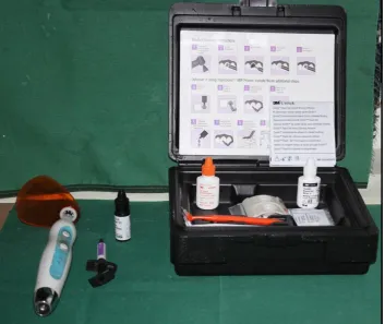
Working model -tray assemblies were soaked in water for 1 hour to dissolve the separating agent. After removal, trays with brackets were cleaned using a detergent solution in an ultrasonic cleaner as described by Sondhi.3 A two-part (A -B) chemically cured bonding adhesive (Sondhi Rapid -Set Indirect Bonding Adhesive, 3M Unitek, St Paul, Minn) was used during IDB. Part A was applied to the patient model teeth and part B to the custom composite pads on brackets.
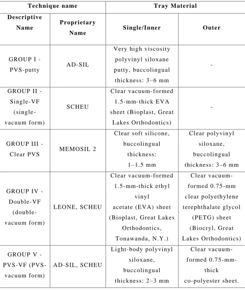
Table 1: Tray Descriptions for Indirect Bonding Techniques
Technique name Tray Material
Descriptive
Name Proprietary
Name Single/Inner Outer
GROUP I -
PVS-putty AD-SIL
Very high viscosity polyvinyl siloxane putty, buccolingual
thickness: 3 –6 mm
-
GROUP II - Single-VF (single- vacuum form) SCHEU Clear vacuum-formed 1.5-mm-thick EVA sheet (Bioplast, Great
Lakes Orthodontics)
-
GROUP III -
Clear PVS MEMOSIL 2
Clear soft silicone, buccolingual thickness: 1–1.5 mm Clear polyvinyl siloxane, buccolingual thickness: 3 –6 mm
GROUP IV - Double-VF (double-vacuum form) LEONE, SCHEU Clear vacuum-formed 1.5-mm-thick ethyl vinyl
acetate (EVA) sheet (Bioplast, Great Lakes
Orthodontics, Tonawanda, N.Y.)
Clear vacu um-formed 0.75 -mm clear polyeth ylene terephthalate glycol
(PETG) sheet (Biocryl, Great Lakes Orthodontics) GROUP V -
PVS-VF (PVS - vacuum form)
AD-SIL, SCHEU
Light-body polyvinyl siloxane,
buccolingual thickness: 2 –3 mm
Clear vacuum-formed 0.75
-mm-thick
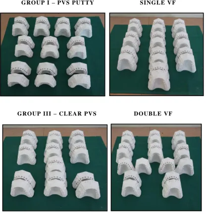
Fig 1: FIVE GROUPS OF IDB TECHNIQUE
WORKING AND PATIENT MODELS
GROUP I – PVS PUTTY SINGLE VF
GROUP III – CLEAR PVS DOUBLE VF
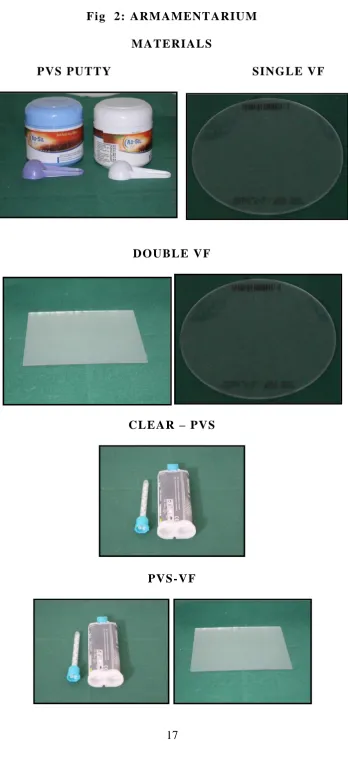
Fig 2: ARMAMENTARIUM
MATERIALS
PVS PUTTY SINGLE VF
DOUBLE VF
CLEAR – PVS
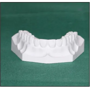
[image:28.595.125.474.63.822.2]Fig 3
WORKING MODEL
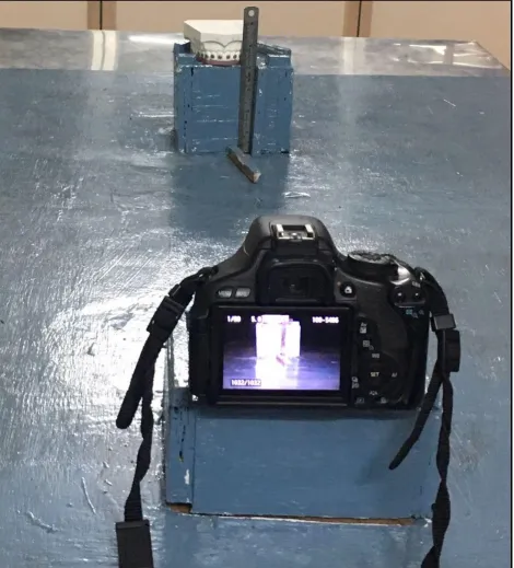
Fig 6: JIG WITH POSITIONING BLOCKS FOR PHOTOGRAPHIC
[image:32.595.75.546.128.648.2]Fig 9: PVS PUTTY
BRACKETS BONDED ON WORKING MODEL
IDB TRAY
[image:35.595.165.433.591.767.2]Fig 10: SINGLE - VF
BRACKETS BONDED ON WORKING MODEL
IDB TRAY
[image:36.595.166.437.588.767.2]Fig 11: PVS CLEAR – MEMOSIL
BRACKETS BONDED ON WORKING MODEL
IDB TRAY
Fig 12 : Double VF
BRACKETS BONDED ON WORKING MODEL
IDB TRAY
[image:38.595.141.456.591.766.2]Photographic Measurements: Mesiodistal and Occluso gingival
Bonded teeth on both working and patient models were photographed individually using a Canon EOS 600D camera with EFS 18-55mm Macrolens set on manual with an F -stop of 40 at 1:500 of a second.JPEG images (300 to 300 dpi) were imported into Adobe Photoshop Elements 10 (Adobe Systems Inc, San Jose, Calif) and magnified 8.5 times. For each bracket, mesiodistal (M -D) (X-axis) and occlusogingival(O -G) (Y-axis) measurements were made as follows,
A software -constructed grid was calibrated so that the distance between the gridlines equaled 1.00 mm.
The image was rotated so that the horizontal and vertical lines outlining the corner of the facial reference notch closest to the bracket paralleled the gridlines.
The origin of the grid was made coincident with the intersection of the lines outlining the corner of the reference notch.
Two points (A, B) were selected to coincide with the intersection of the inside of the mesial and distal tie wings with the occlusal edge of the scribe line base, except for the molars, where the inner occlusal corners of the tie wings were used.
For each point, x - and y-coordinate values were recorded to the nearest 0.01 mm.
Caliper Measurements: Buccolingual
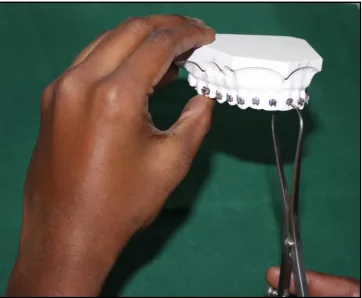
Manual caliper were used to measure buccolingual (B-L) ( Z-axis ) bracket positions as follows:
Two points (C, D) were selected at the gingival and occlusal ends of the bracket scribe line, ex cept for molars, where the points were located at the intersection of the gingival and occlusal scribe lines with the bracket base.
One tip of a manual caliper was placed at the depth of the lingual reference notch, and the other tip was aligned with each of the points.
Measurements were recorded to the nearest 0.01 mm.
RESULTS
Statistical Analysis
To assess the accuracy of each technique, bracket coordinate value differences for each tooth between working and patient model pairs (n = 10) were analyzed using paired t -tests. For technique comparisons, analyses were conducted for all teeth grouped, ant erior teeth, posterior teeth of the arch using analysis of variance followed by Tukey post hoc comparisons. Significance was set to P < .05.
PAIRED t-tests
GROUP I - PVS PUTTY
VARIABLES
BEFORE AFTER t -
Test
P -
Value
RESULT
N MEAN ±SE MEAN ±SE
MD-1A 10 2.64 0.0178 2.59 0.0315 1.77 0.1114 NS
MD-1B 10 2.16 0.0083 2.13 0.0285 1.08 0.3092 NS
OG-1A 10 4.17 0.0285 4.04 0.0657 2.99 0.0152 *
OG-1B 10 4.10 0.0329 3.98 0.0655 2.70 0.0245 *
BL-1A 10 6.94 0.0178 6.72 0.0817 2.68 0.0252 *
BL-1B 10 6.35 0.0140 6.17 0.0858 2.18 0.0572 NS
GROUP II - SINGLE - VF
VARIABLES
BEFORE AFTER t -
Test
P -
Value
RESULT
N MEAN ±SE MEAN ±SE
MD-1A 10 2.62 0.0255 2.40 0.0643 3.47 0.0070 **
MD-1B 10 2.18 0.0290 2.00 0.0541 3.28 0.0095 **
OG-1A 10 4.25 0.0370 3.69 0.0992 6.40 0.0001 **
OG-1B 10 4.22 0.0340 3.67 0.0978 6.40 0.0001 **
BL-1A 10 6.90 0.0243 6.32 0.1734 3.51 0.0066 **
GROUP III - CLEAR - PVS
VARIABLES
BEFORE AFTER t -
Test
P -
Value
RESULT
N MEAN ±SE MEAN ±SE
MD-1A 10 2.63 0.0184 2.63 0.0136 0.00 1.0000 NS
MD-1B 10 2.16 0.0231 2.17 0.0215 1.00 0.3434 NS
OG-1A 10 4.25 0.0294 4.21 0.0252 3.00 0.0150 *
OG-1B 10 4.17 0.0329 4.13 0.0285 3.00 0.0150 *
BL-1A 10 6.74 0.0271 6.73 0.0306 1.00 0.3434 NS
BL-1B 10 6.24 0.0256 6.23 0.0265 1.00 0.3434 NS
GROUP IV - DOUBLE - VF
VARIABLES
BEFORE AFTER t -
Test
P -
Value
RESULT
N MEAN ±SE MEAN ±SE
MD-1A 10 2.63 0.0186 2.56 0.0352 1.56 0.1527 NS
MD-1B 10 2.16 0.0150 2.13 0.0311 1.18 0.2695 NS
OG-1A 10 4.20 0.0241 4.01 0.0624 2.96 0.0161 *
OG-1B 10 4.14 0.0299 3.96 0.0724 2.85 0.0191 *
BL-1A 10 6.84 0.0262 6.72 0.0788 1.82 0.1018 NS
GROUP V - PVS - VF
VARIABLES
BEFORE AFTER t -
Test
P -
Value
RESULT
N MEAN ±SE MEAN ±SE
MD-1A 10 2.68 0.0150 2.64 0.0127 1.50 0.1679 NS
MD-1B 10 2.18 0.0167 2.18 0.0111 0.00 1.0000 NS
OG-1A 10 4.10 0.0188 4.08 0.0158 1.86 0.0957 NS
OG-1B 10 4.08 0.0215 4.06 0.0178 1.96 0.0811 NS
BL-1A 10 6.73 0.0178 6.73 0.0178 1.00 0.3434 NS
BL-1B 10 6.11 0.0206 6.11 0.0208 1.00 0.3434 NS
P > 0.05 – NS – Not Significant
P < 0.05 - * - Significant
P < 0.01 - ** - Highly Significant
MD – Mesiodistal – X-axis
OG – Occlusogingival – Y-axis
ANALYSIS OF VARIANCE
N Mean Std.
Deviation
Std.
Error
F-value P-value
B-MD-1A
PVSP 10 2.6417 .05625 .01779
1.352 0.266 SVF 10 2.6167 .08051 .02546
MEMO 10 2.6333 .05827 .01843 DVF 10 2.6250 .05893 .01863 PVSVF 10 2.6750 .04730 .01496 Total 50 2.6383 .06211 .00878
A-MD-1A
PVSP 10 2.5917 .09977 .03155
7.166 0.000 SVF 10 2.4000 .20337 .06431
MEMO 10 2.6333 .04303 .01361 DVF 10 2.5583 .11146 .03525 PVSVF 10 2.6417 .04025 .01273 Total 50 2.5650 .14215 .02010
B-MD-1B
PVSP 10 2.1583 .02635 .00833
0.357 0.838 SVF 10 2.1750 .09171 .02900
MEMO 10 2.1583 .07297 .02307 DVF 10 2.1583 .04730 .01496 PVSVF 10 2.1833 .05270 .01667 Total 50 2.1667 .06070 .00858
A-MD1B
PVSP 10 2.1250 .09001 .02846
4.874 0.002 SVF 10 2.0000 .17123 .05415
MEMO 10 2.1667 .06804 .02152 DVF 10 2.1250 .09821 .03106 PVSVF 10 2.1833 .03514 .01111 Total 50 2.1200 .11804 .01669
B-OG1A
PVSP 10 4.1667 .09001 .02846
4.989 0.002 SVF 10 4.2458 .11695 .03698
A-OG1A
PVSP 10 4.0420 .20787 .06574
9.775 SVF 10 3.6917 .31378 .09923
MEMO 10 4.2125 .07966 .02519 DVF 10 4.0083 .19720 .06236 PVSVF 10 4.0792 .04988 .01577 Total 50 4.0067 .25497 .03606
B-OG1B
PVSP 10 4.0958 .10403 .03290
0.331 SVF 10 4.2208 .10767 .03405
MEMO 10 4.1667 .10393 .03287 DVF 10 4.1417 .09461 .02992 PVSVF 10 4.0833 .06804 .02152 Total 50 4.1417 .10548 .01492
A-OG1B
PVSP 10 3.9833 .20713 .06550
7.629 SVF 10 3.6667 .30932 .09782
MEMO 10 4.1250 .09001 .02846 DVF 10 3.9583 .22906 .07244 PVSVF 10 4.0583 .05625 .01779 Total 50 3.9583 .24972 .03532
B-BL1C
PVSP 10 6.9417 .05625 .01779
16.854 SVF 10 6.8958 .07670 .02426
MEMO 10 6.7375 .08573 .02711 DVF 10 6.8417 .08287 .02621 PVSVF 10 6.7292 .05642 .01784 Total 50 6.8292 .11016 .01558
A-BL1C
PVSP 10 6.7208 .25839 .08171
3.661 SVF 10 6.3208 .54823 .17337
B-BL-1D
PVSP 10 6.3458 .04414 .01396
27.071 0.000 SVF 10 6.4333 .07658 .02422
MEMO 10 6.2375 .08110 .02565 DVF 10 6.2542 .09097 .02877 PVSVF 10 6.1125 .06529 .02065 Total 50 6.2767 .12980 .01836
A-BL-1D
PVSP 10 6.1667 .27146 .08584
2.207 0.083 SVF 10 5.9000 .48201 .15242
MEMO 10 6.2292 .08391 .02653 DVF 10 6.1417 .19165 .06060 PVSVF 10 6.1083 .06573 .02079 Total 50 6.1092 .27892 .03945
P > 0.05 – NS – Not Significant
P < 0.05 - * - Significant
P < 0.01 - ** - Highly Significant
A – After tray transfer
B – Before tray transfer
MD – Mesiodistal – X-axis
OG – Occlusogingival – Y-axis
BAR CHART
Chart 1 : Mean Difference in M -D dimension – X axis – Point A
Chart 2 : Mean Difference in M -D dimension – X axis – Point B -0.0500
0.0000 0.0500 0.1000 0.1500 0.2000 0.2500
PVSP SVF MEMO DVF PVSVF
-0.0200 0.0000 0.0200 0.0400 0.0600 0.0800 0.1000 0.1200 0.1400 0.1600 0.1800
Chart 3 : Mean Difference in O -G dimension – Y-axis – Point A
Chart 4 : Mean Difference in O -G dimension – Y-axis – Point B 0.0000
0.1000 0.2000 0.3000 0.4000 0.5000 0.6000
PVSP SVF MEMO DVF PVSVF
0.0000 0.1000 0.2000 0.3000 0.4000 0.5000 0.6000
Chart 5 : Mean Difference in B -L dimension – Z-axis – Point A
Chart 6 : Mean Difference in B -L dimension – Z-axis – Point B 0.0000
0.1000 0.2000 0.3000 0.4000 0.5000 0.6000
PVSP SVF MEMO DVF PVSVF
0.0000 0.1000 0.2000 0.3000 0.4000 0.5000 0.6000
RESULTS
Method Error
The method error was 0.07 mm for photographic measurements and 0.01 mm for caliper measurements.
Debonded Brackets
Eleven of the 600 brackets that were transferred debonded during tray removal: two with PVS putty, eight with single VF, one with double VF.
Comparisons between Working and Patient Models
DISCUSSION
The goal of the study was to compare the bracket transfer accuracy of five different indirect bonding tray transfer material. Ideal maxillary model was taken for this study to minimize the variables related to arch form, tooth shape and crowding. Henceforth assessment is made in ideal maxillary arch and reference notches are made in distolabial line angle in the labial surface and lingually in middle third of the tooth for measurement purpose.
The transferred brackets were phot ographed and saved as JPEG images and imported to Adobe photoshop CS6 and measurements are taken in mesiodistal (M -D) and occlusogingival (O -G) direction. Five techniques were compared and bracket transfer accuracy was found between the five groups and it was found that single VF and double VF has less accuracy in transferring the brackets because of less thickness of tray material and increased elongation of ethyl vinyl acetate (EVA) sheets in anteriors
Each technique had at least one tooth with significant differences i n bracket position; however, many differences were small, suggesting the need to set thresholds of clinical significance. Armstrong et al.3 7 reported that changes of > 0.25 mm for upper central and lower incisor brackets and > 0.5 mm for remaining teeth wo uld be clinically significant. For this study, a 0.13 -mm discrepancy in any direction was deemed clinically significant, so if brackets on adjacent teeth were misplaced by 0.13 mm in opposite directions, tooth position discrepancies would be .0.25 mm. Sing le-VF and double -VF showed the greatest number of teeth with clinically significant differences, whereas PVS -VF had none.
Two measurement points were used per dimension to determine whether the bracket position changes were rotational or purely linear. Ten teeth (at least one per technique) had significant differences in only one of two points, indicating that bracket rotations had occurred.
Significant differences in the B -L measurements on tooth no:21 (point C) and on tooth no:16 (point D) wi th single -VF likely relate to
the large differences that were also present in the O -G direction, causing angulation changes in the B -L line of measurement when using calipers. This is inherent to the use of calipers and may be considered a weakness of the study.
were significantly less accurate than the others in th e O-G direction, a result also evident for anterior but not for posterior teeth. For posterior teeth, O -G accuracy was significantly lower for single -VF versus double -PVS, and although potentially clinically significant, the mean change (0.15 mm) was consi derably smaller than mean O -G changes for double -VF and single -VF with anterior teeth (0.29 mm and 0.33 mm, respectively). In the B -L direction, posterior teeth showed significantly lower accuracy for Clear -PVS versus PVS -VF and double -VF, but the small me an change (0.11 mm) is likely not clinically significant.
The poorer O -G accuracy for anterior teeth with double -VF and single-VF can be attributed to increased elongation of the ethyl vinyl acetate (EVA) sheets over the longer anterior crowns, resulting in relatively decreased anterior tray thickness. Ryokawa et al. found significant decreases in material thickness and elastic modulus (decreased rigidity) for EVA following vacuum -forming. Although double -VF has an outer polyethylene terephthalate glycol (PETG) tray, it is not fused with the inner EVA tray and is trimmed occlusal to the gingival bracket wings, away from all heights of contour, 12 and thus does not cover the entire O -G dimension of the brackets.
the O-G direction, could be a result of the greater sensitivity of these techniques to differences in hand pressure.
The three techniques with the highest bracket transfer accuracy used polyvinyl siloxane (addit ion silicone) as the material in direct contact with both brackets and tooth surfaces. Additional silicone impression materials have been shown to have excellent dimensional stability, superior recovery from deformation (elastic recovery), and high rigidit y. Additionally, all silicone trays were thicker buccolingually than nonsilicone trays, contributing to increased rigidity.
SUMMARY AND CONCLUSION
All IDB techniques investigated had at least one tooth with statistically significant differences in bracket position, but the most clinically significant differences were found with the single-VF and double -VF techniques.
Changes in bracket position were not purely linear; some were rotational.
When the five techniques were compared, bracket transfer accuracy was comparable for the silicone -based techniques.
BIBLIOGRAPHY
1. Thomas M. Graber, Robert L. Vanarsdall, Katherine W.L Vig Orthodontics Current Principles and Techniques. 2000.
2. Michael G. Buonocore. A simple method of increasing the adhesion of acrylic filling materials to enamel surfaces. Journal Dent Res 1955; 34:849.
3. Anoop Sondhi. Bonding in the new millennium: Reliable and consistent bracket placementwith indirect bonding. WJO, Vol 2, No:2, 2001.
4. Silverman E, Cohen M, Gianelly AA, Dietz VS. A universal direct bonding system for both metal and plastic brackets. Am J Orthod. 1972; 62:236 –244.
5. Thomas RG. Indirect bonding: simplicity in action. J Clin Orthod. 1979;13:93 –106.
6. Michael J. Aguirre, Gregory J. King, Joseph M. Waldron. Assessment of bracket placement and bond strength when comparing direct bond ing to indirect bonding techniques. AJODO Oct 1982, Vol82 (4):269 -276.
8. Koo BC, Chung CH, Vanarsdall RL. Comparision of the accuracy of bracket placement between direct and indirect bonding techniques. AJODO. 1999;116:346 -351.
9. Rainer R. Miethke, Birte Melson. Effect of variation in tooth morphology and bracket position on first and third order correction with preadjusted appliances. AJODO, Vol 116, No 3. Sep 1999.
10.Arndt Klocke, Dr med dent, Jianmin Shi, Barbel Kahl -Nieke, Ulrich Bismayer. In vitro evaluation of a moisture -active adhesive for indirect bonding. Angle orthod, Vol 73, No 6, 2003.
11.Arndt Klocke, Dr med dent, Jianmin Shi, Barbel Kahl -Nieke, Ulrich Bismayer. In vitro investigation of indirect bonding with a hydrophilic primer. Angle orthod, vol 73, No 4, 2003.
12.Arndt Klocke, Dr med dent, Drazen Tadic, Farhad Vaziri, Barbel Kahl Nieke. Cus tom base presaging in indirect bonding. Angle Orhod, Vol 74, No 1, 2004.
13.Pablo Echarri. Double transfer trays for indirect bonding. JCO, Vol 38:No:1: (8 -13) 2004.
15.Jacob Daub, David Berzins, Brandon James Linn, Thomas Gerard Bradley. Bond strength of Direct and Indirect bonded brackets after thermocycling. Angle orthod, Vol 76, N o 2, 2006.
16.John T. Kalange Prescription -based precision full arch indirect bonding. Semin orthod, Vol 13, No1 (March), 2007: 19 -42.
17.Duncan W. Higgins. Indirect bonding with Light -cured adhesive and a hybrid tray transfer. Semin orthod, Vol 13, No 1 (March) , 2007:64-68.
18.Nir Shpack, Silvia Geron, Ioannis Floris, Moshe Davidovitch, tamar Brosh, Alexander Dan Vardimon. Bracket placement in Lingual vs labial systems and Direct vs Indirect Bonding. Angle orthod, Vol 77, No 3, 2007.
19.Zachrisson, Bjorn U. Direct bon ding in orthodontic treatment and retention a post -treatment evaluation. EJO. Apr 2007, Supplement1,Vol29:28 -34.
20.David Armstrong, Gang Shen, Peter Petocz, M. Ali Darendeliler. A comparision of accuracy in bracket positioning between two techniques – localizing the centre of the clinical crown and measuring the distance from the incisal edge. EJO 29 (2007) 430-436.
22.Mark joiner. In house precision bracket place ment with the indirect bonding technique. AJODO June 2010.
23.S. Madhusudhan, S. M. Laxmikanth, Pradeep Chandra Shetty. A Newly Simplified Indirect Bonding Technique. Indian Journal of Dental Sciences.
24.Mauricio Mezomo, Edurado Martinelli Santayana de Lima, Luciane m.de Menezes, Andre Weissheimer. Indirect bonding with thermal glue and brackets with positioning jigs. Progress In Orthodontics 12 (2011) 180 -185.
25.Domenico Dalessandri, Michela Dalessandri, Stefano Bonetti, luca Visconti, Corrado Paganelli. Effectiv eness of an indirect bonding technique in reducing plaque accumulation around braces. Angle orthod, Vol 82, No 2, 2012.
26.Lylian Kazumi Kanashrio, Julissa Janet Robles -Ruiz, Ana lidia Ciamponi, Igor Studart Medeiros, Gladys Cristina Dominguez, Solange Mongelli de Fantini. Effect of adhesion boosters on indirect bonding. Angle orthod, Vol 84, No1, 2014.
27.Katiyar R. A simplified indirect bonding technique. Vol 4, Issue 4, 103-106,2014
29.P Premanand, A J Shankar. An In -vivo comparision of the efficacy between direct and indirect bonding methods. Internet Scientific Publications.
30.Sudhir Sharma, Pradeep Tandon, Vinay K Chugh. A comparision of shear bond strength of orthodontic brackets bonded with four different orthodontic adhesives. Journal of orthodontic Science.
31.Fahad F. Alsulaimani. Effect of Lactic acid etching on bonding effectiveness of orthodontic bracket after water storage. ISRN Dentistry.
32.Lincoln Issamu Nojima, Adriele Silveria Araujo, Matheus Alves Junior. Indirect orthodontic bonding - a modified technique for improved efficiency and precision. Dental Press J Orthod. 2015 May-Jun;20(3).
33.M Swetha, Vinaya S Pal, N Sanjay, S Nan dhini. Indirect vs Direct bonding – A shear bond strength Comparision: An in vitro study. The Journal of Contemporary Dental Science
34.Breuning KH, Dekkers L, Beeftink M, Schols JH, Wolke J. Bonding metal brackets on tooth surface. Dentistry,2014.
36.Thorsten Grunheid, Mi chael S. Lee, Brent E. Larson. Transfer accuracy of vinyl polysiloxane trays for indirect bonding. Angle orthod. Vol 86: Isuue 3: 468 -474.
37.Armstrong D, Shen G, Petocz P, Ali Darendeliler M. A comparison of accuracy in bracket positioning between two techniques—localizing the centre of the clinical crown and measuring the distance from the incisal edge. Eur J Orthod. 2007;29:430 –436.
38.Bhatnagar S, Dhandapani G, Bagga DK. Evaluation of bond failures and shear bond strength in indirect bonding technique using 4 different transfer tray materials —an in vitro study. J Indian Dent Assoc. 2011;5:595 –598.
39.Ryokawa H, Miyazaki Y, Fujishima A, Miyazaki T, Maki K. The mechanical properties of dental thermoplastic materials in a simulated intraoral environment. Orthod Wave s. 2006; 65:64 –72.