0022-538X/07/$08.00⫹0 doi:10.1128/JVI.01917-06
Copyright © 2007, American Society for Microbiology. All Rights Reserved.
Involvement of Hsp90 in Assembly and Nuclear Import of Influenza
Virus RNA Polymerase Subunits
䌤
Tadasuke Naito,
1Fumitaka Momose,
2Atsushi Kawaguchi,
1and Kyosuke Nagata
1*
Department of Infection Biology, Graduate School of Comprehensive Human Sciences and Institute of Basic Medical Sciences,
University of Tsukuba, 1-1-1 Tennoudai, Tsukuba 305-8575, Japan,1and Department of Infection Control and Immunology,
Kitasato Institute for Life Sciences, Kitasato University, 5-9-1 Shirokane, Minato-ku, Tokyo 108-8641, Japan2
Received 2 September 2006/Accepted 10 November 2006
Transcription and replication of the influenza virus RNA genome occur in the nuclei of infected cells through the viral RNA-dependent RNA polymerase consisting of PB1, PB2, and PA. We previously identified a host factor designated RAF-1 (RNA polymerase activating factor 1) that stimulates viral RNA synthesis. RAF-1 is found to be identical to Hsp90. Here, we examined the intracellular localization of Hsp90 and viral RNA polymerase subunits and their molecular interaction. Hsp90 was found to interact with PB2 and PB1, and it was relocalized to the nucleus upon viral infection. We found that the nuclear transport of Hsp90 occurs in cells expressing PB2 alone. The nuclear transport of Hsp90 was in parallel with that of the viral RNA polymerase binary complexes, either PB1 and PB2 or PB1 and PA, as well as with that of PB2 alone. Hsp90 also interacted with the binary RNA polymerase complex PB1-PB2, and it was dissociated from the PB1-PB2 complex upon its association with PA. Furthermore, Hsp90 could form a stable PB1-PB2-Hsp90 complex prior to the formation of a ternary polymerase complex by the assembly of PA in the infected cells. These results suggest that Hsp90 is involved in the assembly and nuclear transport of viral RNA polymerase subunits, possibly as a molecular chaperone for the polymerase subunits prior to the formation of a mature ternary polymerase complex.
The influenza A virus contains eight segmented and nega-tive-stranded RNAs as its genome. The viral RNAs (vRNA) are associated with the viral RNA-dependent RNA polymer-ase subunits (PB1, PB2, and PA) and nucleoprotein (NP), forming structurally distinct viral ribonucleoprotein (vRNP) complexes (36). The vRNP complex is a basic unit for active transcription and replication. Transcription and replication of vRNA occur in the nuclei of infected cells. The PB1 subunit plays a central role in the catalysis of the polymerization of the RNA chain. It contains amino acid motifs that are common to RNA-dependent RNA polymerases and RNA-dependent DNA polymerases (2). The PB2 subunit is required for the transcription of vRNA. It binds to the methylated cap-1 struc-ture of host RNAs, and the capped oligonucleotide RNA is endonucleolytically cleaved by the PB1 subunits (8, 15). The resultant 10- to 13-nucleotide-long capped RNA fragment serves as a primer for viral mRNA synthesis. Genetic analyses suggest that the PA subunit is required for vRNA replication (14). The PA subunit induces a generalized proteolytic process (23, 34), and it is involved in the assembly of the polymerase subunits (13).
In negative-strand RNA viruses, RNA-dependent RNA polymerases are present in the virion. Purified vRNP com-plexes or RNA polymerases catalyze transcription from vRNA in vitro; however, the vRNP complexes alone are not sufficient for genome replication or for the efficient transcription of viral RNAs. Some of the paramyxoviruses and rhabdoviruses have
been shown to require host factors for efficient RNA synthesis in vitro. Tubulin is involved in the transcription of vesicular stomatitis virus, Sendai virus, and measles virus (20, 21, 28). Actin and-catenin stimulate viral RNA synthesis by the viral RNA polymerase of the human parainfluenza virus type 3 (3, 10). Heat shock protein 72/73 (Hsp72/73) stimulates the virus RNA polymerase activity of the canine distemper virus and measles virus (29). Hsp60 and translation elongation factor-1␣ bind to a transcriptase complex of vesicular stomatitis virus (5, 38). In the case of influenza virus, several host factors, such as NP- or PA-interacting factors, have been isolated (12, 31). Nucleoprotein-interacting protein 1 (NPI-1) and NPI-3 were identified using theSaccharomyces cerevisiaetwo-hybrid system (39). These two proteins were shown to mediate the nuclear import of NP (31). A human cellular protein, namely, hCLE, interacts with the PA subunit (12).
By using an in vitro RNA synthesis assay system, we identified host factors that stimulate influenza virus RNA synthesis from uninfected HeLa cell nuclear extracts; these host factors were designated RAF-1 (RNA polymerase activating factor 1) and RAF-2 (18, 19). RAF-1 is found to be identical to Hsp90. RAF-2 consists of two subunits, namely, RAF-2p48 and RAF-2p36. RAF-2p48 has also been identified as NPI-5, BAT-1, or UAP56. Hsp90 interacts with the PB2 subunit through the N-terminal chaperone domain and the middle region that contains a highly acidic domain; the virus RNA synthesis stimulatory activity of Hsp90 depends on this acidic domain of the middle region (19). RAF-2p48/NPI-5/BAT-1/UAP56 is well characterized as a splic-ing factor belongsplic-ing to the DEAD-box family of RNA-dependent ATPases (42). Furthermore, RAF-2p48 has been identified as NPI-5, an NP-interacting protein in a yeast two-hybrid screen of a mammalian cDNA library (32). RAF-2p48 binds to the free NP and promotes NP-RNA complex formation (18).
* Corresponding author. Mailing address: Department of Infection Biology, Graduate School of Comprehensive Human Sciences and Institute of Basic Medical Sciences, University of Tsukuba, 1-1-1 Ten-nodai, Tsukuba 305-8575, Japan. Phone and fax: (81) 298-53-3233. E-mail: knagata@md.tsukuba.ac.jp.
䌤Published ahead of print on 22 November 2006.
1339
on November 8, 2019 by guest
http://jvi.asm.org/
Hsp90 is a cellular molecular chaperone that belongs to the Hsp family (33, 35, 37). Hsp90 is an abundant and highly conserved protein and is essential for viability in eukaryotes. It has a housekeeping function that contributes to the folding, activation, and assembly of a variety of proteins, such as tran-scription factors, steroid receptors, and protein kinases. These substrates of Hsp90 are involved in cell cycle regulation and signal transduction. Hsp90 functions in cooperation with other chaperones and cochaperones, such as Hsp70, Hop, p50/ CDC37, Aha1, p23, Hip, and PA28. Hsp90 is regulated by Hsp90 cochaperones in substrate recognition, the ATPase cy-cle, and chaperone function. Further, a number of viral pro-teins, including the core protein 4a of the vaccinia virus, the sigma protein of the reovirus, the simian virus 40 T antigen, NS2/3 of the hepatitis C virus, and reverse transcriptase of the hepatitis B virus, have been identified as targets of Hsp90 (11, 17, 40, 43).
We have previously reported that Hsp90 binds to PB2 and is relocalized to the nucleus after influenza virus infection (19). Here, we attempted to examine the physiological implications of this phenomenon. We demonstrated that the expression of PB2 results in the nuclear relocalization of Hsp90, suggesting that Hsp90 is imported into the nucleus through the nuclear localiza-tion signal of PB2 (22) by its binding to PB2. We found that Hsp90 also interacts with the PB1 subunit and the binary RNA polymerase complex PB1-PB2 and modulates the interaction of PA with PB1. In contrast, Hsp90 was not bound to the mature RNA polymerase complex PB2-PB1-PA. We discussed the func-tion of Hsp90 as a host factor that is involved in the nuclear transport and assembly of viral RNA polymerase subunits.
MATERIALS AND METHODS
Vector construction.For construction of mammalian expression vectors for influenza virus polymerase subunits, cDNAs corresponding to the full-length PB1, PB2, PA, PB1 with a FLAG tag at its C terminus (PB1cFLAG), PB2 with a hemagglutinin (HA) tag at its C terminus (PB2cHA), PB2 with a Myc tag at its C terminus (PB2cMyc), PA with a FLAG tag at its C terminus (PAcFLAG), and PB1 devoid of the regions between amino acids 1 and 39 (PB1⌬N) were ampli-fied by PCR from a pcDNA-PB1, pcDNA-PB2, or pcDNA-PA (25) containing viral cDNAs derived from influenza virus A/Puerto Rico/8/34 (A/PR/8) as tem-plates using the following sets of primers: PB1-FOR (5⬘-GATCCCGGGCATA TGGATGTCAATCCGACCTTAC-3⬘) and PB1-REV (5⬘-GAACTCGAGAAG CTTATTTTTGCCGTCTGAGCTCT-3⬘) for PB1, 5⬘-GCGGATCCCATATGA TGGATACTGTCAACAGGAC-3⬘and PB1-REV for PB1⌬N, PB1-FOR and 5⬘-TCACTACTTGTCGTCGTCATCCTTGTAGTCTTTTTGCCGTCTGAGC TCTT-3⬘for PB1cFLAG, PB2-FOR (5⬘-CGCGGATCCCGGGCGGCCGCCA CCATGGAAAGAATAAAAGAACTAAGAAATCT-3⬘) and 5⬘-CGCGCTCG AGCTAATTGATGGCCATCCGAATTC-3⬘for PB2, PB2-FOR and 5⬘-TCAT TAAGCGTAATCTGGAACATCGTATGGGTAATTGATGGCCATCCGAA TTCTTTTGG-3⬘for PB2cHA, 5⬘-GAAAGAATAAAAGAACTAAGAAATCT AATGTCGCAGTC-3⬘ and 5⬘-TTGATGGCCATCCGAATTCTTTTGGTC G-3⬘for PB2cMyc, PA-FOR (5⬘-CGCGGATCCGCGGCCGCCACCATGGAA GATTTTGTGCGACAATGCTTC-3⬘) and 5⬘-TAGGATCCGCTAGCTCAATG CATGTGTAAGGAAGG-3⬘for PA, and PA-FOR and 5⬘-TCACTTGTCGTCGT CATCCTTGTAGCTCAATGCATGTGTAAGGAAGG-3⬘for PAcFLAG. Then, amplified PB1 and PB1⌬N PCR products were digested with NdeI and HindIII and blunted with Klenow fragment (TaKaRa). The amplified PB2 PCR product was digested with BamHI and XhoI and blunted with Klenow fragment. The amplified PA PCR product was digested with NdeI and BamHI and blunted with Klenow fragment. The amplified PB1cFLAG, PB2cHA, and PAcFLAG PCR products were phosphorylated with T4 polynucleotide kinase. The treated frag-ments were cloned into the XhoI site of pCAGGS which had been treated with Klenow fragment (27). The amplified PB2cMyc PCR products were phosphory-lated with T4 polynucleotide kinase and cloned into pBSIIKSP-cMyc (containing the Myc tag sequence for construction of a protein fused to the Myc tag), which
had been treated with digested NcoI and blunted with Klenow fragment. The resultant plasmid was designed pBSIIKSP-PB2cMyc. PB2cMyc fragments were excised from pBSIIKSP-cMyc plasmid by digestion with BamHI and EcoRI and then blunted with Klenow fragment. The fragment was cloned into the EcoRI site of pCAGGS which had been treated with Klenow fragment. The plasmid containing human Hsp90␣cDNA was a kind gift from I. Yahara. The insert DNA was amplified by PCR with human Hsp90␣cDNA as the template using a set of primers, 5⬘-CGGGATCCGTCGACATGCCTGAGGAAACCCAGAC-3⬘ and 5⬘-CGGGATCCGTCGACTTAGTCTACTTCTTCCATGCGTGA-3⬘. The amplified DNA fragment was digested with SalI and inserted into SalI-digested pBluescript II plasmid containing the FLAG tag sequence (pBSII-FLAG) (16). The resultant plasmid was designated pBSII-FLAG-Hsp90␣. The FLAG-tagged Hsp90␣gene fragment was excised from pBSII-FLAG-Hsp90␣by BamHI and XhoI and blunted with Klenow fragment. The fragment was cloned into the XhoI site of pCAGGS which had been treated with Klenow fragment.
Cells, virus infection, and transfection.Monolayer cultures of HeLa cells and MDCK cells were maintained at 37°C in minimal essential medium (MEM) (Nissui) containing 10% fetal calf serum (Cell Culture Technologies) and used for all experiments in this study. For infection, monolayer cultures of HeLa cells and MDCK cells in 60-mm-diameter dishes were washed twice with serum-free MEM. The cells were infected with influenza A/PR/8 virus at a multiplicity of infection (MOI) of 5 to 10 PFU per cell. After virus adsorption at 37°C for 1 h, the cells were washed with serum-free MEM and incubated at 37°C for 5 h with the growth medium (MEM containing 10% fetal calf serum). Transfection of HeLa cells with plasmids was carried out by using the calcium phosphate-mediated method (4). HeLa cells were grown on a 60-mm-diameter dish at 50% confluence and transfected with the calcium phosphate-DNA suspension. At 6 h posttransfection, the medium was replaced with fresh growth medium, and cells were further incubated for 24 h.
Indirect immunofluorescence assay.HeLa cells were grown on glass coverslips and infected with influenza virus or transfected with plasmids where indicated. One day after transfection, the cells were fixed with phosphate-buffered saline (PBS) containing 4% paraformaldehyde and then permeabilized with PBS containing 0.1% NP-40. The coverslips were soaked in 1% nonfat dry milk in PBS. The coverslips were then incubated for 30 min with a primary antibody. After incubation, the coverslips were washed three times for 5 min each time with PBS containing 0.1% NP-40 and then incubated for 30 min with secondary antibodies, either Alexa Fluor 488-conjugated donkey anti-mouse antibody (1/1,000; Molecular Probe), Alexa Fluor 488-conjugated donkey anti-rabbit antibody (1/1,000; Molecular Probe), Alexa Fluor 568-conjugated goat anti-mouse antibody (1/1,000; Molecular Probe), or Al-exa Fluor 568-conjugated goat anti-mouse antibody (1/1,000; Molecular Probe). Coverslips were washed with PBS containing 0.1% NP-40 and incubated for 10 min with 10 mM 4⬘,6⬘-diamidino-2-phenylindole dihydrochloride (DAPI). Coverslips were finally mounted on glass plates, and cells were observed by using a confocal laser-scanning microscope (Carl Zeiss).
Immunoprecipitation assay.Transfected or infected cells were washed twice with PBS and collected by centrifugation. Cell pellets were resuspended in lysis buffer (20 mM Tris-HCl [pH 7.9], 100 mM NaCl, 30 mM KCl, 1 mM EDTA, 0.1% NP-40, 20 mM sodium molybdate). After sonication, homogenates were centrifuged at 10,000⫻gat 4°C for 5 min. The supernatant fraction was used as extracts for assays. Cell extracts were mixed and incubated at 4°C for 4 h with specific antibody or antiserum. Then, protein A-Sepharose beads were mixed and rotated at 4°C for 2 h. The beads were washed three times with lysis buffer, and immunoprecipitated proteins were separated by 7.5% sodium dodecyl sulfate-polyacrylamide gel electrophoresis (SDS-PAGE), followed by Western blotting with anti-Hsp90 (MBL), anti-PB1, anti-PB2, or anti-PA antibodies. Proteins fused to the FLAG or Myc tag were detected by anti-FLAG (Sigma) and anti-Myc (Nacalai) antibodies. Hsp90 was detected by a rat anti-Hsp90 alpha monoclonal antibody (MBL) or rabbit anti-Hsp90 polyclonal antibody (a gener-ous gift from Y. Miyata and E. Nishida, Kyoto University).
Cross-linking of proteins.Infected or mock-infected cells were washed twice with PBS and collected by centrifugation. Cell pellet fractions were resuspended in PBS. DSP (dithiobis[succinimidylpropionate]) (PIERCE Biotechnology) was added to the suspension at a final concentration of 1 mM. DSP is a thiol-cleavable and primary amine-reactive cross-linker. A covalent amide bond is formed when theN-hydroxysuccinimide ester conjugation reagent reacts with primary amines. The reaction mixture was incubated at 25°C for 30 min. After incubation, stop solution (1 M Tris-HCl [pH 7.9]) was added at a final concen-tration of 10 mM, and the reaction mixture was incubated further for 15 min. Cross-linked proteins are reversed by boiling at 95°C for 3 min with 145 mM -mercaptoethanol in SDS-PAGE sample dye (2% SDS, 62.5 mM Tris-HCl [pH 6.8], and 10% glycerol).
on November 8, 2019 by guest
http://jvi.asm.org/
Affinity purification of proteins.Whole-cell extracts prepared from HeLa cells coexpressing PB1 and PB2cHA and those expressing PAcFLAG were mixed and incubated for 6 h with HA (3F10) monoclonal antibody (Roche) and anti-FLAG M2 monoclonal antibody (Sigma), respectively. Then, protein A-Sepha-rose beads were added to the mixture and incubated for 1 h with rotation. Beads were washed three times with a buffer (20 mM Tris-HCl [pH 7.9], 10% glycerol, 1 mM EDTA, 0.2% NP-40, 20 mM sodium molybdate) containing 300 mM NaCl, and bound proteins were eluted from protein A-Sepharose beads by incubation for 1 h with 0.2 mg/ml of HA peptide (Sigma) or FLAG peptide (Sigma) in the same buffer. Recombinant Hsp90 with a glutathioneS-transferase (GST) tag at the C terminus was prepared fromEscherichia coliBL21 transformed with pET-Hsp90␣-GST (19). The recombinant protein was purified according to the manufacturer’s instructions (Amersham Biosciences).
Viral RNA synthesis and GST pull-down assays.GST-tagged recombinant Hsp90 and GST (500 ng) were fixed on 10l (bed volume) of glutathione-Sepharose beads (Amersham Biosciences). The binding reaction was carried out at 37°C for 60 min in a solution in a final volume of 100l. The solution contained the following: 50 mM HEPES-NaOH (pH 7.9); 3 mM MgCl2; 50 mM KCl; 2.5 mM dithiothreitol; 500M each of ATP, GTP, CTP, and UTP; 500M ApG; and protein-bound beads in the presence or absence of 5l of vRNP (150 ng of NP equivalent). vRNP was purified from virions as previously described (41). After adsorption, the beads were washed twice with NETN buffer contain-ing 20 mM Tris-HCl (pH 7.9), 100 mM NaCl, 1 mM EDTA, and 0.5% NP-40. Protein bound to the affinity beads were separated by 7.5% SDS-PAGE, followed by Western blotting with anti-PB1, anti-PB2, or anti-PA antibodies.
Antibodies. Rabbit polyclonal antibodies against PB1, PB2, and PA were generated by immunization of 2-month-old female rabbits (Japan White; Tokyo Jikken Doubutsu) with 250 mg of hexahistidine-tagged PB1 (His-PB1) fragment, His-PB2 fragment, and His-PA fragment, respectively, in Freund’s complete adjuvant (Sigma). Preparation of anti-PA antibody was described previously (13). The His-PB1 protein fragment spanning the PB1 region between amino acid positions 1 and 377 with an additional hexahistidine tag at its N terminus was prepared for immunization as an antigen. The DNA fragment corresponding to
this PB1 region was amplified from pcDNA-PB1 by PCR with specific primers, PB1-FOR and 5⬘-GCGCCTCGAGCTACTAATCGATGCTTGCTAGCATTT C-3⬘. The PCR product was phosphorylated with T4 polynucleotide kinase and digested with NdeI. The fragment was cloned between NdeI and BamHI sites of pET-14b which had been treated with Klenow fragment. The His-PB2 fragment spanning the PB2 region between amino acid positions 17 and 258 with an additional hexahistidine tag at its N terminus was prepared for immunization as an antigen. The DNA fragment corresponding to this PB2 region was amplified from pcDNA-PB2 by PCR with specific primers, i.e., 5⬘-GCGCCTCGAGCGC GAGATACTCACAAAAACC-3⬘and 5⬘-GCGCCTCGAGCTACTAGCTTTG ATCAACATCATCATT-3⬘. The PCR product was digested with XhoI and cloned into XhoI-digested pET-14b plasmid. These plasmids were used for transformation ofE.coliBL21. Recombinant proteins recovered as insoluble fractions were solubilized with guanidine buffer (20 mM Tris-HCl [pH 7.4], 6 M guanidine hydrochloride, and 500 mM NaCl). Solubilized recombinant proteins were purified using Ni-nitrilotriacetic acid resin in the presence of 6 M guanidine hydrochloride according to the recommended protocol of the manufacturer (Novagen). The animals were boosted three times with 150 mg of each protein in Freund’s incomplete adjuvant at 2-week intervals. Every antibody was used for immunological methods by appropriate dilution, so that the titer of each diluted antibody was exactly the same as the others in Western blotting. Rat anti-PB1, rat anti-PB2, and rat anti-PA antibodies were prepared as described previously (13).
RESULTS
Intracellular localization of viral polymerases and Hsp90.
[image:3.585.113.472.70.335.2]Transcription of the viral mRNAs and replication of the viral genome occur in the nucleus. Therefore, the viral RNA poly-merases were expected to be imported into the nucleus from the cytoplasm in the infected cells. At 5 h postinfection, PB2 was present in the nucleus (Fig. 1B). As already known, the
FIG. 1. Subcellular localization of viral RNA polymerases and Hsp90. HeLa cells were infected with influenza A/PR/8 virus at a multiplicity of infection of 10 (B and D) or mock infected (A and C). At 5 h postinfection (hpi), infected or mock-infected cells were fixed and stained with rat anti-PB2 (A and B) and rabbit anti-Hsp90 (C and D) antibodies. HeLa cells were transfected with plasmids encoding PB2 (E and H), PB1 (F and I), and PA (G and J). At 24 h after transfection, cells were fixed and stained with rat anti-PB2 (E), rat anti-PB1 (F), rat anti-PA (G), and rabbit anti-Hsp90 (H, I, and J) antibodies. (K) Cells were counted, and the localization pattern of Hsp90 and polymerase subunits in transfected cells was determined. The number of cells showing each localization pattern was expressed as the percentage of the total cell number (PB1 [n⫽80], PB2 [n⫽38], PA [n⫽83], and mock infected [n⫽50]).
on November 8, 2019 by guest
http://jvi.asm.org/
majority of Hsp90 in uninfected cells was localized in the cytoplasm (Fig. 1C). In contrast, in the infected cells, Hsp90 accumulated in the nucleus (Fig. 1D). Since Hsp90 binds to PB2 (19), it is possible that the nuclear localization of Hsp90 is mediated by the nuclear transport of PB2. Therefore, we ex-amined the effect of expression of each viral polymerase sub-unit on the subcellular localization of Hsp90 in cells trans-fected with each viral polymerase subunit expression plasmid. When PB2 was singly expressed, it was present mostly in the nuclei of the cells (Fig. 1E). It is interesting to note that Hsp90 was predominantly present in the nuclei of the cells expressing PB2 (Fig. 1E and H). PB1 or PA was present in both the cytoplasm and nucleus in the absence of other polymerase
subunits (Fig. 1F and G). In cells expressing either PB1 or PA, a majority of Hsp90 was localized in the cytoplasm (Fig. 1I and J), and a part of Hsp90 was localized in the nuclei in cells expressing PB1. Figure 1K shows a statistical summary of the intracellular localization patterns of viral polymerase subunits and Hsp90; these data support the abovementioned notion. These results suggest that the expression of PB2 results in the relocalization of Hsp90 to within the nucleus, and PB1 may have the same effect but with less efficiency.
Intracellular localization of viral polymerases and Hsp90 in cells cotransfected with a combination of expression plasmids.
[image:4.585.125.459.66.464.2]The functional viral RNA polymerase complex is formed by the binding of PB1 to both PB2 and PA; however, PB2 does
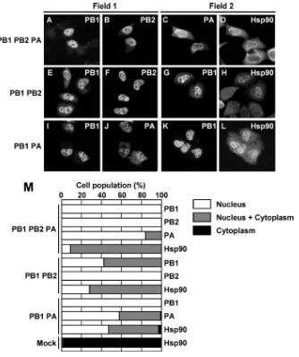
FIG. 2. Subcellular localization of viral RNA polymerases and Hsp90 in cotransfected cells. HeLa cells were cotransfected with a combination of plasmids encoding viral polymerases PB1, PB2, and PA (A to D). At 24 h after transfection, cells were fixed and stained with rat anti-PB1 (A), rabbit anti-PB2 (B), rat anti-PA (C), and rabbit anti-Hsp90 (D) antibodies. Due to the limitation in the species variety of antibodies, two different fields were stained with two different combinations of antibodies. The localization of polymerase subunits and Hsp90 in cells coexpressing either PB1 and PB2 (E to H) or PB1 and PA (I to L) was examined. At 24 h after transfection, cells were fixed and stained with rat anti-PB1 (E, G, I, and K), rabbit anti-PB2 (F), rabbit anti-PA (J) and rabbit anti-Hsp90 (H and L) antibodies. (M) Cells were counted, and the localization pattern of Hsp90 and polymerase subunits in cotransfected cells was determined. The number of cells showing each localization pattern was expressed as a percentage of the total cell number (for PB1-PB2-PA,n⫽232 for PB1,n⫽261 for PB2,n⫽175 for PA, andn⫽204 for Hsp90; for PB1-PB2,
n⫽78 for PB1,n⫽220 for PB2, andn⫽142 for Hsp90; for PB1-PA,n⫽75 for PB1,n⫽147 for PA, andn⫽66 for Hsp90; and for mock infected,n⫽50 for Hsp90).
on November 8, 2019 by guest
http://jvi.asm.org/
not directly bind to PA (30). We examined the effects of two possible binary complexes, namely, PB1-PB2 and PB1-PA, on the localization of Hsp90 in cells expressing combinations of two viral RNA polymerase subunits. In cells expressing all three subunits, i.e., PB1, PB2, and PA, Hsp90 was localized in the nucleus (Fig. 2A to D and M), similar to that in the infected cells (Fig. 1A to D). We found that the coexpression of PB2 with PB1 facilitates the nuclear transport of PB1, pos-sibly through PB1-PB2 interaction (compare Fig. 1F with Fig. 2E and F and refer to Fig. 2M). However, we detected a trace but distinct signal for PB1 in the cytoplasm (Fig. 2E). We examined the effect of the ratio of the expression level between PB1 and PB2 on the nuclear localization of PB1 (Fig. 3A to N and O). PB1 was localized in both the nucleus and cytoplasm of cells expressing PB1 alone (Fig. 1F and 3A). The nuclear accumulation of PB1 increased as the expression level of PB2
[image:5.585.104.477.74.435.2]increased (Fig. 3A to O), while PB2 was predominantly present in the nuclei in all cells (Fig. 3H to N). Figure 3P shows the expression level of PB1 and PB2 under the conditions em-ployed. The low-level expression of PB1 and PB2 (Fig. 3P, lanes 2 and 6) could be detected by longer development of the firm (data not shown). These results suggest that the nuclear accumulation of PB1 could be promoted by the formation of a PB1-PB2 binary complex. Fodor and Smith reported that the efficient accumulation of PB1-green fluorescent protein (GFP) in the nucleus requires the coexpression of PA, while the coexpression of PB2 does not affect the localization of PB1-GFP (9). The carboxyl-terminal region of PB1 binds to the amino-terminal region of PB2 (30). It is possible that PB1-GFP has only a weak ability to interact with PB2 because of the effect of the fusion of GFP with the PB1 carboxyl terminus. Hsp90 accumulated in the nucleus of cells expressing both PB1
FIG. 3. Nuclear accumulation of PB1 depends on PB2. HeLa cells were cotransfected with a combination of plasmids encoding viral polymerases PB1 and PB2 (A to N). The ratios of the amounts of transfected plasmids are indicated above the panels. At 24 h after transfection, cells were fixed and stained with rat anti-PB1 (A to G) and rabbit anti-PB2 (H to N) antibodies. (O) Cells were scored for the localization pattern of PB1 in cells cotransfected with PB1 and PB2. The number of cells showing each localization pattern was expressed as a percentage of the total cell number (n⫽131 for cells transfected with a PB1:PB2 ratio of 100:0,n⫽201 for 90:10 ratio,n⫽107 for 75:25 ratio,n⫽168 for 50:50 ratio,
n⫽119 for 25:75 ratio,n⫽68 for 10:90 ratio, andn⫽101 for 0:100 ratio). N.D., not detected. (P) Western blotting analyses of expressed proteins. HeLa cells were transfected with plasmids encoding PB1 and PB2. The ratios of the amounts of transfected plasmids are indicated above the lanes. Cell lysates were separated by 7.5% SDS-PAGE and analyzed by immunoblotting using rabbit anti-PB1 and anti-PB2 antibodies. Template activating factor-I(TAF-I), a nuclear protein, is also shown as a control (24).
on November 8, 2019 by guest
http://jvi.asm.org/
and PB2 (Fig. 2H and M). Therefore, it is suggested that the nuclear accumulation of Hsp90 depends on its interaction with PB2 or the PB1-PB2 subcomplexes. In cells expressing PB1 and PA, both subunits were observed mainly in the nucleus (Fig. 2I, J, and M). This is in good agreement with the previous report that the complete nuclear localization of PA is induced by the coexpression of PB1 (9, 26). These results suggest that the nuclear accumulation of PA could be promoted by the formation of a PB1-PA binary complex. Note that Hsp90 is accumulated in the nucleus of cells expressing both PB1 and PA (Fig. 2L and M). These staining patterns show that the nuclear transport of the viral RNA polymerase binary com-plexes, either PB1 and PB2 or PB1 and PA, as well as that of PB2 alone, induces the nuclear accumulation of Hsp90.
[image:6.585.46.282.69.470.2]Association of Hsp90 with PB1, PB2, and the PB1-PB2 sub-complex.To examine whether Hsp90 interacts with each viral polymerase subunit, binary complexes, and/or ternary com-plexes, we performed immunoprecipitation assays using lysates prepared from HeLa cells transfected with the expression plas-mids for viral RNA polymerases and antibodies against PB1, PB2, and PA. Western blot analyses revealed that PB2 or PB1 alone interacts with Hsp90 in transfected cells (Fig. 4A, lanes 3 and 6). In contrast, Hsp90 was not immunoprecipitated with PA (Fig. 4A, lane 9). These results indicated that PB1 and PB2 but not PA interact with Hsp90. In cells expressing both PB1 and PB2, PB1 and Hsp90 were coimmunoprecipitated with PB2 using anti-PB2 antibody (Fig. 4B, lane 7), suggesting the presence of trimolecular complexes among PB1, PB2, and Hsp90. However, a considerably smaller amount of Hsp90 was found to interact with PB2 and PB1 in cells expressing all three subunits (Fig. 4B, lane 8). We also carried out two-step immu-noprecipitation assays using anti-FLAG and anti-Myc antibod-ies (Fig. 4C). First, we performed immunoprecipitation assays using anti-FLAG antibody and lysates prepared from cells cotransfected with combinations of the expression plasmids PB1cFLAG and PB2cMyc and PA. In cells expressing both PB1cFLAG and PB2cMyc, PB1cFLAG interacted with Hsp90 (Fig. 4C, lane 8). PB2cMyc and Hsp90 were coimmunoprecipi-tated with PB1cFLAG by anti-FLAG antibody (Fig. 4C, lane 9). However, Hsp90 was not found to interact with PB1cFLAG and PB2cMyc in cells expressing all three subunits, including PA (Fig. 4C, lane 10). Next, the immunoprecipitated proteins were eluted from anti-FLAG agarose beads by using a FLAG peptide. We carried out the second immunoprecipitation assay using anti-Myc antibody and the eluted proteins (Fig. 4C, lanes 11 to 13). PB2cMyc interacted with PB1cFLAG and Hsp90 in
FIG. 4. Interaction of Hsp90 with viral RNA polymerases in co-transfected cells. (A) HeLa cells were co-transfected with plasmids en-coding PB1, PB2, and PA. At 24 h after transfection, cell lysates were prepared and incubated with rabbit antipolymerase antibodies (anti-PB2 [lane 3], anti-PB1 [lane 6], and anti-PA [lane 9]) or rabbit preim-mune serum (lanes 2, 5, and 8) of protein A-Sepharose beads. Immu-noprecipitated proteins were visualized by immunoblotting using rat anti-PB2, rat anti-PB1, rat anti-PA, and rat anti-Hsp90 antibodies. Lanes 1, 4, and 7 contain 10% of the input of transfected HeLa cell lysates. I.P.w/Pol, immunoprecipitation with antipolymerase anti-body. (B) HeLa cells were transfected with plasmids encoding PB1, PB2, and PA. HeLa cells expressing PB2 (lanes 2 and 6), PB2 and PB1 (lanes 3 and 7), or PB2, PB1, and PA (lanes 4 and 8) were prepared by transfection of corresponding combinations of plasmids, respectively. The cell lysates were incubated with rabbit anti-PB2 antibody (lanes 5 to 8) and protein A-Sepharose beads. The input (10%) is shown in lanes 1 to 4. The band intensities of PB2 and Hsp90 in lanes 7 and 8 were analyzed using NIH Image (data not shown). The amount of the precipitated PB2 was slightly less in lane 8 than that in lane 7. How-ever, the difference in the amount of precipitated Hsp90 in lane 7 and lane 8 was several times higher than the different in the amount of PB2 in lane 7 and lane 8. (C) HeLa cells were transfected with plasmids encoding PB1cFLAG, PB2cMyc, and PA. HeLa cells expressing PB1cFLAG (lanes 3, 8, and 11), PB2cMyc (lanes 2 and 7), PA (lanes 1 and 6), PB1cFLAG and PB2cMyc (lanes 4, 9, and 12), or PB1cFLAG, PB2cMyc, and PA (lanes 5, 10, and 13) were prepared by transfection of corresponding combinations of plasmids. The input (10%) is also shown in lanes 1 to 5. The cell lysates were incubated with
anti-FLAG conjugated to agarose beads (Sigma) (lanes 6 to 13). Pro-teins bound to PB1cFLAG (lanes 6 to 10) were eluted with FLAG peptide. Then, the second immunoprecipitation assays were carried out by anti-Myc-conjugated agarose beads (Nacalai) using eluted pro-teins (lanes 11 to 13). The amount of propro-teins used for the second immunoprecipitation assays was eight times more than that for lanes 8 to 10. (D) HeLa cells were transfected with plasmids encoding PB1, PA, and FLAG-Hsp90. HeLa cells expressing FLAG-Hsp90 (lanes 1 and 4), FLAG-Hsp90 and PB1 (lanes 2 and 5), or FLAG-Hsp90, PB1, and PA (lanes 3 and 6) were prepared. Cell lysates were incubated with mouse anti-FLAG antibody (lanes 4 to 6) and protein A-Sepharose beads. The input (10%) is shown in lanes 1 to 3.
on November 8, 2019 by guest
http://jvi.asm.org/
the eluted proteins in the absence of PA expression (Fig. 4C, lane 12). In contrast, in cells expressing all three viral polymer-ase subunits, Hsp90 was not found to be associated with poly-merases (Fig. 4C, lane 13). These results suggest that Hsp90 interacts with PB1-PB2 binary complexes but that it may be dissociated when the PB1-PB2 binary complexes mature into ternary polymerase complexes by interacting with PA. We also analyzed the interaction among Hsp90, PB1, and PA. PB1 was associated with FLAG-Hsp90 in cells expressing both PB1 and FLAG-Hsp90 (Fig. 3D, lane 5). However, following the ex-pression of PA, PB1 was not found to be associated with FLAG-Hsp90 (Fig. 3D, lane 6), indicating that the formation of PB1-PA binary complexes drastically reduces the interaction between Hsp90 and PB1. Alternatively, this can be interpreted
as the dissociation of the PB1-Hsp90 complexes by PA. On the basis of these results, we propose that Hsp90 is replaced by PA in PB1-PB2-Hsp90 complexes and that the ternary viral poly-merase complexes are concomitantly formed.
Release of the PB1-PB2 complexes from Hsp90 by PA. To examine the above possibility, we carried out in vitro interac-tion assays. First, to test whether Hsp90 binds directly to the PB1-PB2 complexes in vitro, PB1 complexed with PB2 con-taining an HA tag at the C-terminal region (PB2cHA) was purified from HeLa cells expressing both proteins by using anti-HA monoclonal antibody and incubated with either puri-fied recombinant Hsp90 containing a C-terminal GST tag (Hsp90cGST) or GST alone (Fig. 5). We found that PB1-PB2cHA complexes bind to Hsp90cGST (Fig. 5, compare lanes 2 and 6), indicating that Hsp90 can be directly associated with PB1-PB2 complexes. The addition of increasing amounts of purified PAcFLAG to PB1-PB2cHA-Hsp90cGST complexes resulted in the dissociation of PB1-PB2cHA complexes from Hsp90cGST (Fig. 5, lanes 7 to 9). These results indicate that PB1-PB2 complexes bind directly to Hsp90 and PB1-PB2 com-plexes are released from Hsp90 by PA.
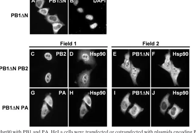
[image:7.585.132.454.468.686.2]Effect of the PB1-PA interaction on Hsp90 localization.We generated a PB1 mutant lacking PA binding activity by dele-tion of the amino-terminal region of PB1 and attempted to study the effect of mutant PB1 on the nuclear localization of Hsp90 and PA. The PB2 binding site of PB1 was present in its C-terminal region (30); hence, PB1⌬N could bind to PB2. PB1⌬N alone was mainly localized in the cytoplasm (Fig. 6A). We found that PB1⌬N accumulates in the nucleus in the pres-ence of PB2 (Fig. 6C and E) and Hsp90 is localized in the nucleus (Fig. 6D and F), as observed in cells expressing both PB1 and PB2 (Fig. 2E and F). On the other hand, in cells expressing PB1⌬N and PA, each subunit was not relocalized to the nucleus, possibly because these subunits could not interact
FIG. 5. Release of PB1-PB2 complexes from Hsp90 by the addition of PA. PB1-PB2cHA and PAcFLAG were affinity purified from cell lysates prepared from cells transfected with expression plasmids for either both PB1 and PB2cHA or PAcFLAG alone using anti-HA and anti-FLAG monoclonal antibodies, respectively. The proteins bound to the beads were subsequently eluted by HA epitope and FLAG epitope peptides. A trimolecular complex of PB1-PB2cHA-Hsp90cGST was recon-stituted at 30°C for 90 min by mixing PB1-PB2cHA (50 ng) (lanes 2 to 9) with GST (100 ng) (lanes 2 to 5) or Hsp90cGST (100 ng) (lanes 6 to 9). The mixtures were further incubated at 30°C for 90 min in the absence (⫺) and presence of PAcFLAG (50 ng [lanes 3 and 7], 500 ng [lanes 4 and 8], and 2,500 ng [lanes 5 and 9]) (the molecular ratios of the added PA to PB1-PB2cHA-Hsp90cGST were as follows: 30:1 [lane 7], 300:1 [lane 8], and 1,500:1 [lane 9]). After the GST pull-down assay, proteins were separated by 7.5% SDS-PAGE and analyzed by immunoblotting.
FIG. 6. Interaction of Hsp90 with PB1 and PA. HeLa cells were transfected or cotransfected with plasmids encoding PB1 N-terminal deletion mutant (PB1⌬N) (A and B), PB1⌬N and PB2 (C to F), or PB1⌬N and PA (G to J). At 24 h after transfection, cells were fixed and stained with rat anti-PB1 (A, E, and I), rat anti-PB2 (C), rat anti-PA (G), and rabbit anti-Hsp90 (D, F, H, and J) antibodies. The cells were also stained with DAPI (B).
on November 8, 2019 by guest
http://jvi.asm.org/
with each other (Fig. 6G and I). In the same cells, Hsp90 was localized only in the cytoplasm (Fig. 6H and J). These results suggest that the nuclear accumulation of Hsp90 is promoted by the nuclear transport of PB1 and PA through the formation of PB1-PA complexes.
Hsp90 accumulated in the nuclei of cells expressing both PB1 and PA (Fig. 2L and M). However, it appears likely that Hsp90 could not interact with PB1 in cells coexpressing PB1 and PA (Fig. 4D). We reexamined the interaction between PB1 and Hsp90 and their localization in cells expressing dif-ferent levels of the PA subunit. PB1cFLAG interacted with Hsp90, and both these proteins were localized in the cytoplasm and nucleus (Fig. 7A and D and Fig. 7M, lane 10). The dom-inant nuclear accumulation of PB1cFLAG and Hsp90 oc-curred and depended on the increasing amounts of expressed PA (Fig. 7B, C, E, F, H, I, K, and L). In cells coexpressing
PB1cFLAG and PA, the level of interaction between PB1 and Hsp90 was markedly reduced by PA in a dose-dependent man-ner (Fig. 7M, lanes 10 to 12). These results suggested that the nuclear accumulation of Hsp90 depends on the nuclear trans-port of the PB1-PA subcomplexes. It is possible that Hsp90 transiently interacts with the PB1-PA subcomplexes prior to or during nuclear transport through its interaction with PB1, and after nuclear transport, Hsp90 could be dissociated from the PB1-PA subcomplexes in the nucleus.
Predominant interaction of Hsp90 with premature viral polymerase complexes.We examined whether the association of Hsp90 with viral polymerase subcomplexes is required for the assembly of active polymerase complexes. For this purpose, we usedts53, a temperature-sensitive mutant virus containing mutations in the PA gene. Recently, we reported thatts53 viral polymerases do not form functional ternary polymerase com-plexes at the nonpermissive temperature (13). The change of Leu to Pro at amino acid position 226 in the PA subunit is responsible for the temperature sensitivity of thets53 virus. We compared the efficiency of the interaction between Hsp90 and viral polymerase subunits at the permissive temperature (34°C) with that at the nonpermissive temperature (39.5°C) ints 53-infected cells. Here, in order to detect premature polymerases, such as polymerase subcomplexes, and to avoid the exchange of subunits during the experiments, we prepared lysates using DSP, a reversible protein cross-linker. Figure 8A indicated that DSP does not affect the mobility of PB1 and PB2 on the SDS-polyacrylamide gel after cross-linking and reversion of cross-linking by the SDS-PAGE sample dye containing 2-mer-captoethanol (see Materials and Methods). Figure 8B shows that Hsp90 is associated with PB1 and PB2 in the lysates prepared fromts53-infected cells at 39.5°C (lanes 5 and 6). When thets53-infected cells were incubated at 39.5°C for 6 h and the temperature was shifted down from 39.5°C to 34°C, the interaction level between Hsp90 and viral polymerases was markedly reduced (Fig. 8B, compare lanes 11 and 12 with lanes 5 and 6). PA was not detected in the fractions precipitated with anti-Hsp90 antibody at both permissive and nonpermissive temperatures (data not shown); this is possibly due to the formation of ternary polymerase complexes and stable PB1-PB2-Hsp90 complexes, respectively. These results suggest that Hsp90 is associated with the premature viral polymerases prior to the formation of active polymerase complexes at the non-permissive temperature ints53-infected cells. At the permissive temperature, the ts53 viral polymerase subunit forms active polymerase complexes. Thus, it is quite likely that the interac-tion of Hsp90 with viral polymerase subunits is required to maintain the polymerase subunits active prior to the formation of mature vRNP complexes.
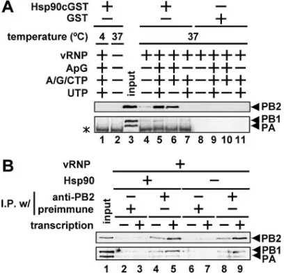
[image:8.585.67.254.66.354.2]Effects of the interactions between viral polymerases and Hsp90 on RNA synthesis.We reported that Hsp90 was a stim-ulatory host factor involved in influenza virus RNA synthesis (19). We examined the effects of interactions between viral polymerases and Hsp90 on the RNA synthesis reaction in vitro. We carried out GST pull-down assays using a recombi-nant Hsp90cGST protein and purified vRNP subjected to the RNA synthesis reaction. A small amount of PB2 was found to be bound to Hsp90cGST in the absence of ribonucleotides and the ApG dinucleotide primer used for the initiation of RNA
FIG. 7. Effect of PA on nuclear transport of Hsp90 with PB1. HeLa cells were transfected with PB1cFLAG (500 ng) with 300 ng (A, D, G, and J) and 200 ng (B, E, H, and K) of empty vector or 100 ng (B, E, H, and I) and 300 ng (C, F, I, and L) of the plasmid expressing PA. At 24 h after transfection, cells were fixed and stained with mouse anti-FLAG (A to C and G to I), rabbit anti-Hsp90 (D to F), and rabbit anti-PA (J to L) antibodies. (M) HeLa cells were transfected with plasmids encoding PB1cFLAG and PA. HeLa cells expressing PB1cFLAG (500 ng [lanes 2 to 4, 6 to 8, and 10 to 12]), PA (100 ng [lanes 3, 7, and 11] or 300 ng [lanes 4, 8, and 12]), and empty plasmid (800 ng [lanes 1, 5, and 9], 300 ng [lanes 2, 6, and 10], and 200 ng [lanes 3, 7, and 11]) were prepared. Cell lysates were incubated with mouse anti-FLAG-conjugated agarose beads (lanes 9 to 12) and mouse im-munoglobulin G agarose beads (Sigma) (lanes 5 to 8). Each protein was detected by Western blotting with mouse FLAG, rabbit anti-Hsp90, and rabbit anti-PA antibodies. The input (10%) is shown in lanes 1 to 4. I.P.w/ mouse IgG, immunoprecipitation with mouse im-munoglobulin G.
on November 8, 2019 by guest
http://jvi.asm.org/
synthesis (Fig. 9A, compare lanes 4 and 8). When Hsp90cGST was mixed with vRNP and the mixture was incubated under RNA synthesis conditions, the level of PB2 bound to Hsp90 markedly increased (Fig. 9A, lane 5). Hsp90cGST also bound strongly to PB2 in the limited elongation reaction lacking UTP (Fig. 9A, lane 6) (19). However, in the absence of the ApG primer, Hsp90cGST did not bind to PB2 (Fig. 9A, lane 7). In addition, PB2 could not be detected under the low tempera-ture condition (Fig. 9A, lane 1). These results indicated that the binding level of Hsp90 to PB2 is increased under the RNA synthesis condition. Furthermore, a low but distinct PB1 signal could be detected in the RNA synthesis condition (Fig. 9A, lane 5). Quantitative analyses using NIH Image indicated that the PB1 band intensity in Fig. 9A, lane 5 is three times more than that in lanes 4, 6, and 7 (data not shown). Therefore, these results suggest that Hsp90 is associated with PB2, and a small amount of the PB2-PB1 complexes might be dissociated from vRNP by RNA synthesis (see Discussion). In addition, we carried out immunoprecipitation assays using the anti-PB2
an-tibody and vRNP under the RNA synthesis condition in the presence or absence of Hsp90 (Fig. 9B). The three polymerase subunits were coimmunoprecipitated by the anti-PB2 antibody in the absence of the RNA synthesis condition (Fig. 9B, lanes 4 and 8). In the presence of RNA synthesis, the amounts of PB2 and PB1 immunoprecipitated by the anti-PB2 antibody increased, while the amount of PA was unchanged (Fig. 9B, lanes 5 and 9). It is worthwhile to note that the amount of immunoprecipitated polymerase subunits in the absence of Hsp90 was virtually the same as that in the presence of Hsp90. These results suggest that Hsp90 binds to PB2 predominantly and PB1 binds less effectively than PB2, as it falls off vRNP during RNA synthesis (see Discussion).
DISCUSSION
[image:9.585.44.285.67.266.2]In this report, we have shown the interaction of Hsp90 with the influenza virus RNA polymerases and its possible involve-ment in nuclear transport and assembly of polymerase sub-units. The viral polymerase complexes comprising PB1, PB2, and PA are formed by the binding between PB1 and PB2 and between PB1 and PA (30). Fodor and Smith reported that PB1
FIG. 8. Interaction of Hsp90 withts53 viral RNA polymerases at the nonpermissive temperature. (A) MDCK cells were infected with
[image:9.585.313.525.69.270.2]ts53 virus at a MOI of 10 and incubated at 34°C. At 6 hours postin-fection (hpi), the cells were collected and resuspended in PBS. Cells were incubated at 25°C for 30 min in the presence (⫹) or absence (⫺) of DSP at a final concentration of 1 mM followed by incubation. The cross-linking reaction was stopped by the addition of 1 M Tris-HCl (pH 7.9) at a final concentration of 10 mM. Cell lysates were separated by 7.5% SDS-PAGE and analyzed by immunoblotting using rabbit anti-PB1 and anti-PB2 antibodies. A protein marker is shown in lane 1. (B) MDCK cells were infected withts53 virus at a MOI of 10 and incubated at 39.5°C. At 6 hpi, cycloheximide (CHX) was added at the concentration of 100g/ml, and then temperature was shifted down to the permissive temperature (34°C) (lanes 7 to 12) or kept at the nonpermissive temperature (39.5°C) (lanes 1 to 6). After incubation for 30 min or 90 min, the cells were collected and resuspended in PBS. Proteins were cross-linked by the addition of 0.1 M DSP at a final concentration of 1 mM, followed by incubation at 25°C for 30 min. The cross-linking reaction was stopped by the addition of 1 M Tris-HCl (pH 7.9) at a final concentration of 10 mM. Cell lysates were prepared and incubated with a rabbit anti-Hsp90 antibody and protein A-Sepha-rose beads. Immunoprecipitated proteins were separated by 7.5% SDS-PAGE and analyzed by immunoblotting. The input (10%) is shown in lanes 1 to 3 and lanes 7 to 9. Immunoprecipitation with (I.P.w/) input or anti-Hsp90 is indicated.
FIG. 9. Interaction between viral polymerase and Hsp90 on RNA synthesis. (A) GST pull-down assays. GST pull-down assays (details in Materials and Methods) were carried out with recombinant Hsp90cGST or GST under RNA synthesis conditions using vRNP (150 ng of NP equivalents). Reaction conditions (the presence [⫹] and absence [⫺] of various proteins or nucleoside triphosphates) are indi-cated above each lane. Eluted and input (25%) vRNP (lane 3) were loaded onto 7.5% SDS-polyacrylamide gels, and Western blotting analysis was carried out using rabbit anti-PB2, anti-PB1, and anti-PA antibodies. A nonspecific signal derived from a Hsp90cGST fraction is indicated by the asterisk. (B) Immunoprecipitation assays. The RNA synthesis reaction was carried out using vRNP (150 ng of NP equiva-lents) under the RNA synthesis condition at 37°C (lanes 3, 5, 7, and 9) in the presence (⫹) or absence (⫺) of Hsp90 (150 ng). In lanes 2, 4, 6, and 8, the reaction mixture lacked ApG and nucleoside triphosphates, so that no RNA synthesis occurred. After the reaction, the reaction mixtures were subjected to immunoprecipitation assays with (I.P.w/) protein G-Sepharose beads and either rabbit anti-PB2 antibody (lanes 4, 5, 8, and 9) or rabbit preimmune serum (lanes 2, 3, 6, and 7). Immunoprecipitated proteins were separated by 7.5% SDS-PAGE and analyzed by Western blotting. The input (25%) is shown in lane 1.
on November 8, 2019 by guest
http://jvi.asm.org/
and PA interact with each other in the cytoplasm and are transported into the nucleus as a dimer, while PB2 is trans-ported into the nucleus as a monomer (9). They suggested that PB2 binds to PB1-PA in the nucleus. We have previously shown that Hsp90 is relocalized to the nucleus upon viral infection (19). Here, we found that Hsp90 is transported into the nucleus with PB2 or with a PB1-PB2 binary complex. We identified Hsp90 as a stimulatory host factor by using an in vitro virus RNA synthesis system, in which Hsp90 stabilizes the viral polymerase activity by binding at least to PB2 (19). There-fore, we assume that the nuclear accumulation of Hsp90 through its interaction with viral polymerases is important in terms of regulation of virus RNA synthesis in the nucleus.
The majority of Hsp90 is present in the cytoplasm of unin-fected cells (Fig. 1C). In cells expressing PB2, both PB2 and Hsp90 were shown to accumulate in the nucleus (Fig. 1E and H). This suggests that Hsp90 is transported into the nucleus through its interaction with PB2 (Fig. 10A). It is shown that PB1 and PA interact with each other in the cytoplasm and could be transported into the nucleus as a complex (Fig. 2I and J). Thus, it is quite likely that Hsp90-PB2 interacts with PB1-PA in the nucleus to form a ternary complex, with the concomitant release of Hsp90 from PB2 (Fig. 10A). An alter-native process for viral polymerase assembly is also possible because a complex of PB1 and PB2 was found in the cytoplasm, and it could be transported into the nucleus as a complex (Fig. 2E and F). The efficient nuclear accumulation of PB1 appears to be dependent on its binding to PB2 (Fig. 10B). It is reported that in the early phases of infection, PB1 and PB2 are present in the nucleus, whereas PA is localized in the cytoplasm (1). Even in unstressed cells, Hsp90 exists as an abundant protein (at the level of 1% to 2% of total cellular proteins), and newly synthesized PB1 and PB2 may interact with Hsp90 in the early phases of infection. Subsequently, a PB1-PB2 complex
associ-ated with Hsp90 is transported into the nucleus (Fig. 10B). Upon the progression of infection, the amount of the PA subunit in the nucleus increases. The results of reconstitution experiments indicated that the PB1-PB2 complexes bind di-rectly to Hsp90 and that this interaction is dissociated by PA (Fig. 5). This assembly process might occur in infected cells when PA is transported into the nucleus, although it is unclear whether PA alone or PA associated with other molecules is transported into the nucleus (Fig. 10B). In addition, we cannot rule out the possibility that the ternary polymerase complex is formed from the PB2-PB1-Hsp90 and PB1-PA complexes. Re-cently, Deng et al. reported that RanBP5 (Ran binding protein 5) interacts with either PB1 alone or with a the PB1-PA sub-complex but not with the PB1-PB2 subsub-complex or with the PB2-PB1-PA complex (6). Viral RNA polymerase may use various host factors for the nuclear transport and functional assembly of different kinds of polymerase subcomplexes and subunits.
We examined the role of Hsp90 in cells infected with thets53 virus. At the nonpermissive temperature, thets53 viral poly-merase complexes are structurally and functionally different from those at the permissive temperature. Further, at the non-permissive temperature,ts53 polymerases do not form the ac-tive viral polymerase structure (13). We showed that Hsp90 is associated with the viral polymerases at the nonpermissive temperature (Fig. 8). However, following a shift from the non-permissive temperature to the non-permissive temperature, viral polymerases are free of Hsp90. These results indicated that Hsp90 might function as a molecular chaperone for viral poly-merases until the PA function is recovered in ts53-infected cells. Thus, it is possible that Hsp90 is required for the main-tenance of PB1 and PB2 prior to the assembly with PA. Im-munoprecipitation assays using cell extracts prepared from cells labeled with [35S]methionine and [35S]cysteine by
pulse-chase and anti-Hsp90 antibody revealed that Hsp90 interacts with the newly synthesized (pulse-labeled) viral polymerase subunits, while during chasing with cold amino acids, this com-plex was lost (data not shown). This supports the notion that Hsp90 may play an important role as a molecular chaperone during polymerase assembly.
We analyzed the interaction between Hsp90 and viral poly-merases under the RNA synthesis condition using vRNP in which trimolecular complexes are thought to be bound (Fig. 9). Hsp90 bound to PB2 predominantly and PB1 less effectively than PB2 during the RNA synthesis reaction. Since in the limited elongation condition lacking UTP, Hsp90 was capable of stimulating RNA synthesis (19), it is possible that Hsp90 plays a role during early stages of the elongation reaction. In the limited elongation condition, Hsp90 interacted with PB2 (Fig. 9). Hsp90 may facilitate the dissociation of RNA poly-merases from vRNP and stabilize the polymerase subunits during their translocation between RNA templates.
[image:10.585.62.262.68.247.2]It is not known which forms of polymerase complexes, in-cluding ternary complexes, binary subcomplexes, or each sub-unit, bind to viral RNAs. Recently, it was shown that the PB1-PA complex binds to the vRNA promoter of the viral genome prior to the formation of a functional viral polymerase complex by assembly with PB2 (7). At present, it is not known whether Hsp90 is involved in this type of assembly pathway that includes the viral RNA in infected cells.
FIG. 10. A model for Hsp90-related nuclear transport and assem-bly of the influenza virus polymerases. (A) PB1 and PA interact with each other in the cytoplasm and translocate into the nucleus as a complex. PB2 is transported into the nucleus with Hsp90. (B) Hsp90 interacts with PB1, PB2, and a PB1-PB2 subcomplex, and then a PB1-PB2-Hsp90 complex is formed and transported into the nucleus. The ternary polymerase complex is assembled with nuclear trans-ported viral polymerase subunits and/or subcomplexes, and concomi-tantly Hsp90 is released from subunits and/or subcomplexes.
on November 8, 2019 by guest
http://jvi.asm.org/
ACKNOWLEDGMENTS
We thank Y. Miyata (Kyoto University), E. Nishida (Kyoto Univer-sity), and K. Tobita (Jichi Medical School) for their generous gifts of anti-Hsp90 antibody (Y. Miyata and E. Nishida) andts53 virus (K. Tobita).
This work was supported in part by a grant-in-aid from the Ministry of Education, Culture, Sports, Science, and Technology of Japan (to K.N.), Kitasato University Research Grant for Young Researchers (to F.M.), and Research Fellowships of the Japanese Society for the Pro-motion of Science (JSPS) (to A.K.).
REFERENCES
1.Akkina, R. K., T. M. Chambers, D. R. Londo, and D. P. Nayak.1987. Intracellular localization of the viral polymerase proteins in cells infected with influenza virus and cells expressing PB1 protein from cloned cDNA. J. Virol.61:2217–2224.
2.Biswas, S. K., and D. P. Nayak.1994. Mutational analysis of the conserved motifs of influenza A virus polymerase basic protein 1. J. Virol.68:1819– 1826.
3.Bose, S., and A. K. Banerjee.2004. Beta-catenin associates with human parainfluenza virus type 3 ribonucleoprotein complex and activates tran-scription of viral genome RNA in vitro. Gene Expr.11:241–249. 4.Chen, C., and H. Okayama.1987. High-efficiency transformation of
mam-malian cells by plasmid DNA. Mol. Cell. Biol.7:2745–2752.
5.Das, T., M. Mathur, A. K. Gupta, G. M. Janssen, and A. K. Banerjee.1998. RNA polymerase of vesicular stomatitis virus specifically associates with translation elongation factor-1 alphabetagamma for its activity. Proc. Natl. Acad. Sci. USA95:1449–1454.
6.Deng, T., O. G. Engelhardt, B. Thomas, A. V. Akoulitchev, G. G. Brownlee, and E. Fodor.2006. Role of Ran binding protein 5 in nuclear import and assembly of the influenza virus RNA polymerase complex. J. Virol. 80: 11911–11919.
7.Deng, T., J. Sharps, E. Fodor, and G. G. Brownlee.2005. In vitro assembly of PB2 with a PB1-PA dimer supports a new model of assembly of influenza A virus polymerase subunits into a functional trimeric complex. J. Virol. 79:8669–8674.
8.Fechter, P., L. Mingay, J. Sharps, A. Chambers, E. Fodor, and G. G. Brown-lee.2003. Two aromatic residues in the PB2 subunit of influenza A RNA polymerase are crucial for cap binding. J. Biol. Chem.278:20381–20388. 9.Fodor, E., and M. Smith.2004. The PA subunit is required for efficient
nuclear accumulation of the PB1 subunit of the influenza A virus RNA polymerase complex. J. Virol.78:9144–9153.
10.Gupta, S., B. P. De, J. A. Drazba, and A. K. Banerjee.1998. Involvement of actin microfilaments in the replication of human parainfluenza virus type 3. J. Virol.72:2655–2662.
11.Hu, J., D. Flores, D. Toft, X. Wang, and D. Nguyen.2004. Requirement of heat shock protein 90 for human hepatitis B virus reverse transcriptase function. J. Virol.78:13122–13131.
12.Huarte, M., J. J. Sanz-Ezquerro, F. Roncal, J. Ortin, and A. Nieto.2001. PA subunit from influenza virus polymerase complex interacts with a cellular protein with homology to a family of transcriptional activators. J. Virol. 75:8597–8604.
13.Kawaguchi, A., T. Naito, and K. Nagata.2005. Involvement of influenza virus PA subunit in assembly of functional RNA polymerase complexes. J. Virol. 79:732–744.
14.Krug, R. M., M. Ueda, and P. Palese.1975. Temperature-sensitive mutants of influenza WSN virus defective in virus-specific RNA synthesis. J. Virol. 16:790–796.
15.Li, M. L., P. Rao, and R. M. Krug.2001. The active sites of the influenza cap-dependent endonuclease are on different polymerase subunits. EMBO J. 20:2078–2086.
16.Miyaji-Yamaguchi, M., K. Kato, R. Nakano, T. Akashi, A. Kikuchi, and K. Nagata.2003. Involvement of nucleocytoplasmic shuttling of yeast Nap1 in mitotic progression. Mol. Cell. Biol.23:6672–6684.
17.Miyata, Y., and I. Yahara.2000. p53-independent association between SV40 large T antigen and the major cytosolic heat shock protein, HSP90. Onco-gene19:1477–1484.
18.Momose, F., C. F. Basler, R. E. O’Neill, A. Iwamatsu, P. Palese, and K. Nagata.2001. Cellular splicing factor RAF-2p48/NPI-5/BAT1/UAP56 inter-acts with the influenza virus nucleoprotein and enhances viral RNA synthe-sis. J. Virol.75:1899–1908.
19.Momose, F., T. Naito, K. Yano, S. Sugimoto, Y. Morikawa, and K. Nagata. 2002. Identification of Hsp90 as a stimulatory host factor involved in influ-enza virus RNA synthesis. J. Biol. Chem.277:45306–45314.
20.Moyer, S. A., S. C. Baker, and S. M. Horikami.1990. Host cell proteins required for measles virus reproduction. J. Gen. Virol.71:775–783. 21.Moyer, S. A., S. C. Baker, and J. L. Lessard.1986. Tubulin: a factor
neces-sary for the synthesis of both Sendai virus and vesicular stomatitis virus RNAs. Proc. Natl. Acad. Sci. USA83:5405–5409.
22.Mukaigawa, J., and D. P. Nayak.1991. Two signals mediate nuclear local-ization of influenza virus (A/WSN/33) polymerase basic protein 2. J. Virol. 65:245–253.
23.Naffakh, N., P. Massin, and S. van der Werf.2001. The transcription/repli-cation activity of the polymerase of influenza A viruses is not correlated with the level of proteolysis induced by the PA subunit. Virology285:244–252. 24.Nagata, K., S. Saito, M. Okuwaki, H. Kawase, A. Furuya, A. Kusano, N.
Hanai, A. Okuda, and A. Kikuchi.1998. Cellular localization and expression of template-activating factor I in different cell types. Exp. Cell Res.240:274– 281.
25.Neumann, G., T. Watanabe, H. Ito, S. Watanabe, H. Goto, P. Gao, M. Hughes, D. R. Perez, R. Donis, E. Hoffmann, G. Hobom, and Y. Kawaoka. 1999. Generation of influenza A viruses entirely from cloned cDNAs. Proc. Natl. Acad. Sci. USA96:9345–9350.
26.Nieto, A., S. de la Luna, J. Barcena, A. Portela, J. Valcarcel, J. A. Melero, and J. Ortin.1992. Nuclear transport of influenza virus polymerase PA protein. Virus Res.24:65–75.
27.Niwa, H., K. Yamamura, and J. Miyazaki. 1991. Efficient selection for high-expression transfectants with a novel eukaryotic vector. Gene108:193– 199.
28.Ogino, T., M. Iwama, J. Kinouchi, Y. Shibagaki, T. Tsukamoto, and K. Mizumoto.1999. Involvement of a cellular glycolytic enzyme, phosphoglyc-erate kinase, in Sendai virus transcription. J. Biol. Chem.274:35999–36008. 29.Oglesbee, M. J., Z. Liu, H. Kenney, and C. L. Brooks.1996. The highly inducible member of the 70 kDa family of heat shock proteins increases canine distemper virus polymerase activity. J. Gen. Virol.77:2125–2135. 30.Ohtsu, Y., Y. Honda, Y. Sakata, H. Kato, and T. Toyoda.2002. Fine mapping
of the subunit binding sites of influenza virus RNA polymerase. Microbiol. Immunol.46:167–175.
31.O’Neill, R. E., and P. Palese.1995. NPI-1, the human homolog of SRP-1, interacts with influenza virus nucleoprotein. Virology206:116–125. 32.Palese, P. W., P. T. Wolff, and R. E. O’Neill.1997. Host-viral protein-protein
interactions in influenza virus replication, p. 327–340.InM. A. McCrae, J. R. Saunders, C. J. Smyth, and N. D. Stow (ed.), Molecular aspects of host-pathogen interaction. Society for General Microbiology Symposium 55. Cambridge University Press, Cambridge, United Kingdom.
33.Pearl, L. H., and C. Prodromou.2006. Structure and mechanism of the hsp90 molecular chaperone machinery. Annu. Rev. Biochem.75:271–294. 34.Perales, B., J. J. Sanz-Ezquerro, P. Gastaminza, J. Ortega, J. F. Santaren,
J. Ortin, and A. Nieto.2000. The replication activity of influenza virus polymerase is linked to the capacity of the PA subunit to induce proteolysis. J. Virol.74:1307–1312.
35.Picard, D.2002. Heat-shock protein 90, a chaperone for folding and regu-lation. Cell. Mol. Life Sci.59:1640–1648.
36.Portela, A., and P. Digard.2002. The influenza virus nucleoprotein: a mul-tifunctional RNA-binding protein pivotal to virus replication. J. Gen. Virol. 83:723–734.
37.Pratt, W. B., and D. O. Toft.2003. Regulation of signaling protein function and trafficking by the hsp90/hsp70-based chaperone machinery. Exp. Biol. Med. (Maywood)228:111–133.
38.Qanungo, K. R., D. Shaji, M. Mathur, and A. K. Banerjee.2004. Two RNA polymerase complexes from vesicular stomatitis virus-infected cells that carry out transcription and replication of genome RNA. Proc. Natl. Acad. Sci. USA101:5952–5957.
39.Wang, P., P. Palese, and R. E. O’Neill.1997. The NPI-1/NPI-3 (karyopherin ␣) binding site on the influenza A virus nucleoprotein NP is a nonconven-tional nuclear localization signal. J. Virol.71:1850–1856.
40.Waxman, L., M. Whitney, B. A. Pollok, L. C. Kuo, and P. L. Darke.2001. Host cell factor requirement for hepatitis C virus enzyme maturation. Proc. Natl. Acad. Sci. USA98:13931–13935.
41.Yamanaka, K., A. Ishihama, and K. Nagata.1990. Reconstitution of influ-enza virus RNA-nucleoprotein complexes structurally resembling native vi-ral ribonucleoprotein cores. J. Biol. Chem.265:11151–11155.
42.Zhao, R., J. Shen, M. R. Green, M. MacMorris, and T. Blumenthal.2004. Crystal structure of UAP56, a DExD/H-box protein involved in pre-mRNA splicing and mRNA export. Structure12:1373–1381.
43.Zhao, Y. G., R. Gilmore, G. Leone, M. C. Coffey, B. Weber, and P. W. Lee. 2001. Hsp90 phosphorylation is linked to its chaperoning function. Assembly of the reovirus cell attachment protein. J. Biol. Chem.276:32822–32827.