A Dissertation on
AN ANALYTICAL STUDY OF VARIOUS REFRACTIVE
ERRORS FOR PROBABLE OCULAR ASSOCATIONS
Dissertation submitted to
THE TAMILNADU Dr.M.G.R. MEDICAL UNIVERSITY
CHENNAI - 600 032
in partial fulfillment of the regulations for the award of the degree of
M.S. DEGREE IN OPHTHALMOLOGY
COIMBATORE MEDICAL COLLEGE
COIMBATORE
DECLARATION
I hereby declare that this dissertation entitled “ANALYTICAL STUDY OF VARIOUS REFRACTIVE ERRORS FOR PROBABLE OCULAR ASSOCIATONS’’ is a bonafide and genuine research work carried out by me under the guidance of Dr. S PADMANABAN M.S., D.O Associate Professor, Department of Ophthalmology, Coimbatore Medical College & Hospital, Coimbatore.
This is submitted to The Tamilnadu Dr.M.G.R. Medical University, Chennai in partial fulfillment of regulations required for the M.S Ophthalmology, Branch III Degree Examination to be held in May 2019.
Date:
CERTIFICATE
This is to certify that the dissertation entitled “ANALYTICAL STUDY OF VARIOUS REFRACTIVE ERRORS FOR PROBABLE
OCULAR ASSOCIATIONS” is a bonafide and research work done
by Dr. DIVYA .N Post Graduate in M.S. Ophthalmology under my
direct guidance and supervision to my satisfaction in partial fulfillment of the requirement for the degree of Master of Surgery in Ophthalmology, Branch III .
Date : Chief Guide
Department of Ophthalmology
Date : Head of the Department
Department of Ophthalmology Coimbatore Medical College, Coimbatore.
Date : The Dean,
CERTIFICATE –II
This is to certify that this dissertation work titled " ANALYTICAL
STUDY OF VARIOUS REFRACTIVE ERRORS FOR PROBABLE OCULAR ASSOCIATONS’’ " of the candidate Dr. DIVYA .N with
registration number 221613201 for the award of M.S. DEGREE IN
OPHTHALMOLOGY. I personally verified the urkund.com website for
the purpose of plagiarism check. I found that the uploaded thesis file
contains from introduction to conclusion 82 pages and result shows 3%
percentage of plagiarism in the dissertation.
ACKNOWLEDGEMENT
It gives me a great pleasure and satisfaction in completing this
dissertation. Firstly, I would like to express my thanks to our Dean,
Dr B Asokan, M.S., M.Ch., for permitting me to do this research work.
I would like to convey my sincere thanks and heartfelt gratitude to
my guide Dr S Padmanaban M.S., D.O., HOD and Associate Professor,
Department of Ophthalmology, Coimbatore Medical College Hospital, Coimbatore for his valuable guidance and support which helped me to complete this project on time.
I would like to convey my thanks to Dr V Thaiyal Nayaki M.S.,
D.G.O., Associate Professor, Department of Ophthalmology, Coimbatore
Medical College Hospital, Coimbatore for her valuable guidance and support for completing this dissertation.
I would like to express my thanks to Assistant Professors
Dr C Jeevakala M.S., D.O., Dr J Saravanan M.S., Dr P Sumathi
M.S., Dr K Malligai D.O., D.N.B., Dr P Mohanapriya M.S., Dr K
Sathya M.S., Dr V Karthikeyan M.S., and Dr M Haripriya M.S., for
I take this opportunity to express my wholehearted gratitude to my family and colleagues who have helped me in all my endeavours and supported me to complete this project.
ABBREVATIONS
IOP – Intra Ocular Pressure
POAG – Primary Open Angle Glaucoma
KC – Keratoconus
RP – Retinitis Pigmentosa
PACG – Primary Angle Closure Glaucoma
BRVO – Branch Retinal Vein Occlusion
NA-AION – Non -Arteritic Anterior Ischemic optic neuropathy
CDR – Cup Disc Ratio
CONTENTS
S.No TITLE Page No
1 INTRODUCTION 1
2 REVIEW OF LITERATURE 29
3 AIM AND OBJECTIVES 49
4 MATERIALS AND METHODS 50
5 RESULTS AND OBSERVATIONS 53
6 DISCUSSION 75
7 SUMMARY 79
8 CONCLUSION 81
9 BIBLIOGRAPHY 83
ANNEXURES
PROFORMA 96
CONSENT FORM 98
LIST OF TABLES
S.No TITLE Page No
1. DISTRIBUTION OF DIAGNOSIS 54
2. AGE WISE DISTRIBUTION OF PATIENTS 56
3. SEX WISE DISTRIBUTION OF PATIENTS 57
4. AGE WISE DISTRIBUTION OF 5 OCULAR
DISORDERS
58
5. PERCENTAGE WISE AGE DISTRIBUTION
OF 5 OCULAR DISORDERS
59
6. CHI SQUARE TESTS -AGE 60
7. SEX WISE DISTRIBUTION OF 5 OCULAR
DISORDERS
61
8. PERCENTAGE WISE DEX DISTRIBUTION
OF 5 OCULAR DISORDERS
62
9. CHI SQUARE TESTS - SEX 63
10. DISTRIBUTION OF REFRACTIVE ERRORS
AMONG THE OCULAR DISORDERS
63
11. GONIOSCOPY GRADINGS OF RIGHT EYE
IN 5 OCULAR DISORDERS
65
12. CHI SQUARE TEST FOR GONIO GRADINGS
OF RIGHT EYE
67
13. GONIOSCOPY GRADINGS OF LEFT EYE IN
5 OCULAR DISORDERSC
14. CHI SQUARE TEST FOR GONIO GRADINGS OF LEFT EYE
70
15. MEAN AXIAL LENGTH MEASUREMETNS
IN THE ABOVE 5 OCULAR DISORDERS
71
16. ANOVA RESULTS FOR AXIAL LENGTH OF
BOTH EYES IN 5 OCULAR DISORDERS
72
17. CUP:DISC RATION OF BOTH EYES WITH 5
OCULAR DISORDERS
73
18. ANOVA RESULTS FOR CUP:DISC
RATIO(CDR) FOR BOTH EYES WITH OCULAR DISORDERS
LIST OF CHARTS
S.No TITLE Page No
1. DISTRIBUTION OF DIAGNOSIS 55
2. AGE WISE DISTRIBUTION 56
3. SEX WISE DISTRIBUTION 57
4. AGE WISE DISTRIBUTION OF DISORDERS 59
5. SEX WISE DISTRIBUTION OF 5 OCULAR
DISORDERS
62
6. DISTRIBUTION OF REFRACTIVE ERRORS
AMONG THE OCULAR DISORDERS
64
7. GONIOSCOPY GRADINGS OF RIGHT EYE
WITH 5 OCULAR DISORDERS
66
8. GONIOSCOPY GRADINGS OF LEFT EYE IN
5 OCULAR DISORDERS
69
9. MEAN AXIAL LENGTH DISTRIBUTION
AMONG 5 OCULAR DISORDERS CUP: DISC RATIO OF BOTH EYES WITH 5 OCULAR DISORDERS
71
10. CUP: DISC RATIO OF BOTH EYES WITH 5
OCULAR DISORDERS
1
INTRODUCTION
When the parallel rays of the light strike a physiologically normal eye they are refracted so as to converge upon the retina where they focus, forming a circle of least confusion without any accommodation, the condition is termed as Emmetropia.
The opposite condition of emmetropia, wherein the parallel rays of light are not focused exactly upon the retina with the eye in a state of rest, is therefore much the more common; such an eye has a refractive error. Anisometropia the condition in which the refractive conditions are unequal in both eyes.
2
TYPES OF REFRACTIVE ERRORS:
There are three main types of refractive errors: the principal focus is formed either in front or behind the retina. If the eye is relatively too short, the principal focus is formed behind the retina, a condition known as Hypermetropia. If the eye is relatively too long, the principal focus if formed in front of retina, the term Myopia is used. Alternatively, the refractive system may be such that no single focus is formed, instead of that two focal lines are formed, in that case Astigmatism is present.
3
The above refractive anomalies can be caused by various conditions
1. The position of the elements of the system
a. Axial Myopia: the antero-posterior diameter of the eye is too
long and the retina is far away from the optical system. It is the commonest form.
b. Axial Hypermetropia: the antero-posterior diameter of the
eye is too short and the retina is too near to the optical system
4
2. Anomalies of the refractive surfaces
a. Curvature Hypermetropia: the curvature of the cornea or lens
is too small
b. Curvature Myopia: the curvature of the cornea or lens is too
great
c. Astigmatism: the curvature is too irregular, varying in
different meridians
3. Obliquity of the elements of the system
a. Lenticular obliquity: if the lens is placed obliquely or
subluxated, it will result in astigmatism
b. Retinal obliquity: the posterior pole of the eye may be place
obliquely, as when if bulges backwards in a staphyloma in high myopia.
4. Anomalies of the refractive index
5
too high, that is more of lens, the refraction will again be less results in index hypermetropia.
b. Index Myopia: if the index of refraction of the aqueous is too
high or that of the vitreous is too low, there will be an index myopia
5. Absence of an element of the system
Absence of the crystalline lens in the patellar fossa, a condition known as aphakia produces hypermetropia
THE DEGREE OF REFRACTIVE ERROR
The strength of lens required to alter the vergence of the parallel rays so that the eye now focuses them on retina.
6
1. An emmetropic eye
2. A myopic eye, parallel rays being focused in front of retina
3. When looking at near object, the divergent rays are focused upon the retina. R is the far point.
4. A similar divergence may be given to the parallel rays by a
concave lens so that a focus is again formed upon the retina
7
1. An emmetropic eye: parallel rays of light come to a focus upon the
retina
2. A hypermetropic eye: parallel rays of light come to a focus behind
the retina
3. A hypermetropic eye: parallel rays of light are brought to a focus upon the retina by increasing the refractivity by accommodation. The normal lens, a, becomes more convex, b.
8
THE MEASUREMENT OF THE AXIAL LENGTH
When an ultrasonic beam is passed through the eye, peaks are seen in the echogram representing the reflections from the various ocular interfaces; with the obtained measurements, the distances between these surfaces and the axial length of the eye can be calculated. The average axial length of the eye ball is 22-24mm. A 1 mm error in axial length measurement results in a refractive error of approximately 2.35D in
23.5mm eye[2]. 1.75D/mm decline in refractive error seen in 30mm eye
but rises to 3.75D/mm in 20 mm eye. Therefore, accuracy in axial length measurement is more important in short eyes than in long eyes.
Simple refractive errors are largely hereditarily determined due to co-ordinated combination of essentially normal elements of the optical system of the eye. These simple refractive errors are not progressive beyond the amount included within normal development and associated with good vision and needs optical correction alone.
9
PREVALENCE OF REFRACTIVE ERRORS
Dandona et al[3] studied about prevalence of refractive errors among urban population in South India and reported that 42.16% had some form of refractive errors with prevalence of 17.8% myopia and 18.8% hypermetropia in the 40- to 49- year age group, 29.6% myopia and 39.6% hyperopia in the 50- to 59- year age group, 44.8% and 29.7% in the 60 to 60- age group and 50% and 30.4% in the >70 year age group for myopia and hyperopia respectively.
Prevalence of refractive errors in rural South Indian population
studied by Prema Raju et al[4] showed 49.40% of people had refractive
errors in which 26.99% myopia and 18.70% hyperopia and 58.70% had astigmatism.
MYOPIA
10
Distant objects cannot be seen clearly; only divergent rays will meet on retina. Thus, in order to see clearly, an object must be brought close to the eye, so that the rays coming from it are rendered sufficiently divergent. The far point (punctum remotum) is the point at which the objects can be seen distinctly. In the myopic eye it is at finite distance and in emmetropic eye it is at infinity. If placing a diverging lens in front of the eye, the parallel rays will be more divergent and come to focus on retina.
11
ETIOLOGY
In great majority of cases, certainly in the higher degrees, myopia is axial.
Curvature myopia may be associated with an increase in the curvature of cornea or one or both surfaces of the lens. An increase of 1 mm results in a refractive change of 6D[1]. usually cornea is flatter than normal in this condition because this factor has no influence on the occurrence of the usual type of axial myopia. Pronounced increase in corneal curvature occur only in diseased conditions such as ectasias or conical corneas.
Increase in lenticular curvature is rare. In corresponding to the state of the cornea, the lens is flat to correct the error. Marked degrees of myopia occur in anterior and posterior lenticonus.
A change in the refractive index of aqueous or vitreous can never be so great as to exercise any appreciable effect. Changes in the lens can certainly induce myopia. In diabetic myopia there is decrease in refractive index of cortex. An increased refractivity of the nucleus is responsible for the myopia found with incipient cataract.
12
Simple myopia is physiological variant of the normal such that they found in large population. The refractive power does not corelate with the axial length of the eye and it is usually minimal to
moderate(<6.00D) and onset is during childhood and adolescence[5].
Myopia progresses throughout adolescent period and slows or stable in early second decade.
Pathological myopia or progressive myopia clearly of more serious nature determined by hereditary and post-natal factors, may come on very early in life. The main determining factor was weakness of the sclera and its consequent inability to withstand the intraocular pressure without giving way and stretching leads to fundus changes. They are usually due to genetic developmental defect
13
Genetic factors (play major role) General growth process
(plays minor role)
More growth of retina
Stretching of sclera
Increased axial length
Degeneration of choroid
Degeneration of retina
Degeneration of vitreous
14
Picture depicts that the deformation affects the posterior pole of the eyeball in comparison with emmetropic eye.
SYMPTOMS AND SIGNS:
1. Blurred distance vision
2. Half shutting of the eyes may be complained by the parents
3. Asthenopic symptoms
4. Prominent eyeballs
5. Deep anterior chamber
6. Fundus changes in pathological myopia: large optic disc with
15
Foster-Fuch’s spot, lattice degeneration, total chorioretinal atrophy, posterior staphyloma and vitreous degeneration.
The myopic crescent
16
SYNDROMES ASSOCIATED WITH MYOPIA
Ocular Diseases:
1. Congenital glaucoma
2. Retinopathy of prematurity
3. Retinitis pigmentosa
4. Cataract
5. Keratoconus
6. Pesudomyopia
17
Multisystem diseases:
1. Uncontrolled Diabetes Mellitus
2. Marfan syndrome
3. Stickler syndrome
4. Ehlers Danlos syndrome
5. Weill-Marchesani syndrome
6. Knobloch syndrome
HYPERMETROPIA
Hypermetropia or long-sightedness is by far the commonest of all refractive anomalies and indeed forms a stage in normal development. At birth practicaly all eyes are hypermetropic to the extent of 2.5 to 3.0 D and as the growth of the body proceeds the antero-posterior of the eyeball lengthens until, when adolescence is passed, the eye should theoretically emmetropic.
18
is therefore made up of circles of diffusion and is consequently blurred and indistinct due to the relative shortening of the eyeball.
HYPERMETROPIA
As a rule, the degree of shortening is not great and rarely exceeds 2mm. Each mm shortening represents approximately 3 Diopters of refractive change and thus hypermetropia of over 6 Diopters is uncommon.
ETIOLOGY:
19
Curvature hypermetropia occurs due to flattening of the refractive surfaces. An increase in 1 mm in its radius of curvature produces a hypermetropia of 6D.
Index hypermetropia usually manifests itself as a decrease in the effective refractivity of the lens and responsible for the hypermetropia in old age.
Aphakia also results in marked hypermetropia.
THE ACCOMMODATION IN HYPERMETROPIA
Contraction of the ciliary muscle in the act of accommodation increases the refractive power of the lens so that it can correct some amount of hypermetropia.
The amount which is corrected normally by ciliary tone is latent hypermetropia. The remaining uncorrected portion is manifest
hypermetropia. Manifest hypermetropia consists of facultative
hypermetropia(overcome by an effort of accommodation) and absolute hypermetropia(which cannot overcome by accommodation)
20 Symptoms:
1. Asthenopic symptoms
2. Defective vision for more near than distance
Signs:
1. Visual acuity varies with the degree of hypermetropia and
power of accommodation
2. Size of the eyeball may be normal or short as a whole
3. Cornea may be slightly smaller than normal
4. Anterior chamber is shallow and angle is narrow
5. Pseudopapillitis of the disc
6. Shorter anteroposterior length of the eyeball in A scan.
DISORDERS AND SYNDROMES ASSOCIATED WITH
HYPERMETROPIA
1. Albinism
2. WAGR (Wilm’s tumour, Aniridia, Genitourinary abnormalities,
Mental retardation)
3. Senior Loken syndrome\
21
ASTIGMATISM
22
Cylindrical lens acting in one plane perpendicular to its axis and changes the refraction of rays that they are brought to a focus at the same distance as those of the other meridian, when the whole image becomes a point.
ETIOLOGY
Curvature astigmatism: most frequently in the cornea and usually congenital, the most common is vertical curve is greater than horizontal (direct astigmatism) present in 68% of children at the age of 4 years, 95% at the age of 7. This tends to disappear in advancing age and reverse itself causes reverse astigmatism (inverse astigmatism).
Index astigmatism: occurs in the lens with patients suffering from diabetes, nuclear sclerosis.
23
TYPES OF ASTIGMATISM
1. Regular astigmatism: the two principal meridians are at right
angles.
a. Simple astigmatism: one of the foci falls upon the retina and
other may fall in front or behind the eyeball, so that one meridian is emmetropic and other is either myopic or hypermetropic. (Simple Myopic and Simple Hypermetropic astigmatism)
b. Compound astigmatism: both foci falls either in front of
retina or behind to it. The state of refraction is then entirely myopic or hypermetropic. Compound myopic astigmatism and compound hypermetropic astigmatism.
c. Mixed astigmatism: one foci in front of retina and other falls
behind the retina, so that the refraction is hypermetropic in one direction and myopic in the other.
24
Against the rule astigmatism: the horizontal meridian is more curved than the vertical meridian.
2. Irregular astigmatism: irregular change of refractive power in
different meridia.
3. Oblique astigmatism: two principal meridia are not horizontal
and vertical, though these are at right angles to each other (eg.45° and 135°)
4. Bioblique astigmatism: two principal meridia are not right
25
TYPES OF ASTIGMATISM
THE APPEARANCE OF THE IMAGE IN ASTIGMATISM
A cross with a vertical (VV1) and horizontal (HH1) limb is the object. Corneal astigmatism of the direct type is present wherein the vertical diameter (C1) is more curved than the horizontal.
26
TREATMENT
MYOPIA TREATMENT:
Optical treatment of myopia constitutes prescription of appropriate concave lens, so that clear image will be formed on the retina.
Mode of prescribing concave lens are spectacles and contact lens.
Surgical treatment includes radial keratatomy, photorefractive
keratectomy, Laser Insitu Keratomileusis, Laser Subepithelial
Keratomileusis, Epi- Laser Insitu Keratomileusis, Phakic lens implantation and Extraction of clear lens and orthokeratology.
Newer methods include intracorneal ring segments and small incision lenticule extraction and femtosecond laser assisted lenticule extraction.
27
HYPERMETROPIA TREATMENT:
Constitutes prescription of convex lenses in the form of spectacles and contact lens, so that they clear image will be formed on the retina.
Holmium laser thermoplasty, hyperopic photorefractive
keratectomy, hyperopic Laser Insitu Keratomileusis, conductive keratoplasty will be the options available for hypermetropia.
ASTIGMATISM
Astigmatism optical treatment comprises of prescribing appropriate cylindrical lens. Spectacles with full correction of cylindrical power and appropriate axis should be used for distance and near vision.
Toric contact lenses are used for correction of higher degrees of astigmatism.
28
REVIEW OF LITERATURE
Association between myopia and ocular conditions
Glaucoma
Glaucoma is a chronic, progressive optic neuropathy caused by a group of ocular conditions, that lead to damage of the optic nerve results of loss of visual function. The most common and known risk factor is raised intraocular pressure. Evidence shows that lowering the IOP slows the progression of glaucoma or reduces the risk of development of
glaucoma[6,9,10]. In addition to the raised intraocular pressure, some other
factors that play a role in glaucoma pathogenesis are age, gender, race, refractive errors, hereditary and systemic factors[11].
High myopia is a risk factor for the development and the
progression of Primary Open Angle Glaucoma[7].
Pathogenesis of POAG:
29
and decreasing axoplasmic flow in the initial stages. Later, directly damaging the nerve fibre bundles via significant backward displacement and compaction of the laminar plates that narrows the openings. This leads to the death of retinal ganglion cells and axons resulting in neuro
retinal rim thinning and excavation of optic nerve head[8].
In the vascular theory, due to mechanical compression of vessels at the laminar cribrosa and decreased flow, the perfusion pressure of the optic nerve head may be affected. A fall in perfusion pressure may additionally be caused by systemic factors such a hypotension, vasospasm and acute blood loss.
Intraocular pressure optic nerve head perfusion
Mechanical damage ischemia
Axoplasmic flow
Ganglionic cell death
30
Optic nerve head may be structurally more susceptible to glaucomatous damage due to changes in connective tissue structure and
arrangement[7] and reduced RNFL thickness[12] in high myopic patients.
The Blue Mountains Eye Study[13] found a strong relationship
between myopia and glaucoma. The Barbados Eye Study[14] showed
myopic refraction was one of the several risk factors for the development of POAG in adult black people.
The Beaver Dam Eye Study[15], 60% of myopic individuas were likely to have glaucoma than those with emmetropia. In Asian
Populations, the Singapore Malays Eye Study[16] showed that patients
with moderate or high myopia (>4D) had 3 times risk of developing POAG when compared with Emmetropia.
In Beijing Eye Study[17] in China marked to high myopia(>6D) may be a risk factor for development of glaucomatous optic neuropathy.
However, not all studies found significant relationships between myopia and POAG. Choa et al[18] did not find axial length to be a risk
factor for visual field loss. Ocular hypertension Treatment Study[19]
31
The variety of mechanisms including increased susceptibility of optic nerve head to raised intraocular pressure and increased effect of shearing forces due to increased axial length. Axial myopic patients may have weaker scleral support at the optic nerve results in greater
susceptibility of optic nerve to glaucomatous damage[7].
Myopic patients have slightly higher IOPs than emmetropic or hyperopic eyes and thinner central corneal thickness[13, 20] . The Beaver
Dam Study[15] demonstrated that a myopic refraction was correlated with
increasing IOP at baseline.
Jonas JB et al[21]studied about Anisometropia and degree of optic
nerve damage in Chronic open angle glaucoma and found that optic nerve damage appears to be more pronounced in high myopic eyes.
Keratoconus
32
It can be graded by the highest axis of corneal power on keratometry as mild (<48D), moderate(48-54D) or severe keratoconus (>54D)[22]. Severe keratoconus is more frequent followed by moderate
and mild. Presentation is around 2nd decade with female predominance,
mostly sporadic only 10% of offspring with develop keratoconus as autosomal dominant transmission with incomplete penetrance. Systemic associations include Marfan syndrome, Ehlers Danlos syndrome, Down syndrome, Osteogenesis Imperfecta, Vernal Keratoconjuctivitis, Aniridia, Retinitis Pigmentosa, Leber congenital amarousis.
Most common symptom is unilateral impairment of vision due to progressive myopia and irregular astigmatism.
As there is increase in corneal curvature, the degree of astigmatism also increased resulting in a reduction of visual acuity[27].
Signs:
1. Progressive corneal protrusion in a cone configuration with
thinning maximal at the apex.
2. Munson sign – bulging of lower lid
33
4. Direct ophthalmoscopy from a distance shows oil droplet reflex
5. Slit lamp biomicroscopy shows very fine, vertical, deep stromal
stress lines – Vogt striae
6. Fleischer ring – epithelial iron deposits
7. Deep keratometry readings
8. Symmetrical bow tie pattern in corneal topography
Qasim KF et al[23] showed that simple myopia may be early
presentation of keratoconus which needs attention and thorough evaluation. Also they found that compound myopic astigmatism with the rule is the most common refractive error among keratoconus patients followed by simple myopic astigmatism, mixed astigmatism and simple myopia.
Scheer SE et al[24 ] found that the posterior segment length is
significantlygreater in the keratoconus group than in the emmetropic
group (16.78 mm versus 16.03 mm). In the keratoconus group, the cornea
is responsiblefor 60% of the myopia. Thus they concluded
34
Ernst BJ et al[25] found in a study that the axial length, anterior chamber depth, and posterior segment length were all significantly longer in the keratoconus patients. The mean axial length was 24.40 in KC patients and 23.24 mm in emmetropic patients due to longer posterior segment.
Anibal Cruz-Becerril et al[26] also concluded that compound myopic astigmatism was the commonest refractive error among keratoconic patients.
Retinitis Pigmentosa
Retinitis Pigmentosa or pigmentary retinal dystrophy, a clinically and genetically diverse group of inherited diffuse retinal degenerative diseases initially predominantly affecting the rod photoreceptors with later degeneration of cones (rod-cone dystrophy).
Most common hereditary fundus dystrophy with X linked recessive form is the least common but more severe form with complete blindness by the 3rd or 4th decades. Autosomal Recessive form also having worse prognosis. Sporadic cases may have a more favourable prognosis with
retention of central vision until 6th decade or later. Autosomal Dominant
35
In 20% to 30 % of cases, RP if often atypical or associated with a systemic disorder.
The classic triad of Retinitis Pigmentosa:
1. Bilateral mid peripheral intraretinal perivascular ‘bone-spicule’ pigmentary changes and RPE atrophy
2. Arteriolar attenuation
3. Waxy pallor of the disc
Others signs and complications:
1. Macular atrophy, epiretinal membrane formation and cystoid
macular edema
2. Myopia is common[28]
3. Optic disc drusen
4. Female carriers of the X linked Recessive form may have
normal fundi or show a golden-metallic(tapetal reflex) at the macula.
5. Posterior subcapsular cataract – common in all forms of RP
36
7. Keratoconus(uncommon)
8. Posterior Vitreous Detachment
A study by Sieving A Paul, Fishman GA[29] about Refractive
errors of retinitis pigmentosa patients showed that myopia was found in 75.0% of 268 eyes of RP patients and in 95% of 41 eyes of X-linked RP patients.
Morchini G et al[30] done the study in retinitis pigmentosa patients
and showed that Myopia was found in the 79% of theeyes, associated in
the 60% within the rule astigmatism; the 10% of them were affected by
hypermetropia and4% by astigmatism greater than 2.5D.
Chassine et al[31] found that Autosomal Recessive RP patients with RP1 mutations have myopia.
Albinism
37
problems reflecting its regulatory role in the development of neural tissue[32].
Oculocutaneous albinism may be positive or tyrosinase-negative, usually autosomal recessive pattern.
Signs:
1. Visual acuity is usually <6/60 due to foveal hypoplasia
2. Pendular and horizonal nystagmus
3. Pink eyed appearance due to translucent iris
4. Fundus lacks pigment and shows large choroidal vessels
5. Optic nerve hypoplasia[32, 33]
6. The optic chiasm has fewer uncrossed fibres from each eye
fibres than normal
7. High refractive errors of various types, a positive angle kappa squint and absent stereopsis.
Tyrosinase-positive oculocutaneous albinism associated with Chediak-Higashi syndrome, Hermansky-Pudlak syndrome, Waardenburg
38
Ocular Albinism is usually X linked Recessive form, involvement is predominantly ocular with normal hair and skin.
The refractive errors included both high myopia[33,34] and high
hypermetropia[35,36,37] and highly astigmatic being mainly corneal in
origin and with the rule astigmatism.
Christine F. Wildsoet et al[37] also found that albino patients were rarely emmetropic and included both high myopia or high hypermetropia. Also they showed that axial length was correlated highly with vertical meridian and corneal curvature, implying that larger eyes had flatter corneas.
Andre Omgbwa Eballe et al[38]identified myopic astigmatism and
myopia were the most common refractive errors among albino patients.
Myogo et al[39] reported in study that 61.9% of albino patients develop myopic astigmatism.
Dry Eye Syndrome
39
extremely common condition that accounts for 25% of patients visits[40], particularly postmenopausal women and the elderly.
The prevalence of the dry eye is about 5% to 30% in the population age 50 years or older, the report given by Dry Eye Workshop(DEWS)
2007[41] with a basic division into aqueous-deficient and evaporative
types.
Aqueous-deficient:
Sjogren Syndrome Dry Eye (primary or secondary)
Non- Sjogren Syndrome Dry Eye
Lacrimal deficiency – primary or secondary
Lacrimal duct obstruction – trachoma, cicatricial pemphigoid
Reflex hyposecretion- sensory or motor block
Evaporative:
Intrinsic – meibomian gland deficiency, disorders of the lid aperture, low blink rate and drug action
40 Symptoms and Signs:
Feeling of dryness, grittiness and burning sensation, stringy discharge, transient blurring of vision, redness and crusting of the lids, conjunctival redness and keratinization and decreased tear film break up time.
Diagnostic tests include Tear Film Breakup time(TBUT), Schirmer’s test, Corneal Fluorescein staining, Rose Bengal staining, tear lysosome and lactoferrin test and keratography.
Rania M Fahmy, Amal Aldarwesh[42] found that the prevalence of dry eye in emmetropes was 24.6%, myopia(36.5%) and in hypermetropia (17.4%). The anterior corneal surface changes as well as the elongation of eyeball in myopics may contribute to development of dryness[43,44,45,46].
Wang et al[47] reported high prevalence of Dry Eye Syndrome among myopic teenagers(18.95%)using keratograph.
Albinism
41
Some studies report that hypermetropia is the most common refractive error among albino patients and some others report that myopia is the commonest form. All the refractive errors were axial in origin.
Christine F. Wildsoet et al[37] showed that increasing trend of hypermetropia in albinism patients due to terms of “meridional emmetropization”.
Nathan J et al[48] Stark N.[49] reported in studies that hypermetropia is common form.
Yahalom et al[50] reported astigmatism and hypermetropia as the most common refractive errors.
Eballe AO et al[51], Ebana Mvogo C et al[52], Ebana Mvogo C et
al[53] all reported that the most common refractive error among the albino
patients is hypermetropia.
Primary Angle Closure Glaucoma(PACG)
42
chambers[57,58] suggested that in that eyes the prevalence for angle closure
glaucoma is high[59,60,61].
The anterior chamber depth is affected by age, gender, race,
ethnicity suggests a potential role for genetic influences. Nongpiur ME,
Khor CC, Jia H, et al.[62 ] reported that ABCC5 genetic variant influences the anterior chamber depth and the risk of Primary Angle Closure Glaucoma among Asians.
Senthil S et al[63] and Van Romunde SH et al[64] did not found out significant relationship between hyperopia and Primary Angle closure Glaucoma.
Branch Retinal Vein Occlusion(BRVO)
Retinal vein thrombosis is strongly associated with well known age related local and systemic factors
43
Risk factors:
1. Age: >50% of cases occur in patients older than 65
2. Hypertension and Dyslipidemia
3. Glaucoma
4. Oral contraceptive pill
5. Smoking, Dehydration, Myeloproliferative disorders,
thrombophilia and inflammatory disorders.
The other ocular risk factors for the development of BRVO are refractive hyperopia, elevated intraocular pressure and shorter axial length[65,66,73].
Goldstein M et al[67] stated that the mean axial length was 22.82±0.94 and in the fellow eyes it was 23.05±0.87. affected eyes axial length was significantly less than the fellow eyes. They also found that there was no significant difference in keratometric readings and mean IOP between the two groups.
44
Timmerman et al[69] found a shorter axial length in 24 patients with BRVO that compared with control group.
Cekic et al[70] and Simons and Brucker et al[71] showed that no difference in axial length among the affected eyes and fellow eyes and
also with control group. Cekic et al[70] suggested that shorter axial length
is associated with impairment in blood flow in the central retinal vein and further reduction of flow in distal tributaries and particularly in arteriovenous crossing sites contributes to the occurrence of branch retinal vein occlusion. This is more promising in the superotemporal quadrant as there is more arteriovenous crossings in that quadrant.
Suzuki et al[71] demonstrated the smaller retinal caliber vessels
accounts for the reduced flow in hypermetropic eyes and Opremcak and
Bruce et al[72] explained about the beneficial effects of retinal vein sheathotomy.
Non-Arteritic Anterior Ischemic Optic Neuropathy (NA-AION)
NA-AION is caused by occlusion of the short posterior ciliary arteries resulting in partial or total infarction of the optic nerve head.
45
1. Structural crowding of the optic nerve head so that the
physiological cup is either very small or absent[74]
2. Hypertension – more common among NA-AION patients
3. Diabetes mellitus and hyperlipidemia
4. Collagen vascular disorders, APLA syndrome,
hyperhomocysteinaemia,
5. Sudden hypotensive episodes, sleep apnea syndrome, cataract
surgery and erectile dysfunction syndrome
Usually over the age of 50 presents with sudden painless monocular visual loss, altitudinal field defect, dyschromotopsia and diffuse or sectoral hyperaemic disc swelling.
Feist Rm, Ticho BH, Shapiro MJ et al[75] and Aritirk N, OGe Y,
Erkan D et al[76] stated that hypermetropic eyes and shorter axial length eyes have small lamina cribrosa and very narrow scleral canal leads to the crowding of the nerve fibres and retinal vessels in the optic canal.
Katz B spencer WH[77] reported that NA-AION patients are
46
Pahor A, Pahor D[78] and Pahor D, Gracner B[79] confirmed that
moderate hyperopia could be a predisposing factor for the development of NA-AION.
Khalil Ghasemi Falavarjani M.D., et al[80] found that axial length and refractive error may not be a risk factor for the development of NA-AION.
Keratoconus:
Qasim KF et al[23] and Anibal Cruz-Becerril et al[26] reported that compound myopic astigmatism with the rule is the most common refractive error among keratoconus patients followed by simple myopic astigmatism, mixed astigmatism and simple myopia.
Scheer SE et al[24 ] found that the posterior segment length is
significantlygreater in the keratoconus group than in the emmetropic
group (16.78 mm versus 16.03 mm). In the keratoconus group, the cornea
is responsiblefor 60% of the myopia. Thus they concluded
that Keratoconus present a significantrelationship with axial myopia.
47
Pterygium
Pterygium is a triangular fibrovascular subepithelial ingrowth of degenerative bulbar conjunctival tissue over the limbus onto the cornea. Risk factors are pinguecula, hot climates, ultraviolet exposure, chronic surface dryness. Histopathology shows elastotic degenerative changes in vascularized subepithelial stromal collagen.
Patients presents with irritation and grittiness and progressive pterygium may interfere with vision by obscuring the visual axis or inducing astigmatism.
Flattening of the corneal curvature along the horizontal meridian by the pterygium leads to with the rule astigmatism.
Amsler has demonstrated that the curvature of cornea is altered
even before the pterygium invades the optical zone of cornea[81].
Lin and Stern[84] found a significant correlation between corneal astigmatism and pterygium size. If the pterygium exceeds >45% of the radius it induces significant degrees of astigmatism.
48
Maheshwari S.[82] reported that corneal curvature changes with pterygium and even after removal of pterygium. The mechanism for flattening was thought that the actual tear meniscus seen between corneal and the elevated pterygium causing an apparent flattening of normal curvature of cornea.
Heena Radadia[83] showed in the study that with the rule
astigmatism was seen in 80% of patients, against the rule in 13.33% of patients and 6.66% had oblique astigmatism.
49
AIM AND OBJECTIVES
AIM:
50
MATERIALS AND METHODS
DESIGN, METHODOLOGY AND TECHNIQUES
STUDY DESIGN
This is an analytical study of involving 100 patients with certain ocular disorders to find out their refractive errors.
STUDY SETTING
Study were conducted at the Department of Ophthalmology, Coimbatore Medical College Hospital, Coimbatore.
STUDY PERIOD
One year period – January 2017 to December 2017
STUDY POPULATION
100 Patients attending the Ophthalmology OPD in Coimbatore Medical College Hospital were included in the study based on selection criteria.
The study included the 5 ocular disorders that we most commonly encountered in the OPD are
51
2. Primary Angle Closure Glaucoma
3. Dry Eye
4. Branch Retinal Vein Occlusion
5. Non-Arteritic Anterior Ischemic Optic Neuropathy
Inclusion Criteria:
1. Patients with ocular disorders in the age group of 15-70 years
Exclusion Criteria:
1. Children and patients aged >70 years
2. Patients with all types of cataract
3. Patients associated with systemic illness
4. Ocular trauma patients and with previous refractive surgeries
5. Patients with ocular nerve palsies
STUDY METHODS
Informed consent was obtained from the patients selected for the study
52
Clinical Examination includes:
1. Uncorrected Visual Acuity (UCVA)
2. Best Corrected Visual Acuity (BCVA) detected by Objective
and Subjective correction followed by Post Mydriatic Testing.
3. Intraocular Pressure (IOP) measurement by Non-Contact
Tonometer
4. Anterior segment examination by Slit Lamp Biomicroscopy
5. Cup: Disc Ration (CDR) detected by doing Fundus examination
with Direct, +90 D and Indirect Ophthalmoscopy
6. Axial Length measurement was done by A scan
53
RESULTS
54
RESULTS AND OBSERVATION
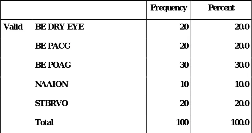
[image:68.595.102.543.389.623.2]Among the 100 patients that included in the study, 30 were known Primary Open Angle Glaucoma patients (POAG), 20 Primary Angle Closure Glaucoma (PACG), 20 were Dry eye patients, 20 Supero Temporal Branch Retinal Vein Occlusion 46(STBRVO) patients and 10 were diagnosed with Non-Arteritic Anterior Ischemic Optic Neuropathy (NA-AION).
TABLE – 1: DISTRIBUTION OF DIAGNOSIS
Frequency Percent
Valid BE DRY EYE 20 20.0
BE PACG 20 20.0
BE POAG 30 30.0
NAAION 10 10.0
STBRVO 20 20.0
55
56
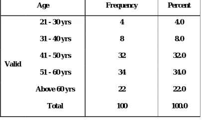
TABLE – 2: AGE WISE DISTRIBUTION OF 100 PATIENTS
Age Frequency Percent
Valid
21 - 30 yrs 4 4.0
31 - 40 yrs 8 8.0
41 - 50 yrs 32 32.0
51 - 60 yrs 34 34.0
Above 60 yrs 22 22.0
Total 100 100.0
57
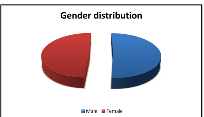
TABLE – 3: SEX WISE DISTRIBUTION OF 100 PATIENTS
Sex Frequency Percent
Valid
Male 51 51.0
Female 49 49.0
Total 100 100.0
Among 100 patients, 51 were male and 49 were female patients
58
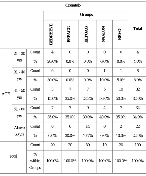
TABLE – 4: AGE WISE DISTRIBUTION OF 5 OCULAR
DISORDERS
Crosstab
Groups
Total
BEDRYEYE BEPACG BEPOAG NAAION
BRVO
AGE
21 - 30 yrs
Count 4 0 0 0 0 4
% 20.0% 0.0% 0.0% 0.0% 0.0% 4.0%
31 - 40 yrs
Count 6 0 0 1 1 8
% 30.0% 0.0% 0.0% 10.0% 5.0% 8.0%
41 - 50 yrs
Count 3 7 7 5 10 32
% 15.0% 35.0% 23.3% 50.0% 50.0% 32.0% 51 - 60
yrs
Count 7 7 9 4 7 34
% 35.0% 35.0% 30.0% 40.0% 35.0% 34.0% Above
60 yrs
Count 0 6 14 0 2 22
% 0.0% 30.0% 46.7% 0.0% 10.0% 22.0%
Total
Count 20 20 30 10 20 100
% within Groups
59
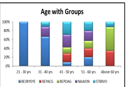
TABLE – 5: PERCENTAGE WISE AGE DISTRIBUTION OF 5
OCULAR DISORDERS
Age in years
B E D R Y E Y E B E P A C G B E P O A G N A A IO N B R V O
21 - 30 yrs 20.0% 0.0% 0.0% 0.0% 0.0%
31 - 40 yrs 30.0% 0.0% 0.0% 10.0% 5.0%
41 - 50 yrs 15.0% 35.0% 23.3% 50.0% 50.0%
51 - 60 yrs 35.0% 35.0% 30.0% 40.0% 35.0%
Above 60 yrs 0.0% 30.0% 46.7% 0.0% 10.0%
60
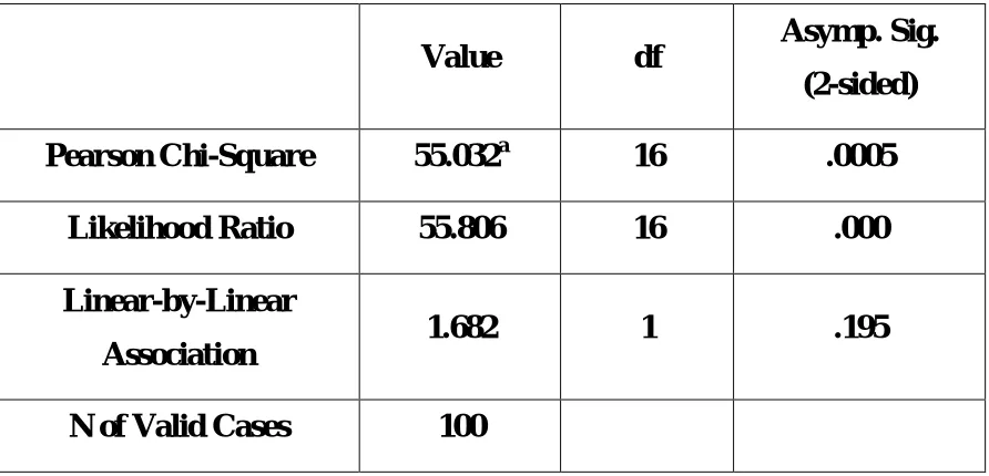
TABLE – 6: CHI-SQUARE TESTS - AGE
Value df Asymp. Sig.
(2-sided)
Pearson Chi-Square 55.032a 16 .0005
Likelihood Ratio 55.806 16 .000
Linear-by-Linear
Association 1.682 1 .195
N of Valid Cases 100
The above mentioned 5 ocular disorders associated with old age with
61
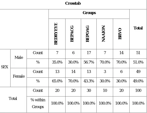
TABLE – 7: SEX WISE DISTRIBUTION OF 5 OCULAR
DISORDERS
Crosstab
Groups
Total
BEDRYEYE BEPACG BEPOAG NAAION
BRVO
SEX
Male
Count 7 6 17 7 14 51
% 35.0% 30.0% 56.7% 70.0% 70.0% 51.0%
Female
Count 13 14 13 3 6 49
% 65.0% 70.0% 43.3% 30.0% 30.0% 49.0%
Total
Count 20 20 30 10 20 100
% within Groups
62
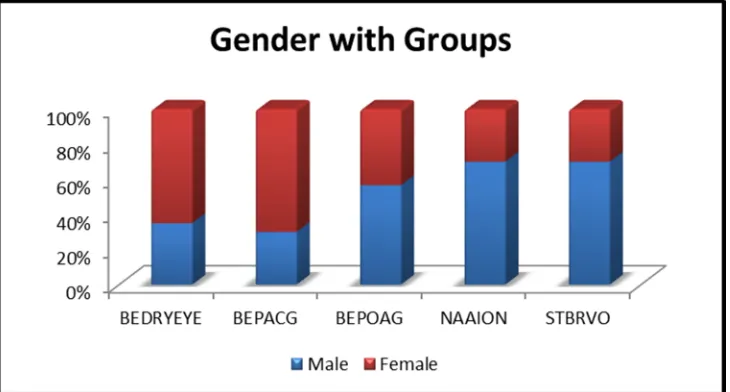
TABLE 8: PERCENTAGE WISE SEX DISTRIBUTION OF 5
OCULAR DISORDERS
SEX BEDRYEYE BEPACG BEPOAG NAAION BRVO
Male 35.0% 30.0% 56.7% 70.0% 70.0%
Female 65.0% 70.0% 43.3% 30.0% 30.0%
CHART – 5: SEX WISE DISTRIBUTION OF 5 OCULAR
63
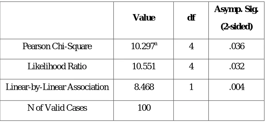
TABLE – 9: CHI-SQUARE TESTS - SEX
Value df
Asymp. Sig.
(2-sided)
Pearson Chi-Square 10.297a 4 .036
Likelihood Ratio 10.551 4 .032
Linear-by-Linear Association 8.468 1 .004
N of Valid Cases 100
TABLE -10: DISTRIBUTION OF REFRACTIVE ERRORS
AMONG THE OCULAR DISORDERS
DISORDERS REFRACTIVE ERRORS TOTAL
Emmetropia Myopia Hypermetropia
POAG - 22 8 30
PACG - 5 15 20
STBRVO - - 20 20
NA AION - - 10 10
DRY EYE 5 11 4 20
[image:77.595.96.538.418.709.2]64
CHART- 6: DISTRIBUTION OF REFRACTIVE ERRORS
AMONG THE OCULAR DISORDERS
0 5 10 15 20 25
POAG PACG STBRVO NA AION DRY EYE
65
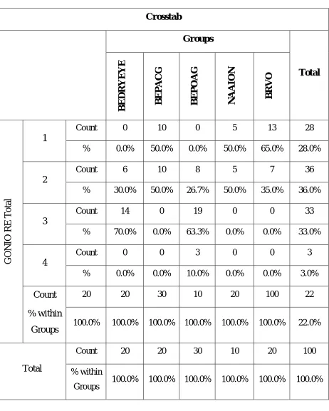
TABLE 11: GONIOSCOPY GRADINGS OF RIGHT EYE IN 5
OCULAR DISORDERS
Crosstab
Groups
Total
BEDRYEYE BEPACG BEPOAG NAAION
BRVO
GONIO
RE
Total
1
Count 0 10 0 5 13 28
% 0.0% 50.0% 0.0% 50.0% 65.0% 28.0%
2
Count 6 10 8 5 7 36
% 30.0% 50.0% 26.7% 50.0% 35.0% 36.0%
3
Count 14 0 19 0 0 33
% 70.0% 0.0% 63.3% 0.0% 0.0% 33.0%
4
Count 0 0 3 0 0 3
% 0.0% 0.0% 10.0% 0.0% 0.0% 3.0%
Count % within
Groups
20 20 30 10 20 100 22
100.0% 100.0% 100.0% 100.0% 100.0% 100.0% 22.0%
Total
Count 20 20 30 10 20 100
% within
66
From this table, we found out that 70% of patients with dry eyes had grade 3 angle in RE in gonioscopy, almost half of the patients with PACG had grade 1. 63.3% of POAG patients had grade 3 angle. 65% of patients with STBRVO had grade 1 and 50% of patients with NAAION had grade 2 and remaining 50% had grade 1 in their RE.
CHART – 7: GONIOSCOPY GRADINGS OF RIGHT EYE WITH 5
67
TABLE – 12: CHI SQUARE TEST FOR GONIO GRADINGS OF
RIGHT EYE
Value df
Asymp. Sig.
(2-sided)
Pearson Chi-Square 71.691a 12 .0005
Likelihood Ratio 94.814 12 .000
N of Valid Cases 100
Right eye of both POAG and Dry Eye disorder had > grade 2 in gonioscopy with higher significance of p<0.0005
68
TABLE – 13: GONIOSCOPY GRADINGS OF LEFT EYE IN 5
OCULAR DISORDERS
Crosstab
Groups
Total
BEDRYEYE BEPACG BEPOAG NAAION
BRVO G O N IO L E T ot al 1
Count 0 10 0 5 13 28
% 0.0% 50.0% 0.0% 50.0% 65.0% 28.0%
2
Count 6 10 8 5 7 36
% 30.0% 50.0% 26.7% 50.0% 35.0% 36.0%
3
Count 14 0 19 0 0 33
% 70.0% 0.0% 63.3% 0.0% 0.0% 33.0%
4
Count 0 0 3 0 0 3
% 0.0% 0.0% 10.0% 0.0% 0.0% 3.0%
Count
% within
Groups
20 20 30 10 20 100 22
100.0% 100.0% 100.0% 100.0% 100.0% 100.0% 22.0%
Total
Count 20 20 30 10 20 100
% within
Groups 100.0% 100.0% 100.0% 100.0% 100.0% 100.0%
69
CHART 8: GONIOSCOPY GRADINGS OF LEFT EYE IN 5
70
TABLE 14: CHI SQUARE TEST FOR GONIO
GRADINGS OF LEFT EYE
Value df
Asymp. Sig. (2-sided)
Pearson Chi-Square 71.691a 12 .0005
Likelihood Ratio 94.814 12 .000
N of Valid Cases 100
Left Eye of both POAG and Dry Eye disorder had > grade 2 in gonioscopy with higher significance of p<0.0005
71
TABLE – 15: MEAN AXIAL LENGTH MEASUREMENTS IN THE
ABOVE 5 OCULAR DISORDERS
DISORDERS RIGHT EYE LEFT EYE
DYE EYE 23.96 24.00
POAG 22.50 22.67
PACG 24.79 24.89
NA AION 22.39 21.98
STBRVO 21.90 22.46
CHART – 9: MEAN AXIAL LENGTH DISTRIBUTION AMONG 5
72
TABLE – 16: ANOVA RESULTS FOR AXIAL LENGTH OF
BOTH EYES IN 5 OCULAR DISORDERS
Sum of
squares df
Mean
Square F Sig.
AL RE
Between
Groups 135.267 4 33.817 35.414 .0005
Within
Groups 90.716 95 .955
Total 225.983 99
AL LE
Between
Groups 121.092 4 30.273 33.965 .0005
Within
Groups 84.674 95 .891
Total 205.766 99
Both POAG and dry eyes with longer axial length with p value <0.0005
73
TABLE – 17: CUP: DISC RATIO OF BOTH EYES WITH 5
OCULAR DISORDERS
DISORDERS RIGHT EYE LEFT EYE
DRY EYE 0.31 0.30
PACG 0.54 0.55
POAG 0.62 0.59
NA AION 0.11 0.11
STBRVO 0.32 0.32
CHART – 10 : CUP: DISC RATIO OF BOTH EYES WITH 5
74
TABLE – 18: ANOVA RESULTS FOR CUP:DISC RATIO(CDR)
FOR BOTH EYES WITH OCULAR DISORDERS
Sum of
Squares df
Mean
Square F Sig.
CDR RE
Between
Groups 2.964 4 .741 120.210 .0005
Within
Groups .586 95 .006
Total 3.550 99
CDR LE
Between
Groups 2.708 4 .677 118.426 .0005
Within
Groups .543 95 .006
Total 3.252 99
Both eyes of NA AION had small cup with higher significant p value <0.0005.
75
DISCUSSION
In 100 patients, 30 were known patients of Primary Open Angle Glaucoma, 20 Primary Angle Closure Glaucoma patients, 20 Dry Eye patients and 20 Supero Temporal Branch Retinal Vein Occlusion patients and 10 patients with Non-Arteritic Anterior Ischemic Optic Neuropathy.
Primary Open Angle Glaucoma (POAG):
Among 30 POAG patients, 46.7%(n=14) patients were above 60 years of age and 56.7%(n=17) were males and 43.3%(n=13) were females.
In those 30 patients 22(73%) had myopic refraction, showing the strong relationship between myopia and open angle glaucoma. This is
also supported by The Blue Mountains Eye Study, The Barbados Eye
Study and Singapore Malays Eye Study.
The Beaver Dam Eye Study showed that 60% of myopic
individuals were likely to have glaucoma than those with emmetropia.
76
All 22 patients had longer axial length (>24mm) with p value <0.0005 and all patients showed higher cup: disc ratio in their fundus examination.
Primary Angle Closure Glaucoma (PACG):
In 20 patients of PACG, 7(35%) were in age group 41-50 years, 7(35%) were in age group 51-60 years, 6(30%) were>60 years of age.
65% of patients were males in PACG category.
In this study we found that 15 of patients with PACG had hypermetropia and grade 1 angle in gonio with shallow anterior chamber implying that hypermetropia had a significant association with PACG as
supported by Lowe RF, Xu L et al and Ling Shen PhD et al.
Dry Eye:
In 20 cases of Dry Eye 50% of patients were below 40 years of age and 65% of patients with Dry Eye were found to be females.
5(25%) were emmetropic, 11(55%) were myopic and 4(20%) were found to be hypermetropic in Dry Eye Patients and high prevalence of dry
eye in myopic patients is supported by Wang et al andRania M Fahmy,
77
Branch Retinal Vein Occlusion (BRVO):
50% of patients with BRVO were in the age group 41-50 years and 70% were males among this group.
All 20(100%) patients of BRVO were found be Hypermetropic.
65% of patients with BRVO had grade 1 angle in gonioscopic examination.
BRVO eyes had shorter axial length in this study (mean AL- 21.90 mm) showed that significantly shorter axial length in affected eyes with
higher significant p value(<0.0005) and this fact is supported by Ariturk
et al, Timmerman et al and Goldstein M et al.
Non-Arteritic Anterior Ischemic Optic Neuropathy (NA-AION):
From the results, we found that NAAION usually occurs in the middle age(60%). In this group, 7(70%) were males and 3(30%) were females with all patients had refractive error of hypermetropia which is
supported by Katz B spencer WH.
Affected eyes had AL of 21.98 mm.
78
Feist Rm, Ticho BH, Shapiro MJ et al[75] and Aritirk N, OGe Y,
79
SUMMARY
This study titled “An Analytical study of various refractive errors for probable ocular associations” is a hospital based analytical study.
The aim of the study was to find out the association between refractive errors and certain ocular disorders and use it as a screening procedure to find the refractive errors.
In total of 100 patients, 30 patients were known Primary Angle Closure Glaucoma and 73% had myopic refractive error and longer axial length.
20 patients were in the Primary Angle Closure Glaucoma group of which 75% had hypermetropic refractive error and shallow anterior chamber.
In this study, 20 people were cases of Dry Eye with 55% of patients were myopic.
80
81
CONCLUSION
Uncorrected refractive errors are the second cause of reversible blindness and cause of low vision worldwide. Refractive errors affect all age groups and about 60% were aged above 40 years.
Myopia was associated with an increased prevalence of all forms of open angle glaucoma and ocular hypertension, whereas hyperopia was associated with an increased prevalence of angle closure glaucoma. Thus, high myopic and hypermetropic patients should be screened for glaucoma at closer intervals.
Patients with moderate hypermetropia should receive detailed examination and recognition of other possible risk factors to minimise the risk of NA-AION and BRVO.
82
83
BIBLIOGRAPHY
1. Duke Elder’s Practice of Refraction chapter 5th Myopia. 10th edi. PP:53.
2. American Academy of Ophthalmology The Eye M.D. Association.
Basic and clinical science course. Optics, refraction and contact lenses. Section 3. American Academy of Ophthalmology 2013-2014: 200
3. Dandona L, Dandona R, Naduvilath TJ et al. Refractive erros in an
urban population in Southern India: the Andhra Pradesh Eye Disease Study. Invest Ophthalmol Vis Sci. 1999;40:2810-2818
4. Prema Raju, S.Ve Ramesh, Hemamalini Arvind et al. Prevalence of
Refractive Errors in a Rural South Indian Population. Invest
Ophthalmol Vis Sci. 2004 December;45(12) 4268-4272.
5. Friedman NJ, Kaiser PK. Essentials of Ophthalmology.
Philadelphia, PA: Elsevier Inc;2007:253-254
6. Sia DI, Edussuriya K, Sennanayake S, Senaratne T, Selva D,
84
7. Marcus MW, de Vries MM, Junoy Montolio FG, Jansonius NM.
Myopia as a risk factor for open angle glaucoma: a systematic review and meta-analysis. 2011;118(10):1989-94e2
8. Flammer J, Orgul S, Costa VP, Orzalesi N, Krieglstein GK, Serra
LM, Renard JP, Stefansson E. The impact of ocular blood flow in glaucoma. 2002;21(4):359-393.
9. Higginbotham EJ. Ocular hypertension treatment study.
2009;127(2):213-215
10. Keltner JL, Johnson CA, Cello KE, Bandermann SE, Fan J, Levine
RA, Kass MA, Gordon MO; Ocular Hypertension Treatment Study Group. Visual field quality control in the Ocular Hypertension Treatment Study (OHTS). 2007;16(8):665-669
11. Blumen Ohana E, Blumen MB, Bluwol E, Derri M, Chabolle F,
Nordmann JP. Primary open angle glaucoma and snoring: prevalence of OSAS. 2010;127(5):159-164
12. Chang RT. Myopia and glaucoma. 2011;51(3): 53-63
13. Mitchell P, Hourihan F, Sandbach J, Wang JJ. The relationship
85
14. Sommer A, Tielsch JM. Risk factors for open angle glaucoma: The
Barbados Eye Study. Arch ophthalmol 1996;114(2):235
15. Wong TY, Klein BE, Klein R, Knudtson M, Lee KE. Refractive
errors, intra ocular pressure, and glaucoma in a white population. 2003;110(1):211-217
16. Perera SA, Wong TY, Tay WT, Foster PJ, Saw SM, Aung T.
Refractive error, axial dimensions, and primary open angle
glaucoma: The Singapore Malay Eye Study. Arch ophthalmol
2010;128(7):900-905
17. Xu L, Wang Y, Wang S, Jonas JB. High myopia and glaucoma
susceptibility the Beijing Eye Study. 2007;114(2):216-220
18. Chao DL, Shrivastava A, Kim DH, Lin H, Singh K. Axial length
does not correlate with degree of visual field loss in myopic Chinese individuals with glaucomatous appearing optic nerves. 2010;19():509-13
19. Gordon MO, Beiser JA, Brandt JD, Heuer DK, Higginbotham EJ,
Johnson CA, Keltner JL, Miller JP, Parrish RK 2nd, Wilson MR, Kass MA. The Ocular Hypertension Treatment Study: baseline factors that predict the onset of primary open angle glaucoma. Arch ophthalmol 2002;120(6): 714-720; discussion829-830
20. Detry-Morel M. Is myopia a risk factor for glaucoma? J Fr
86
21. Jonas JB, Martus P, Budde WM. Anisometropia and degree of
optic nerve damage in chronic open angle glaucoma. Am J
Ophthalmol 2002; 134(4):547-551
22. Brad Bowling. Kanski’s Clinical Ophthalmology a systemic
approach, chapter 6th Cornea, 8th (edn), pp: 213-214
23. Qasim KF and Shahad A. Prevalence of Refractive Errors in
Patients with Keratoconus among Sample of Iraqi Population. J Ophthalmol 2017, 2(4): 000134.
24. Scheer SE, Touzeau O, Moreal C, Kopito R, Allouch C Laroche.
Relation between keratoconus and axial myopia. Inves Oph Vis Sci 20003 May 44;1310
25. Benjamin J. Ernst; Hugo Y. Hsu. Keratoconus Association with
Axial Myopia: A Prospective Biometric Study. Eye & Contact Lens: Science & Clinical Practice. 37(1):2-5, JAN 2011
26. Aníbal Cruz-Becerril, Alejandra Valdivia, Raúl Peralta, Ruth N
DomínguezFernández, Marco a Castro-reyes (2015) Prevalence of refractive errors in Mexican patients with keratoconus. Dovepress J Clinical optometery 7: 39-44.
27. Valdez-García JE, Sepúlveda R, Salazar-Martínez JJ,
87
28. Brad Bowling. Kanski’s Clinical Ophthalmology a systemic
approach, chapter 15th Hereditary Fundus Dystrophies, 8th (edn),
pp: 646-647
29. Sieving A Paul, Fishman GA. Refractive errors of retinitis
pigmentosa patients, British Journal of Ophthalmology
1978,62,163-167.
30. Morichini G, Salvator S, Montaldi F, Vingolo EM. Refractive
errors of RP patients. Inves Ophthal Vis Sci 2007 May 48:3746
31. Chassine T, Bocquet B, Daien V, Avila-Fernandez A, Ayuso C,
Collin RW, Corton M, Hejtmancik JF, van den Born Ll, Klevering BJ, Riazuddin SA, Sendon N, Lacrox A, Meunier I, Harnel CP. Autosomal recessive retinitis pigmentosa with RP1 mutations is associated with myopia. Br J Ophthalmol 2015 Oct;99(10):1360-5.
32. Lyle WM, Sangster JO, Williams TD. Albinism: an update and
review of the literature. J Am Optom Assoc.1997;68:623-645
33. Spedick MJ, Beauchamp GR. Retinal vascular and optic nerve
abnormalities in albinism. J Pediatr Ophthalmol Strabismus. 1986;23(2):58–63.
34. Dickerson CM, Abadi RV. Corneal topography of humans with