CASE REPORT
Macular edema with serous retinal
detachment post-phacoemulsification
followed by spectral domain optical coherence
tomography: a report of two cases
Hui Xiao, Xing Liu
*and Xinxing Guo
Abstract
Background: Macular edema and detachment at the first day after an uneventful cataract surgery is very rare, and has been reported previously with the use of high concentrations of intra-cameral cefuroxime. However, we hereby reported two cases of macular edema with extensive serous retinal detachment the first day after an uneventful phacoemulsification with intra-cameral injection of a standard dose of cefuroxime during the procedure.
Case presentation: A 68-year-old female and a 63-year-old male without any special history both underwent an uneventful phacoemulsification surgery and 1 mg/0.1 ml of cefuroxime solution was injected into the anterior chamber at the end of the procedure. Macular edema with extensive serous retinal detachment around macula and optic disc area were observed the first day after surgery. Without surgical intervention, a quick recovery of the macular edema and retinal detachment was observed by spectral domain optical coherence tomography 1 week later in both cases.
Conclusion: We presume that the retina injury in the two cases may be attributed to cefuroxime toxicity even under a use of a standard dose. But the retinal damages are restorable and routine anti-inflammatory treatment is enough.
Keywords: Macular edema, Retinal detachment, Cefuroxime, Phacoemulsification
© 2015 Xiao et al. This article is distributed under the terms of the Creative Commons Attribution 4.0 International License (http://creativecommons.org/licenses/by/4.0/), which permits unrestricted use, distribution, and reproduction in any medium, provided you give appropriate credit to the original author(s) and the source, provide a link to the Creative Commons license, and indicate if changes were made. The Creative Commons Public Domain Dedication waiver (http://creativecommons.org/ publicdomain/zero/1.0/) applies to the data made available in this article, unless otherwise stated.
Background
Macular edema is one of the most common compli-cations after cataract surgery that causes unfavorable visual outcomes and usually occurs in the surgical eye 4–16 weeks after the procedure [1, 2]. Acute macular edema with retinal detachment after cataract surgery is very rare, and has been reported previously with the use of high concentrations of intra-cameral cefuroxime [3, 4]. Cefuroxime is commonly used during phacoemulsifica-tion procedure [5] and has been proved to be safe with a standard dose previously [6–8]. However, we hereby report two cases of macular edema with extensive serous retinal detachment that was immediately detected by
spectral domain optical coherence tomography (SD-OCT) the first day after an uneventful phacoemulsifica-tion with intra-cameral injecphacoemulsifica-tion of standard doses of cefuroxime during the procedure. We presume that the retina injury in the two cases may be attributed to cefuro-xime toxicity even under a use of a standard dose.
Case presentation Case 1
A 68-year-old female had an uncomplicated phacoemul-sification surgery with folded in-the-bag intraocular lens (IOL) implantation in her left eye. Her systemic and oph-thalmic histories were unremarkable. No diabetic, uveitis or any other remarkable retinal history was found prior to the surgery. Preoperatively, the refractive errors of her right and left eyes were −4.0 and −4.5 diopters (D), with
axial lengths of 24.12 and 24.37 mm, respectively. The
Open Access
*Correspondence: drliuxing@163.com
best-corrected visual acuity was 20/33 in the right and 20/40 in the left eye. The patient’s anterior segment and fundus were normal in both eyes, as revealed by regular examination. The surgery was performed using an Infiniti phacoemulsification unit (Alcon, Inc.). The nucleus chop-ping time was 5.8 s, and the average power was 6.8 %. A +18 D folded IOL (Acrysof SN60AT Alcon, Inc) was implanted in-the-bag. The surgery was completed without complications. At the end of the procedure, 1 mg/0.1 ml of cefuroxime solution was injected into the anterior chamber. We perform our dilution in the oper-ating room: the nurse takes 750 mg of preservative-free vial cefuroxime and adds 7.5 ml of balanced salt solution (BSS), and then, the surgeon takes 0.1 ml of this first solu-tion and adds another 0.9 ml of BSS to obtain the second solution. 0.1 ml of the second solution is finally injected in the anterior chamber of the patient. The patient is therefore supposed to receive 0.1 ml of 10 mg/ml solu-tion of intra-cameral cefuroxime. The total length of the surgical time was approximately 10 min.
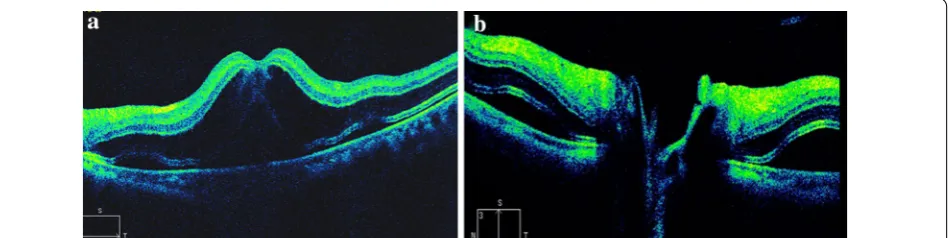
The first day after the operation, the visual acuity in her left eye was 20/200. There were no signs of remark-able inflammation or abnormality in the anterior seg-ment, as well as in the vitreous. Fundus examination showed no foveal reflection in the macula. Diffuse reti-nal edema affected most of the posterior pole. Retireti-nal wrinkles were found around the macula and disc area. No significant abnormality was found in the peripheral retina. SD-OCT (Carl Zeiss Meditec, Dublin, CA, USA) scanning was immediately performed and showed mac-ular edema, especially at the outer nuclear layer, with extensive shallow serous retinal detachment around macula and optic disc area (Fig. 1). The retinal thickness of the fovea was 750 μm. No significant abnormality was found in the choroids and the subfoveal choroidal thickness was 350 μm. Vitreomacular traction was not found in the SD-OCT image. Topical dexamethasone 0.1 %/tobramycin 0.3 % (Tobradex®) eye drops and
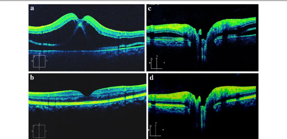
pranoprofen (Senju Pharmaceutical Co. Ltd) were pre-scribed four times a day. After 1 week of treatment, the patient’s vision in her left eye had improved to 20/20. The macular retina was scanned with the same area by SD-OCT and the image showed that the retinal thickness of the fovea returned within a normal range (194 μm), and the subfoveal choroidal thickness seemed not changed a lot (about 347 μm). The macular edema and subretinal fluid were absorbed completely. The inte-grated ellipsoid zone was preserved in the outer retina (Fig. 2). No recurrence of macular edema or retinal detachment was noted at the last follow-up (4 months post-operative).
Case 2
A 63-year-old male underwent an uneventful phaco-emulsification surgery with folded in-the-bag IOL implantation in the left eye. The systemic and ophthal-mic histories were unremarkable. Preoperatively, the refractive error of his left eye was −2.75 D with an axial length of 23.93 mm. The best corrected visual acuity was 20/200. The right eye had IOL implanted 1 year ago with good visual acuity of 20/20. Findings of anterior segment and fundus examination were normal in both eyes. The phacoemulsification surgery was performed by the same doctor as in case 1. The nucleus chopping time was 32 s and the average power was 16.8 %. A +20.0 D folded IOL (Acrysof SN60AT Alcon, Inc) was implanted in-the-bag. The surgery was completed without complications. At the end of the procedure, 1 mg/0.1 ml of cefuroxime solu-tion (diluted as case 1) was also injected into the anterior chamber. The total length of the surgical time was about 12 min.
The first day after the operation, the visual acuity in his left eye was finger count. No remarkable inflamma-tions or abnormalities were found in the anterior seg-ment and vitreous by slit-lamp examination. Fundus manifestation was similar to case 1. SD-OCT image also
[image:2.595.65.540.579.698.2]showed the same macular edema as case 1 (Fig. 3a). The retina thickness of the fovea was 794 μm. No signifi-cant abnormality was found in the choroids. The same drugs as in case 1 were adopted four times a day. After 1 week treatment, the patient’s visual acuity improved to 20/20. SD-OCT revealed macular edema and sub-retinal fluid were absorbed completely. The integrated ellipsoid zone was preserved in the outer retina (Fig. 3b). The retinal thickness of the fovea returned within a nor-mal range (174 μm). No recurrence of macular edema or retinal detachment was noticed until the last follow-up (3 months after surgery).
Conclusions
With modern cataract surgical techniques, the inci-dence of post-surgical cystoid macular edema (CME) has decreased to 0.1–2.35 % [9]. Several mechanisms may contribute to such macular edema, including the effects of vitreoretinal traction, light damage, produc-tion of prostaglandins and intraoperative complicaproduc-tions [1, 10]. The rate of macular edema after cataract surgery is increased in the presence of diabetic retinopathy and uveitis [11, 12]. However, the two cases were notable for its unremarkable retinopathy and lack of history of diabe-tes or uveitis.
Fig. 2 SD-OCT images of case 1 One week post-operation. Macular edema and subretinal fluid was absorbed and the central foveal thickness resumed to normal (a). The subretinal fluid was also absorbed around optic disc (b)
[image:3.595.57.541.256.378.2] [image:3.595.59.537.427.658.2]Jurecka et al. [13] found a positive statistical correla-tion between the real phacoemulsificacorrela-tion time and the increase in macular retinal thickness after surgery. In the present cases, the real phacoemulsification time was not long, and the average power was low.
Cefuroxime toxicity may be one of the cause of macular edema and detachment. The recommended dose of intra-cameral cefuroxime injection is 0.1 ml of 10.0 mg/ml solu-tion. The fact that excessive cefuroxime solution injections into the anterior chamber can cause early serous macular detachment and edema has been reported previously [3, 4]. The reported dose has been varied from 20 to 50 mg/ ml. However, recently, Kontos et al. [14] reported a case with acute serous macular detachment and macular edema after a standard dose of subconjunctival cefuro-xime injection in the phacoemulsification. Faure et al. [15] even reported a case occurred retinal toxicity the second day after surgery with a standard dose of intra-cameral cefuroxime injection in France. In the present two cases in China, early macular edema and extensive retinal detach-ment were found immediately the first day after surgery with a standard dose of intra-cameral cefuroxime injec-tion at the end of the phacoemulsificainjec-tion. The visual loss was earlier in the present two cases than that of Faure et al. report. Though the visual loss time after surgery had little difference, the manifestations of these cases were similar. The interval time between the present two cases was about one month. No abnormality was found dur-ing the drug dilution process. Thus, we presume that the retina injury in the two cases may be also attributed to cefuroxime toxicity even under a use of a standard dose.
In these two cases, the location of the edema was unu-sual: Typically retinal edema was located in the outer plexiform layer. However, in these cases the outer plexi-form layer appeared to be spared and the outer nuclear layer had large edema. There was extensive subretinal fluid without debris. These OCT characteristics were similar to the manifestation that has been identified in OCT of the retinal toxicity caused by excessive cefuroxime solution injections [3, 4], and might provide a marker for cefuro-xime toxicity. The mechanism of this pattern of edema is unclear. The electroretinogram (ERG) results of animal experiments [16] and human clinical observation [15] prompted cefuroxime was toxic to retina, and may effect the Müller cell function. Previous study [3, 4] reported that fluorescein angiograms (FA) showed diffuse leakage with-out abnormal retinal perfusion in cefuroxime toxic eyes and indicated that the blood–retinal barrier at the retinal pigment epithelium (RPE) may be disrupted. It is a limita-tion that the FA was not obtained in the present two cases, but the SD-OCT images may suggest that the primary lesions were localized at the outer retinal and RPE.
Clear vitreous haze has been reported in ocular toxicity after intra-cameral injection of very high doses of cefuro-xime during cataract surgery [4]. But, no sign of remarka-ble inflammation in the vitreous was found in the present cases. No abnormality in vitreous has also been reported by Buyukyildiz in two cases of retinal toxicity caused by 2 mg/0.1 ml cefuroxime intra-cameral injection [3]. The dose of cefuroxime injection was much higher in Delyfer et al. study [4] than the presented cases and Buyukyildiz et al. cases [3]. The different doses of cefuroxime injec-tion during cataract surgery may lead to the different findings in vitreous.
Topical nonsteroidal anti-inflammatory drugs and cor-ticosteroids have been reported to be effective and safe therapy for preventing post-surgical ocular inflamma-tory and macular edema [17–20]. Thus combination of nonsteroidal anti-inflammatory drugs and corticoster-oids was applied in the present cases topically as routine anti-inflammation treatment after phacoemulsification. The SD-OCT image revealed a quick recovery from the macular edema without any special surgical intervention 1 week later. Delyfer et al. [4] also reported that retinal injury and visual dysfunction induced by intra-cameral excessive cefuroxime injection were able to recover to normal without surgery intervention after 6 weeks. The recovery time was shorter in the present cases than pre-vious report. That may be due to the much lower con-centration of cefuroxime solution used in the two cases. These results suggest that early macular edema with extensive serous retinal detachment which may be attrib-uted to cefuroxime toxicity are restorable. Routine anti-inflammatory treatment is sufficient and do not require excessive interventions.
Consent
Written informed consent was obtained from the patients for publication of this case report and any accompanying images.
Abbreviations
SD-OCT: spectral domain optical coherence tomography; OCT: optical coher-ence tomography; IOL: intraocular lens; CME: cystoid macular edema; FA: fluorescein angiograms; RPE: retinal pigment epithelium.
Authors’ contributions
XH has made substantial contributions to analysis and interpretation of data; has been involved in drafting the manuscript or revising it critically for important intellectual content; and has given final approval of the version to be published. LX and GXX have been involved in revising it critically for important intellectual content; and have given final approval of the version to be published. All authors read and approved the final manuscript.
Acknowledgements
Competing interests
The authors declare that they have no competing interests.
Received: 10 December 2014 Accepted: 27 October 2015
References
1. Gulkilik G, Kocabora S, Taskapili M, Engin G. Cystoid macular edema after phacoemulsification: risk factors and effect on visual acuity. Can J Oph-thalmol. 2006;41:699–703.
2. Rossetti L, Autelitano A. Cystoid macular edema following cataract surgery. Curr Opin Ophthalmol. 2000;11:65–72.
3. Buyukyildiz HZ, Gulkilik G, Kumcuoglu YZ. Early serous macular detachment after phacoemulsification surgery. J Cataract Refract Surg. 2010;36:1999–2002.
4. Delyfer MN, Rougier MB, Leoni S, Zhang Q, Dalbon F, Colin J, Korobelnik JF. Ocular toxicity after intracameral injection of very high doses of cefuro-xime during cataract surgery. J Cataract Refract Surg. 2011;37:271–8. 5. Barry P, Seal DV, Gettinby G, Lees F, Peterson M, Revie CW, ESCRS
Endophthalmitis Study Group. ESCRS study of prophylaxis of postopera-tive endophthalmitis after cataract surgery; preliminary results from a European multicenter study. J Cataract Refract Surg. 2006;32:407–10. 6. Lam PT, Young AL, Cheng LL, Tam PM, Lee VY. Randomized controlled
trial on the safety of intracameral cephalosporins in cataract surgery. Clin Ophthalmol. 2010;8:1499–504.
7. Gupta MS, McKee HD, Saldana M, Stewart OG. Macular thickness after cataract surgery with intracameral cefuroxime. J Cataract Refract Surg. 2005;31:1163–6.
8. Hann JV, Lee LR. Macular thickness after cataract surgery with intracam-eral cefuroxime. J Cataract Refract Surg. 2006;32:545.
9. Loewenstein A, Zur D. Postsurgical cystoid macular edema. Dev Ophthal-mol. 2010;47:148–59.
10. Agange N, Mosaed S. Prostaglandin-induced cystoid macular edema following routine cataract extraction. J Ophthalmol. 2010. doi:10.1155/2010/690707.
11. Kim SJ, Equi R, Bressler NM. Analysis of macular edema after cataract surgery in patients with diabetes using optical coherence tomography. Ophthalmology. 2007;114:881–9.
12. Bélair ML, Kim SJ, Thorne JE, Dunn JP, Kedhar SR, Brown DM, Jabs DA. Incidence of cystoid macular edema after cataract surgery in patients with and without uveitis using optical coherence tomography. Am J Ophthalmol. 2009;148:128–35.
13. Jurecka T, Bátková Z, Ventruba J. Macular edema after an uncomplicated cataract surgery. Cesk Slov Oftalmol. 2007;63:262–73.
14. Kontos A, Mitry D, Althauser S, Jain S. Acute serous macular detachment and cystoid macular edema after uncomplicated phacoemulsification using standard dose subconjunctival cefuroxime. Cutan Ocul Toxicol. 2013. doi:10.3109/15569527.2013.835817.
15. Faure C, Perreira D, Audo I. Retinal toxicity after intracameral use of a standard dose of cefuroxime during cataract surgery. Doc Ophthalmol. 2014. doi:10.1007/s10633-014-9465-7.
16. Shahar J, Zemel E, Perlman I, et al. Physiological and toxicological effects of cefuroxime on the albino rabbit retina. Invest Ophthalmol Vis Sci. 2012;21(53):906–14.
17. Asano S, Miyake K, Ota I, Sugita G, Kimura W, Sakka Y, Yabe N. Reducing angiographic cystoid macular edema and blood-aqueous barrier disrup-tion after small-incision phacoemulsificadisrup-tion and foldable intraocular lens implantation: multicenter prospective randomized comparison of topical diclofenac 0.1% and betamethasone 0.1%. J Cataract Refract Surg. 2008;34:57–63.
18. Miyake K, Ota I, Miyake G, Numaga J. Nepafenac 0.1% versus fluo-rometholone 0.1% for preventing cystoid macular edema after cataract surgery. J Cataract Refract Surg. 2011;37:1581–8.
19. Romac I, Gabrić N, Dekaris I, Barisić A. Resolution of pseudophakic cystoid macular edema with combination therapy of topical corticosteroids and nonsteroidal anti-inflammatory drugs. Coll Antropol. 2011;35:281–4. 20. Zur D, Fischer N, Tufail A, Monés J, Loewenstein A. Postsurgical cystoid
macular edema. Eur J Ophthalmol. 2011;21(suppl):S62–8.
Submit your next manuscript to BioMed Central and take full advantage of:
• Convenient online submission
• Thorough peer review
• No space constraints or color figure charges
• Immediate publication on acceptance
• Inclusion in PubMed, CAS, Scopus and Google Scholar
• Research which is freely available for redistribution