R92
Introduction
Mammary gland development during the pregnancy cycle is characterized by successive phases of cell growth, dif-ferentiation, high metabolic activity and apoptosis. At the ultrastructural level this includes dramatic changes in tissue architecture, involving ductal epithelial branching and morphogenesis, invasion of tissue compartments,
vas-cularization and subsequent organized remodelling. These events are highly reproducible and strictly controlled at the transcriptional level by circulating hormones and locally derived factors [1]. Thus, many transcription factors have been shown either to directly affect this developmental program or to exhibit altered activity at specific stages in the pregnancy cycle [1].
CAM = cell adhesion molecule; C/ebp = CAAT-enhancer binding protein; IGF = insulin-like growth factor; IL = interleukin; LIF = leukaemia inhibitory factor; LPS = lipopolysaccharide; NF-κB = nuclear factor-κB; Stat = signal transducer and activator of transcription.
Research article
Gene expression profiling of mammary gland development
reveals putative roles for death receptors and immune mediators
in post-lactational regression
Richard WE Clarkson
1, Matthew T Wayland
2, Jennifer Lee
2, Tom Freeman
2and
Christine J Watson
11Department of Pathology, University of Cambridge, Cambridge, UK 2MRC-HGMP Resource Centre, Hinxton, UK
Correspondence: Richard WE Clarkson (e-mail: [email protected])
Received: 22 Sep 2003 Revisions requested: 12 Nov 2003 Revisions received: 15 Nov 2003 Accepted: 21 Nov 2003 Published: 18 Dec 2003 Breast Cancer Res 2004, 6:R92-R109 (DOI 10.1186/bcr754)
© 2004 Clarkson et al., licensee BioMed Central Ltd (Print ISSN 1465-5411; Online ISSN 1465-542X). This is an Open Access article: verbatim copying and redistribution of this article are permitted in all media for any purpose, provided this notice is preserved along with the article's original URL.
See related Research article: http://breast-cancer-research.com/content/6/2/R75 and related Commentary: http://breast-cancer-research.com/content/6/2/89
Abstract
Introduction In order to gain a better understanding of the molecular processes that underlie apoptosis and tissue regression in mammary gland, we undertook a large-scale analysis of transcriptional changes during the mouse mammary pregnancy cycle, with emphasis on the transition from lactation to involution.
Method Affymetrix microarrays, representing 8618 genes, were used to compare mammary tissue from 12 time points (one virgin, three gestation, three lactation and five involution stages). Six animals were used for each time point. Common patterns of gene expression across all time points were identified and related to biological function.
Results The majority of significantly induced genes in involution were also differentially regulated at earlier stages in the pregnancy cycle. This included a marked increase in inflammatory mediators during involution and at parturition, which correlated with leukaemia inhibitory factor–Stat3 (signal
transducer and activator of signalling-3) signalling. Before involution, expected increases in cell proliferation, biosynthesis and metabolism-related genes were observed. During involution, the first 24 hours after weaning was characterized by a transient increase in expression of components of the death receptor pathways of apoptosis, inflammatory cytokines and acute phase response genes. After 24 hours, regulators of intrinsic apoptosis were induced in conjunction with markers of phagocyte activity, matrix proteases, suppressors of neutrophils and soluble components of specific and innate immunity.
ConclusionWe provide a resource of mouse mammary gene expression data for download or online analysis. Here we highlight the sequential induction of distinct apoptosis pathways in involution and the stimulation of immunomodulatory signals, which probably suppress the potentially damaging effects of a cellular inflammatory response while maintaining an appropriate antimicrobial and phagocytic environment.
Keywords:apoptosis, immunity, involution, mammary, microarray
R93 Our laboratory has focused on the role of transcription
factors in postlactational regression of the gland after weaning. Involution can be divided into at least two phases [2–4], broadly comprising the following: an initial reversible phase whereby the gland maintains its gross morphology but undergoes a substantial increase in the rate of epithelial cell apoptosis [5]; and a secondary irre-versible phase, which involves the destruction of base-ment membrane by matrix metalloproteinases, phagocytic clearance of milk, and apoptotic bodies and alveolar col-lapse [6]. Immune cells are present at all stages of mammary development, including involution [7], but the precise role of the immune system during postlactational regression has yet to be fully established. Many genes have been shown to be differentially regulated during invo-lution [5,8,9]. However, there has been no comprehensive analysis of gene expression in mammary involution.
Microarray analysis has had a major impact on our under-standing of the transcriptional basis of complex biological systems. Normal tissue development and homeostasis has been studied in a variety of mouse tissues, including retina [10], liver [11], pancreas [12], uterus [13] and mammary gland [14].
The few microarray studies of normal mouse mammary gland described in the literature have either focused on early stages in the developmental cycle [14], or have used mammary data as a tool for illustrating methods of data analysis [15,16].
In the present study we applied a microarray approach to study the transcriptional expression (mRNA levels) of 8618 mouse genes in mammary gland during the preg-nancy cycle. We focused on five time points in involution but we also included earlier time points in the pregnancy cycle to establish the specificity of these involution-related genetic changes. We provide the raw microarray data files and processed genelists for online analysis or download (see the Mammary Apoptosis and Development Group Home Page: www.path.cam.ac.uk/~madgroup). Here we highlight specific aspects of the microarray data that are pertinent to involution by identifying common patterns of gene expression and relating them to specific biological functions. Thus, we demonstrate the sequential activation of death receptor genes followed by components of the mitochondrial (intrinsic) pathway after weaning, suggest-ing that different apoptosis mechanisms are employed at different phases of involution. We also highlight a promi-nent role for immune related genes in involution, equating this with phagocytic clearance of apoptotic cells and the maintenance of an antimicrobial environment during milk stasis. We provide evidence that the proapoptotic, acute phase transcription factor Stat3 (signal transducer and activator of transcription-3) [17,18] may simultaneously suppress a potentially damaging cellular infiltrate. A similar
study conducted in Balb/c mice and described in the accompanying paper in this issue [19] confirms proinflam-matory gene expression in involution. Together, these data provide a useful online resource for studying the activity of specific genes in mammary involution.
Method
Mouse husbandry
Six-week-old C57/Bl/6 mice were obtained from Harlan Laboratories (Bicester, UK). At 8 weeks females were either mated or culled for virgin mammary glands. Postpar-tum, a minimum of six pups per suckling female was ensured by cross-fostering where appropriate. Females used in involution time points were force weaned at 10 days of lactation. Lactating and involuting mammary glands were monitored daily for signs of localized inflam-mation or mastitis. Two out of 90 animals were excluded from the study for this reason. The study was approved by the local ethics committee and complies with the Helsinki Declaration.
Harvesting mammary gland RNA
A total of 12 stages of adult mouse mammary gland devel-opment were selected for this study: 8-week-old virgin; 5 days, 10 days and 15 days of gestation (in which day 1 was the first day postcoitum); 0 days (first day post-partum), 5 days and 10 days of lactation; and 12, 24, 48, 72 and 96 hours after forced weaning. All mammary glands were harvested between 11:00 and 13:00 to limit circadian effects. A total of six animals were used per time point (three per hybridization). In each case a single abdominal gland was removed following excision of lymph nodes and immediately frozen in liquid nitrogen. RNA was extracted from frozen tissue using Trizol reagent (Invitro-gen, Paisley, UK) followed by additional column purifica-tion (Rneasy; Qiagen, Crawley, UK). Briefly, three samples of mammary tissue from the same time point were ground under liquid nitrogen, and 20 mg of each was pooled into 1.5 ml Trizol reagent. Resuspended RNA samples were then passed through purification columns according to the manufacturer’s instructions and RNA integrity was moni-tored with Lab-on-a-chip (Agilent, West Lothian, UK) before use in microarray analysis.
Microarray hybridization and data analysis
RNA 5µg was labelled according to manufacturers proto-cols (Affymetrix, High Wycombe, UK) and hybridized to Affymetrix MGU74ver2a chips representing 12 488 tran-scripts or 8618 genes. Computation of expression values was performed using the perfect match/mismatch model implemented in dChip [20].
R94
Genetics, Redwood City, CA, USA). Two filters were applied to the complete data set of 12 488 transcripts. First, 755 transcripts (6%) were removed following elimi-nation of genes whose mean signal intensities were below 20 normalized intensity units in at least 11 of the 12 time point conditions. Second, 5149 transcripts (44%) were removed on the basis that they exhibited no significant change in mean signal intensity across all time points (Welch ANOVA P= 0.05, using the Benjamini and Hochberg False Discovery Rate controlling procedure). A separate lactation/involution specific time course gene set was established by applying the same cutoff filters to all 12 488 transcripts using 5 day and 10 day lactation time points, and 12, 24, 48, 72 and 96 hour involution time points.
Both data sets were clustered (K means) according to expression pattern similarity across all 12 conditions (com-plete time course) or seven conditions (involution time course) using standard correlation. Nine K mean classifi-cations were performed for each data set, starting with 10 clusters and increasing by five incrementally to 50 clus-ters. The optimal number of clusters was determined empirically based on highest observed variability and redundancy between similar clusters.
All genes were annotated according to known function using the Gene Ontology Consortium categories [21]: bio-logical process, cellular component and molecular function. Onto-Express [22] was used to determine whether clusters of genes with similar expression profiles were enriched in specific GO functional categories. Based on the genes present on the GeneChip, Onto-Express calculated the expected number of occurrences of each functional cate-gory in each cluster. The probability that each functional category was over-represented in a cluster was derived using a binomial model and the Pvalues were corrected for multiplicity using the Bonferroni method. Thus, Onto-Express provided information about the statistical signifi-cance of each of the pathways and categories represented by the genes in each cluster (these data are available online as Additional files 1 and 2).
Archived data
All genelists, including expression profile clusters, ontolog-ical definitions and all raw data .cel files, fulfilling MIAME criteria, were uploaded to ArrayExpress (http://www.ebi. ac.uk/arrayexpress/) and are also available through GeNet (http://genet.hgmp.mrc.ac.uk:8080/servlet/GeNet) for down-load and online analysis. (For information on accessing data through GeNet or from Breast Cancer Researchsee our website: www.path.cam.ac.uk/~madgroup/). Gene lists use Affymetrix probe_ID and accession number as transcript identifiers. In the body of the text and figures, gene/transcripts are named according to the Mouse Genome Database [23].
Results
We analyzed the expression of over 12 000 transcripts, or approximately one-third of the protein encoding capacity of the mouse genome, at 12 different time points in the mammary gland pregnancy cycle. We focused our analy-sis on the changes in gene expression that occur during the first 4 days of postlactational regression, harvesting at five time points during this period. We also included three time points in lactation, three in gestation and a single time point in nulliparous animals in order to distinguish between involution-specific genes and genes that were also highly expressed at other times in the pregnancy cycle. All raw and normalized data files were submitted to GeNet and made available for download or online analysis through our website (www.path.cam.ac.uk/~madgroup/).
Expression of 6796 transcripts (54% of the transcripts assayed) significantly changed across the 12-point time course. This suggested that approximately half of the protein encoding content of the mouse genome was regu-lated in mammary gland at some point during the preg-nancy cycle. Among the genes that did not significantly change (nonsignificant profiles genelist in GeNet) were a number of recognized ‘housekeeping’ genes, including 18s ribosomal and α-actin genes. However, other genes that are ubiquitously expressed in some cell types (e.g. GAPDH and cyclophilins) exhibited differential expression in the mammary time course. This reflects a combination of factors, including changes in tissue composition and dramatic shifts in cellular metabolism and macromolecular synthesis throughout the mammary cycle, in particular during lactation.
In order to verify that the microarray time course reflected expected global expression patterns in vivo, we compared our results with the known mammary expression patterns of 32 genes described in the literature, all of which exhibited the expected expression profiles (see Additional file 3).
Expression profile analysis of the pregnancy cycle (12-point time course)
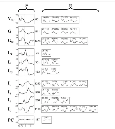
It was expected that a proportion of the 6797 differentially expressed transcripts would be specific to involution, whereas others may have additional roles at earlier stages in the pregnancy cycle. In order to identify these subsets of involution related genes, the expression patterns of all differentially expressed transcripts were compared and subsequently grouped according to similarity into 35 gene profile clusters. Some clusters exhibited superficially similar patterns (Fig. 1). In order to simplify the classifica-tion of expression patterns observed in the time course, these similar profiles were pooled, resulting in 11 broadly distinct expression profiles (Fig. 1a).
cluster of 651 transcripts (5% of all transcripts on the array) were maximally expressed in virgin mammary gland (VSL). Two clusters with a total of 2317 transcripts (19%) were maximally expressed in gestation (G and GSL) and
[image:4.612.121.525.95.548.2]three clusters (529 transcripts, 4%) were maximally induced during lactation (LT, L and LG). Four clusters (3113 transcripts, 25%) exhibited maximum expression during involution (IL, IT, IP and IG) whereas one cluster R95
Figure 1
included 187 transcripts (1%) that were uniformly expressed throughout the pregnancy cycle (PC). Further details on the nomenclature of these combined clusters are provided in the legend to Fig. 1. Genelists for each combined cluster and the 35 primary clusters are available online (see Method section, above).
Six clusters consisted of transcripts that were either maxi-mally expressed during involution (IL, IT, IP and IG) or specifically suppressed following weaning (L and LG), sug-gesting that a proportion of these genes may play impor-tant roles in regression of the mammary gland. These six clusters consisted of 3463 transcripts, or 28% of all tran-scripts on the array. Only one group of 518 genes exhib-ited involution-specific expression, all of which were transiently activated within 24 hours after weaning (IT). It was expected that a proportion of these represent genes that may contribute to the initial phase of cell death char-acteristic of the reversible first stage of involution. All other involution related clusters exhibited elevated expression at earlier stages in the pregnancy cycle. For example, one group of 236 involution related genes (IP) exhibited an additional peak of expression specifically at parturition. This suggested that many transcriptionally regulated processes in involution were common to gestation (IG), parturition (IP) or lactation (IL).
Correlation of gene function with gene expression pattern
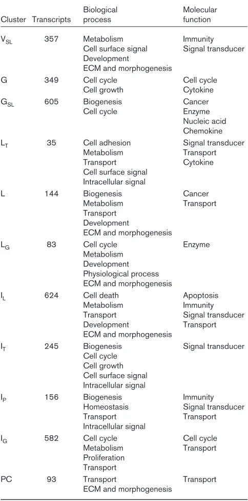
Of the 6747 differentially expressed transcripts, 3273 (49%) encoded proteins with known function. In order to establish what proportion of the transcripts in each cluster from Fig. 1a shared common biological functions, tran-scripts with known function were assigned to categories as defined by the Gene Ontology Consortium (www.geneontology.org), and statistically significant asso-ciations between gene function and each cluster were identified using Onto-Express [22]. The significant associ-ations (corrected P< 0.05) are listed in Table 1. Further details on the distribution of functionally related genes between these clusters are available in Additional file 4.
[image:5.612.314.556.130.618.2]Unexpectedly, similar proportions of apoptosis related genes were expressed in each of the 11 clusters; the only significant correlation being with the IL cluster, in which maximal gene expression occurred at 12 hours of involu-tion. However, a number of other biological processes were significantly linked to particular stages of mammary development, and were consistent with our current under-standing of the physiology of the mammary gland. Thus, during gestation there was a significant increase in the expression of cell cycle regulatory genes (G, GSLand IG) and a concomitant increase in nucleic acid and macromol-ecular synthesis (‘biogenesis’ and ‘metabolism’ in Table 1). By lactation, the proportion of cell cycle genes had diminished, to be replaced by an increase in the R96
Table 1
Relationship between gene expression pattern and gene function in the 12-point developmental time course
Biological Molecular Cluster Transcripts process function
VSL 357 Metabolism Immunity
Cell surface signal Signal transducer Development
ECM and morphogenesis
G 349 Cell cycle Cell cycle
Cell growth Cytokine
GSL 605 Biogenesis Cancer
Cell cycle Enzyme
Nucleic acid Chemokine LT 35 Cell adhesion Signal transducer
Metabolism Transport
Transport Cytokine
Cell surface signal Intracellular signal
L 144 Biogenesis Cancer
Metabolism Transport Transport
Development
ECM and morphogenesis
LG 83 Cell cycle Enzyme
Metabolism Development Physiological process ECM and morphogenesis
IL 624 Cell death Apoptosis
Metabolism Immunity Transport Signal transducer Development Transport ECM and morphogenesis
IT 245 Biogenesis Signal transducer Cell cycle
Cell growth Cell surface signal Intracellular signal
IP 156 Biogenesis Immunity
Homeostasis Signal transducer
Transport Transport
Intracellular signal
IG 582 Cell cycle Cell cycle
Metabolism Transport Proliferation
Transport
PC 93 Transport Transport
ECM and morphogenesis
number of genes involved in development and differenti-ated function, including fatty acid biosynthesis and other metabolic processes (LG, L and IL). These three clusters should therefore contain genes that are important for ter-minal differentiation and transition to a secretory pheno-type (genelists for each cluster are available online; see Method section, above).
Morphogenesis and tissue remodelling are characteristic features of the mammary gland both during pregnancy and late in postlactational regression. We observed sig-nificant correlations between morphogenesis and three clusters with broadly similar expression profiles (LG, L and IL), each being characterized by increased gene expres-sion during gestation and lactation followed by a marked and progressive decline at the lactation–involution boundary. A significant proportion of remodelling-related genes were also observed in the VSLcluster, which exhib-ited an inverse pattern to the three profiles described above. Because the transcripts in the VSLcluster were, by definition, distinct from those in the LG, L and ILgroups, it is possible that the former may contain remodelling related genes that contribute to mammary regression whereas the latter group of three clusters contains genes that are involved in invasion and remodelling of the fat pad during pregnancy.
The major differences between these two groups of genes were the high number of cell adhesion molecules (CAMs; e.g. Ceacam-10 and -11, integrins α5 and β3, Vcam1, Ncam1, Pecam and Cadherin-8) and bone morphogenic protein (Bmp5 and Bmp7) genes in clusters LG, L and IL compared with the relatively high proportion of matrix com-ponents (e.g. laminin-α2, -α4 and -β2; procollagen types Iα1, Iα2, IVα1, IVα2, Vα1, VIα1, VIα2, VIα3, XIVα1 and XV; fibronectin-1 and decorin), matrix metalloproteinases and inhibitors (Mmp9, Mmp14, Timp3) and fibroblast growth factors (Fgf1, Fgf2) in the VSLcluster.
A strong statistical relationship existed between involu-tion and immune-related genes (Table 1 and Fig. 2). Of the four clusters exhibiting maximal gene expression during involution, three were significantly associated with inflammation (IT), the acute phase response (IPand IL), or humoral immunity (IP). Of the remaining eight clus-ters, VSL, whose transcripts exhibited a modest increase during involution (and were linked with remodelling), was associated with innate cellular defence (Fig. 2). Immune genes were most prevalent in the IP cluster, in which transcripts were transiently expressed at parturi-tion and maximally expressed by 48 hours of involuparturi-tion (Fig. 2). Of the 57 immune-related genes present in the IP cluster (24% of known genes in the cluster), 43 (18%) were immunoglobulin genes, seven (3%) were acute phase protein genes and seven (3%) were inflam-matory mediators.
Acute phase gene expression correlates with Stat3 activity We have previously shown that the IL-6 family cytokine leukaemia inhibitory factor (LIF) induced phosphorylation (and activation) of Stat3 in mammary gland during involu-tion, and that this transcriptional signalling pathway was important for the induction of apoptosis in the early involut-ing gland [17,24]. Stat3 is also a recognised mediator of R97
Figure 2
the acute phase response, and because acute phase related genes exhibited a parturition/involution expression profile (Fig. 2) we assessed the relationship between Stat3 activity and acute phase gene expression.
Acute phase proteins are classified according to the tran-scriptional signalling pathways that regulate their expres-sion [25]. Thus, class I acute phase genes are activated by cooperative binding of CAAT-enhancer binding protein (C/ebp)β, C/ebpδ and/or nuclear factor (NF)-κB (and in some cases also Stat3) to specific sites in their promot-ers. Class II genes are activated by Stat3 alone. Class I genes were induced at parturition and involution (similar to the IPprofile). Class II genes were transiently, and specifi-cally, expressed at the onset of involution (similar to the IT profile). We previously described Stat3 and NF-κB activi-ties in the mammary pregnancy cycle [24,26], whereas Cebp gene expression in mammary gland has also been
described [27]. We compared the expression of these transcription factors and found that Stat3and both Cebp genes in addition to phosphorylated Stat3 DNA binding activity [17] correlated with class I acute phase gene expression (Fig. 3).
Expression profile analysis of lactation/involution (seven-point time course)
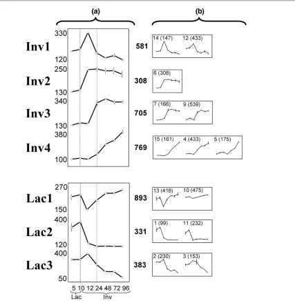
In order to define patterns of gene expression pertinent to the lactation–involution transition, irrespective of expres-sion in pregnancy, differentially expressed genes were reclustered using a truncated time course involving lacta-tion and involulacta-tion time points only (Fig. 4). A total of 4103 transcripts (33% of all transcripts) exhibited significant changes in expression across the seven time points, namely 5 days and 10 days of lactation, and 12, 24, 48, 72 and 96 hours of involution. These profiles were sorted according to similarity into 15 clusters (Fig. 4b). Similar clusters were then combined to produce seven basic pro-files that differed according to the kinetics of gene induc-tion or suppression during lactainduc-tion and involuinduc-tion (Fig. 4a). Thus, four patterns of activation were observed: a rapid but transient increase in gene expression 12 hours after weaning (Inv1); a rapid activation, which was maximal by 12 hours and sustained for up to 4 days (Inv2); a slightly delayed induction, maximal by 24/48 hours, and sustained for at least 96 hours after weaning; and a gradual increase in expression, which peaked at or beyond 96 hours (Inv4). Similarly, three patterns of sup-pression were observed: a transient decrease in transcript levels that mirrored the pattern observed in Inv1 (Lac1); a rapid loss of expression, reaching baseline levels at 12/24 hours and remaining low for up to 96 hours (Lac2); and a delayed reduction in expression, originating from a peak in expression at 12 hours involution and reaching a minimum beyond 96 hours (Lac3).
These patterns inferred three phases of gene expression during involution: rapid changes within the first 12 hours (Inv1, Inv2, Lac1, Lac2), delayed expression by 24–48 hours (Inv3), and prolonged changes extending beyond 4 days (Inv4, Lac3).
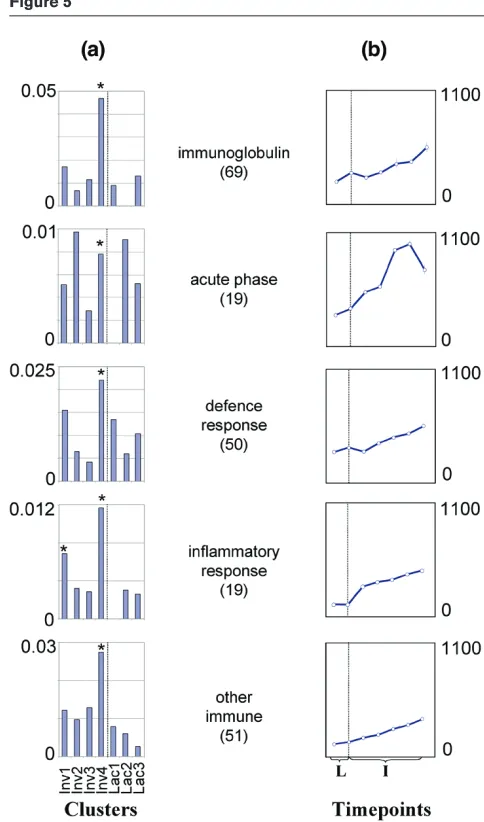
[image:7.612.59.288.88.416.2]Consistent with our analysis of the complete 12-point time course, we found that involution related gene expression was associated with an overall increase in immune gene expression (Table 2 and Fig. 5; also see Additional file 5). There was an initial transient increase in proinflammatory genes (Inv1). However, the association was most pro-nounced with transcripts exhibiting delayed kinetics (Inv4), in which known genes involved in innate immunity, antimi-crobial defence and inflammation were all preferentially expressed. The delayed immunological changes also coin-cided with a significant increase in apoptosis markers (see below) and factors that affect tissue architecture, includ-ing genes encodinclud-ing structural proteins (decorin, laminins, R98
Figure 3
Nid1, collagen 1α1, 1α2, 3α1 and 4α2) and matrix asso-ciated proteins (Mmp3, Mmp12, Gelsolin, uPa and Adamts1), thus corroborating previous reports of the role of matrix proteases in remodelling of the regressing mammary gland [2]. Lactation specific expression, as expected, was associated with downregulation of differen-tiation and metabolic processes after weaning (Lac2 and Lac3; Table 2).
Cytokine expression highlights two immune phases during involution
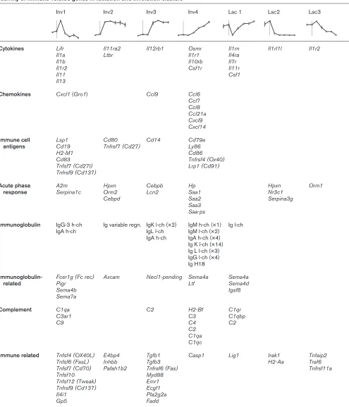
[image:8.612.112.539.98.534.2]In order to gain further insight into the immune responses associated with mammary involution, we listed the cytokines, chemokines and immune-related genes associ-ated with each involution profile from Fig. 4 (Table 3). This confirmed the two phases of immune-related gene expres-sion illustrated by Fig. 5: an initial transient expresexpres-sion of R99
Figure 4
proinflammatory cytokines and TNF superfamily genes fol-lowed by delayed expression of monocyte and lymphoid chemokines and immunoglobulin genes.
Thus, an initial burst of cytokine and cytokine receptor expression was observed within 12 hours of forced weaning (Inv1; Table 3), which included the acute-phase mediators LIF receptor (Lifr), IL-11 (Il11) and the neu-trophil chemoattractant Gro1 (Cxcl1), and the proinflam-matory mediators IL-1a, IL-1b and IL-13 (Il1a, Il1b and Il13). This was accompanied by a transient decrease in other cytokine receptors (including Il11r) and an inhibitor of IL-1 receptor signalling (Il1rn), suggesting that specific proinflammatory signals may be suppressed at the tran-scriptional level.
[image:9.612.310.552.91.502.2] [image:9.612.55.295.131.433.2]One expected outcome of the transient cytokine expres-sion was an infiltration of inflammatory cells, in particular, neutrophils due to the expression of Cxcl1. Analysis of immune cell specific antigen expression demonstrated a R100
Table 2
The relationship between gene expression pattern and gene function in the seven-point lactation/involution time course
Biological Molecular Cluster Transcripts process function
Inv1 292 Transport Immunity
Cell surface signal Enzyme Intracellular signal Structural
Growth factors
Inv2 153 Transport Enzyme
Inv3 336 Biogenesis Apoptosis
Metabolism Enzyme
Development Nucleic acid Signal transducer Structural Inv4 420 Cell adhesion Apoptosis Cell surface signal Immuntiy
ECM and Enzyme
morphogenesis Signal transducer Structural Cytokine Lac1 374 Intracellular signal
Lac2 163 Biogenesis Enzyme
Metabolism Nucleic acid Transport Transport ECM and morphogenesis
Lac3 188 Cell adhesion Signal transducer Transport Transport Development Cytokine
Growth factors Transcripts representing genes with known function were classified by molecular function and biological process, according to the ontological definitions of the Gene Ontology Consortium. The total numbers of known transcripts classified in this way are shown for each cluster. A statistical test for nonrandom distribution of these functional groups (taking into account the total number of transcripts in each category) was performed on each cluster using Onto-Express. All statistically significant associations (corrected P< 0.05) are listed for each cluster using the same ontological definitions used in Table 1 and available on GeNet. Additional information on the details of these significant associations and a graphical representation of all ontologies are available as Additional files 2 and 5. ECM, extracellular matrix.
Figure 5
R101
Table 3
Identity of immune-related genes in lactation and Involution clusters
Inv1 Inv2 Inv3 Inv4 Lac 1 Lac2 Lac3
Cytokines Lifr Il11ra2 Il12rb1 Osmr Il1rn Il1rl1l Il1r2
Il1a Ltbr Il1r1 Il4ra
Il1b Il10rb Il7r
Il1r2 Csf1r Il11r
Il11 Csf1
Il13
Chemokines Cxcl1(Gro1) Ccl9 Ccl6
Ccl7 Ccl8 Ccl21a Cxcl9 Cxcl14
Immune cell Lsp1 Cd80 Cd14 Cd79a
antigens Cd19 Tnfrsf7 (Cd27) Ly86
H2-M1 Cd86
Cd83 Tnfrsf4 (Ox40)
Tnfsf7 (Cd27l) Lrp1 (Cd91)
Tnfrsf9 (Cd137)
Acute phase A2m Hpxn Cebpb Hp Hpxn Orm1
response Serpina1c Orm2 Lcn2 Saa1 Nr3c1
Cebpd Saa2 Serpina3g
Saa3 Saa-ps
Immunoglobulin IgG-3 h-ch Ig variable regn. IgK l-ch (×2) IgM h-ch (×1) Ig l-ch
IgA h-ch IgL l-ch IgM l-ch (×2)
IgA h-ch IgA h-ch (×4) Ig K l-ch (×14) Ig L l-ch (×3) IgG l-ch (×4) Ig H18
Immunoglobulin- Fcer1g (Fc rec) Axcam Necl1-pending Sema4a Sema4a
related Pigr Ltf Sema4d
Sema4b Igsf8
Sema7a
Complement C1qa C2 H2-Bf C1qr
C3ar1 C3 C1qbp
C9 C4 C2
C2 C1qa C1qc
Immune related Tnfsf4 (OX40L) E4bp4 Tgfb1 Casp1 Lig1 Irak1 Tnfaip2
Tnfsf6 (FasL) Inhbb Tgfb3 H2-Aa Traf6
Tnfsf7 (Cd70) Pafah1b2 Tnfrsf6 (Fas) Tnfrsf11a
Tnfsf10 Myd88
Tnfsf12 (Tweak) Emr1
Tnfrsf9 (Cd137) Ecgf1
Il4i1 Pla2g2a
Gp5 Fadd
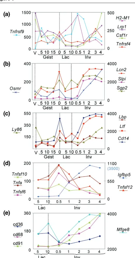
marked increase in the monocyte/macrophage markers Lrp, Csf1r and Cd14 (Table 3 and Fig. 6a), but no evi-dence for an accompanying increase in neutrophils or eosinophils (polymorphonuclear leucocyte markers Fut4, Fcgr3, Caecam1, Il8rb and Tnfrsf5 were not induced during involution; data not shown). This is consistent with previous observations of the immune cell complement in regressing murine mammary gland [28] and supports the hypothesis that neutrophil numbers are suppressed in involuting tissues in order to prevent subsequent tissue damage [28,29]. Interestingly, the gene for oncostatin M receptor (Osmr) was upregulated in involution (Fig. 6b). In models of inflammation oncostatin M is produced locally to prevent proinflammatory cytokines such as IL-8 from recruiting neutrophils, thus attenuating the inflammatory response [30]. We also confirm a previous report that ute-rocalin (Lcn2) and Sgp2, a subunit of clusterin, are upreg-ulated during involution and parturition [19], because their expression correlates with the IP profile (Fig. 6b). Utero-calin, an acute-phase response gene that is regulated by Stat3 [31,32], prevents neutrophil accumulation possibly through targeted destruction of neutrophils [29], whereas clusterin protects cells from the damaging effects of neu-trophils [33,34]. Furthermore, secretory leukocyte pro-tease inhibitor (Slpi), a potent inhibitor of neutrophil proteases [35], and a Stat3-induced gene (Boland M and coworkers, unpublished data) was also markedly upregu-lated during involution (Fig. 6b). Thus, although the tran-scriptional effects of the acute-phase mediators LIF–Stat3 and C/ebpβ/δare clearly evident (Fig. 3), the immunologi-cal outcome at the cellular level is likely to be minimised by coexpression of anti-inflammatory factors such as these.
Following the initial ‘spike’ in inflammatory cytokine expres-sion, six different chemokine genes were slowly induced over the 4-day period of involution (Inv4; Table 3). These included chemoattractants for monocytes and macrophages (Ccl6, Ccl7, Ccl8and Cxcl14) and lympho-cytes (Ccl6, Ccl21a, Cxcl9and Cxcl14). This chemokine induction was accompanied by a marked increase in immunoglobulin gene expression (Fig. 5, Table 3). Consis-tent with these observations, the monocyte/macrophage markers Lrp1, Csf1rand Cd14, and the lymphocyte anti-gens Tnfrsf9, H2-M1and Tnfrsf4 were increased during involution (Table 3 and Fig. 6a).
[image:11.612.64.293.94.530.2]Soluble innate defence factors are induced in involution In addition to the immunoglobulins, genes that encode a number of innate soluble defence factors were highly expressed in involution (Table 3 and Fig. 6c). These soluble factors possess antimicrobial properties that are secreted to protect against mastitis during the vulnerable period of milk stasis [36]. Thus, the iron chelator lactoferrin (Ltf), which is found at high levels in the milk of humans and cattle [37,38], is induced with kinetics similar to those of the antibody transcripts described above (Fig. 5). Comple-R102
Figure 6
Involution related expression profiles: apoptosis and immune related genes. (a)Expression profiles of differentially expressed immune cell antigens, illustrating the relative proportions of immune cell subtypes in mammary gland during the complete pregnancy cycle.
Monocyte/macrophage markers: Lrp1 = Cd91; Csf1r = Cd115. Lymphocyte markers: Tnfrsf9=Cd137; H2-M1=Mb1; Tnfrsf4=OX40. (b)Profiles of differentially expressed
ment components are prevalent at all stages during involu-tion (Table 3). These factors are found at high levels in mastitic milk and in normal bovine mammary glands during involution, and are thought to play a dual role in opsoniza-tion and lysis of bacteria and as a proinflammatory mediator [39,40]. Cd14 was significantly upregulated in involution and was also highly expressed during parturition (Fig. 6c). This antigen is expressed on bovine mammary polymor-phonuclear neutrophils and macrophages [41] and plays an integral role in suppressing bacterial infections in response to endotoxin. Furthermore, Lbp [42] and Ly86 [43], which in conjunction with Cd14augment responses to lipopolysaccharide (LPS), were also upregulated. The significant regulation of these genes in uninfected mammary gland confirms a role for soluble defence factors in normal development of mouse mammary gland.
Involution is associated with a transient induction of death receptor genes
Involution is characterized by extensive apoptosis of epithelial cells, which occurs both before and during the tissue remodelling process. By subdividing the first 4 days of involution into three discrete phases of gene expres-sion, we demonstrated a link between the expression of apoptosis-related genes and the delayed onset of extra-cellular matrix degradation and alveolar collapse at 48 hours (Inv3 and Inv4; Table 2). However, we were unable to identify a significant increase in the number of apoptosis genes expressed at the onset of involution. One explanation for this was that specific combinations of cell
death genes, rather than an increase in the number of genes activated, influenced apoptosis at the onset of invo-lution. In order to address this, all cell death related genes found to be regulated during involution were grouped according to their lactation/involution expression pattern (Table 4). Regulators (Bax, BclX, Mcl1, mXIAP and Apaf-1) and effectors (caspases) of classical apoptosis and immune cell-mediated killing (granzyme A, granzyme B, Fadd, Fas, Fas ligand) were regulated with different kinet-ics during involution and were relatively evenly distributed across the four involution clusters (Table 4).
However, we observed a strong bias in the expression of extrinsic (death receptor) and intrinsic (mitochondrial) apoptosis genes, in which extrinsic-related components were generally induced within the first 24 hours of involu-tion whereas intrinsic genes, with the excepinvolu-tion of Hrk and Diva, were not maximally transcribed until at least 96 hours.
[image:12.612.58.571.117.336.2]Thus, six ligands of the TNF superfamily (Tnf, Tnfsf4, Tnfsf6, Tnfsf7, Tnfsf10 and Tnfsf12) were specifically induced in involution and all were transiently activated at 12 hours after weaning (Inv1; Fig. 6d). Four of these ligands (death receptor ligands: Fas ligand, TNF-α, TWEAK and TRAIL) are responsible for activating the five principal mammalian death receptor (extrinsic) apoptosis pathways through their cognate receptors Fas, TNFR-1, TNFR-2, DR3 and DR4. Two of these receptors (Fasand Tnfrsf1a) were also induced, and maximally coexpressed, within 24 hours of weaning (Inv3; Table 4). This correlates R103
Table 4
Cell death-related genes in lactation and involution clusters
Inv1 Inv2 Inv3 Inv4 Lac 1 Lac2 Lac3
Cell survival Prdx2 Nfkb2 Api5 Bcl2l (BclX) Psap Akt1
Bcl10 Ank3 Mcl1 Birc4(mXIAP) Dad1
Psap Gfi1
Cell death Hrk Bcl2l10 (Diva) Fsp27 Igfbp5 Tnip1 (ABIN1) Tnfrsf11a(Rank)
Gzmb Casp4 (casp11) Bax Casp1 Eya2 Cidea
Nkx3-1 Casp12 Casp7 Foxm1 Tial1 Dffb
Tnfsf6(FasL) Gadd45b Serpinb5 Apaf1 Dapk3 Perp-pending
Lsp1 Pdcd6 Pdcd6 Gzma Foxb1 Traf6
Tnfsf4(OX40L) Cdkn1a (p21) Pdcd5 Tia1 Rbl2 Tnfsf9
Tnfsf7 (Cd27l) Unc5 Tnfrsf6 (Fas) Phlda1 Tnfsf5 (TRAP) Tnfsf10 (Trail) Foxb2 Fadd Tnfrsf4 (Ox40)
Tnfsf12 (Tweak) Mycs (s-myc) Myd88 Tgfb1 Tnfrsf9 (Cd137) E4bp4 Tnfrsf1a
Tnf (Tnfa) Ngfa Ltbr
R104
with previous studies in mammary gland of two of these ligand–receptor pairs [44,45]. NF-κB activity also corre-lated with the rapid activation of these TNF superfamily ligands. DNA binding activity of this transcription factor is markedly upregulated within 3 hours of forced involution and is suggested to promote survival of a subpopulation of mammary epithelial cells [26]. Thus Nfkb2 and activators of NF-κB (Tnfsf12, Bcl10, Tnfrsf11a, Traf6 and Tnfsf10) were upregulated, whereas an inhibitor of NF-κB function (Tnip1) was inhibited during the initial 24 hours following forced weaning, when NF-κB activity was induced [26]. Previous studies demonstrating the direct activation of NF-κB by TNF superfamily ligands in mammary epithelial cells [26,46,47] therefore suggests that the observed tran-sient increase in these ligands may be functionally relevant.
In contrast to the rapid response of extrinsic factors, intrin-sic apoptosis genes were predominantly (but not exclu-sively) expressed at least 24 hours after forced weaning. Proapoptotic and antiapoptotic members of the Bcl-2 family (Bax, Mcl1and Bcl2l) were induced together with the downstream effector of the intrinsic pathway Apaf1. Although the precise mechanism(s) of cell death in mammary gland have yet to be established, the trends in apoptosis gene expression identified here hint that extrin-sic (death receptor) and intrinextrin-sic death programmes may predominate at different times. A marked induction of Igfbp5 complemented the delayed induction of intrinsic apoptosis genes (Fig. 6d). The kinetics of this potent inducer of mammary apoptosis and involution [48] sug-gests that it probably contributes to the stimulation of intrinsic cell death at this time.
Apoptotic bodies initially shed into the lumen and the residual dying cells left in the epithelium are believed to be cleared from the mammary gland by nonprofessional phagocytes and interstitial macrophages, respectively [28]. Receptors and ligands that mediate recognition and engulfment of apoptotic cells include members that are common to both types of phagocytic cell (e.g. Cd36, Lrp1 and Mfge8) and those specific to professional macrophages (e.g. Cd14and Cd68). The genes for each of these factors (except Cd36, which exhibits an initial downregulation before a modest increase by 96 hours) are induced with similar kinetics (Fig. 6e). This is indicative of the increasing demands placed on the mammary gland to clear dead/dying cells during regression, and also prob-ably reflects the increasing numbers of macrophages occupying the gland during the first 4 days of involution.
Discussion
Microarray analysis of large sequence verified cDNA probe sets provides a powerful tool for analyzing complex biological systems because it can extract patterns of gene expression from a significant proportion of the total genomic content of an organism. In this study we
simulta-neously analyzed approximately 12 000 transcripts, repre-senting 8618 unigene clusters, or about one-third of the protein encoding capacity of the mouse, to determine basic expression patterns in adult mammary gland during the pregnancy cycle.
Although a previous study of approximately 5000 tran-scripts described gene expression in early phases of the pregnancy cycle [14], we focused our analysis on the first 4 days after forced weaning, using time points from earlier stages in development to establish the specificity of these involution-related genetic changes. In the accompanying paper, Stein and coworkers [19] describe a similar study in Balb/c animals. Despite differences in the methods of analysis, their findings on the activation of inflammatory mediators and monocyte/lymphocyte markers in involution correlate with the results described here, further verifying our microarray data. Thus, strain differences did not appre-ciably influence the immune responses associated with involution.
The principal objective of the present study was to provide a comprehensive set of gene expression data from late stages of mouse mammary development to be used as an online resource for further analysis and/or download. The raw microarray data files and processed genelists from the study are therefore available for download or online analy-sis (see www.path.cam.ac.uk/~madgroup/).
Here we have outlined the common patterns of gene expression across the developmental time course and have correlated these patterns with biological function. Thus, as expected, pregnancy was associated with the induction of genes that are involved in cell proliferation, differentiation and tissue morphogenesis. This was superceded in lactation by genes involved in biosynthetic (fatty acid synthesis) and metabolic pathways, correlating with the findings of an earlier microarray study of the preg-nancy cycle [14]. We have not attempted to provide a detailed account of the genetic changes that occur early in the pregnancy cycle; however, the data are available online for further scrutiny.
approximately 48 hours characterized by increased apop-tosis and loss of differentiated function, followed by matrix degradation and alveolar collapse. The pattern of gene expression appeared to correlate with this cellular model. We observed a swift increase in specific apoptosis tran-scripts (Inv1; Table 4) and a subsequent decline in differen-tiated function by 48 hours (Inv3; Table 2), followed by an increase in factors affecting tissue architecture (Inv4; Table 2) and phagocyte function (Fig. 6). Further detailed analysis of the Inv3 cluster, which exhibits maximal expres-sion at 48 hours, may elucidate underlying mechanisms that are responsible for the transition to phase two involution.
Transcriptional regulation of apoptosis in involution: two phases of death?
Our data also provide provisional evidence for the exis-tence of a two-phase apoptotic process in involution and which also coincides with the two phases previously described [2]. While the number of apoptosis genes was similar in each involution cluster, there was a pronounced bias in the distribution of extrinsic and intrinsic apoptosis genes. The initial spike of activity consisted of four death receptor ligands, namely Tnf(TNF-α) Tnfsf6(Fas ligand), Tnfsf10 (TRAIL) and Tnfsf12 (TWEAK). These ligands bind to the principal death receptors Fas, TNFR-1, TNFR-2, DR3 and DR4, which initiate apoptosis in cell mediated immune cell death and in a variety of physiologi-cal and pathologiphysiologi-cal situations. These data confirm previ-ous observations that Fas and Fas ligand are upregulated within a day of involution in mouse mammary gland, and that the deletion of these genes results in delayed regres-sion and loss of apoptosis [44]. Deregulation of Fas sig-nalling through aberrant Akt and NF-κB activity is believed to play a role in the immune escape of breast cancers and in resistance to chemotherapy [49]. TNF-α also induces NF-κB signalling to prevent apoptosis in a subpopulation of cultured mammary epithelial cells [26,47], and has been shown to be downregulated late in involution (day 7); however, it has not been studied at earlier time points [50]. Furthermore, elevated IL-10 dependent TRAIL expression (and its receptor DR4) has previously been demonstrated in involution [45].
In contrast to the transient activation of death receptor ligands, components of mitochondrial (or intrinsic) apopto-sis exhibit sustained, often delayed induction. Thus the caspase 9 effector, Apaf1, and Bax were induced with delayed kinetics. Costimulation of the survival factors Mcl1 and Bcl2l (BclXl) suggests that the transcriptional regula-tion of Bax may be counterbalanced, and therefore con-trolled, by expression of Bcl-2 homologues. Suppression of BclX, for example, promotes death during mammary involution [51], whereas delayed induction of Bax has pre-viously been demonstrated [52]. Regulation of Bcl homo-logues was not exclusively restricted to late stage involution. Hrkand Boo/Divawere differentially expressed
within 24 hours. Hrk is a proapoptotic factor that is tran-scriptionally suppressed by progesterone signals in breast cancer cell lines [53]. The function of Boo/Diva, which has only previously been described in mouse ovary, is poorly defined [54]. Cross-talk between death receptor pathways and Bcl-2 regulatory factors is well established [55] and may explain the early activation of these two Bcl homo-logues. Probable upstream initiators of a putative delayed apoptotic mechanism include the loss of survival signals, such as insulin-like growth factor (IGF)-1 [48,56], and the destruction of extracellular matrix–integrin contacts leading to an anoikis-type death [6,57]. The dramatic induction of the proapoptotic mammary protein IGF-binding protein 5 proposed previously [48] coincides with the onset of second phase involution (Fig. 6d, Table 4) and the upregulation of metalloproteinases [2,6]. Both IGF-1 and integrins signal through Akt, which was also downreg-ulated in involution (Table 4). Loss of this signal could then induce Bax [57]. Our data suggest that apoptosis is induced in involution following the specific transcriptional activation of a subset of proapoptotic genes encoding death receptors, and that delayed expression of inhibitors of cell survival (e.g. IGF-binding protein 5) induce alternate apoptosis pathways during the second phase of involu-tion. This hypothesis warrants further investigainvolu-tion.
Integral to the apoptotic process is the phagocytic clear-ance of apoptotic bodies by macrophages. It has been sug-gested that monocytic macrophages are recruited to the mammary gland to supercede phagocytic epithelial cells in the clearance of milk and dead cells [28]. We have demon-strated monocyte specific (Cd14 and Cd68) and nonspe-cific phagocyte receptors during involution, supporting a role for monocytic phagocytes in mammary involution.
Multiple roles for immune cells in mammary involution Measurements of the cellular content in milk from involut-ing glands of sheep, pigs and cattle indicate an early rise in polymorphonuclear neutrophils, a subsequent increase in macrophages and low numbers of eosinophils, B cells and T cells [7,28]. However, the immune cell complement of human and rodent mammary gland is poorly defined. It is believed that these phagocytes are recruited to the gland to perform several functions in phagocytic clearance and cellular immunity.
R106
for normal development [58]. A role for these cells in phagocytic clearance during involution is emphasized by the concomitant increase in two receptors (Cd14 and Cd91) and the milk factor Mgfe8, which mediate recogni-tion of apoptotic cells by macrophages (Fig. 6c,e).
Recruitment of cells of the monocytic lineage comprises an inflammatory response. However, in the absence of a persistent bacterial infection, this does not occur in the regressing mammary gland, even though inflammatory cytokines and downstream mediators are activated, pre-sumably to perform additional roles during involution. During chronic inflammatory diseases such as rheumatoid arthritis or inflammatory bowel disease, uncontrolled accu-mulation of neutrophils yield aberrantly elevated levels of inflammatory compounds, which become major contribu-tors to tissue damage [60,61] Thus, regulation of neu-trophil recruitment into inflammatory sites and their clearance are critical processes that ensure effective host defence without tissue injury. In mammary gland, the LIF–Stat3 signaling pathway is known to be crucial for appropriate regression during early involution by initiating epithelial apoptosis [17,24]. With the concomitant down-regulation of IL-11 receptor (Table 3) and lack of a signifi-cant increase in IL-6 or its receptor (data not shown), it is also likely to be the predominant protagonist of acute phase signalling. This is confirmed by previous observa-tions that C/ebpβ and C/ebpδ are both transcriptional targets of Stat3 in vivo[24] (Boland M, unpublished data) and that the profiles of Class I acute phase genes corre-lated precisely with phosphorycorre-lated Stat3 activity [24] and Stat3, C/ebpβ and C/ebpδ expression (Fig. 3). Class II acute-phase response genes exhibit specific and transient expression in involution. It is thought that these genes respond transiently to proinflammatory signals [25] and therefore may respond in a similar manner to the rapid activation of Stat3 at the onset of involution. Why they are not induced at parturition is unclear, although a molecular switch that determines the specificity of signalling path-ways downstream of LIF has been proposed [24].
Thus, inhibitory mechanism(s) must exist to prevent the accumulation of potentially damaging cellular infiltrates in response to LIF. In a previous study of involution in uterus and mammary gland, the acute phase protein uterocalin (Lcn2) was shown to be induced [29]. It was proposed that one role of this protein in involuting mammary gland and uterus was to eliminate infiltrating neutrophils by inducing apoptosis [62] and therefore to prevent subse-quent tissue damage [63]. We are able to confirm signifi-cant activation of uterocalin expression in mammary involution (Fig. 6b) and the concomitant absence of neu-trophil antigens (Table 3). In conjunction with the induction of uterocalin, clusterin (Sgp2), oncostatin M receptor (Osmr) and secretory leukocyte inhibitory protein (Slpi) were also increased (Fig. 6b). Clusterin protects cells
against oxidative and chemical damage [33,34] and has been suggested to be coexpressed with uterocalin as part of a localized acute-phase response in order to limit the damaging effects of residual neutrophils in involuting mammary gland [29,63]. SLPi, an antichymotrypsin, also contributes to the elimination of neutrophil function, whereas Osmr may possess similar anti-inflammatory effects through the sequestration of proinflammatory cytokines. Osmr, Slpi and Sgp2 are all directly induced by Stat3 in mammary epithelial cells (Boland M, unpublished data) [64], whereas uterocalin expression conformed with Stat3 activity (Fig. 3 and 6b). A putative anti-inflammatory role for Stat3 was suggested in our previous study of Stat3-deficient mammary glands [17], in which we observed an increase in mastitis in the absence of Stat3. A similar protective mechanism may exist during parturi-tion, when the LIF–Stat3 signal is also active [24,65]. We are currently investigating the possibility that LIF–Stat3 signalling is responsible for the induction of these four genes in mammary gland, therefore confirming a dual role for LIF in involuting mammary gland. In addition to the direct effects of these anti-inflammatory acute phase genes, immunosuppressive factors including transforming growth factor-β (Inv3; Table 3) expressed by phagocytic macrophages are likely also to contribute to the suppres-sion of an inflammatory response [64,66].
Soluble defence factors are induced during involution Soluble defence factors and cells that are involved in humoral immunity are thought to be induced to prevent mastitis during this extended period of milk stasis. For example, the iron chelator lactoferrin is induced in mammary involution (Fig. 6c), which plays a role in mammary defence by protecting against coliform infection [36]. Indeed, low levels of lactoferrin is a predisposing factor for mastitis in humans [38]. Immunoglobulins not only have an important role in supplementing milk but are also required to facilitate opsonization by macrophages and neutrophils (IgM and IgG2) [67] and the direct sequestration of bacteria (IgA).
Conclusion
We highlighted two general trends in gene expression during mouse mammary involution: an increase in immune-related genes and a shift in the identity of apoptosis com-ponents. The increase in immune genes correlated with previously described roles for immune cells in involuting mammary gland, namely antimicrobial defence, cytokine signalling and phagocytic clearance. The proposed change in the cell death machinery based on extrinsic and intrinsic apoptosis gene expression has not previously been observed. This putative mechanism clearly needs further investigation. One common link between apoptosis and immune responses in mammary gland is the previ-ously defined role for LIF and Stat3. We are currently investigating the possibility that Stat3 mediates the anti-inflammatory effects vital for the protection of the mammary gland in addition to its recognized role as a proapoptotic factor in mammary gland.
Additional files
Competing interests
None declared.Acknowledgements
This work was supported in part by the Lucas Walker Trust of the Department of Pathology, University of Cambridge. RWEC is a Lucas Walker Research Fellow of the Department of Pathology, University of Cambridge.
References
1. Visvader JE, Lindeman GJ: Transcriptional regulators in mammary gland development and cancer.Int J Biochem Cell Biol2003, 35:1034-1051.
2. Lund LR, Romer J, Thomasset N, Solberg H, Pyke C, Bissell MJ, Dano K, Werb Z: Two distinct phases of apoptosis in mammary gland involution: proteinaseindependent and -dependent pathways.Development1996, 122:181-193. 3. Alexander CM, Selvarajan S, Mudgett J, Werb Z: Stromelysin-1
regulates adipogenesis during mammary gland involution.J Cell Biol 2001, 152:693-703.
4. Li M, Liu X, Robinson G, Bar-Peled U, Wagner KU, Young WS, Hennighausen L, Furth PA: Mammary-derived signals activate programmed cell death during the first stage of mammary gland involution.Proc Natl Acad Sci USA1997, 94:3425-3430. 5. Strange R, Li F, Saurer S, Burkhardt A, Friis RR: Apoptotic cell death and tissue remodelling during mouse mammary gland involution.Development 1992, 115:49-58.
6. Streuli CH, Gilmore AP: Adhesion-mediated signaling in the regulation of mammary epithelial cell survival. J Mammary Gland Biol Neoplasia 1999, 4:183-191.
7. Paape MJ, Shafer-Weaver K, Capuco AV, Van Oostveldt K, Bur-venich C: Immune surveillance of mammary tissue by phago-cytic cells.Adv Exp Med Biol 2000, 480:259-277.
8. Clarkson RW, Watson CJ: NF-kappaB and apoptosis in mammary epithelial cells. J Mammary Gland Biol Neoplasia 1999, 4:165-175.
9. Wilde CJ, Knight CH, Flint DJ: Control of milk secretion and apoptosis during mammary involution.J Mammary Gland Biol Neoplasia1999, 4:129-136.
10. Diaz E, Yang YH, Ferreira T, Loh KC, Okazaki Y, Hayashizaki Y, Tessier-Lavigne M, Speed TP, Ngai J: Analysis of gene expres-sion in the developing mouse retina.Proc Natl Acad Sci USA 2003, 100:5491-5496.
11. Arai M, Yokosuka O, Chiba T, Imazeki F, Kato M, Hashida J, Ueda Y, Sugano S, Hashimoto K, Saisho H, Takiguchi M, Seki N: Gene expression profiling reveals the mechanism and pathophysi-ology of mouse liver regeneration. J Biol Chem 2003, 278: 29813-29818.
12. Chiang MK, Melton DA: Single-cell transcript analysis of pan-creas development.Dev Cell2003, 4:383-393.
13. Tan Y, Li X, Piao Y, Sun X, Wang Y: Global gene profiling analy-sis of mouse uterus during the oestrus cycle. Reproduction 2003, 126:171-182.
14. Master SR, Hartman JL, D’Cruz CM, Moody SE, Keiper EA, Ha SI, Cox JD, Belka GK, Chodosh LA: Functional microarray analysis of mammary organogenesis reveals a developmental role in adaptive thermogenesis.Mol Endocrinol 2002, 16:1185-1203. 15. Lemkin PF, Thornwall GC, Walton KD, Hennighausen L: The
microarray explorer tool for data mining of cDNA microarrays: application for the mammary gland.Nucleic Acids Res 2000, 28:4452-4459.
16. Phang TL, Neville MC, Rudolph M, Hunter L: Trajectory cluster-ing: a non-parametric method for grouping gene expression time courses, with applications to mammary development. Pac Symp Biocomput 2003, 5:351-362.
17. Chapman RS, Lourenco PC, Tonner E, Flint DJ, Selbert S, Takeda K, Akira S, Clarke AR, Watson CJ: Suppression of epithelial apoptosis and delayed mammary gland involution in mice with a conditional knockout of Stat3. Genes Dev 1999, 13: 2604-2616.
18. Alonzi T, Maritano D, Gorgoni B, Rizzuto G, Libert C, Poli V: Essential role of STAT3 in the control of the acute-phase response as revealed by inducible gene inactivation (correc-tion of activa(correc-tion) in the liver.Mol Cell Biol 2001, 21:
1621-1632. R107
The following Additional files are available online:
Additional file 1
Table showing all significant ontological associations (corrected P< 0.05) with combined clusters and primary clusters from the 12-point timecourse from Fig. 1. See http://breast-cancer-research.com/content/ supplementary/bcr754-S1.doc
Additional file 2
Table showing all significant ontological associations (corrected P< 0.05) with combined clusters and primary clusters from the 7-point timecourse from Fig. 4. See http://breast-cancer-research.com/content/ supplementary/bcr754-S2.doc
Additional file 3
Table showing the relative expression of genes previ-ously identified as differentially regulated during the pregnancy cycle are compared to the relative expression of the same genes in our microarray study.
See http://breast-cancer-research.com/content/ supplementary/bcr754-S3.xls
Additional file 4
A graphical representation of the relationship between gene expression pattern and gene function in the 12-point developmental time course.
See http://breast-cancer-research.com/content/ supplementary/bcr754-S4.tiff
Additional file 5
A graphical representation of the relationship between gene expression pattern and gene function in the seven-point lactation/involution time course.
19. Stein T, Morris JS, Davies CR, Weber-Hall SJ, Duffy MA, Heath VJ, Bell AK, Ferrier RK, Sandilands GP, Gusterson BA: Involution of the mouse mammary gland is associated with an immune cascade and an acute phase response, involving LBP, CD14 and STAT3.Breast Cancer Res2004, 6:R75-R91.
20. Li C, Wong WH: Model-based analysis of oligonucleotide arrays: expression index computation and outlier detection. Proc Natl Acad Sci USA 2001, 98:31-36.
21. Ashburner M, Ball CA, Blake JA, Botstein D, Butler H, Cherry JM, Davis AP, Dolinski K, Dwight SS, Eppig JT, Harris MA, Hill DP, Issel-Tarver L, Kasarskis A, Lewis S, Matese JC, Richardson JE, Ringwald M, Rubin GM, Sherlock G: Gene ontology: tool for the unification of biology. The Gene Ontology Consortium.Nat Genet 2000, 25:25-29.
22. Khatri P, Draghici S, Ostermeier GC, Krawetz SA: Profiling gene expression using onto-express.Genomics2002, 79:266-270. 23. Blake JA, Richardson JE, Bult CJ, Kadin JA, Eppig JT: MGD: the
Mouse Genome Database.Nucleic Acids Res2003, 31: 193-195.
24. Kritikou EA, Sharkey A, Abell K, Came PJ, Anderson E, Clarkson RW, Watson CJ: A dual, non-redundant, role for LIF as a regu-lator of development and STAT3-mediated cell death in mammary gland.Development2003, 130:3459-3468.
25. Poli V: The role of C/EBP isoforms in the control of inflamma-tory and native immunity functions.J Biol Chem1998, 273: 29279-29282.
26. Clarkson RW, Heeley JL, Chapman R, Aillet F, Hay RT, Wyllie A, Watson CJ: NF-kappaB inhibits apoptosis in murine mammary epithelia.J Biol Chem2000, 275:12737-12742.
27. Seagroves TN, Krnacik S, Raught B, Gay J, Burgess-Beusse B, Darlington GJ, Rosen JM: C/EBPbeta, but not C/EBPalpha, is essential for ductal morphogenesis, lobuloalveolar prolifera-tion, and functional differentiation in the mouse mammary gland.Genes Dev1998, 12:1917-1928.
28. Monks J, Geske FJ, Lehman L, Fadok VA: Do inflammatory cells participate in mammary gland involution?J Mammary Gland Biol Neoplasia 2002, 7:163-176.
29. Ryon J, Bendickson L, Nilsen-Hamilton M: High expression in involuting reproductive tissues of uterocalin/24p3, a lipocalin and acute phase protein.Biochem J2002, 367:271-277. 30. Wahl AF, Wallace PM: Oncostatin M in the anti-inflammatory
response.Ann Rheum Dis 2001, Suppl 3:iii75-iii80.
31. Orabona C, Dumoutier L, Renauld JC: Interleukin-9 induces 24P3 lipocalin gene expression in murine T cell lymphomas. Eur Cytokine Netw2001, 12:154-161.
32. Liu Q, Ryon K, Nilsen-Hamilton M: Uterocalin: a mouse acute phase protein expressed in the uterus around birth. Mol Reprod Dev 1997, 46:507-514.
33. Miyake H, Nelson C, Rennie PS, Gleave ME: Acquisition of chemoresistant phenotype by overexpression of the anti-apoptotic gene testosterone-repressed prostate message-2 in prostate cancer xenograft models.Cancer Res 2000, 60: 2547-2554.
34. Viard I, Wehrli P, Jornot L, Bullani R, Vechietti JL, Schifferli JA, Tschopp J, French LE: Clusterin gene expression mediates resistance to apoptotic cell death induced by heat shock and oxidative stress.J Invest Dermatol1999, 112:290-296. 35. Smith CE, Johnson DA: Human bronchial leucocyte proteinase
inhibitor. Rapid isolation and kinetic analysis with human leu-cocyte proteinases.Biochem J 1985, 225:463-472.
36. Sordillo LM, Streicher KL: Mammary gland immunity and masti-tis susceptibility.J Mammary Gland Biol Neoplasia 2002, 7: 135-146.
37. Goodman RE, Schanbacher FL: Bovine lactoferrin mRNA: sequence, analysis, and expression in the mammary gland. Biochem Biophys Res Commun 1991, 180:75-84.
38. Breton-Gorius J, Mason DY, Buriot D, Vilde JL, Griscelli C: Lacto-ferrin deficiency as a consequence of a lack of specific gran-ules in neutrophils from a patient with recurrent infections. Detection by immunoperoxidase staining for lactoferrin and cytochemical electron microscopy.Am J Pathol 1980, 99: 413-428.
39. Rainard P, Poutrel B: Deposition of complement components on Streptococcus agalactiae in bovine milk in the absence of inflammation.Infect Immun 1995, 63:3422-3427.
40. Riollet C, Rainard P, Poutrel B: Differential induction of comple-ment fragcomple-ment C5a and inflammatory cytokines during
intra-mammary infections with Escherichia coliand Staphylococcus aureus.Clin Diagn Lab Immunol 2000, 7:161-167.
41. Paape MJ, Lilius EM, Wiitanen PA, Kontio MP, Miller RH: Intra-mammary defense against infections induced by Escherichia coliin cows.Am J Vet Res 1996, 57:477-482.
42. Wright SD, Ramos RA, Tobias PS, Ulevitch RJ, Mathison JC: CD14, a receptor for complexes of lipopolysaccharide (LPS) and LPS binding protein.Science 1990,249:1431-1433. 43. Miyake K: Innate recognition of lipopolysaccharide by CD14
and toll-like receptor 4-MD-2: unique roles for MD-2. Int Immunopharmacol 2003, 3:119-128.
44. Song J, Sapi E, Brown W, Nilsen J, Tartaro K, Kacinski BM, Craft J, Naftolin F, Mor G: Roles of Fas and Fas ligand during mammary gland remodeling. J Clin Invest 2000, 106: 1209-1220.
45. Sohn BH, Moon HB, Kim TY, Kang HS, Bae YS, Lee KK, Kim SJ: Interleukin-10 up-regulates tumour-necrosis-factor-alpha-related apoptosis-inducing ligand (TRAIL) gene expression in mammary epithelial cells at the involution stage. Biochem J 2001, 360:31-38.
46. Cao Y, Bonizzi G, Seagroves TN, Greten FR, Johnson R, Schmidt EV, Karin M: IKKalpha provides an essential link between RANK signaling and cyclin D1 expression during mammary gland development.Cell 2001, 107:763-775.
47. Varela LM, Stangle-Castor NC, Shoemaker SF, Shea-Eaton WK, Ip MM: TNFalpha induces NFkappaB/p50 in association with the growth and morphogenesis of normal and transformed rat mammary epithelial cells. J Cell Physiol 2001, 188: 120-131.
48. Marshman E, Green KA, Flint DJ, White A, Streuli CH, Westwood M: Insulin-like growth factor binding protein 5 and apoptosis in mammary epithelial cells.J Cell Sci 2003, 116:675-682. 49. Toillon RA, Descamps S, Adriaenssens E, Ricort JM, Bernard D,
Boilly B, Le Bourhis X: Normal breast epithelial cells induce apoptosis of breast cancer cells via Fas signaling. Exp Cell Res 2002, 275:31-43.
50. Varela LM, Ip MM: Tumor necrosis factor-alpha: a multifunc-tional regulator of mammary gland development. Endocrinol-ogy 1996,137:4915-4924.
51. Walton KD, Wagner KU, Rucker EB III, Shillingford JM, Miyoshi K, Hennighausen L: Conditional deletion of the bcl-x gene from mouse mammary epithelium results in accelerated apoptosis during involution but does not compromise cell function during lactation.Mech Dev 2001, 109:281-293.
52. Metcalfe AD, Gilmore A, Klinowska T, Oliver J, Valentijn AJ, Brown R, Ross A, MacGregor G, Hickman JA, Streuli CH: Developmen-tal regulation of Bcl-2 family protein expression in the involut-ing mammary gland.J Cell Sci 1999, 112:1771-1783. 53. Ory K, Lebeau J, Levalois C, Bishay K, Fouchet P, Allemand I,
Therwath A, Chevillard S: Apoptosis inhibition mediated by medroxyprogesterone acetate treatment of breast cancer cell lines.Breast Cancer Res Treat 2001, 68:187-198.
54. Russell HR, Lee Y, Miller HL, Zhao J, McKinnon PJ: Murine ovarian development is not affected by inactivation of the bcl-2 family member diva.Mol Cell Biol 2002, 22:6866-6870. 55. Cory S, Adams JM: The Bcl2 family: regulators of the cellular
life-or-death switch.Nat Rev Cancer 2002, 2:647-656. 56. Campana WM, Darin SJ, O’Brien JS: Phosphatidylinositol
3-kinase and Akt protein 3-kinase mediate IGF-I- and prosaptide-induced survival in Schwann cells.J Neurosci Res 1999, 57: 332-341.
57. Wang P, Valentijn AJ, Gilmore AP, Streuli CH: Early events in the anoikis program occur in the absence of caspase activation.J Biol Chem 2003, 278:19917-19925.
58. Gouon-Evans V, Rothenberg ME, Pollard PW: Postnatal mammary gland development requires macrophages and eosinophils.Development 2000, 7:2269-2282.
59. Pollard JW, Hennighausen L: Colony stimulating factor 1 is required for mammary gland development during pregnancy. Proc Natl Acad Sci USA 1994, 91:9312-9316.
60. Dallegri F, Ottonello L: Tissue injury in neutrophilic inflamma-tion.Inflamm Res 1997, 46:382-391.
61. Pillinger MH, Abramson SB: The neutrophil in rheumatoid arthritis.Rheum Dis Clin North Am 1995, 21:691-714.
![Figure 3described [27]. We compared the expression of thesetranscription factors and found that Stat3 and both Cebp](https://thumb-us.123doks.com/thumbv2/123dok_us/8278041.282627/7.612.59.288.88.416/figure-described-compared-expression-thesetranscription-factors-stat-cebp.webp)