T E C H N I C A L N O T E
Open Access
Ring cell migration assay identifies distinct effects
of extracellular matrix proteins on cancer
cell migration
Hui Chen
1and Josephine Nalbantoglu
1,2,3*Abstract
Background:Alterations in cell migration are a hallmark of cancer cell invasion and metastasis.In vitroassays commonly used to study cell migration, including the scratch wound healing assay, Boyden chamber assay, and newly developed advanced systems with microfluidics, each have several disadvantages.
Findings:Here we describe an easy and cost-effectivein vitroassay for cell migration employing cloning rings to create gaps in the cell monolayer (“ring cell migration assay”). The assay was used to quantitate innate differences in cell motility and the effect of various extracellular matrix proteins on migration of five cancer cell lines: U87 and U251N glioma cells, MDA-MB-231and MCF-7 breast cancer cells, and HeLa cervical cancer cells. Interestingly, collagen was a general promoter of cell migration for all five cancer cell lines, without affecting cell proliferation.
Conclusions:Taken together, the ring cell migration assay is an easy, convenient and cost-effective assay to study cell migrationin vitro.
Keywords:Cell migration assay, Cloning ring, Extracellular matrices
Findings Introduction
Metastasis is the leading cause of mortality for patients suffering from cancer. During metastasis, abnormal cells with genomic heterogeneity may undergo epithelial-to-mesenchymal transition (EMT) and phenotypically exhibit increased motility and invasiveness as compared to nor-mal cells [1-3]. Thus, increased cell migration is a priming process for cancer cells to invade and metastasize during cancer progression. Therefore, evaluating cancer cell mo-tility is a critical step in studying mechanism of cancer cell invasion and metastasis.
Extracellular matrix (ECM) proteins provide a structural basis supporting cell migration; they can also either func-tion as a reservoir for growth factors, or directly bind to re-ceptors such as integrins to activate cellular signaling pathways specifically driving cell migration [4-6]. Therefore,
ECM plays critical roles in cancer cell invasion and metas-tasis [7-10]. However, it is mostly unknown how various ECM proteins affect cell migration of different tissue-specific cancer cell types.
The different techniques used to assess cell migration and invasion have been described and critically evaluated in recent review articles [11,12]. There are several con-ventional in vitro assays for studying cancer cell migra-tion, including scratch wound healing assay [13] and Boyden chamber assay [14]. While these assays have ad-vantages either in ease of performance (scratch assay) or in mimicking in vivo chemoattractant gradients for cell migration (Boyden assay), they also have many disadvan-tages. For example, scratch wound healing assay is not applicable to every type of cancer cell as some mono-layers remain hard for scratching and cells may be dam-aged during the wounding, while Boyden chamber assay is difficult to reproduce as it is dependent on the number of cells seeded and only provides endpoint data of cell migra-tion. Additional assays have been configured to overcome some of these problems, such as the cell exclusion zone assay in which cells are cultured on microfabricated sten-cils [15] or in the presence of silicone stoppers which are
* Correspondence:josephine.nalbantoglu@mcgill.ca 1
Division of Experimental Medicine, Department of Medicine, McGill University, Montreal, Quebec, Canada
2
Department of Neurology & Neurosurgery, McGill University, Montreal, Quebec, Canada
Full list of author information is available at the end of the article
removed at cell confluence [16], generating cell-free areas with well-defined linear borders. However, microfabrica-tion is not available to all laboratories, and care must be taken to prevent cell entry under the stopper when silicone inserts are used. Thus, an easy, convenient and cost-effective assay with a high level of reproducibility is required for rapid assessment of cell migration.
Here we describe an easy assay for cell migration, based on the principles of the cell exclusion assay, and which we have termed“ring cell migration assay”as it uses a cloning ring to establish the gap between two parts of a mono-layer. We provide detailed procedures to perform the assay. We tested five cancer cell lines of different tissue origin to verify the assay’s ability to distinguish differences in cancer cell motility and to analyze the effect of various extracellular matrix proteins on cancer cell migration.
Materials and methods
Cell culture and reagents
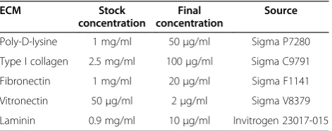
Two glioma cell lines U87 and U251N, two breast cancer cell lines MDA-MB-231 and MCF-7, and HeLa cervical cancer cells were obtained from American Type Cell Collection (ATCC) (Manassas, VA) and routinely main-tained in Dulbecco’s modified Eagle’s medium (DMEM) with 10% fetal bovine serum (FBS) supplemented with 100 μg/ml of penicillin-streptomycin. Pyrex cloning rings of 8 mm × 8 mm (Catalog No. CLS 31668-125EA) used in the ring cell migration assay were made by Corning and were purchased from Sigma-Aldrich. The XTT (2,3-Bis-(2-Methoxy-4-Nitro-5-Sulfophenyl)-2H-Tetrazolium-5-Carboxanilide) (Catalog No. X4251) used in cell proliferation assay was purchased from Sigma-Aldrich. The sources and coating concentrations of ECM proteins are listed in Table 1.
Ring cell migration assay
Before use, the rings preserved in alcohol solution were briefly flamed using tweezers. The rings were placed ver-tically in each well of six-well plates, either coated with ECM proteins or not, with a maximum of three rings in each well keeping appropriate distance from each other. Cell suspensions were added carefully to both sides of
each ring. Depending on the cell line, the cell number varied between 5,000 and 10,000 cells inside the ring, and between 250,000 to 300,000 cells outside the ring, reach-ing about 80% confluency when cells were attached. After cells attached firmly, which may take four to eight hours, the rings were carefully removed straight up using twee-zers followed by washing with 1 × Phosphate-Buffered Sa-line (PBS) and preserving in alcohol solutions for future uses. The cells were washed gently with PBS to remove cell debris while avoiding detachment of the cells. The cells were replenished with fresh DMEM containing 10% FBS and 100μg/ml of penicillin-streptomycin. The gaps of the cell rings were observed under microscope (Leica Microsystems, Wetzlar, Germany) and photographs were taken at various, marked sites along the rings at the start of assay (T0); between 4 and 6 positions were marked for imaging per ring. At different time points, e.g. 24 (T1) or 48 (T2) hours, photographs were taken again of the remaining gap, at the same pre-specified positions along the rings as at T0.
Image analysis
The areas of the gaps were measured using Image J soft-ware (National Institutes of Health, Bethesda, MD). The pictures of gaps taken at different time points were opened in Image J. The distance pixels were set as“400” and the unit of length was set as “mm”. The shapes of the gap areas were selected using the “freehand selec-tions”and the area was measured.
XTT proliferation assay
Five thousand cells were plated into each well of 96-well plates coated with or without indicated ECM protein. The cells were kept in the 37°C incubator supplied with 5% CO2. After 24, 48 or 72 hours incubation, the medium of
cells was removed and the cells were washed with 1 × PBS. Fifty microliters of XTT solution were added to each well and the plates were kept in the 37°C incubator for four hours. The O.D. (450 nm) of each well was determined by Multiskan EX Microplate Photometer (Thermo Scientific, Waltham, MA).
Statistical analysis
The differences in cell migration due to the effects of the different ECM proteins were determined by one-way ANOVA analysis using Prism software (GraphPad). The difference was considered as significant when p < 0.05 after Bonferroni correction for multiple testing.
Results
Set-up for the ring cell migration assay
[image:2.595.57.293.623.716.2]Although simple and easy, the conventional scratch assay has been shown to be very difficult in generating good and reliable scratches for many cell lines, including U87 cells.
Table 1 ECM proteins used in this study
ECM Stock
concentration
Final concentration
Source
Poly-D-lysine 1 mg/ml 50μg/ml Sigma P7280 Type I collagen 2.5 mg/ml 100μg/ml Sigma C9791 Fibronectin 1 mg/ml 20μg/ml Sigma F1141 Vitronectin 50μg/ml 2μg/ml Sigma V8379 Laminin 0.9 mg/ml 10μg/ml Invitrogen 23017-015
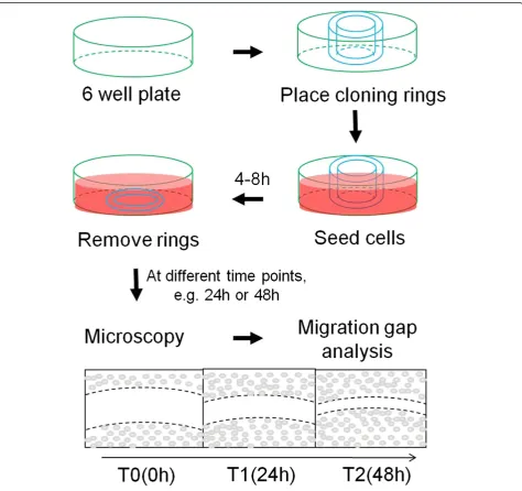
To keep the advantage and to overcome the shortcoming, we sought to develop an easy assay for cell migration. As described in Materials and Methods and shown in Figure 1, the ring cell migration assay constitutes a very simple set-up of experiments by using cloning rings, which are placed before seeding cells and removed after cells attached. The gaps formed before cells migrated allowed all cell lines to be analyzed using this assay. Although the cloning rings create ring-shaped gaps, it was difficult and inconvenient to monitor the entire gap under the microscope. Instead, we marked part of the ring at various sites and tracked them at various time points, mimicking an exclusion zone
[image:3.595.62.537.253.699.2]assay (Additional file 1: Figure S1). This allowed us to ob-tain more data points from single rings when each part of the ring gap was measured. In addition, we found that data points in each experiment could be increased by placing up to three rings in each well of six-well plates, making the assay more reproducible and efficient. We found that cell confluency did not significantly affect the migration rate of cells (data not shown) and that the difference in cell motil-ity of the different cell lines was independent of cell con-fluency; for example, U87 cells at lower confluency migrated faster than U251N cells did at higher confluency (Figure 2), suggesting that cell motility is an intrinsic
property of migrating cells. Thus, the ring cell migration assay is a simple, cost-effective and efficient way to analyze motility of cells.
Cancer cell motility as visualized during ring cell migration assay
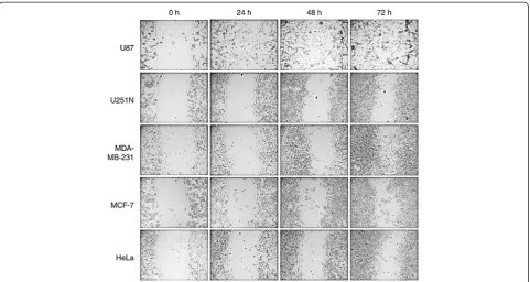
To test whether ring cell migration can distinguish dif-ferences in cell motility among different cell lines, two sets of cancer cells, i.e. glioma cells (U87 and U251N) and breast cancer cells (MDA-MB-231 and MCF-7), with different migratory potential were used in this assay and compared to HeLa cells. All five cell lines were processed in the ring cell migration assay as de-scribed above. At different time points, i.e. 0 h, 24 h, 48 h and 72 h, photographs were taken of the remaining gap from the same sites along the ring. As shown in Figure 2, while the gap area of all five cell lines was the same at the start of the assay (indicating the uniform thickness of the cloning ring), the rate of closing of the gaps differed significantly with increasing time. It was clear that the U87 glioma cells migrated faster than U251N cells even at 24 h, and more so at the 72 h time point. Similarly, the MDA-MB-231 breast cancer cells migrated faster than MCF-7 cells. Thus, the ring cell migration assay can be used to distinguish the motility of different cancer cell lines.
Quantitation of cancer cell migration using the ring cell migration assay
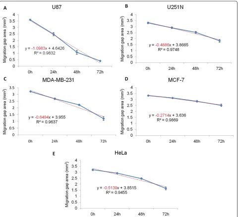
Data obtained from photomicrographs such as those shown in Figure 2 can yield quantitative comparisons be-tween cell lines. To do so, a group of gap areas from the same sites at individual time points were measured and plotted against time, and a trend line and a R2value were calculated for each plot. As shown in Figure 3, the slope of the trend line for U87, U251N, MDA-MB-231, MCF-7 and HeLa cells was −1.0983, −0.4886, −0.6494, −0.2714 and −0.5139 mm2/h respectively. Thus, the motility of each cell line could be compared according to the slope value, i.e. U87 > MDA-MB-231 > HeLa > U251N > MCF-7 cells. Therefore, the motility of cancer cells can be quanti-fied and compared in the ring cell migration assay.
Effect of ECM proteins on cancer cell migration
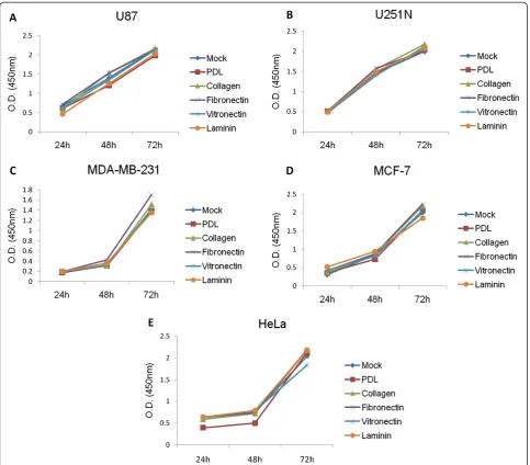
[image:4.595.58.540.88.344.2]individual time points, suggesting that these did not affect the proliferation of the cancer cells. Since there were no differences when proliferation assays were carried out with cultures that were subconfluent at the beginning, it can be presumed that differences in proliferation rate will not in-fluence the results of the ring migration assay in which the cell monolayer is more likely to be confluent and inhibited by contact.
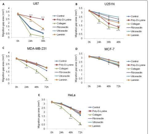
To evaluate the effect of ECM proteins on cancer cell migration, various proteins were each coated onto the
[image:5.595.59.540.88.526.2]the utility of this assay on assessing the effect of ECM components on short-term cell migration.
Discussion
We present a newly developed, simple and quantitative in vitro cell migration assay that can also be applied to evaluate the effect of various extracellular matrix com-ponents on coated plates. This assay is a variation of the cell exclusion zone assay whereby a cell-free zone is cre-ated without disturbance of the cell monolayer. The most critical steps in this assay are the placement and removal of the cloning rings before and after cell
seeding, respectively. As shown in Figure 2, differences in cell motility of the various cancer cell lines can be ob-served at 24 h or even earlier (data not shown), suggesting that this assay may be adapted to shorter time periods, es-pecially when combined with time-lapse videomicroscopy.
[image:6.595.57.541.88.512.2]and release of cellular factors during the scratch process. As well, the ring migration assay can be applied to cells which do not produce ideal scratches (such as the U87 gli-oma cells) and scratch-refractory monolayers. Other individually-developed assays for analyzing cell migration include the circle wound-healing assay [17] which uses a drill bit to make round-shaped wounds in a cell monolayer, and suffers from many of the same limita-tions as the scratch wound healing assay. Even the more advanced techniques such as the electric cell-substrate
impedance sensing method [18] which produces uni-form gaps in the cell monolayer with a pulse of high current still lead to cell death, producing cell remnants that may interfere with proper wound closing.
[image:7.595.58.543.89.525.2]observed by time-lapse videomicroscopy (data not shown). Furthermore Boyden assays are more costly because commercially available transwell plates must be used instead of the much cheaper cloning rings which can be recycled many times. New developments include microfluidics-based cell migration apparatuses that use microchannels with mixed flow of microfluids to study cell movement [19]. In the microfluidic system, cells are seeded in microchambers and streams of media with and without chemoattractants are applied in paral-lel. Combined with time-lapse microscopy, these assays provide insight into the migration behavior of cells, allowing single cells to be tracked and the direction and velocity of each cell to be calculated. However, these so-phisticated systems are expensive and may be unafford-able for many labs.
We took advantage of the ability to obtain quantita-tive measures to study the effect of ECM proteins on the migration of the different cell lines. The ECM con-stitutes an important component of the microenviron-ment during cancer cell invasion and metastasis [8,9]. Collagen is known to play a critical role in tumorigen-esis [20] and angiogentumorigen-esis [21].Our results suggest that collagen functions as a general promoter for glioma, breast and cervical cancer cell migration (Figure 5). In contrast, Ohtaka et al. showed that laminin had a stronger impact on promoting colon cancer cell migration than other extracellular matrix proteins such as collagen [22], while pancreatic cancer cell migration was positively in-fluenced by collagen, as well as fibronectin and laminin [23]. These studies point to the diverse roles that ECM proteins play in regulating migration of tissue-specific cancer cells. Interestingly, collagen, as well as other ECM proteins, did not affect the proliferation rate of any of the cancer cell lines used here (Figure 4) but had distinct effects on cell migration (Figure 5), suggesting that these proteins may activate different signaling pathways specific for cell migration, such as signaling though integrins [6]. The observed differences on the effect of ECM proteins on the migration of the various cell lines might be due to tissue-specific expression of receptors that bind to and are activated by different ECM proteins [4].
In summary, the ring cell migration assay is an easy, cost-effective method for quantitative analysis of cell migration in vitro. It is adaptable to both short and long-term evaluation periods using time-lapse micros-copy. Although the assay as described here does not in-volve chemotactic gradients, and is limited to analyzing autonomous cell migration, modifications can be envis-aged, for example, through implantation of matrigel containing chemokines within the ring, in order to cre-ate the chemotactic gradient. Thus, the ring migration assay may have broad applications in different fields.
Additional files
Additional file 1: Figure S1.Representation of the gap of the U251N glioma cells at 24 h after removal of the cloning ring.
Additional file 2: Figure S2.Slope as an indicator of distinct effects of extracellular matrices on cell migration. Ring cell migration assay was performed on U87, U251N, MDAMB231, MCF7 and HeLa cells grown on the indicated extracellular matrices and the slope was measured from Figure 5. One-way ANOVA Dunnett’s Multiple Comparison Test: Compare all columns vs. Control, N = 2. **p < 0.001, **p < 0.01 and *p < 0.05.
Abbreviations
ANOVA:Analysis of variance; ATCC: American Type Cell Collection;
DMEM: Dulbecco’s modified Eagle’s medium; EMT: Epithelial-to-mesenchymal transition; ECM: Extracellular matrix; FBS: Fetal bovine serum; O.D.: Optical density; PBS: Phosphate-buffered saline; XTT: 2,3-Bis-(2-Methoxy-4-Nitro-5-Sulfophenyl)-2H-Tetrazolium-5-Carboxanilide.
Competing interests
The authors declare that they have no competing interests.
Authors’contributions
HC and JN conceived and designed this study. HC carried out experiments and collected data. HC and JN analyzed data and wrote the manuscript. Both authors read and approved the final manuscript.
Acknowledgements
The authors thank Dr. Jean-Jacques Lebrun (Royal Victoria Hospital, McGill University) for generously providing MDA-MB-231 and MCF-7 cells. This study was supported by the Cancer Research Society Inc. to JN.
Author details 1
Division of Experimental Medicine, Department of Medicine, McGill University, Montreal, Quebec, Canada.2Department of Neurology &
Neurosurgery, McGill University, Montreal, Quebec, Canada.3Montreal Neurological Institute, McGill University, 3801 University Street, Montreal, Quebec H3A 2B4, Canada.
Received: 9 August 2013 Accepted: 17 March 2014 Published: 27 March 2014
References
1. Chiang AC, Massague J:Molecular basis of metastasis.N Engl J Med2008,
359(26):2814–2823.
2. Kraljevic Pavelic S, Sedic M, Bosnjak H, Spaventi S, Pavelic K:Metastasis: new perspectives on an old problem.Mol Cancer2011,10:22. 3. Spano D, Heck C, de Antonellis P, Christofori G, Zollo M:Molecular
networks that regulate cancer metastasis.Semin Cancer Biol2012,
22(3):234–249.
4. Hynes RO:The extracellular matrix: not just pretty fibrils.Science2009,
326(5957):1216–1219.
5. Ungefroren H, Sebens S, Seidl D, Lehnert H, Hass R:Interaction of tumor cells with the microenvironment.Cell Commun Signal2011,9:18. 6. Hood JD, Cheresh DA:Role of integrins in cell invasion and migration.
Nat Rev Cancer2002,2(2):91–100.
7. Rowe RG, Weiss SJ:Navigating ECM barriers at the invasive front: the cancer cell-stroma interface.Annu Rev Cell Dev Biol2009,25:567–595. 8. Joyce JA, Pollard JW:Microenvironmental regulation of metastasis.
Nat Rev Cancer2009,9(4):239–252.
9. Friedl P, Alexander S:Cancer invasion and the microenvironment: plasticity and reciprocity.Cell2011,147(5):992–1009.
10. Wolf K, Friedl P:Extracellular matrix determinants of proteolytic and non-proteolytic cell migration.Trends Cell Biol2011,21(12):736–744. 11. Kramer N, Walzl A, Unger C, Rosner M, Krupitza G, Hengstschläger M, Dolznig H:In vitro cell migration and invasion assays.Mutat Res2013,
752(1):10–24.
12. Zimmermann M, Box C, Eccles SA:Two-dimensional vs. three-dimensional in vitro tumor migration and invasion assays.Methods Mol Biol2013,
13. Liang CC, Park AY, Guan JL:In vitro scratch assay: a convenient and inexpensive method for analysis of cell migration in vitro.Nat Protoc
2007,2(2):329–333.
14. Chen HC:Boyden chamber assay.Methods Mol Biol2005,294:15–22. 15. Poujade M, Grasland-Mongrain E, Hertzog A, Jouanneau J, Chavrier P, Ladoux B, Buguin A, Silberzan P:Collective migration of an epithelial monolayer in response to a model wound.Proc Natl Acad Sci U S A2007,
104(41):15988–15993.
16. Gough W, Hulkower KI, Lynch R, McGlynn P, Uhlik M, Yan L, Lee JA:A quantitative, facile, and high-throughput image-based cell migration method is a robust alternative to the scratch assay.J Biomol Screen2011,
16(2):155–163.
17. Kam Y, Guess C, Estrada L, Weidow B, Quaranta V:A novel circular invasion assay mimics in vivo invasive behavior of cancer cell lines and distinguishes single-cell motility in vitro.BMC Cancer2008,8:198. 18. Lo CM, Keese CR, Giaever I:Monitoring motion of confluent cells in tissue
culture.Exp Cell Res1993,204(1):102–109.
19. Rhoads DS, Nadkarni SM, Song L, Voeltz C, Bodenschatz E, Guan JL:Using microfluidic channel networks to generate gradients for studying cell migration.Methods Mol Biol2005,294:347–357.
20. Ortiz-Urda S, Garcia J, Green CL, Chen L, Lin Q, Veitch DP, Sakai LY, Lee H, Marinkovich MP, Khavari PA:Type VII collagen is required for Ras-driven human epidermal tumorigenesis.Science2005,307(5716):1773–1776. 21. Kalluri R:Basement membranes: structure, assembly and role in tumour
angiogenesis.Nat Rev Cancer2003,3(6):422–433.
22. Ohtaka K, Watanabe S, Iwazaki R, Hirose M, Sato N:Role of extracellular matrix on colonic cancer cell migration and proliferation.
Biochem Biophys Res Commun1996,220(2):346–352.
23. Ryschich E, Khamidjanov A, Kerkadze V, Buchler MW, Zoller M, Schmidt J:
Promotion of tumor cell migration by extracellular matrix proteins in human pancreatic cancer.Pancreas2009,38(7):804–810.
doi:10.1186/1756-0500-7-183
Cite this article as:Chen and Nalbantoglu:Ring cell migration assay identifies distinct effects of extracellular matrix proteins on cancer cell migration.BMC Research Notes20147:183.
Submit your next manuscript to BioMed Central and take full advantage of:
• Convenient online submission
• Thorough peer review
• No space constraints or color figure charges
• Immediate publication on acceptance
• Inclusion in PubMed, CAS, Scopus and Google Scholar
• Research which is freely available for redistribution