Replication to Wild-Type PGC-1
␣
/Liver-Specific PGC-1
␣
Upregulation
Wenxia Yao,a,bHua Cai,a,bXinlei Li,bTing Li,aLongbo Hu,aTao Penga,b
Laboratory of Viral Immunology, State key Laboratory of Respiratory Disease, Guangzhou Institutes of Biomedicine and Health, Chinese Academy of Sciences, Guangzhou, Chinaa; Sino-French Hoffmann Institute of Immunology, Guangzhou Medical University, Guangzhou, Chinab
ABSTRACT
Hepatitis C virus (HCV) causes not only severe liver problems but also extrahepatic manifestations, such as insulin resistance
(IR). Wild-type peroxisome proliferator-activated receptor gamma coactivator 1 alpha (WT-PGC-1␣) is essential in hepatic
glu-coneogenesis and has recently been demonstrated to link HCV infection to hepatic insulin resistance (IR). A recent study has
characterized a novel human liver-specific PGC-1␣(L-PGC-1␣) transcript, which is proposed to reflect human adaption to more
complex pathways. However, the effect of HCV infection on L-PGC-1␣expression and the mechanism by which HCV modulates
WT-PGC-1␣/L-PGC-1␣remain unclear. In this study, we showed that HCV infection upregulated both WT-PGC-1␣and
L-PGC-1␣, which further promoted HCV production. The upregulation of both PGC-1␣isoforms depended on HCV RNA
repli-cation. By using promoter-luciferase reporters, kinase inhibitors, and dominant negative mutants, we further observed that the
HCV-induced upregulation of WT-PGC-1␣was mediated by the phosphorylation of cyclic AMP (cAMP)-responsive element-binding
protein (CREB), whereas that of L-PGC-1␣was mediated by CREB phosphorylation and forkhead box O1 dephosphorylation.
More-over, HCV infection induced endoplasmic reticulum (ER) stress, and pharmacological induction of ER stress upregulated
WT-PGC-1␣/L-PGC-1␣and phosphorylated CREB. In contrast, pharmacological inhibition of HCV-induced ER stress impaired WT-PGC-1␣/
L-PGC-1␣upregulation along with decreased phosphorylated CREB. The correlation of hepatic mPGC-1␣with ER stress was further
confirmed in mice. Overall, HCV infection upregulates both WT-PGC-1␣and L-PGC-1␣through an ER stress-mediated,
phosphory-lated CREB-dependent pathway, and both PGC-1␣isoforms promote HCV production in turn.
IMPORTANCE
HCV causes not only severe liver problems but also extrahepatic manifestations, such as insulin resistance (IR). As a key
regula-tor in energy metabolism, wild-type PGC-1␣(WT-PGC-1␣), has recently been demonstrated to link HCV infection to hepatic
IR. A recent study has characterized a novel human liver-specific PGC-1␣(L-PGC-1␣), which reflects human adaption to more
complex pathways. However, the effect of HCV infection on L-PGC-1␣expression and the mechanism by which HCV regulates
WT-PGC-1␣/L-PGC-1␣remain unclear. In this study, we showed that HCV infection upregulated both WT-PGC-1␣and
L-PGC-1␣, which further promoted HCV production. WT-PGC-1␣upregulation was mediated by CREB phosphorylation,
whereas L-PGC-1␣upregulation was mediated by CREB phosphorylation and FoxO1 dephosphorylation. HCV-induced ER
stress mediated WT-PGC-1␣/L-PGC-1␣upregulation and CREB phosphorylation. Overall, this study provides new insights into
the mechanism by which HCV upregulates WT-PGC-1␣/L-PGC-1␣and highlights the novel intervention of HCV-ER
stress-PGC-1␣signaling for HCV therapy and HCV-induced IR therapy.
H
epatitis C virus (HCV) infection has become a serious health issue associated with substantial morbidity and mortality (1). The World Health Organization estimates that approximately 185 million people are or have been infected with HCV worldwide (2). HCV causes not only severe liver problems but also extrahepatic manifestations, such as insulin resistance (IR) and type 2 diabetes mellitus (T2DM). In patients with chronic HCV, the achievement of sustained virological response with drugs prevents the develop-ment ofde novoIR (3). HCV-infected patients also have increased risk of T2DM compared with noninfected controls and hepatitis B virus-infected controls (4). These studies suggest that HCV di-rectly contributes to the development of IR and T2DM.Peroxisome proliferator-activated receptor gamma coactiva-tor 1 alpha (PGC-1␣) is an inducible transcription factor coacti-vator that controls cellular energy metabolism (5). Cumulative studies link altered PGC-1␣signaling to IR and T2DM. PGC-1␣ induced in liver during fasting promotes hormone-stimulated gluconeogenesis (6). PGC-1␣is robustly upregulated in diabetic liver (7), increasing hepatic glucose production. PGC-1␣serves as
a metabolic sensor; hence, PGC-1␣expression is finely regulated to meet energy demands. Several levels of regulation have been implicated, including transcriptional regulation and posttransla-tional modifications (8). The transcriptional regulation of PGC-1␣ by the cyclic AMP (cAMP)-responsive element-binding protein (CREB) is a common pattern in the metabolic adaptions of the liver to gluconeogenic status in response to glucagon and gluco-corticoids (7,9) or in response to free fatty acids (10). Alternative splicing or transcription initiation represents another mode of
regu-Received27 April 2014Accepted6 May 2014 Published ahead of print15 May 2014 Editor:M. S. Diamond
Address correspondence to Tao Peng, [email protected].
W.Y. and H.C. contributed equally to this article.
Copyright © 2014, American Society for Microbiology. All Rights Reserved.
doi:10.1128/JVI.01202-14
on November 7, 2019 by guest
http://jvi.asm.org/
lation. A recent study has characterized a novel human liver-specific PGC-1␣(L-PGC-1␣) transcript (11), which results from alternative promoter usage. The previously described typical form of PGC-1␣is referred to as wild-type PGC-1␣(WT-PGC-1␣).
Disparate promoter usage suggests different transcriptional regulation between L-PGC-1␣ and WT-PGC-1␣. The pro-moter study by Felder et al. (11) suggests that forkhead box O1 (FoxO1) plays a greater role in L-PGC-1␣transcription than in WT-PGC-1␣ transcription. Besides, like WT-PGC-1␣ mRNA, L-PGC-1␣mRNA increases in response to glucocorticoids and CREB signaling. L-PGC-1␣is located mainly in the nucleus and is identical to WT-PGC-1␣except for a deletion of the N-terminal 127 amino acids (11). Therefore, L-PGC-1␣shows overlapped coactivation properties compared with those of WT-PGC-1␣ (11). There are also functional differences between L-PGC-1␣and WT-PGC-1␣. For example, only WT-PGC-1␣coactivates liver X receptor alpha (LXR␣) at the promoter of sterol regulatory element-binding transcription factor 1c (SREBP-1c) (11). L-PGC-1␣ transcript homologues are not detected in livers of the rat, mouse, dog, and monkey (11), whereas all the main interacting domains of WT-PGC-1␣are highly conserved in many chordate species (12). Thus, L-PGC-1␣is proposed to reflect an adaption to more complex pathways in humans. Although substantial progress has been made in the study of WT-PGC-1␣, the function and regula-tion of L-PGC-1␣remain largely unknown.
Endoplasmic reticulum (ER) is the cellular organelle responsi-ble for lipid metabolism, protein processing, and Ca2⫹storage.
Interference with ER functions by conditions such as high-fat feeding, virus infection, and glucose deprivation induces a stress response, known as ER stress. Upon sensing ER stress, cells activate a unique intracellular signaling pathway unfolded protein response (UPR) to cope with this insult. ER stress and the activated UPR have been associated with various diseases, including liver diseases (13). Replication of the HCV genome occurs at the so-called ER-associated membranous webs (14). Studies usingin vitroandin vivo experimen-tal models (15,16) and clinical liver biopsy (17) have indicated that HCV induces ER stress. HCV further utilizes cellular re-sponses to ER stress to promote HCV persistence and patho-genesis. For instance, HCV-induced ER stress has been impli-cated in the pathogenesis of HCV-related IR (18,19).
The important function of WT-PGC-1␣in the pathogenesis of IR and T2DM suggests that HCV infection may upregulate liver WT-PGC-1␣expression and further promote hepatic IR. Shlomai et al. (20) showed that WT-PGC-1␣is robustly upregulated by HCV infection and that elevated WT-PGC-1␣links HCV infec-tion to hepatic IR. However, the effect of HCV infecinfec-tion on L-PGC-1␣expression remains unknown, and the mechanism by which HCV infection modulates WT-PGC-1␣/L-PGC-1␣ re-mains unclear. In this study, we investigated whether L-PGC-1␣is also regulated by HCV infection and whether the modulation of WT-PGC-1␣/L-PGC-1␣is associated with ER stress.
In the current study, we found that HCV infection upregulates both WT-PGC-1␣and L-PGC-1␣to promote HCV production. We also found that the HCV infection-induced upregulation of both WT-PGC-1␣and L-PGC-1␣depends on HCV RNA repli-cation and CREB phosphorylation. Furthermore, HCV-induced ER stress has an intermediary function in the upregulation of both PGC-1␣isoforms. Finally, the demonstrated efficacy of 4-phenyl-butyric acid (PBA), an ER stress inhibitor, in blocking HCV pro-vides a new perspective for HCV therapy.
MATERIALS AND METHODS
Cells.Huh7, its derivative Huh7.5.1, and Hep3B cells were grown in Dul-becco’s minimal essential medium (DMEM) supplemented with 10% fe-tal bovine serum (FBS; HyClone, Beijing, China) and 1% penicillin-strep-tomycin. Huh7-sgHCV1b and Huh7.5.1-sgJFH2a cells, which harbor genotype 1b and 2a subgenomic replicons, respectively, were maintained in the medium described above and supplemented with 400g/ml of G418 and 2g/ml of puromycin. 3#, 4#, 6#, and 7# represent the respec-tive subgenomic replicon clones of Huh7.5.1-sgJFH2a cells. The cells were maintained at 37°C in a humidified atmosphere containing 5% CO2.
Plasmids.Promoter-luciferase reporter vectors were generated in the pGL4.17 backbone (Promega). pL-PGC-1␣-Prom-Luc and pWT-PGC-1␣-Prom-Luc were constructed by cloning 3.518-kb and 2.614-kb up-stream fragments into pGL4.17, respectively. Two mutants of the L-PGC-1␣promoter region, namely, pL-PGC-1␣-Prom-GRE-Mut and pL-PGC-1␣-Prom-FoxO1-site-Mut, were created from pL-PGC-1␣ -Prom-Luc by using site-directed mutagenesis (11). Three mutants of the WT-PGC-1␣promoter region were also constructed by using site-di-rected mutagenesis. pWT-PGC-1␣-Prom-IRS-Mut was mutated with all the three insulin response sequences (IRSs) (21), and pWT-PGC-1␣ -Prom-CRE-Mut and pWT-PGC-1␣-Prom-MEF2-site-Mut were con-structed as previously described (22). pRL-SV40-Renilla reporter plasmid (Promega) served as a transfection control.
Plasmids expressing HCV proteins (except NS5B) were generated with the lentiviral vector (pRlenti) tagged with Flag sequence at the C terminus. Hemagglutinin (HA)-NS5B was constructed by cloning NS5B-encoding fragments into the plasmid pReceiver-M06 with 3 copies of the HA se-quence at the N terminus. pRlenti was also used to generate the expression plasmids L-PGC-1␣and WT-PGC-1␣with HA tags at the N terminus. To express short hairpin RNA (shRNA) targeting WT-PGC-1␣/L-PGC-1␣, we used the pSuper retroviral plasmid that was created as described in reference23by using the following primers: for shPGC-1␣1#, 5=-GATC CCCCCAACACTCAGCTAAGTTATTCAAGAGATAACTTAGCTGAGT GTTGGTTTTTA-3=and 5=-AGCTTAAAAACCAACACTCAGCTAAGT TATCTCTTGAATAACTTAGCTGAGTGTTGGGGG-3= (20), and for shPGC-1␣2#, 5=-GATCCCCCCGTTATACCTGTGATGCTTTTTCAAG AGAAAAGCATCACAGGTATAACGGTTTTTA-3=and 5=-AGCTTAAA AACCGTTATACCTGTGATGCTTTTCTCTTGAAAAAGCATCACAGGT ATAACGGGGG-3=.
The expression plasmid pCREB was generated by C-terminal Flag-tagged CREB cloned in vector pCR3.1 (Invitrogen) in our laboratory (24). The pCREB133 vector, which contains a serine-to-alanine mutation cor-responding to amino acid 133, was created from pCREB using site-di-rected mutagenesis.
Antibodies and reagents.The following primary antibodies were used for Western blotting: monoclonal mouse anti-PGC-1␣antibody (4C1.3; Calbiochem), polyclonal rabbit anti-PGC-1␣antibody (ab54481; Ab-cam), monoclonal mouse anti-HCV core antibody (C7-50; AbAb-cam), monoclonal mouse anti-HCV NS3 (H23; Abcam), monoclonal mouse phospho-CREB antibody (10E9; Santa Cruz), monoclonal mouse CREB antibody (X-12; Santa Cruz), polyclonal rabbit phospho-ATF2 (Thr71) antibody (no. 9221; Cell Signaling Technology), monoclonal rabbit phos-pho-FoxO1 (Thr24) antibody (4G6; Beyotime, China), monoclonal rab-bit FoxO1 antibody (C29H4; Beyotime), rabrab-bit Bip antibody (Beyotime), rabbit histone H3 antibody (Beyotime, China), polyclonal rabbit-actin antibody (MultiSciences, China), and glyceraldehyde-3-phosphate dehy-drogenase (GAPDH) antibody (Kang Xiang, Shanghai, China). A goat anti-mouse antibody (Chemicon) and a goat anti-rabbit antibody (Chemicon), both conjugated with horseradish peroxidase, were used as secondary antibodies.
Naive Huh7.5.1 cells were treated with 1M thapsigargin (Tg) or 1.5 g/ml of tunicamycin (Tm) for 8 h to induce ER stress. HCV-infected Huh7.5.1 cells were treated with 6 mM PBA for 24 h to inhibit ER stress. HCV-infected Huh7.5.1 cells were treated with H89 for 24 h, SB203580 for 24 h, or wortmannin for 12 h to inhibit kinase activities. PBA (Sigma)
on November 7, 2019 by guest
http://jvi.asm.org/
and H89 (Sigma) stocks were prepared in double-distilled water. Tg (Sigma), Tm (Fermentek), SB203580 (Sigma), and wortmannin (Sigma) were prepared in dimethyl sulfoxide.
RNA isolation, reverse transcription, XBP1 PCR, and real-time PCR.Total RNA was extracted from cells using TRIzol (Invitrogen) ac-cording to the manufacturer’s instructions. RNA was reverse transcribed by Moloney murine leukemia virus reverse transcriptase (Fermentas) in the presence of random hexamers (TaKaRa). The reaction mixture was incubated for 10 min at 25°C and for 1 h at 42°C and then the reaction was stopped by heating at 95°C for 5 min. The mRNA fragments of XBP1 and actin were amplified under the following PCR conditions with cDNA: 38 cycles of 94°C for 30 s, 58°C for 30 s, and 72°C for 1 min. Previously reported primers for XBP1 and actin were used (18). We analyzed three fragments representing spliced, unspliced, and hybrid XBP1 on a 2% aga-rose gel. The cDNA was used to perform SYBR-PCR using a SYBR Premix
Ex Taqkit (TaKaRa). The primers used for real-time PCR are listed in
Table 1. The housekeeping 18S rRNA or GAPDH gene served as an
inter-nal control. All reactions were run in triplicate using the CFX96 Touch real-time PCR detection system (Bio-Rad).
Preparation of cell extracts and Western blotting.Proteins were ex-tracted from cells using radioimmunoprecipitation assay buffer supple-mented with protease inhibitors (Sigma), with GAPDH/-actin as an in-ternal control. Proteins were prepared using a nuclear and cytoplasmic protein extraction kit (Beyotime, China), with histone H3 as an internal control. For Western blotting, equal amounts of protein extracts boiled with a sodium dodecyl sulfate (SDS) loading buffer containing
-mercap-toethanol (140 mM) were subjected to SDS-polyacrylamide gel electro-phoresis and then transferred onto a nitrocellulose membrane (Portran; Whatman). The membrane was subsequently incubated with the respec-tive primary antibodies. Horseradish peroxidase-conjugated secondary antibodies were used to visualize the respective proteins with an enhanced chemiluminescence detection system (Millipore).
Luciferase reporter assays.Huh7.5.1 cells or HCV-infected Huh7.5.1 cells that were prepared in a 96-well tissue culture plate at a density of 1.5⫻104cells per well were cotransfected with pWT-PGC-1␣ /L-PGC-1␣-Prom-Luc reporter plasmid and pRL-SV40-Renilla. After 72 h, lucif-erase activity was determined using a Dual-Glo luciflucif-erase assay system kit (E2920; Promega). Firefly andRenillaluciferase activities were measured with luminometer (Turner Biosystems, USA). Firefly luciferase activity was normalized toRenillaluciferase activity for each sample.
In vitrotranscription of HCV RNA, HCV preparation, and titration. Plasmid pFL-J6/JFH/Jc1, used for the generation of infectious HCV-Jc1 (25), was linearized with XbaI and then purified by ethanol precipitation. Then, 1 g of linearized plasmid was transcribed in vitro with a MEGAscript T7 kit (Ambion, Austin, TX) for the large-scale production of RNA. For RNA transfection, Huh7.5.1 cells were transfected with 24g of HCV-Jc1 RNA using Lipofectamine 2000 reagent (Invitrogen). Super-natants collected at 4 to 5 days posttransfection (dpt) were centrifuged at low speed to remove cell debris. Clarified viral supernatants were ali-quoted and then stored at⫺80°C until use. HCV-Jc1 titers were deter-mined as previously described (26). TheEGFPgene was inserted into the C terminus of NS5A-encoding sequence within the HCV-Jc1 genome to generate an HCV reporter virus termed HCV-Jc1EGFP. The adapted HCV-Jc1EGFP was generated and prepared for stocks in our laboratory as previously described (27).
To determine the effect of PGC-1␣overexpression on HCV produc-tion, Huh7.5.1 cells were infected with the lentivirus carrying the coding sequence of WT-PGC-1␣or L-PGC-1␣or the pRlenti control. After 12 h, Huh7.5.1 cells were infected with HCV-Jc1EGFP or HCV-Jc1 at a multi-plicity of infection (MOI) of 0.02. The HCV-Jc1EGFP or HCV-Jc1 virus in the supernatant was collected at 72 h postinfection, and then the titer of the HCV-Jc1EGFP virus was determined by both limiting dilution assay and flow cytometry (FCM) and that of the HCV-Jc1 virus was determined only by limiting dilution assay. To determine the effect of PGC-1␣ knock-down on HCV production, Huh7.5.1 cells were infected with the retrovi-rus carrying the shRNA expression cassette against PGC-1␣(shPGC-1␣ 1# or shPGC-1␣2#) or a nontarget control (shRNA-Ctrl). After 12 h, Huh7.5.1 cells were infected with HCV-Jc1EGFP or HCV-Jc1 at an MOI of 0.02. The HCV-Jc1EGFP or HCV-Jc1 in the supernatant was collected at 96 h postinfection, and the titer was determined as mentioned above. For the titration of supernatant virus by limiting dilution assay, viral su-pernatants collected at 72 h or 96 h postinfection were clarified and inoc-ulated on Huh7.5.1 cells by endpoint dilution. At 72 h postinfection, Huh7.5.1 cells were fixed with 4% paraformaldehyde and then immuno-stained for HCV core antigen. Stained foci were counted and used to calculate the titer of focus-forming units (FFU)/ml. To determine the titer of HCV in the supernatant by flow cytometry, approximately 3 l (PGC-1␣knockdown experiment) or 5l (PGC-1␣overexpression ex-periment) of viral supernatants was obtained to inoculate naive Huh7.5.1 cells in 24-well tissue culture plates; the infected cells were quantified for flow cytometry at 72 h postinfection. The percentage of NS5A-EGFP-positive cells was used to calculate the infectivity of HCV. For the intra-cellular infectivity of HCVcc, the pellets of HCV-infected cells were resus-pended in DMEM containing 10% FBS and then subjected to three cycles of freezing and thawing to induce cell lysis. The samples were then centri-fuged for 5 min at 3,000 rpm to remove cell debris; afterward, superna-tants were collected to determine infectivity.
[image:3.585.40.284.81.416.2]RNA extraction from mouse liver.Male Leprdb/dbmice were on a C57BL/6 genetic background, and they were maintained at the Animal Center of Guangzhou Institutes of Biomedicine and Health according to the guidelines of the Institutional Animal Committee. Livers isolated TABLE 1Sequences and positions of primers used in this study
Gene (GenBank
accession no.) Primer Position PCR product size (bp) HCV 5=UTRa TCTGCGGAACCGGTGAGTA 147⫹19b 150
TCAGGCAGTACCACAAGGC 296⫺19 L-PGC-1␣
(HQ695733.1)
TCCCTCTGTTGCCTTTGTG 144⫹19 93 TAACCCCATGCCATCCAT 236⫺18 WT-PGC-1␣
(NM_013261.3)
TGAAAAAGCTTGACTGGCG 87⫹19 133 AAGATCTGGGCAAAGAGGC 219⫺19 PEPCK
(NM_001018073.1)
TATTATGACCCGACTGGGGA 699⫹20 143 TCAGGGTTTTCTCTGGGTTG 841⫺20 CREB
(NM_004379.3)
GTGTTACGTGGGGGAGAGAA 163⫹20 110 GGCTCCAGATTCCATGGTC 272⫺19 ATF2 (NM_001880.3) GGTGCTTTGTAAACACGGCT 85⫹20 104
GCAGTCCTTTCTCAAGTTTCC 188⫺21 FoxO1 (NM_002915) GAGGGTTAGTGAGCAGGTTAC 2,352⫹21 217
AGTCCTTATCTACAGCAGCAC 2,568⫺21 GAPDH
(NM_002046.4)
GAAGGTGAAGGTCGGAGTC 180⫹19 226 GAA GATGGTGATGGGATTTC 405⫺20 18S (NR_003286.2) GGTGAAATTCTTGGACCGGC 958⫹20 196
GACTTTGGTTTCCCGGAAGC 1,153⫺20 mPGC-1␣
(NM_008904.2)
AATTGAAGAGCGCCGTGT 2,152⫹18 127 TCCATCATCCCGCAGATT 2,278⫺18 mBip
(NM_001163434.1)
ACCAAGACATTTGCCCCA 562⫹18 147 TTTGGTTGCTTGTCGCTG 708⫺18 Murine actin
(NM_007393.3)
CTCCTCCTGAGCGCAAGTACTC 1,071⫹22 91 TCCTGCTTGCTGATCCACATC 1,161⫺21
aUTR, untranslated region. b
The first value is the nucleotide position of the 5=terminus of the primer; the second represents the number of the nucleotide bases of the primer.
on November 7, 2019 by guest
http://jvi.asm.org/
from 16-week-old mice were stored in liquid nitrogen until use. Total RNA was extracted from mouse liver using a total RNA isolation kit (San-gon Biotech, China) according to the manufacturer’s instructions.
RESULTS
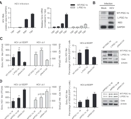
HCV infection upregulates WT-PGC-1␣and L-PGC-1␣to
pro-mote HCV production. A previous study showed that
WT-PGC-1␣is upregulated following HCV infection (20). To investi-gate this phenomenon in more detail, gene expression level was analyzed by infecting Huh7.5.1 cells with HCV-Jc1. As the HCV RNA level continuously increased from 1 to 7 days postinfection (dpi) (Fig. 1A, left), the mRNA levels of both WT-PGC-1␣and L-PGC-1␣significantly increased (14-fold and 4-fold at 7 days, respectively) (Fig. 1A, right). Western blot analysis showed that HCV infection increased the protein levels of both WT-PGC-1␣
and L-PGC-1␣(approximately 91 kDa and 77 kDa, respectively) (Fig. 1B). These results indicate that aside from WT-PGC-1␣, HCV infection also induces L-PGC-1␣elevation.
We further investigated whether the elevated WT-PGC-1␣ /L-PGC-1␣affects HCV infection in turn. Huh7.5.1 cells were trans-duced with PGC-1␣-overexpressing lentiviruses or PGC-1␣ -si-lencing retroviruses, followed by inoculation with adapted HCV-Jc1EGFP or HCV-Jc1. The extracellular HCV-Jc1EGFP ti-ter was quantified by both endpoint dilution and flow cytometry (FCM). The overexpression of both PGC-1␣isoforms increased extracellular HCV-Jc1EGFP titer by limiting dilution assay (Fig. 1C, left) and by FCM (Fig. 1C, middle) and simultaneously in-creased HCV core expression (Fig. 1C, right). In contrast, knock-down of WT-PGC-1␣/L-PGC-1␣with specific shRNAs signifi-cantly impaired extracellular HCV-Jc1EGFP titer and HCV core
A
B
D
0 5 10 15 ** ***C
HCV in fe ct io n
1dpi 3dpi 5dpi 7dpi
0 1 2 3 4 HCV R NA (re la tiv e v a lu e t o G A P D H ) 0 1 2 3 4 5 ** Infection
Mock HCV
GAPDH WT-PGC-1α
L-PGC-1α
NS3
1dpi3dpi 5dpi 7dpi 1dpi 3dpi 5dpi 7dpi
0 5 10 15 20 WT-PGC-1α L-PGC-1α (n o rm a liz
ed to 1dpi
) Per c entage of N S 5 A-EG F P ( + ) c e lls * P e rc e n ta g e o f NS5A-EG F P ( + ) c e lls ) shPGC-1α 1# shPGC-1α 2
# WT-P GC -1α L-PG C-1α G ene m R N A Relative WT-PGC-1α L-PGC-1α Core β-Actin WT-PGC-1α L-PGC-1α Core β-Actin pRle nti-C trl pRle nti-C trl shRNA-Ctr l shRNA-Ctr l Ex tr a . H C V ti te r HCV-Jc1EGFP Ex tr a . H C V ti te r HCV-Jc1EGFP WT -PGC -1α L-P GC-1α pRle nti-C trl WT -PGC -1α L-P GC-1α pRle nti-C trl 0 300 600 900 1200 0 500 1000 1500 ** ** HCV-Jc1EGFP HCV-Jc1 *** *** E x tr a . H C V tite r (F F U /m
l) Extr
a . H C V ti te r ( F F U /m l) shPGC-1α 1
# shPGC-1α2# shRNA-Ctr l shPGC-1α 1#
shPGC-1α 2#
shRNA-Ctrl 0 300 600 900 1200 0 500 1000 1500 ** ** HCV-Jc1EGFP HCV-Jc1 ** p=0.08 E x tr a . H C V tite r ( F F U /m
l) Ex
tr a . H C V ti te r ( F F U /m l)
FIG 1WT-PGC-1␣and L-PGC-1␣are upregulated by HCV infection and promote HCV production. (A) Analysis of HCV RNA levels and WT-PGC-1␣and L-PGC-1␣mRNA levels after infection of Huh7.5.1 cells with HCV-Jc1 (MOI, 0.02) for different times. (B) Western blot analysis for detecting the expression of WT-PGC-1␣and L-PGC-1␣at 7 dpi in Huh7.5.1 cells. (C) Effect of WT-PGC-1␣and L-PGC-1␣overexpression on HCV production and on HCV core protein level. (D) Effect of WT-PGC-1␣/L-PGC-1␣knockdown on HCV production and on HCV core protein level. shRNA-Ctrl, nontargeting shRNA. (C and D) Huh7.5.1 cells were infected with HCV-Jc1EGFP or HCV-Jc1 at an MOI of 0.02. The extracellular HCV-Jc1EGFP titer was quantified by both endpoint dilution and flow cytometry (FCM), and the HCV-Jc1 titer was quantified by only endpoint dilution. In the FCM assay, the extracellular HCV-Jc1EGFP titer was determined as a percentage of NS5A-EGFP-positive cells by using viral supernatants to inoculate naive Huh7.5.1 cells. In all panels, data are presented as means⫾ SEMs (nⱖ3). *,P⬍0.05; **,P⬍0.01; ***,P⬍0.001 (all determined by Student’sttest).
on November 7, 2019 by guest
http://jvi.asm.org/
[image:4.585.79.510.66.457.2]expression (Fig. 1D). And herein both shPGC-1␣ 1# and shPGC-1␣2# did not discriminate between WT-PGC-1␣and L-PGC-1␣(Fig. 1D, right). In addition, overexpression of
WT-PGC-1␣/L-PGC-1␣ and knockdown of WT-PGC-1␣ and
L-PGC-1␣by shPGC-1␣1# have similar effects on the extracellu-lar titer of HCV-Jc1 (Fig. 1C, left, andFig. 1D, left).
Collectively, these results indicate that HCV infection upregu-lates both WT-PGC-1␣and L-PGC-1␣and then the upregulated WT-PGC-1␣/L-PGC-1␣enhances HCV production.
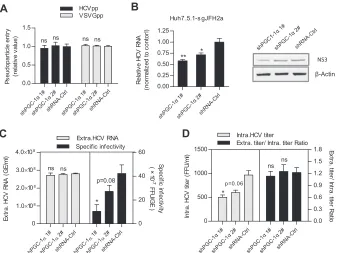
WT-PGC-1␣/L-PGC-1␣is involved in HCV RNA replication
and assembly of infectious virions.To identify in which step of
the HCV life cycle WT-PGC-1␣/L-PGC-1␣is required, multiple virologic assays were performed. As shown inFig. 2A, knockdown of WT-PGC-1␣/L-PGC-1␣with shPGC-1␣1# or shPGC-1␣2# did not affect HCV pseudovirus infection. This result suggests that WT-PGC-1␣/L-PGC-1␣is not required for HCVpp entry. In ad-dition, shPGC-1␣ 1# significantly decreased both intracellular HCV RNA levels and NS3 protein levels in Huh7.5.1-sgJFH2a cells, which harbor HCV subgenomic replicons (Fig. 2B), and shPGC-1␣2# also significantly decreased intracellular HCV RNA levels. This result indicates that WT-PGC-1␣/L-PGC-1␣affects HCV RNA replication.
To further investigate whether WT-PGC-1␣/L-PGC-1␣is in-volved in the late stage of the HCV life cycle, we calculated the specific infectivity (infectious titers divided by HCV RNA copy number) and the ratio of extracellular titer to intracellular titer as we previously described (27). Specific infectivity was calculated to evaluate the efficiency of HCV assembly (28), and the ratio of extracellular titer to intracellular titer was used to evaluate the
secretion of viral particles (27). WT-PGC-1␣/L-PGC-1␣silencing by shPGC-1␣1# significantly reduced the extracellular HCV-Jc1 specific infectivity (Fig. 2C, right), with no effect on the extracel-lular HCV RNA (Fig. 2C, left). Moreover, similarly to the extra-cellular titer (Fig. 1D), the intracellular titer showed similar reduc-tion upon shPGC-1␣1# treatment (Fig. 2D, left), whereas the ratio of extracellular titer to intracellular titer was not changed by shPGC-1␣1# (Fig. 2D, right). shPGC-1␣2# had a slight reduction effect on the extracellular specific infectivity and on the intracellular titer due to its moderate reduction of WT-PGC-1␣/L-PGC-1␣ pro-tein. Taken together, the reduced extracellular specific infectivity sug-gests that the assembly of HCV is regulated by WT-PGC-1␣ /L-PGC-1␣, while the unchanged ratio of extracellular titer to intracellular titer suggests that viral secretion is not affected (Fig. 2F).
Collectively, these observations indicate that WT-PGC-1␣ /L-PGC-1␣is involved in the HCV RNA replication and assembly of HCV.
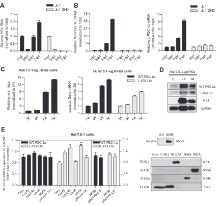
Upregulation of WT-PGC-1␣and L-PGC-1␣depends on
HCV RNA replication.To investigate the mechanism by which
HCV upregulates WT-PGC-1␣/L-PGC-1␣from the HCV point of view, Huh7.5.1 cells were transfected with the full-length HCV-Jc1 RNA for up to 7 days. Full-length HCV-HCV-Jc1 RNA with a lethal mutation in the RNA-dependent RNA polymerase served as a negative control (HCV-Jc1-GND) (29). As shown in Fig. 3A, HCV-Jc1 RNA increased sharply from 3 dpt and then peaked at 7 dpt. In contrast, HCV-Jc1-GND RNA declined continuously from 1 dpt to 7 dpt (Fig. 3A). The mRNA levels of both WT-PGC-1␣and L-PGC-1␣increased in a time-dependent manner in HCV-Jc1 RNA-transfected cells from 3 dpt to 7 dpt but were not
C
B
shPGC-1α 1
#
shPGC-1α 2
#
shPGC-1α 1
#
shPGC-1α 2
#
0.0 0.5 1.0 1.5
HCVpp VSVGpp
P
s
eudopar
tic
le
ent
ry
(re
la
tiv
e
v
a
lu
e
)
ns ns ns ns
D
A
1# α
shPGC-1 α 2#
shPGC-1 shRNA-Ctr
l 1# α
shPGC-1 α 2#
shPGC-1 shRNA-Ctr
l 0
4.0×10⁸
0 20 40 60
ns ns
*
p=0.08
E
x
tr
a
. H
C
V
R
N
A
(
G
E
/m
l)
Sp
e
c
ific
in
fe
c
tiv
ity
( ×
10
-7
FF
U
/G
E
)
Extra.HCV RNA Specific infectivity
Huh7 .5.1-s gJFH2a
shPGC-1α 1
#
shPGC-1α
2#
shRNA-Ctrl
0.00 0.25 0.50 0.75 1.00 1.25
** *
Re
la
tiv
e
HCV
R
NA
(n
or
m
a
liz
e
d
t
o
c
o
ntor
l)
NS3
β-Actin
shPGC-1α
1#
shPGC-1α
2#
shRNA-Ctrl
shPGC-1α 1#
shPGC-1α
2#
shRNA-Ctr l
0 500 1000 1500
0.0 0.3 0.6 0.9 1.2 1.5 1.8 Intra.HCV titer
Extra. titer/ Intra. titer Ratio
* *
p=0.06
ns ns
In
tr
a
. H
C
V
ti
te
r
(F
F
U
/m
l)
E
x
tr
a
. ti
ter
/ I
n
tr
a
. ti
te
r R
a
tio
shRNA-Ctrl shRNA-Ctrl
3.0×10⁸
2.0×10⁸
1.0×10⁸
FIG 2WT-PGC-1␣/L-PGC-1␣is involved in HCV RNA replication and HCV assembly. (A) Effect of WT-PGC-1␣/L-PGC-1␣knockdown on pseudoparticle entry (means⫾SEMs;n⫽3). (B) Effect of WT-PGC-1␣/L-PGC-1␣knockdown on intracellular HCV RNA levels and NS3 protein levels in Huh7.5.1-sgJFH2a cells (means⫾SEMs;n⫽3). (C) Effect of WT-PGC-1␣/L-PGC-1␣knockdown on extracellular HCV RNA copies and specific infectivity. (D) Effect of WT-PGC-1␣/L-PGC-1␣silencing on intracellular HCV titer and the ratio of extracellular titer to intracellular titer. For panels C and D, Huh7.5.1 cells were infected with HCV-Jc1 at an MOI of 0.02. Results are means⫾SEMs of three repetition measurements. For all panels, statistical analyses were performed by using Student’sttest. ns, not significant. * and **, same as forFig. 1.
on November 7, 2019 by guest
http://jvi.asm.org/
[image:5.585.125.463.67.320.2]elevated in HCV-Jc1-GND RNA-transfected cells (Fig. 3B). These results suggest that the expression of WT-PGC-1␣and L-PGC-1␣ is upregulated in a HCV RNA replication-dependent manner. To further define the association between the expression of both PGC-1␣isoforms and HCV RNA replication, we obtained several clones of Huh7.5.1-sgJFH2a cells that support HCV subgenomic RNA replication (clones 3#, 4#, 6#, and 7#). Different adaptive mutations in HCV replicon RNA may contribute to the different HCV RNA levels and NS3 protein levels among the above-listed clones (Fig. 3CandD). As shown inFig. 3C, the mRNA levels of both PGC-1␣isoforms were associated with the HCV RNA levels in these clones; as shown inFig. 3D, the association with the pro-tein level was further confirmed in the 6# and 7# clone cells. These results indicate that the upregulation of WT-PGC-1␣/L-PGC-1␣ depends on HCV RNA replication.
Considering that HCV RNA replication is accompanied by the abundant expression of HCV proteins, we determined whether any of the HCV proteins is responsible for the upregulation of WT-PGC-1␣/L-PGC-1␣. As indicated in Fig. 3E, none of the
HCV proteins enhanced the expression of WT-PGC-1␣ or
L-PGC-1␣except that NS5A had an effect on L-PGC-1␣ eleva-tion. These results indicate that a single HCV protein is not suffi-cient to upregulate WT-PGC-1␣/L-PGC-1␣except NS5A.
HCV-induced upregulation of WT-PGC-1␣is mediated by
CREB phosphorylation.We subsequently explored the
mecha-nism by which HCV upregulates WT-PGC-1␣/L-PGC-1␣from the PGC-1␣ point of view. We investigated WT-PGC-1␣and L-PGC-1␣separately because of their difference in transcriptional regulation.
The regulation of WT-PGC-1␣expression is linked to the
sta-A
C
1dpt3dpt5dpt7dpt 1dpt3dpt5dpt7dpt 0.0
0.5 1.0 1.5 2.0 2.5
(n
o
rm
a
liz
ed to 1dpt
)
1dpt3dpt5dpt7dpt 1dpt3dpt5dpt7dpt 0
9 18 27 36 45 Jc1
Jc1-GND
(n
o
rm
a
liz
ed to 1dpt
)
1dpt3dpt5dpt7dpt 1dpt3dpt5dpt7dpt 0
2 4 6 8 10
Jc1 Jc1-GND
(n
o
rm
a
liz
ed to 1dpt
)
Huh7.5.1-sgJFH2a cells
3# 4# 6# 7#
0 3 6 9 12 15
(n
o
rm
a
liz
ed to 3#)
Huh7.5.1-sgJFH2a cells
3# 4# 6# 7# 3# 4# 6# 7#
0 1 2 3 4
5 WT-PGC-1α
L-PGC-1α
(n
o
rm
a
liz
ed to 3#)
NS3 GAPDH WT-PGC1α
L-PGC1α Huh7.5.1-sgJFH2a
(-) 7# 6#
HCV
RNA
Relative
L-P
G
C
-1α
m
R
N
A
Relative
W
T
-P
G
C-1
α
m
RNA
Relative
G
ene m
R
N
A
Relative
HCV
RNA
Relative
B
D
NS5A NS4B NS3
Core Core C-NS2 NS3/4A NS4B NS5A
56 kDa 27 kDa 70 kDa
21 kDa Huh7.5.1 cells
Core C-NS
2
NS3/4ANS4BNS5A pRle
nti-Ctrl CoreC-NS 2
NS3/4ANS4BNS5A pRle
nti-Ctrl 0.0
0.4 0.8 1.2 1.6
**
Re
la
ti
v
e
m
RNA
e
x
p
re
ss
io
n
t
o
G
A
P
D
H
(N
o
rm
a
li
z
e
d
to
c
o
n
tr
o
l)
E
NS5B 66 kDa
Ctrl NS5B
NS5B
pRceive r-Ctr
l
NS5B
pRceive r-Ctr
l 0.0 0.4 0.8 1.2 1.6 WT-PGC-1α L-PGC-1α WT-PGC-1α
L-PGC-1α
FIG 3Upregulation of both WT-PGC-1␣and L-PGC-1␣depends on HCV RNA replication. HCV RNA levels (A) and WT-PGC-1␣and L-PGC-1␣mRNA
levels (B) were determined after transfecting Huh7.5.1 cells with full-length HCV-Jc1 RNA for different times (means⫾SEMs of triplicate measurements). (C) The association of the mRNA levels of both PGC-1␣isoforms with HCV RNA levels in Huh7.5.1-sgJFH2a cells. 3#, 4#, 6#, and 7# represent the respective subgenomic replicon clones (means⫾SEMs;n⫽3). (D) Association of the protein levels of both PGC-1␣isoforms with HCV NS3 protein levels in Huh7.5.1-sgJFH2a cells. (E) Effect of HCV proteins on WT-PGC-1␣/L-PGC-1␣mRNA expression. Left, effect of HCV protein overexpression on WT-PGC-1␣ and L-PGC-1␣mRNA levels (means⫾SEMs;n⫽2); right, Western blot analysis to detect the expression of HCV protein at 3 days post-lentivirus transduction in Huh7.5.1 cells. The expressed HCV proteins were derived from HCV-Jc1 (25) (genotype 2a). (C-NS2 represents HCV proteins from core to NS2, namely, Core-E1-E2-p7-NS2; NS5B was expressed in the pReceiver plasmid, which was different from other HCV proteins.)
on November 7, 2019 by guest
http://jvi.asm.org/
[image:6.585.75.509.62.466.2]tus of energy stress. Transcription factors, including myocyte en-hancer factor 2 (MEF2), FoxO1, activating transcription factor 2 (ATF2), and CREB, control WT-PGC-1␣expression in response to physiologic stimuli (8). To determine the effect of these tran-scription factors on WT-PGC-1␣elevation by HCV infection, three mutations (MEF2-site-Mut, IRS-Mut, and CRE-Mut) were introduced to the binding sites of these transcription factors within the WT-PGC-1␣promoter (Fig. 4A;Table 2). CRE-Mut was not activated by HCV infection, although a slight but nonsig-nificant increase was detected (Fig. 4B). In contrast, MEF2-site-Mut and IRS-MEF2-site-Mut were significantly activated by HCV infection, similar to the case with WT-PGC-1␣-Prom (Fig. 4B). This result suggests that CRE is important for the HCV infection-induced upregulation of WT-PGC-1␣.
CRE is the binding site for phosphorylated CREB (phospho-CREB) and phosphorylated ATF2 (phospho-ATF2). Given that
the mRNA levels of CREB and ATF2 were not altered following HCV infection (Fig. 4C), we investigated whether HCV
infec-tion enhances WT-PGC-1␣ expression through CREB and
ATF2 phosphorylation. H89 is a phosphokinase A (PKA)-spe-cific inhibitor that inhibits CREB phosphorylation by PKA (Fig. 4D), and SB203580 inhibits ATF2 phosphorylation by p38 mitogen-activated protein kinase (MAPK) (Fig. 4E). WT-PGC-1␣upregulation was significantly inhibited when HCV-infected Huh7.5.1 cells were treated with H89 (Fig. 4D). How-ever, no inhibition was observed after SB203580 treatment (Fig. 4E). These results suggest that phospho-CREB, not
phos-pho-ATF2, is required for WT-PGC-1␣ upregulation.
CREB133, which contains a serine-to-alanine mutation corre-sponding to amino acid 133 of the CREB protein, was con-structed as a dominant negative mutant. As shown inFig. 4F, CREB133 blocked the upregulation of WT-PGC-1␣. Western
A
C
D
CRE CREB
P ATF2 P FoxO1 MEF2
site
MEF2 WT-PGC-1α
*
** *
*
WT-PGC-1α Prom MEF2-site Mut IRS Mut CRE Mut -1317 -1308 -958-567 -324 -102 -95B
P-CREB
NS3
GAPDH
Huh7.5.1-sgJFH2a
(-) 7# 6#
GAPDH core P-CREB
Infection
Mock HCV
Cont rol
H89 08u M
H89 16u M
0.00 0.26 0.52 0.78 1.04
*** ***
(n
or
m
a
liz
e
d
t
o
c
o
nt
ro
l)
E
GAPDH P -CREB 0 8 16 (uM)
H89
WT-PGC-1α Prom
CRE M ut
M EF2-site M ut
IRS M ut
0.0 0.5 1.0 1.5 2.0
HCV M o ck
p= 0.004
p= 0.058
p= 0.005 p= 0.004
** ** **
ns
R
L
U
to S
V
4
0
R
L
u
c
(n
o
rm
a
lize
d
t
o
m
o
ck
in
fe
ct
io
n
)
W
T
-P
G
C
-1
α
m
R
N
A
Relative
Cont rol
SB20358 0 10
uM
SB20 3580 40
uM
0.00 0.25 0.50 0.75 1.00 1.25
Re
la
tiv
e
W
T
-P
G
C
-1
α
m
RN
A
(N
o
rm
a
liz
e
d
t
o
c
o
n
tro
l) 0 10 40 (uM)
SB203580
P -ATF2
β -Actin
F
G
ns ns
1dpi3dpi5dpi7dpi 1dpi3dpi5dpi7dpi
0.0 0.5 1.0 1.5 2.0
ATF2 CREB
R
e
la
tiv
e
G
e
n
e
m
R
N
A
(n
o
rm
a
liz
ed to 1dpi
)
ns ns
Contro l
CR EB
CR EB133
0 1 2 3
*** ***
R
e
la
tiv
e
W
T
-P
G
C
-1
α
m
R
N
A
(N
o
rm
a
liz
e
d
t
o
c
o
n
tr
o
l)
P-CREB
GAPDH
FIG 4HCV-induced WT-PGC-1␣upregulation is mediated by CREB phosphorylation. (A) Schematic of the three WT-PGC-1␣promoter site-specific mutants. IRS, insulin response sequence; CRE, cAMP response element. (B) Different WT-PGC-1␣promoter mutant activities by HCV infection using luciferase reporter assay (means⫾SEMs;n⫽3). (C) mRNA levels of CREB and ATF2 after HCV infection (means⫾SEMs;n⫽3). (D) Effect of H89 on WT-PGC-1␣mRNA elevation after HCV infection. H89 is a PKA-specific inhibitor that inhibits CREB phosphorylation; the effect of H89 on CREB phosphorylation was shown by Western blotting (means⫾SEMs;n⫽3). (E) Effect of SB203580 on WT-PGC-1␣/L-PGC-1␣mRNA levels in HCV-infected Huh7.5.1 cells. SB203580 is a p38 MAPK inhibitor that inhibits the phosphorylation of ATF2; the effect of SB203580 on protein level of phospho-ATF2 (P-ATF2) was shown by Western blotting. Results are means⫾SEMs of three repeat measurements. (F) Effect of CREB133 (a dominant negative mutant) on WT-PGC-1␣mRNA expression (means⫾ SEMs;n⫽2). (G) Western blot analysis for detecting the expression of phospho-CREB (P-CREB) in HCV-infected Huh7.5.1 cells (at 7 dpi) and in Huh7.5.1-sgJFH2a cells. For panels B to G, Huh7.5.1 cells were infected with HCV-Jc1 at an MOI of 0.02. For panels B to F,Pvalues are as follows: *,P⬍0.05; **,P⬍0.01; and ***,P⬍0.001 (all determined by Student’sttest). ns, not significant.
on November 7, 2019 by guest
http://jvi.asm.org/
blot analysis showed that phospho-CREB was dramatically in-creased in HCV-infected Huh7.5.1 (Fig. 4G, left) and Huh7.5.1-sgJFH2a (Fig. 4G, right) cells.
These results strongly suggest that the HCV-induced upregu-lation of WT-PGC-1␣is mediated by CREB phosphorylation.
HCV-induced upregulation of L-PGC-1␣ is mediated by
CREB phosphorylation and FoxO1 dephosphorylation.To
de-termine whether CREB phosphorylation is also important for the HCV infection-induced upregulation of L-PGC-1␣, the
inhibi-tion activity of H89 was analyzed. As shown inFig. 5A, H89 sig-nificantly decreased the upregulation of L-PGC-1␣. CREB133 also blocked the upregulation of L-PGC-1␣(Fig. 5B). These re-sults suggest that the HCV-induced upregulation of L-PGC-1␣is also mediated by CREB phosphorylation.
Aside from CREB phosphorylation, the expression of L-PGC-1␣ is also regulated by FoxO1 and glucocorticoid signaling (11). To verify whether the HCV-induced upregulation of L-PGC-1␣is also mediated by FoxO1 or glucocorticoid signaling, FoxO1-site-TABLE 2Mutant strategies of PGC-1␣promotera
Gene TFBS Position Orientation Length (bp) Original sequence Mutant sequence
WT-PGC-1␣ MEF2-site ⫺1317 to 1308 Direct 10 TTATATTTAG TTCCGAGGAG
IRS1 ⫺961 to⫺955 Direct 7 TATTTTT TCAGGGT
IRS2 ⫺571 to⫺564 Direct 8 TATTTTGT TCAGGGT
IRS3 ⫺328 to⫺320 Direct 9 TTGTTTTGG TTCAGGGTGA
CRE ⫺102 to⫺95 Direct 8 TGACGTCA TAGATCTA
L-PGC-1␣ FoxO1-site ⫺68 to⫺58 Reverse 11 ACATTGTTTGC ACATTCTTTGC
GRE ⫺60 to⫺55 Direct 6 TGTTCT TGACCT
a
Abbreviations: TFBS, transcription factor binding site; CRE, cyclic AMP response element; GRE, glucocorticoid response element; IRS, insulin response sequence.
A
B
C
D
F
FoxO1 site
L-PGC-1α
*
*
L-PGC-1α Prom GRE Mut FoxO1-site Mut GRE-68 -58 -60 -55
E
0.0 0.5 1.0 1.5 2.0
2.5 ***
Cont rol
H89 08u M
H89 16u M
0.0 0.3 0.6 0.9 1.2
* *
(n
or
m
a
liz
e
d
t
o
c
o
nt
ro
l)
L-P
G
C
-1α
m
R
N
A
(n
or
m
a
liz
e
d
t
o
c
o
nt
ro
l)
0.0 0.5 1.0 1.5
2.0 HCV
Mock
L-PGC-1α Prom
GRE Mut
FoxO1-site Mut * *
ns
FoxO1
core
GAPDH Infection
HCV Mock
Total Infection
HCV Mock
FoxO1
β-Actin
FoxO1
Histone H3 Cyto
nuc
G
R
L
U
to S
V
4
0
R
L
u
c
(n
o
rm
a
liz
e
d
t
o
m
o
c
k
in
fe
c
tio
n
)
Relative
L-P
G
C
-1α
m
R
N
A
Relative
H
1dpi 3dpi 5dpi
0.0 0.5 1.0 1.5
(n
o
rm
a
liz
ed to 1dpi
)
ns ns
Fo
x
O
1
m
R
N
A
Relative
Cont rol
CRE B
CREB133
0.0 0.5 1.0 1.5 2.0
** *
Re
la
tiv
e
L
-P
G
C
-1
α
m
RN
A
(N
o
rm
a
liz
e
d
t
o
c
o
n
tr
o
l)
Infection
HCV Mock
P-FoxO1
core
GAPDH Total
FIG 5L-PGC-1␣upregulation by HCV is mediated by CREB phosphorylation and FoxO1 dephosphorylation. (A) Effect of H89 on L-PGC-1␣mRNA
upregulation after HCV infection. The effect of H89 on CREB phosphorylation was shown by Western blotting (means⫾SEMs;n⫽3). (B) Effect of CREB133 on L-PGC-1␣mRNA expression (means⫾SEMs;n⫽2). (C) Schematic of the two L-PGC-1␣promoter site-specific mutants. GRE, glucocorticoid response element. (D) Luciferase reporter assay for detecting different L-PGC-1␣promoter mutant activities by HCV infection (means⫾SEMs;n⫽3). For panels A, B, and D, results were analyzed similarly to those for WT-PGC-1␣as mentioned in the legend toFig. 4. (E) mRNA levels of FoxO1 after HCV infection (mean⫾ SEM;n⫽3) (F) Effect of wortmannin (a PI3K inhibitor that blocks FoxO1 phosphorylation) on L-PGC-1␣mRNA upregulation after HCV infection (means⫾ SEMs;n⫽2). (G) Western blot analysis for detecting the expression of phospho-FoxO1 (Thr 24) (P-FoxO1) at 7 days in HCV-infected Huh7.5.1 cells. (H) Western blot analysis to detect the expression of total, nuclear (nuc), and cytoplasmic (Cyto) FoxO1 at 7 days in HCV-infected Huh7.5.1 cells. For panels A, B, and D to H, Huh7.5.1 cells were infected with HCV-Jc1 at an MOI of 0.02. For pnels A, B, D, E, and F,Pvalues are as follows: *,P⬍0.05, and ***,P⬍0.001 (determined by Student’sttest). ns, not significant.
on November 7, 2019 by guest
http://jvi.asm.org/
[image:8.585.40.546.77.174.2] [image:8.585.103.481.321.628.2]Mut and glucocorticoid response element (GRE)-Mut in the L-PGC-1␣promoter were constructed (Fig. 5C;Table 2). HCV infection significantly increased L-PGC-1␣-Prom and GRE-Mut promoter-luciferase expression, whereas HCV infection had no effect on FoxO1-site-Mut promoter-luciferase expression (Fig. 5D). This result suggests that L-PGC-1␣promoter activation is dependent on the FoxO1 site.
When phosphatidylinositol 3-kinase (PI3K), the kinase sponsible for FoxO1 phosphorylation, is suppressed, FoxO1 is re-tained in the nucleus to activate downstream gene transcription. Given that the mRNA level of FoxO1 was not affected following HCV infection (Fig. 5E), we analyzed the effect of FoxO1 phos-phorylation on L-PGC-1␣upregulation. Wortmannin is a PI3K inhibitor. Treatment of HCV-infected cells with wortmannin increased the mRNA level of L-PGC-1␣(Fig. 5F). This result suggests that HCV infection decreases the level of phosphory-lated FoxO1 (phospho-FoxO1) and then upregulates L-PGC-1␣. Furthermore, Western blot analysis showed that HCV in-fection indeed suppressed the level of phospho-FoxO1 at Thr24 (Fig. 5G), which is critical for FoxO1 nuclear exclusion (30). Accordingly, Western blotting of nuclear and cytoplasmic extracts showed that HCV infection upregulated nuclear FoxO1 expression and downregulated cytoplasmic FoxO1 expres-sion compared with mock infection (Fig. 5H, left) but did not affect the total level of FoxO1 (Fig. 5H, right). Taken together, these results suggest that HCV infection results in decreased FoxO1 phosphoryla-tion, FoxO1 nuclear accumulaphosphoryla-tion, and, eventually, increased L-PGC-1␣expression.
Collectively, these results indicate that the HCV infection-in-duced upregulation of L-PGC-1␣ is mediated by both CREB phosphorylation and FoxO1 dephosphorylation.
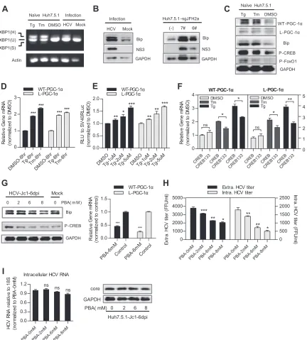
HCV upregulates WT-PGC-1␣/L-PGC-1␣ through ER
stress.Previous studies have shown that ER stress can activate
CREB (18) and that HCV RNA replication can result in ER stress (31). In light of our results, we hypothesized that ER stress medi-ates WT-PGC-1␣/L-PGC-1␣upregulation by HCV infection in a CREB-dependent manner.
We first tested whether HCV infection induces an ER stress response. ER stress can activate UPR via three different sensors: PERK, ATF6, and IRE1 (32). The UPR results in the upregulation of ER-resident molecular chaperones such as BiP/GRP78 and thereby augments the folding capacity of the ER (32). We accord-ingly examined the effect of HCV on IRE1-XBP1 activation and BiP upregulation to indicate ER stress. Once IRE1 is activated, it induces the splicing of the XBP1 mRNA to stimulate the expres-sion of UPR target genes. Splicing of XBP1 mRNA was assayed by RT-PCR: the unspliced mRNA, XBP1(U), generates a 424-bp product, whereas the spliced mRNA, XBP1(S), generates a 398-bp product; XBP1(H) represents the hybrid DNA derived from XBP1(U) and XBP1(S). The appearance of XBP1(H) and/or XBP1(S) indicates XBP1 splicing (15). XBP1(H) was easily de-tected in HCV-infected Huh7.5.1 cells but not in mock-infected Huh7.5.1 cells (Fig. 6A). Both HCV infection in Huh7.5.1 cells and HCV RNA replication in Huh7.5.1-sgJFH2a cells upregulated the protein level of BiP (Fig. 6B). The splicing of XBP1 mRNA and the elevated levels of Bip protein demonstrate the activation of ER stress in these cells. Subsequently, we investigated whether phar-macological induction of ER stress can induce CREB phosphory-lation and WT-PGC-1␣/L-PGC-1␣upregulation. Tg and Tm are ER stress inducers. Treatment of naive Huh7.5.1 cells with Tg and
Tm induced a classical ER stress, as evidenced by XBP1 mRNA splicing and BiP upregulation (Fig. 6AandC). Phospho-CREB protein was simultaneously elevated by Tg and Tm treatment, along with the upregulation of the protein and mRNA levels of WT-PGC-1␣and L-PGC-1␣(Fig. 6CandD). However, Tg and Tm treatment resulted in FoxO1 phosphorylation (Fig. 6C), which is in contrast with the finding that HCV infection resulted in FoxO1 dephosphorylation (Fig. 5G); therefore, HCV-induced L-PGC-1␣upregulation by FoxO1 dephosphorylation is not de-pendent on ER stress. Increasing concentrations of Tg led to a dose-dependent increase in luciferase expression under the con-trol of both PGC-1␣promoters (Fig. 6E). In addition, CREB133 blocked the pharmacological ER stress-induced mRNA upregula-tion of WT-PGC-1␣and L-PGC-1␣(Fig. 6F). Finally, we vali-dated whether inhibition of ER stress can alleviate the HCV infec-tion-induced upregulation of WT-PGC-1␣/L-PGC-1␣. PBA is a chemical chaperone that attenuates ER stress (33). Treatment of HCV-infected Huh7.5.1 cells with PBA markedly reduced the protein levels of Bip and phospho-CREB (Fig. 6G, left) and further impaired the mRNA levels of both WT-PGC-1␣and L-PGC-1␣ (Fig. 6G, right). PBA treatment also inhibited the extracellular titer and intracellular titer of HCV-Jc1 (Fig. 6H). However, both the HCV core protein expression and the intracellular HCV RNA level were not changed after PBA treatment (Fig. 6I), which was consistent with the studies of Huang et al. (34). This result ex-cludes the possibility that the observed inhibition of WT-PGC-1␣/L-PGC-1␣expression by PBA was due to the suppression of HCV RNA replication by PBA.
Huh7.5.1 cells are defective in innate immune response that involves retinoic acid-inducible gene I (RIG-I) (35). Robust HCV replication supported by Huh7.5.1 cells is more likely to cause severe ER stress. Therefore, we determined whether HCV replica-tion in the parental Huh7 cells could also induce significant ER stress to upregulate WT-PGC-1␣/L-PGC-1␣ expression. As shown inFig. 7A, Huh7-sgHCV1b cells, which harbor genotype 1b subgenomic replicons, had higher levels of Bip protein than Huh7 cells, which indicated an ER stress by HCV replication in
Huh7 cells. Moreover, phospho-CREB and WT-PGC-1␣
/L-PGC-1␣ proteins were simultaneously elevated in Huh7-sgHCV1b cells (Fig. 7AandB). The inhibition of ER stress by PBA in Huh7-sgHCV1b cells reduced the protein levels of Bip and phospho-CREB (Fig. 7C) and further impaired the mRNA levels of both PGC-1␣isoforms (Fig. 7D). Treatment of Huh7 cells with Tg and Tm increased the mRNA levels of both WT-PGC-1␣and L-PGC-1␣(Fig. 7E). Taken together, these results suggest that ER stress mediates the HCV replication-induced upregulation of WT-PGC-1␣/L-PGC-1␣in Huh7 cells.
To confirm that WT-PGC-1␣/L-PGC-1␣was upregulated by pharmacological ER stress not restricted to Huh7 and Huh7-de-rived Huh7.5.1 cells, we used another hepatoma carcinoma cell, Hep3B cell, for Tg and Tm treatment. Results showed that the mRNA and protein levels of WT-PGC-1␣and L-PGC-1␣were significantly elevated when treated with Tg and Tm in Hep3B cells (Fig. 7F). To further validate the connection between PGC-1␣ expression and ER stressin vivo, the mRNA levels of hepatic mu-rine Bip (mBip) and mPGC-1␣were analyzed in Leprdb/dbmice, a genetic model of T2DM. Leprdb/dbmice were hyperphagic and obese (36), and Ozcan et al. (37) showed that obesity contributes to the development of type 2 diabetes because obesity causes ER stress. In the present study, results showed that the mRNA levels of
on November 7, 2019 by guest
http://jvi.asm.org/
mPGC-1␣strongly correlated with the mRNA levels of ER stress marker mBip in Leprdb/dbmice (Fig. 7G).
Collectively, these results strongly suggest that ER stress medi-ates the HCV-induced upregulation of WT-PGC-1␣/L-PGC-1␣.
DISCUSSION
In the current study, we demonstrated a previously unrecog-nized function of ER stress in mediating the HCV-induced upregulation of WT-PGC-1␣/L-PGC-1␣. This conclusion is
A
B
C
Infection
HCV Mock Naïve Huh7.5.1
Tg Tm DMSO
WT-PGC-1α L-PGC-1α PBA-6m M Cont rol PBA-6m M Cont rol 0.0 0.5 1.0 1.5 *** *** (n o rm a liz
ed to control)
D
core
GAPDH
Huh7.5.1-Jc1-6dpi 0 2 6 8 PBA( mM) WT-PGC-1α L-PGC-1α
G
H
Ge n e m R N A Relative XBP1(H) XBP1(U) XBP1(S) Actin DMSO-8 hr Tg-8hrTm-8hr DMSO-8 hr Tg-8hrTm-8hr 0 1 2 3 WT-PGC-1α L-PGC-1α *** *** ****** R e la tiv e G e n e m R N A (n o rm a lized to D
M
S
O
)
DMSOTg-1uMTg-2uMTg-5uMDMSOTg-1uMTg-2uMTg-5uM 0.0 0.5 1.0 1.5 2.0 ** * *** ** ** *** RL
U t
o S V 4 0 R L u c (n o rm a liz
ed to D
M S O ) CR EB CRE B13 3 CRE B CR EB13 3 CRE B CRE B13 3 0 1 2 3 4 WT-PGC-1α * * Re la tiv e G e n e m R NA (n o rm a liz e d t o D M S O ) L-PGC-1α DMSO Tm Tg
E
F
Bip GAPDH WT -PGC -1αL-PGC -1α
P -CREB
P -FoxO1 Naïve Huh7.5.1
Tg Tm DMSO
Infection
HCV Mock
NS3
GAPDH Bip
Huh7.5.1 -sgJFH2a
(-) 7# 6#
NS3
GAPDH Bip
Intracellular HCV RNA
PBA-0m M
PBA-2m M
PBA-6m M
PBA-8m M 0.0 0.3 0.6 0.9 1.2 ns ns ns HCV RNA r e la tiv e t o 1 8 S (n o rma liz e d t o P B A -0 mM)
I
PBA-0m M
PBA-2m M
PBA-6m M
PBA-8m M
PBA-0m M PBA-2m M PBA-6m M PBA-8m M 0 1000 2000 3000 4000 5000 0 500 1000 1500 2000 2500 Intra. HCV titer
Extra. HCV titer
** *** * ** ** * E x tr a . H C V ti te r ( F F U /m
l) Intr
a . H C V ti te r ( F F U /m l) CRE B CRE B13 3 CRE B CRE B13 3 CRE B CR EB13
3 0 1 2 3 4 5 * ** ns ns DMSO Tm Tg P -CREB Bip GAPDH HCV-Jc1-6dpi
0 2 6 8 0 PBA( m M ) Mock
FIG 6HCV upregulates WT-PGC-1␣/L-PGC-1␣through ER stress in Huh7.5.1 cells. (A) The activation of ER stress is shown by the activation of the IRE1-XBP1 pathway. Splicing of XBP1 was shown by reverse transcription-PCR (RT-PCR). XBP1(U) and XBP1(S) represent DNA fragments were derived from unspliced and spliced XBP1 RNAs, respectively. XBP1(H) represents the hybrid DNA derived from XBP1(U) and XBP1(S). The actin RNA was also analyzed to serve as an internal control. Thapsigargin (Tg) and tunicamycin (Tm) are pharmacological inducers of ER stress. (B) Effect of HCV infection and HCV RNA replication on Bip protein levels. (C) Effect of Tg and Tm on WT-PGC-1␣and L-PGC-1␣protein levels, Bip protein levels, and CREB and phospho-FoxO1 protein levels. (D) Effect of Tg and Tm on WT-PGC-1␣/L-PGC-1␣mRNA levels. (E) Effect of Tg on WT-PGC-1␣/L-PGC-1␣promoter-luciferase activities in Huh7.5.1 cells. (F) Effect of CREB133 on the pharmacological ER stress-induced WT-PGC-1␣and L-PGC-1␣mRNA upregulation. (G) Effect of PBA on the protein levels of Bip and phospho-CREB (P-CREB) and on HCV-induced WT-PGC-1␣/L-PGC-1␣mRNA upregulation. (H) Effect of PBA on the extracellular titer and intracellular titer of HCV-Jc1. (I) Effect of PBA on the intracellular HCV RNA level and on the HCV core protein level in HCV-Jc1 infected Huh7.5.1 cells. For panels A, B, G, and H, Huh7.5.1 cells were infected with HCV-Jc1 at an MOI of 0.02. For panels D to I, data are presented as means⫾SEMs (n⫽3). *,P⬍0.05; **,P⬍0.01; ***,P⬍0.001 (all determined by Student’sttest). ns, not significant.
on November 7, 2019 by guest
http://jvi.asm.org/
[image:10.585.78.510.64.543.2]supported by the following findings: (i) the expression levels of WT-PGC-1␣/L-PGC-1␣and ER stress markers increased in HCV-infected and HCV RNA-replicating cells; (ii) the upregu-lation of WT-PGC-1␣/L-PGC-1␣depended on CREB
phos-phorylation, and the pharmacological induction of ER stress induced CREB phosphorylation and WT-PGC-1␣/L-PGC-1␣ upregulation; (iii) the pharmacological inhibition of ER stress reduced the HCV-induced WT-PGC-1␣/L-PGC-1␣
upregula-A
G
P-CREB Bip
GAPDH Huh7-sgHCV1b
0 3 6 9 PBA( mM)
C
D
Huh7 cells
DMSO-8 hr
Tg-8hrTm-8hr DMSO-8
hr Tg-8hrTm-8hr
0.0 0.2 0.4 0.6
WT-PGC-1α L-PGC1-1α
G
e
ne ex
pr
es
s
ion
(r
e
la
tiv
e
m
R
N
A
le
v
e
l) *** ***
** *
Hep3B cells
DMSO-8 hr
Tg-8hrTm-8hr DMSO-8
hr Tg-8hrTm-8hr
0.0 0.3 0.6 0.9 1.2
WT-PGC-1α L-PGC-1α
*** **
*** ***
G
e
ne ex
pr
es
s
ion
(r
e
la
tiv
e
m
R
N
A
le
v
e
l)
E
Bip
GAPDH WT-PGC-1α
L-PGC-1α
P-CREB Hep3B
Tg Tm DMSO
F
H
0.8 1.0 1.2 1.4 1.6 0.0
0.2 0.4 0.6 0.8 1.0
r2=0.6077 p=0.0078
m PGC-1α m RNA expression
m
B
ip
m
RNA
e
x
p
re
s
s
io
n
PBA-6m
M
Cont rol
PBA-6m
M
Cont rol
0.00 0.25 0.50 0.75 1.00 1.25
* *
(n
o
rm
a
liz
e
d
t
o
c
o
n
tr
o
l)
WT-PGC-1α L-PGC-1α
G
e
n
e
m
RNA
Relative
L-PGC-1α WT-PGC-1α
CREB FoxO1
PEPCK Pro-viral genes Entry
RNA replicaon
(+) RNA (-) RNA
Assembly and release
HCV-induced IR
ER stress
HCV Producon (+) (-) sgHCV1b
Bip
P-CREB
GAPDH Huh7
GAPDH NS3 WT-PGC-1α
L-PGC-1α Huh7
(+) (-) sgHCV1b
B
FIG 7HCV RNA replication upregulates WT-PGC-1␣/L-PGC-1␣through ER stress in Huh7 cells, and hepatic mPGC-1␣correlates with ER stress marker mBip in mice. (A) The activation of ER stress and CREB is indicated by the high expression levels of Bip and phospho-CREB (P-CREB) in Huh7-sgJFH1b cells, respectively. (B) Western blot analysis for detecting the expression of WT-PGC-1␣and L-PGC-1␣in Huh7-sgJFH1b cells and in Huh7 cells. (C) Effect of PBA on the protein levels of Bip and phospho-CREB (P-CREB) in Huh7-sgJFH1b cells. (D) Effect of PBA on the HCV-induced WT-PGC-1␣/L-PGC-1␣mRNA upregulation in Huh7-sgJFH1b cells. (E) Effect of Tg and Tm on WT-PGC-1␣/L-PGC-1␣mRNA levels in Huh7 cells. (F) Effects of Tg and Tm on WT-PGC-1␣/L-PGC-1␣elevation in Hep3B cells. Left, effects of Tg and Tm on WT-PGC-1␣/L-PGC-1␣mRNA levels in Hep3B cells; right, Western blot analysis for detecting the protein levels of Bip, phospho-CREB (P-CREB), and both WT-PGC-1␣and L-PGC-1␣in Hep3B cells with Tg and Tm treatment. For panels D to F, results are presented as means⫾SEMs of triplicate measurements. (G) Correlation of hepatic mPGC-1␣and mBip transcript levels in Leprdb/dbmice
(individual dot represents one mouse; results are presented as means⫾SEMs of triplicate measurements;n⫽10). (H) A proposed model of the dual effects of WT-PGC-1␣/L-PGC-1␣in HCV-induced insulin resistance and HCV production, and the mechanism by which HCV upregulates WT-PGC-1␣/L-PGC-1␣. Gray and black solid arrows represent the signaling pathways by which HCV upregulates L-PGC-1␣and WT-PGC-1␣, respectively. HCV RNA replication induces ER stress, which further phosphorylates CREB to activate both WT-PGC-1␣and L-PGC-1␣transcription. L-PGC-1␣transcription is also elevated by HCV infection-induced FoxO1 dephosphorylation, which is independent of ER stress. The increased levels of WT-PGC-1␣and L-PGC-1␣promote expression of PEPCK and proviral genes. The increased PEPCK expression could account for HCV-induced IR, and the increased proviral gene expression promotes HCV production. For panels D to F,Pvalues are as follows: *,P⬍0.05; **,P⬍0.01; and ***,P⬍0.001 (all determined by Student’sttest).
on November 7, 2019 by guest
http://jvi.asm.org/
[image:11.585.82.503.67.514.2]tion; and (iv) the mRNA levels of mPGC-1␣strongly correlated with that of mBipin vivo.
Our findings showed that WT-PGC-1␣/L-PGC-1␣was up-regulated by ER stress in Huh7.5.1 cells infected by HCV or in hepatoma carcinoma cells (Huh7, Huh7.5.1, and Hep3B) treated with Tg or Tm and that the mRNA levels of mPGC-1␣strongly correlated with that of mBip in diabetic mice. These studies indi-cate that an ER stress-PGC-1␣signaling pathway exists in livers. A recent study has shown that tribbles 3 (TRB3) mediates ER stress-induced IR in skeletal muscles (38). Another study reported that the muscle-specific overexpression of PGC-1␣in mice, which ex-acerbates IR, is associated with elevated TRB3 expression (39). These two studies suggest that an ER stress-PGC-1␣-TRB3 signal-ing pathway exists in muscles. However, this hypothesis needs further investigation. Nevertheless, combined with our findings, these studies suggest that the ER stress-induced upregulation of WT-PGC-1␣/L-PGC-1␣is a universal mechanism in multiple tis-sues.
In search of the mechanism underlying HCV-associated WT-PGC-1␣elevation, Shlomai et al. (20) speculated that HCV-in-duced oxidative stress promotes WT-PGC-1␣upregulation from the result that treatment of HCV replicon cells with the antioxi-dantN-acetylcysteine resulted in reduction of WT-PGC-1␣levels. ER stress and oxidative stress are intimately interrelated (40). ER stress can promote oxidative stress in cells that support HCV rep-lication (32). Therefore, it is possibly that ER stress induces oxi-dative stress to upregulate WT-PGC-1␣/L-PGC-1␣in HCV-in-fected cells. In addition, the induction of PGC-1␣expression was lower after 8 h of Tg or Tm treatment (Fig. 6C) than after 5 and 7 days of HCV infection (Fig. 1A). This result suggests that the HCV-mediated induction of PGC-1␣expression involves other mechanisms aside from ER stress.
From the PGC-1␣point of view, our results showed that the HCV-induced upregulation of WT-PGC-1␣ was mediated by CREB phosphorylation, whereas that of L-PGC-1␣was mediated by both CREB phosphorylation and FoxO1 dephosphorylation. The activation of PGC-1␣by CREB is a common pattern in the metabolic adaptations of the liver to gluconeogenic status, and HCV infection also exploits this CREB-PGC-1␣signaling to ele-vate both WT-PGC-1␣and L-PGC-1␣, which are further hijacked by HCV for its production.
In the present study, L-PGC-1␣upregulation was also depen-dent on FoxO1 dephosphorylation. This finding is consistent with previous results that FoxO1 has a more important function in L-PGC-1␣transcription than in WT-PGC-1␣transcription and that the mRNA level of L-PGC-1␣strongly correlates with that of FoxO1 in the livers of obese subjects (11). We also observed an effect of NS5A on the elevation of L-PGC-1␣. Deng et al. (41) previously reported that HCV promotes hepatic gluconeogenesis through an NS5A-mediated, FoxO1-dependent pathway, and herein we demonstrated that the HCV-induced upregulation of L-PGC-1␣ depends on FoxO1 dephosphorylation. Taken to-gether, the results show that there may be an NS5A-FoxO1-L-PGC-1␣signaling pathway following HCV infection.
The HCV-induced upregulation of WT-PGC-1␣/L-PGC-1␣ demonstrated dual effects. First, the HCV-induced upregulation of WT-PGC-1␣/L-PGC-1␣ promoted the gluconeogenic re-sponse. Shlomai et al. (20) showed that WT-PGC-1␣induction following HCV infection promotes hepatic gluconeogenesis by increasing glucose-6 phosphatase and glucose production.
Phos-phoenolpyruvate carboxykinase (PEPCK), another rate-limiting enzyme for hepatic gluconeogenesis, is also transcriptionally reg-ulated by PGC-1␣(6,7). In the present study, the overexpression of both WT-PGC-1␣and L-PGC-1␣in Huh7.5.1 cells signifi-cantly increased the mRNA level of PEPCK (data not shown). Although the transcript levels of hepatic L-PGC-1␣ and WT-PGC-1␣are similar in human liver, the mRNA level of hepatic PEPCK is more strongly associated with L-PGC-1␣ than with WT-PGC-1␣(11). The interaction of the PGC-1␣family with hepatocyte nuclear factor 4 alpha (HNF4␣) is crucial in hepatic gluconeogenesis (42), and a direct physical interaction exists be-tween L-PGC-1␣ and HNF4␣ at the PEPCK promoter (11). Therefore, similar to WT-PGC-1␣, L-PGC-1␣also has a vital function in the development of HCV-associated IR and T2DM. Daitoku et al. (21) showed that insulin activates the PI3K signaling pathway, which stimulates FoxO1 phosphorylation, followed by the nuclear exclusion of FoxO1 and the repression of PGC-1␣ transcription. In the present study, the demonstrated FoxO1 de-phosphorylation and L-PGC-1␣elevation by HCV infection may interrupt the insulin signaling pathway, and this interruption may result in IR from another perspective.
Second, WT-PGC-1␣and L-PGC-1␣demonstrated proviral functions in HCV production. HCV particle production relies on very-low-density lipoproteins (VLDLs) (43). PGC-1␣is report-edly important in VLDL assembly (44). We also found that both WT-PGC-1␣and L-PGC-1␣can transcriptionally upregulate sev-eral factors involved in VLDL assembly and release, including apolipoprotein E and cell death-inducing DFFA-like effector B (data not shown). We have previously shown that HNF4␣affects the late stages of HCV life cycle through the VLDL pathway (27). Given that HNF4␣is a critical component of PGC-1␣-mediated hepatic function, we speculate that WT-PGC-1␣and L-PGC-1␣ promote HCV production through their downstream factors as-sociated with the VLDL pathway by interacting with HNF4␣. Studies on this issue are under way in our laboratory.
Emerging lines of evidence indicate that HCV-induced ER stress contributes to HCV persistence and HCV pathogenesis. HCV-induced ER stress and UPR can block innate immunity to promote HCV replication (45, 46). Aside from suppression of innate immune response, our results showed that ER stress in-duced WT-PGC-1␣/L-PGC-1␣ upregulation to enhance HCV RNA replication and HCV assembly, which shall give new insight into the proviral effect of HCV-induced ER stress. Accumulating evidence implicates ER stress-induced cell death as a major con-tributor to many diseases (47); HCV-induced ER stress sensitizes infected cells to apoptosis (48) and accounts for HCV pathogen-esis to some extent. The observed ER stress-PGC-1␣pathway in the present study may account for the pathogenesis of HCV-in-duced IR.
Overall, we propose a model that illustrates the mechanism of WT-PGC-1␣/L-PGC-1␣upregulation by HCV infection and also illustrates the dual effects of WT-PGC-1␣/L-PGC-1␣(Fig. 7H). In this model, HCV infection upregulates both WT-PGC-1␣and L-PGC-1␣ through an ER stress-mediated, phosphorylated CREB-dependent pathway, and both PGC-1␣isoforms promote HCV-induced IR and HCV production in turn through their downstream factors; HCV-induced upregulation of L-PGC-1␣ also depends on FoxO1 dephosphorylation, which is independent of ER stress. A comprehensive understanding of the cross talk among HCV, ER stress, and WT-PGC-1␣/L-PGC-1␣is important
on November 7, 2019 by guest
http://jvi.asm.org/
to identify novel antiviral targets. The dual effects of WT-PGC-1␣/L-PGC-1␣suggest that their inhibitors would elicit therapeu-tic effects for both HCV infection and HCV-associated IR. The therapeutic value and promising safety profile of PGC-1␣ modu-lators have been demonstrated in animal models for different dis-eases (49); thus, the pharmacological regulation of WT-PGC-1␣/ L-PGC-1␣may be a novel approach for HCV therapy. ER stress has also been considered another anti-HCV target because of its intermediary function. PBA, an ER stress inhibitor, can be orally administered and is currently used clinically (50). The demon-strated efficacy of PBA in attenuating WT-PGC-1␣/L-PGC-1␣ upregulation and in blocking HCV production suggests that PBA may serve as another promising anti-HCV therapeutic choice.
ACKNOWLEDGMENTS
We thank Donghai Wu and Tao Nie for providing genetic Leprdb/db mouse samples.
This study was supported by the National Basic Research Program (973) (grant 2010CB530100) and the National Science Foundation (grant 31370204).
REFERENCES
1.Thomas DL.2013. Global control of hepatitis C: where challenge meets opportunity. Nat. Med.19:850 – 858.http://dx.doi.org/10.1038/nm.3184. 2.Mohd Hanafiah K, Groeger J, Flaxman AD, Wiersma ST.2013. Global epidemiology of hepatitis C virus infection: new estimates of age-specific antibody to HCV seroprevalence. Hepatology57:1333–1342.http://dx .doi.org/10.1002/hep.26141.
3.Aghemo A, Prati GM, Rumi MG, Soffredini R, D’Ambrosio R, Orsi E, De Nicola S, Degasperi E, Grancini V, Colombo M.2012. Sustained virological response prevents the development of insulin resistance in pa-tients with chronic hepatitis C. Hepatology56:1681–1687.http://dx.doi .org/10.1002/hep.25867.
4.White DL, Ratziu V, El-Serag HB.2008. Hepatitis C infection and risk of diabetes: a systematic review and meta-analysis. J. Hepatol.49:831– 844. http://dx.doi.org/10.1016/j.jhep.2008.08.006.
5.Finck BN, Kelly DP.2006. PGC-1 coactivators: inducible regulators of energy metabolism in health and disease. J. Clin. Invest.116:615– 622. http://dx.doi.org/10.1172/JCI27794.
6.Yoon JC, Puigserver P, Chen G, Donovan J, Wu Z, Rhee J, Adelmant G, Stafford J, Kahn CR, Granner DK, Newgard CB, Spiegelman BM.2001. Control of hepatic gluconeogenesis through the transcriptional coactivator PGC-1. Nature413:131–138.http://dx.doi.org/10.1038/35093050. 7.Herzig S, Long F, Jhala US, Hedrick S, Quinn R, Bauer A, Rudolph D,
Schutz G, Yoon C, Puigserver P, Spiegelman B, Montminy M.2001. CREB regulates hepatic gluconeogenesis through the coactivator PGC-1. Nature413:179 –183.http://dx.doi.org/10.1038/35093131.
8.Fernandez-Marcos PJ, Auwerx J. 2011. Regula