Phosphoribosyltransferase
Kathryn L. Roche,aMasatoshi Nukui,bBenjamin A. Krishna,c Christine M. O’Connor,cEain A. Murphya,b
aFORGE Life Science, Pennsylvania Biotechnology Center, Doylestown, Pennsylvania, USA
bDepartment of Translational Medicine, Baruch S. Blumberg Institute of Living Science, Institute for Hepatitis
and Virus Research, Doylestown, Pennsylvania, USA
cGenomic Medicine Institute, Lerner Research Institute, Cleveland Clinic, Cleveland, Ohio, USA
ABSTRACT Infections with human cytomegalovirus (HCMV) are highly prevalent in the general population as the virus has evolved the capacity to undergo distinct replication strategies resulting in lytic, persistent, and latent infections. During the latent life cycle, HCMV resides in subsets of cells within the hematopoietic cell com-partment, including hematopoietic progenitor cells (HPCs) and peripheral blood monocytes. Since only a small fraction of these cell types harbor viral genomes dur-ing natural latency, identification and analysis of distinct changes mediated by viral infection are difficult to assess. In order to characterize latent infections of HPCs, we used an approach that involves complementation of deficiencies within the human pyrimidine salvage pathway, thus allowing for conversion of labeled uracil into rUTP. Here, we report the development of a recombinant HCMV that complements the de-fective human pyrimidine salvage pathway, allowing incorporation of thiol contain-ing UTP into all RNA species that are synthesized within an infected cell. This virus grows to wild-type kinetics and can establish a latent infection within two distinct culture models of HCMV latency. Using this recombinant HCMV, we report the spe-cific labeling of transcripts only within infected cells. These transcripts reveal a tran-scriptional landscape during HCMV latency that is distinct from uninfected cells. The utility of this labeling system allows for the identification of distinct changes within host transcripts and will shed light on characterizing how HCMV establishes and maintains latency.
IMPORTANCE HCMV is a significant pathogen that accounts for a substantial amount of complications within the immunosuppressed and immunocompromised. Of partic-ular significance is the capacity of HCMV to reactivate within solid tissue and bone marrow transplant recipients. While it is known that HCMV latency resides within a fraction of HPCs and monocytes, the exact subset of cells that harbor latent viral ge-nomes during natural infections remain uncharacterized. The capacity to identify changes within the host transcriptome during latent infections is critical for develop-ing approaches that therapeutically or physically eliminate latent viral genome con-taining cells and will represent a major breakthrough for reducing complications due to HCMV reactivation posttransplant. In this report, we describe the generation and use of a recombinant HCMV that allows specific and distinct labeling of RNA species that are produced within virally infected cells. This is a critical first step in identifying how HCMV affects the host cell during latency and more importantly, allows one to characterize cells that harbor latent HCMV.
KEYWORDS HCMV, latency, transcriptome, UPRT, cytomegalovirus
Received25 May 2018Accepted3 August 2018
Accepted manuscript posted online8 August 2018
CitationRoche KL, Nukui M, Krishna BA, O'Connor CM, Murphy EA. 2018. Selective 4-thiouracil labeling of RNA transcripts within latently infected cells after infection with human cytomegalovirus expressing functional uracil phosphoribosyltransferase. J Virol 92:e00880-18. https://doi.org/10.1128/JVI.00880-18.
EditorRozanne M. Sandri-Goldin, University of California, Irvine
Copyright© 2018 American Society for Microbiology.All Rights Reserved.
Address correspondence to Eain A. Murphy, [email protected].
on November 6, 2019 by guest
http://jvi.asm.org/
V
iruses that have coevolved with their hosts employ different replication strategies to ensure successful infections. Some of the more common strategies include broad tissue tropism, rapid genomic mutagenesis, robust serial infections, subclinical persistent infections, and the ability to establish a latent infection. Arguably one of the most successful human viruses infecting a majority of the human population is human cytomegalovirus (HCMV) (1). HCMV is a large double-stranded DNA virus that has both a lytic and a latent life cycle (reviewed in reference 2). In general, HCMV infections within healthy individuals are unapparent; however, this virus represents a significant health burden in immuno-naive, immunocompromised, and immunosuppressed pop-ulations (3). Currently, HCMV is the leading cause of congenital birth defects with infections resulting in varied sequelae, including hearing loss, premature birth, learning disabilities, microcephaly, and infant mortality (4). Equally problematic are HCMV infections in patients with weakened immune surveillance (5). HCMV viremia frequently occurs in both soft tissue and organ recipients upon transplant in response to requisite immunosuppressive regimens. As a result, HCMV reactivation can result in systemic viremia, thereby inducing organ rejection and/or graft versus host disease (6).The prevalence of HCMV in the general population is underscored by its capacity to establish a lifelong latent infection within the host coupled with sporadic reactivation events. These reactivation events are limited by the host’s effective adaptive immune response. Thus, HCMV latency is the predominant viral strategy that ensures lifelong infections. HCMV enters a latent state upon infection of myeloid lineage-specific hematopoietic progenitor cells (HPCs) (7), as well as peripheral monocytes (8). During latency, a limited repertoire of viral transcripts is expressed, and the production of infectious progeny is halted (9–13). During conditions of immunological stress coupled with cytokine stimulation, the HCMV lytic cycle initiates and virus production ensues (14). While current antiviral strategies against HCMV exist, they only limit viral DNA replication and packaging of viral progeny, both late stages of the viral lytic cycle (15). Investigations into the characterization of the subpopulations of human cells that harbor latent HCMV genomes are emerging (9, 12, 13). Identifying and characterizing both the host and viral factors required for successful establishment and maintenance of HCMV latency, and reactivation from latency, are of high interest in order to develop interventions that limit or eliminate reactivation in a clinical setting. However, signifi-cant obstacles exist that make these studies difficult. During natural infections of HCMV, only 1 in 5,000 to 1 in 10,000 HPCs are latently infected (16), thereby making isolation and investigation of host cell transcriptional changes within viral genome-containing cells difficult. To begin to characterize HCMV latency, several model systems for the study of this phase of infection have been developed, including ex vivo cultured primary CD34⫹CD38⫺ HPCs (10). Using this model system, ex vivo infectivity with low-passage-number clinical isolates of HCMV results in the establishment of latent infections within a subset of the population and supports lytic reactivation using physiologically relevant stimuli (10, 11). While this system has been influential in understanding HCMV latency, restrictions exist, including heterogeneity of the purified population, donor variation, and sporadic reactivation. To address these limitations, several model systems employing hematopoietic cell lines and differentiated stem cell model systems have since been developed to complement the primary culture systems and thus aid in the characterization of HCMV latency (17–19). These model systems are benefitted by increasedin vitroinfectivity, reduced cost, population homogeneity, and the capacity for genetic manipulation. However, each have their own distinct limita-tions, including incomplete or low reactivation rates (17, 18) and differential surface markers compared to the ex vivo model systems (19, 20). Due to these barriers, characterizing host cell changes in response to HCMV latency are problematic. How-ever, recent advancements in transcript labeling coupled with recombinant viral engi-neering may offer insights into the transcriptional landscape during HCMV latency.
One innovative transcript labeling protocol relies on complementation of the de-fective pyrimidine salvage pathway in higher eukaryotes (21, 22). UMP, a precursor to pyrimidine synthesis and an energy carrier within cells, is synthesizedde novoby the
on November 6, 2019 by guest
http://jvi.asm.org/
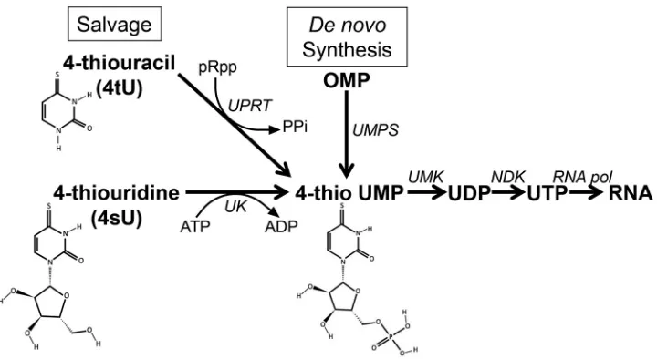
decarboxylation of orotidine 5=-monophosphate catalyzed by UMP synthetase. An alternative source of UMP can arise from the salvage pathway by uridine kinase phosphorylation of uridine or by an enzymatic reaction between phosphoribosyl pyrophosphate and uracil catalyzed by uracil phosphoribosyltransferase (UPRT; Fig. 1) (23). However, in higher eukaryotes, a two-amino-acid substitution within an ATP binding domain renders the UPRT nonfunctional, making this arm of the salvage pathway inoperative (24). By complementing mammalian cells with a functional UPRT, one can restore the defective pyrimidine salvage pathway (21). Addition of 4-thiouracil (4tU) to the media results in the eventual synthesis of thio-rUTP, which is incorporated in all subsequent RNA species within the UPRT-expressing cell, thus providing a suitable platform for purification and enrichment of thiol-containing RNA species.
Here, we report the specific labeling and characterization of RNA species that are synthesized in response to HCMV infection. We generated a recombinant clinical isolate of HCMV that expresses a functional UPRT driven by a promoter that is active during latency. Using this recombinant virus, we are able to specifically 4tU label RNA species synthesized only in cells that harbor the recombinant virus, allowing us to purify these labeled transcripts from RNA transcripts originating prior to infection. This is critical since it resolves many of the limitations involved in profiling transcriptional changes during HCMV latency within mixed populations of cells. Further, this methodology was utilized to profile transcriptional changes observed duringin vitro models of HCMV latency that we subsequently validated in anex vivomodel system. This represents the first report of 4tU labeling of transcripts specific to viral latent infections.
RESULTS
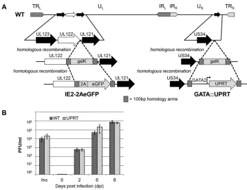
Recombinant HCMV expressingT. gondiiUPRT distinguishes transcription from populations of lytically versus latently infected cells.Infection of HPCs with HCMV is often inefficient and results in a heterogeneous population of both infected and uninfected cells. In addition, sporadic reactivation of HCMV frequently occurs during extended periods within tissue culture (10). Thus, an analysis of transcripts derived solely from latently infected cells is difficult when harvested from a mixed background of both lytic and uninfected cells. To enrich for a population of latently infected cells, we generated a recombinant HCMV utilizing bacterial artificial chromosome (BAC) recombineering techniques (25) in the TB40/E clinical isolate backbone (Fig. 2A). We
FIG 1Salvage andde novosynthesis pathways for UTP synthesis. 4-Thiouracil (4tU) and 4-thiouridine (4sU) are converted to 4-thio UMP by uracil phosphoribosyltransferase (UPRT) or uridine kinase (UK), respectively. The resulting 4-thio UMP is incorporated into newly transcribed cellular RNA by each of the RNA polymerases. Complementation withT. gondiiUPRT is required for 4tU incorporation in mammalian cells via the pyrimidine salvage pathway. Abbreviations: pRpp, phosphoribosyl pyrophosphate; PPi, pyrophosphate; OMP, orotidine-5= -monophosphate; UMPS, UMP synthetase; UMK, UMP kinase; NDK, nucleoside diphosphate kinase.
on November 6, 2019 by guest
http://jvi.asm.org/
[image:3.585.43.403.70.267.2]created a recombinant virus that expresses the fluorescent marker, enhanced green fluorescent protein (eGFP), downstream from the lytically expressed UL122 transcript encoding the essential immediate-early 2 (IE2) protein. The UL122 open reading frame (ORF) was separated from eGFP by the addition of a T2A “self-cleaving” peptide sequence derived fromThosea asigna virus(26), thereby allowing for the synthesis of a concatenated polypeptide of UL122 and eGFP that is subsequently self-cleaved to generate independently functional IE2 and eGFP proteins. In doing so, the expression of eGFP is indicative of IE2 expression, which serves as a marker of lytic gene expres-sion. However, lack of eGFP expression does not distinguish between latently infected and uninfected cells. To analyze transcripts originating from cells harboring latent HCMV genomes versus transcripts from uninfected, “bystander” cells, we cloned the fully functional UPRT gene fromT. gondii, an obligate intracellular eukaryotic parasite, downstream from the GATA2 promoter (Fig. 2A). The human GATA2 promoter drives expression of the transcription factor GATA2, whose expression is critical for inhibiting differentiation of HPCs (27). Thus, use of the GATA2 promoter to drive UPRT transcrip-tion in undifferentiated HPCs allows for UPRT expression under cellular conditranscrip-tions that favor HCMV latency. After generating and verifying the recombinant virus by sequence analyses, we assessed viral growth compared to the parental virus in fibroblasts. We observed no apparent growth defect with the recombinant TB40/E GATA::UPRT IE2-2AeGFP virus compared to the wild-type (WT) virus (Fig. 2B), suggesting that neither the insertion cassette driving UPRT nor the eGFP ORF downstream of IE2 alters this virus’s growth properties.
FIG 2TB40/E GATA::UPRT IE2-2AeGFP displays wild-type growth kinetics in fibroblasts. (A) Schematic representa-tion of the recombinant virus. BAC-derived TB40/Ewt-mCherry (WT) was used to generate TB40/E GATA::UPRT IE2-2AeGFP using standard recombineering techniques. Black arrows indicate unaltered, flanking ORFs. The stop codon of UL122 (white arrow) was replaced in the first recombination event withgalK, which was subsequently replaced with a 2A self-cleaving element in frame with eGFP. A second recombineering event in the United States region of the genome inserted a double-stranded DNA fragment containing the GATA2 promoter and codon-optimized UPRT downstream of US34A. Striped boxes denote conserved homology arms. TRLand TRS, long and short terminal repeats, respectively; ULand US, unique long and short regions, respectively, IRLand IRS, long and short internal repeats, respectively. (B) Fibroblasts (MRC-5) were infected at an MOI ⫽1 PFU/cell with either TB40/Ewt-eGFP (WT) or TB40/E GATA::UPRT IE2-2AeGFP (UPRT). Cell-free virus was collected over 8 d and quantified by TCID50(n⫽3). Inoc, inoculum.
on November 6, 2019 by guest
http://jvi.asm.org/
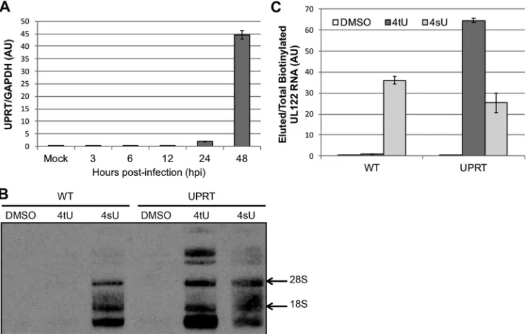
[image:4.585.40.399.68.344.2]HCMV-encoded UPRT allows functional 4tU labeling of viral transcripts in infected fibroblasts.As mentioned above, the addition of the eGFP ORF downstream of IE2 allows us to distinguish and remove the lytically infected cell population from the latently infected population by fluorescence activated cell sorting (FACS). Yet, separat-ing truly latently infected cells from a high frequency of uninfected cells remains problematic. To this end, we established a method to 4tU label host cell transcripts utilizing virus-delivered UPRT. This methodology allows us to isolate thiol-containing rUTP transcripts from cells only in which UPRT is expressed, thereby distinguishing infected from uninfected cells. First, we tested the expression efficiency of the GATA2 driven UPRT in fibroblasts by infecting them with TB40/E GATA::UPRT IE2-2AeGFP at a multiplicity of infection (MOI) of 0.1 PFU/cell and monitoring the expression of UPRT transcription over 48 h. We observed the accumulation of UPRT RNA beginning at 24 h postinfection (hpi) and robust expression at 48 hpi (Fig. 3A). We next evaluated the specificity of UPRT-mediated labeling of RNA. We infected fibroblasts with either the UPRT-expressing virus or wild-type virus in the presence of either 4-thiouracil (4tU) or 4-thiouridine (4sU). Since 4sU can be converted to 4-thio-UMP by uracil kinase (Fig. 1) (28), all cells exposed to 4sU incorporate this label into subsequently transcribed RNA, irrespective of UPRT status. However, only cells with functioning UPRT can integrate 4tU; hence, only the UPRT-expressing virus infected cell population can produce 4tU-labeled RNA. To determine the efficiency of labeling, fibroblasts infected with either TB40/E GATA::UPRT IE2-2AeGFP or TB40/Ewt-eGFP (WT; MOI⫽1 PFU/cell) for 36 h were treated with vehicle (dimethyl sulfoxide [DMSO]), 4tU or 4sU, which was added to the culture media. Total RNA was isolated and subjected to thiol-specific biotinyla-tion. We analyzed the efficiency of 4tU and 4sU incorporation first by using a modified
FIG 3HumanizedT. gondiiUPRT is expressed by the recombinant HCMV and allows for viral transcript enrichment of thiol-RNAs. (A) Fibroblasts were infected with TB40/E GATA::UPRT IE2-2AeGFP (MOI⫽0.1 PFU/cell), and UPRT expression was assessed by RT-qPCR over the indicated times. All samples were analyzed in triplicate and normalized to GAPDH. AU, arbitrary units. (B and C) Fibroblasts were infected with either TB40/Ewt-eGFP (WT) or TB40/E GATA::UPRT IE2-2AeGFP (UPRT) at an MOI⫽1 PFU/cell. At 36 hpi, media containing DMSO, 100⬀M 4-thiouracil (4tU), or 200⬀M 4-thiouridine (4sU) was added for the respective labeling duration (DMSO, 6 h; 4tU, 6 h; 4sU, 2 h). Total RNA was isolated, and 50g was used for thiol-specific biotinylation. (B) Thiol-specific biotinylated RNA (1g) was separated on a denaturing urea gel, transferred to nitrocellulose, and detected by probing with streptavidin-HRP specific antibody. A representative Northern blot is shown. Migration of the ribosomal 28S and 18S RNA species is indicated on the right. (C) Biotinylated RNA (25g) was incubated with streptavidin microbeads and column purified. Equal volumes of eluted RNA were used to generate cDNA, and UL122 expression was assessed by RT-qPCR. Samples were analyzed in triplicate and normalized to total biotinylated RNA reserved prior to streptavidin purification.
on November 6, 2019 by guest
http://jvi.asm.org/
[image:5.585.43.428.73.316.2]Northern blot. Biotinylated RNA samples were separated by denaturing urea polyacryl-amide gel electrophoresis, transferred to nitrocellulose, and probed with a biotin-specific antibody. We observed no detectable biotinylated RNA species following the addition of vehicle (Fig. 3B, DMSO lanes). As we expected, 4sU treatment resulted in significant incorporation of biotin in the RNA isolates of both WT- and UPRT-infected cells. The addition of 4tU resulted in robust labeling of RNA species only in the UPRT-expressing virally infected cells. Importantly, we observed no 4tU incorporation in the WT-infected cells. This demonstrates that virally expressed UPRT within infected human fibroblasts is sufficient to allow for the incorporation of 4tU into the transcrip-tome.
To investigate the specific transcript labeling capabilities of 4tU and 4sU, we further processed the biotinylated RNA from the above samples by streptavidin affinity puri-fication, separating thiol-labeled (bound) and nonlabeled (nonbound) transcripts using a column. We then quantified nascent transcription levels of UL122, which encodes IE2, an essential viral protein expressed during lytic viral replication, in the bound portion of RNA (Fig. 3C). We observed no detectable labeling of UL122 transcripts in the purified RNA from WT- and UPRT-infected cells treated with vehicle alone (DMSO). We consistently detected 4sU-labeled, newly transcribed UL122 in purified RNA from both WT- and UPRT-infected cells. Our results also reveal increased levels of nascent UL122 in 4tU-labeled cells infected with UPRT-expressing HCMV and importantly, little to no labeling in purified RNA from 4tU-treated, WT-infected cells. This demonstrates that in the presence of 4tU, expression of UPRT delivered by the infecting virus results in specific labeling of newly synthesized transcripts, thereby allowing for subsequent affinity enrichment.
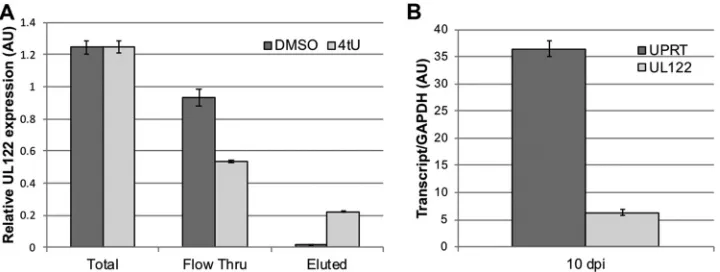
4tU labeling is functional in a Kasumi-3 cell model of HCMV latency. Having demonstrated that GATA2-drivenT. gondiiUPRT is functional in human fibroblasts, we next sought to determine whether UPRT is functional within cells that support HCMV latency. To profile HCMV latent infections, we utilized Kasumi-3 cells, a myeloid-like cell line that mirrors many aspects of HCMV latency observed inex vivomodel systems, including latent transcript expression, viral miRNA expression and reactivation from latency producing infectious progeny (19). We infected Kasumi-3 cells with TB40/E GATA::UPRT IE2-2AeGFP (MOI⫽5 PFU/cell) for 2 days, and treated the cells with either 4tU or vehicle (DMSO) for 6 h prior to total RNA isolation. We reserved a portion of total isolated RNA and used equal amounts of the remaining RNA for biotinylation and streptavidin column purification. We then quantified UL122 transcription in the total, flowthrough (nonlabeled), and eluted (labeled) RNA portions by quantitative reverse transcription-PCR (RT-qPCR). We observed that 4tU treatment did not impact the levels of UL122 transcription in total isolated RNA compared to DMSO treatment (Fig. 4A). Importantly, we detected increased expression of UL122 in the eluate from 4tU-treated cells, compared to those treated with DMSO. This result indicates that nearly 20% of the total UL122 mRNA was newly transcribed and UPRT-infected Kasumi-3 cells are able to efficiently incorporate the 4tU label (Fig. 4A). The extremely low level of UL122 transcript detected in the DMSO-treated cells is likely due to nonspecific RNA binding to streptavidin beads during affinity purification and shows that our stringent labeling method produces very low background transcripts. This result indicates that the virus-mediated expression of UPRT in Kasumi-3 cells results in specific 4tU labeling of actively transcribed RNA within the infected host.
To asses whether the GATA2 promoter allows for sufficient UPRT expression during conditions indicative of HCMV latency, we infected Kasumi-3 cells with TB40/E GATA:: UPRT IE2-2AeGFP (MOI⫽5 PFU/cell) under conditions that lead to latency (19) and isolated total RNA at 10 days postinfection (dpi) to quantify UPRT and UL122 transcrip-tion by RT-qPCR. Our results revealed reduced levels of UL122 compared to GATA2 driven UPRT, indicating that UPRT expression is sustained in Kasumi-3 cells during latent infection for 10 days (Fig. 4B).
TB40/E GATA::UPRT IE2-2AeGFP latent infection of Kasumi-3 cells results in 4tU-labeled RNA for use in whole-cell transcriptome analysis.Since our findings
on November 6, 2019 by guest
http://jvi.asm.org/
revealed that UPRT is both functional and expressed in Kasumi-3 cells during extended periods of latent infection, we next used this labeling system to profile the host transcriptome in latently infected cells. To this end, we infected Kasumi-3 cells with our recombinant TB40/E GATA::UPRT IE2-2AeGFP (MOI ⫽ 5 PFU/cell) for 10 days under conditions that promote latency. As a control, mock-infected Kasumi-3 cells were treated in parallel for the same duration. Next, to eliminate any sporadic, lytically replicating infected cells, we FACS sorted eGFP-positive cells from the eGFP-negative population. eGFP expression is coupled to the synthesis of the lytic gene product, IE2, and thus the eGFP-negative population of cells that remained were a mixture of uninfected and HCMV latently infected cells. After sorting, the isolated eGFP-negative cell population was immediately labeled for 6 h with 4tU, and the mock-infected cells were labeled with 4sU for 2 h. We then isolated, biotinylated, and purified total RNA from 4tU-labeled infected cells and 4sU-labeled mock-infected cells. To confirm that we were monitoring latently infected Kasumi-3 cells, we assayed transcription of the lytic gene, UL123, and the latent HCMV transcript, UL138, by RT-qPCR. Our analyses revealed that the selection of eGFP-negative cells resulted in cell populations enriched for latently infected cells, since there were significantly higher levels of latency-associated UL138 mRNA compared to lytic UL122 mRNA (Fig. 5A). This profile ratio is similar to that previously reported for latently infected Kasumi-3 cells (29) and CD34⫹HPCs (11).
In order to identify and quantify changes in the host transcriptome in response to HCMV latency, we compared 4tU-labeled RNA from latently infected to 4sU-labeled RNA from mock-infected Kasumi-3 cells. We used equal amounts of purified RNA to generate probes suitable for quantifying transcripts by hybridization to an Affymetrix Human Gene 2.0ST transcriptome profiling microarray. We assessed three technical replicates of the transcriptomes of infected and mock-infected cells by monitoring changes in host RNA in response to HCMV latent infection. We observed significant changes in the transcriptional profile of infected Kasumi-3 cells in response to viral infection under conditions that promote latency (Fig. 5B). Of the 24,880 different human transcripts profiled by⬎56,000 probes on the array, we found that 708 were reduced in levels by at least 3-fold. In addition, we observed 155 unique transcripts that increased in expression 3-fold or greater in response to viral infection.
We employed the PANTHER (Protein ANalysis THrough Evolutionary Relationships) classification system to categorize groups of proteins of similar biological function that changed in response to HCMV infection. This analysis revealed that host transcripts involved in distinct biological pathways were altered in response to viral infection (Fig. 5C). Interestingly, we identified a significant number of differentially altered membrane
FIG 4HumanizedT. gondiiUPRT functionally labels viral transcripts in latently infected Kasumi-3 cells. (A) TB40/E GATA::UPRT IE2-2AeGFP-infected Kasumi-3 cells (MOI⫽5 PFU/cell) were labeled at 2 dpi with DMSO or 100M 4tU for 6 h. Total RNA was isolated, biotinylated, and purified with a streptavidin column. Equal volumes of flowthrough and eluted RNA were used to generate cDNA. UL122 expression was quantified by RT-qPCR and normalized to total biotinylated RNA. All samples were analyzed in triplicate. (B) Total RNA was isolated from TB40/E GATA::UPRT IE2-2AeGFP-infected Kasumi-3 cells (MOI⫽5 PFU/cell) at 10 dpi. Equal amounts of total RNA were used to generate cDNA and quantified by RT-qPCR using a standard curve generated from BAC containing UPRT. All samples were analyzed in triplicate and normalized to GAPDH. AU, arbitrary units.
on November 6, 2019 by guest
http://jvi.asm.org/
[image:7.585.43.403.72.208.2]bound transcripts. We found 8 transcripts increased during viral infection (5.1% of total identified transcripts) encode membrane proteins and 51 transcripts (7.2% of total identified transcripts) that encode host membrane proteins were decreased due to viral infection. Distinct alterations in the membrane profile of latently infected cells may offer a mechanism for isolation/depletion of naturally latently infected host cells. In addition, we found a significant number of signaling molecules downregulated in Kasumi-3 cells following latent infection with HCMV (e.g., CCL2 chemokine, BCL2L1 and BCL2A1 signaling molecules, and interleukin OSM). Our results reveal potential
candi-FIG 5Cellular transcripts are specifically dysregulated during HCMV latent infection. Kasumi-3 cells were mock infected (Mock) or infected with TB40/E GATA::UPRT IE2-2AeGFP-positive lytically infected cells (Infected) at an MOI⫽5 PFU/cell. At 10 dpi, eGFP-positive HCMV infected cells were removed by FACS. The mock-infected or remaining eGFP-negative infected cells (presumed to be latently infected) were labeled with 200M 4sU or 100M 4tU, respectively. Total RNA was isolated, biotinylated, and streptavidin purified with the uMACS system. Equal amounts of purified RNA were used to quantify viral transcription by RT-qPCR using UL123 (lytic) and UL138 (latent) specific primers (A) and to generate cDNA probes for hybridization to microarrays (n⫽3) (B). A heat map of the expression profile and clustering of genes with a⬎3-fold change between the average of the three conditions is shown. (C) PANTHER analysis of transcripts that display a⬎3-fold change. Membrane-bound transcripts (lavender) were significantly regulated in response to HCMV. Eight transcripts (5.1% of the total) increased, while 51 transcripts (7.2% of the total) decreased. The “total component hits” reflect the number of entered targets that map to subcategorized classifications by molecular function, biological processes, or cellular component.
on November 6, 2019 by guest
http://jvi.asm.org/
[image:8.585.40.472.70.524.2]dates for identifying natural latently infected cells and offer insight into host cell changes induced during latency.
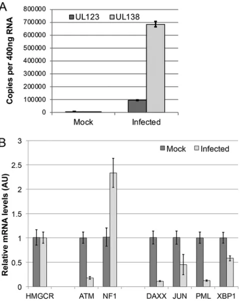
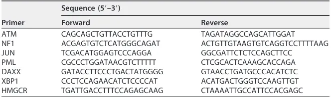
Specific host transcripts are dysregulated during HCMV latent infection of Kasumi-3 cells and CD34ⴙHPCs.Our microarray analysis provides a global assess-ment of transcriptional changes observed during viral infection, however is not as quantitative a method such as RT-qPCR. Therefore, to validate and quantify the transcriptional changes we observed by microarray, we profiled a select set of tran-scripts by RT-qPCR that were significantly altered (⬎3-fold) in response to latent infection of Kasumi-3 cells. To compare transcriptional differences, we used equal amounts of 4tU-labeled RNA or 4sU-labeled RNA from UPRT-expressing latently in-fected and mock-inin-fected Kasumi-3 cells, respectively. Our microarray analysis revealed that the host 3-Hydroxy-3-Methylglutaryl-CoA Reductase (HMGCR) transcript was rela-tively unchanged (⬍5%) between the infected and mock-infected conditions, and thus we used this transcript to normalize our data. Of the significantly altered transcripts we identified by microarray, we chose two representative transcripts that were upregu-lated and four transcripts that demonstrated reduced levels during latency. We found that the ATM serine/threonine kinase (ATM) and neurofibromin 1 (NF1) transcripts were upregulated in latently infected cells, compared to parallel, mock-infected cultures. ATM increased ⬎4-fold during latent infection in the microarray experiment, and in agreement with these results, we detected a⬎3-fold increase in ATM transcription as determined by RT-qPCR (Fig. 6). In addition, the NF1 transcript is expressed at 3-fold-higher levels in latently infected Kasumi-3 cells according to the microarray analysis. In accordance with this result, we found NF1 transcription to be nearly 2-fold higher in latently infected cells by RT-qPCR (Fig. 6). The discrepancies we observed in the exact fold differences between the microarray and RT-qPCR results underscore the need to validate microarray results by more stringent means (e.g., RT-qPCR), as we have done. We also profiled four transcripts that were reduced in expression in the latently infected cells according to the microarray analysis. Our findings from the microarray experiment revealed that the death domain-associated protein (DAXX), Jun proto-oncogene AP-1 transcription factor subunit (JUN), promyelocytic leukemia/TRIM19 (PML), and X-box binding protein 1 (XBP1) transcripts all had greater than 3-fold lower transcriptional expression in the latently infected Kasumi-3 cells than in the mock-infected cells. We indeed confirmed this by RT-qPCR, which revealed that each of these transcripts was reduced in latently infected cells compared to mock-infected cultures, in strong accordance with our microarray results (Fig. 6).
FIG 6Latency-specific cellular transcripts are modulated in Kasumi-3 cells. Kasumi-3 cells were either mock infected or infected with TB40/E GATA::UPRT IE2-2AeGFP (MOI⫽5 PFU/cell) for 10 days. Cells were sorted using FACS and thiol labeled as described for Fig. 5; the total RNA was then isolated, biotinylated, and streptavidin purified. Host cell transcription was profiled by RT-qPCR using transcript-specific primers and plotted as ΔΔCTvalues relative to those of the control transcript, HMGCR (n⫽3). The results show mean fold changes in mRNA levels between infected and mock-infected cells. AU, arbitrary units.
on November 6, 2019 by guest
http://jvi.asm.org/
[image:9.585.86.323.72.230.2]While profiling HCMV latency in Kasumi-3 cells serves as a viable model system (29–31) for monitoring virus-specific changes in the host transcriptome, the “gold standard” for such studies includes a latency culture model utilizing primary,ex vivo cultured cells, such as peripheral monocytes (32) or CD34⫹ HPCs (10, 11). Thus, to confirm our findings from the Kasumi-3 cellin vitromodel of latency, we assessed these transcripts using the CD34⫹ HPC latency model. We infected cord blood-derived CD34⫹HPCs with TB40/Ewt-mCherry at an MOI of 2 PFU/cell and sorted for mCherry expression at 24 hpi. We then returned these cells to culture under conditions that promote viral latency, alongside parallel cultures of mock-infected CD34⫹HPCs. After 10 days, we harvested the cells for RNA extraction. To confirm the cells were latently infected, we evaluated the expression levels of the viral lytic gene, UL123 and the viral latency transcript, UL138. Similar to the latently infected Kasumi-3 cells, we found that UL138 was indeed expressed to higher levels than UL123 in the latently infected CD34⫹ HPCs (Fig. 7A), in accordance with previous findings (31).
In strong agreement with data from our Kasumi-3 microarray analysis, we found that RT-qPCR analyses of transcripts from primary CD34⫹HPCs revealed that the HMGCR transcript was expressed to similar levels in both the mock-infected and the latently infected CD34⫹HPCs (Fig. 7B). We were surprised to find that RT-qPCR analysis of ATM revealed that this transcript is reduced ⬎5-fold in latently infected CD34⫹ HPCs, in contrast to its expression profile in Kasumi-3 cells, in which we observed a 3-fold increase during latency. However, we did detect an increase in NF1 expression in the CD34⫹HPCs that mirrored the observations from our microarray and RT-qPCR analyses of the latently infected Kasumi-3 cells.
FIG 7Cellular transcripts are altered in response to HCMV infection of primary CD34⫹HPCs. Cord
blood-derived CD34⫹HPCs were mock-infected or infected with TB40/Ewt-mCherry and cultured under
latent conditions for 10 days. (A) Total RNA was isolated and used to quantify the expression of viral transcripts UL123 and UL138 by RT-qPCR (n⫽3). (B) Changes in mRNA expression for the indicated genes were quantified by RT-qPCR and are shown as ddCT values relative to the cellular control, HMGCR (n⫽3). The results shown the mean fold changes in mRNA levels between infected and mock-infected cells. AU, arbitrary units.
on November 6, 2019 by guest
http://jvi.asm.org/
[image:10.585.87.328.71.371.2]as well as distinct medium conditions required for the two model systems, which highlight the benefit of validating findings in primary cell systems. Overall, these results suggest that targeted labeling of RNA in latently infected cells, coupled with high-throughput transcriptome analysis techniques, can offer global assessments of host cell changes in response to latent HCMV.
DISCUSSION
While a significant amount of research has revealed distinct aspects of HCMV latency, much remains unknown about this phase of infection. Specifically, what are the true sites of latent viral reservoirs within the hematopoietic compartment? Indeed, during natural latency, distinct lineage-specific subsets of CD34⫹ HPCs, as well as monocytes, harbor the viral genome in its latent state (7, 32); however, a complete characterization of the specific cells that maintain quiescent HCMV in the context of natural infections remains undetermined due to the infrequent numbers of genome positive cells (16). Characterization of the transcriptional landscape within these natu-rally, latently infected cells would allow one to exploit changes to either allow immu-nodepletion of HCMV-positive donor stem cell populations or specifically target latently infected cells therapeutically so as to eliminate transmission of HCMV to transplant recipients. Recent advances in single cell transcriptome sequencing and target enrich-ment sequencing are now being applied to the study of HCMV latency to characterize viral transcripts (9, 12). In this study, however, we focused on a novel approach to characterize changes in the host transcriptome as opposed to the viral transcriptome, using models of latent HCMV infection to identify distinct characteristics of latently infected cells. We exploited complementation of the human pyrimidine salvage path-way, such that only HCMV-infected cells will support specific RNA labeling of newly synthesized transcripts (33). In doing so, we enriched RNA species for use in subsequent high-throughput (i.e., microarray or transcriptome sequencing [RNA-Seq]) or highly specific and quantitative (i.e., RT-qPCR) analyses. We report the generation of a virus that allows for monitoring of lytic gene expression by tethering the expression of a reporter gene, eGFP, to that of the lytic replication regulator, IE2. Expression of IE2 is indicative of cells that are primed to undergo lytic HCMV replication. The generation of a polypeptide of IE2 and eGFP, separated by the short T2A autocleavage linker peptide, resulted in no discernible lytic growth defect and strong eGFP expression, thereby allowing us to monitor productive viral infection. We used this virus as a backbone to generate a virus that also includes T. gondii UPRT gene expression driven by the promoter that facilitates expression of the hematopoietic GATA2 zinc finger transcrip-tion factor. This cassette is expected to be active exclusively in lymphoid cells infected
with HCMV. By complementing the defective human UPRT gene with that from T.
gondii, we successfully and efficiently incorporated thiol groups into pyrimidine-containing transcripts from virus-infected cells. This labeling system, in combination with fluorescent cell sorting to isolate nonlytic cells, afforded us, for the first time, a platform to purify labeled transcripts from cells latently infected with HCMV.
As proof of principle, we utilized this system to monitor changes in host transcripts in response to infection within two different culture models of HCMV latency. We used microarray technology, as this method allows for a direct comparison of transcript
on November 6, 2019 by guest
http://jvi.asm.org/
levels between two different conditions. We infected Kasumi-3 cells, anin vitromodel system for HCMV latency, with our UPRT-expressing recombinant virus under condi-tions that promote viral latency and removed lytic cells, determined by eGFP expres-sion, prior to labeling with 4tU. Mock-infected cells, which lack a functional UPRT, were labeled with 4sU, which is readily incorporated into all pyrimidine-containing tran-scripts irrespective of UPRT status, providing a baseline to monitor host transcriptional changes upon HCMV latency. We report that of the more than 24,000 different genes represented on the microarray, less than 3.5% of the transcripts changed 3-fold or more in response to HCMV infection. This is not overly surprising as viral latency is predicted to result in few host changes, thereby ensuring evasion of host antiviral measures and immune responses. As a validation of the microarray analyses, we performed RT-qPCR analyses of specific transcripts from latently infected Kasumi-3 cells and found that the microarray results accurately predicted changes in transcripts in response to viral infection. Our examination of these host transcripts in the context of the physiologically relevant CD34⫹HPCs revealed that latent infection of these cells mirrored many, but not all of the changes seen in the Kasumi-3 cells. This may be due to several reasons, although the four most likely reasons are discussed here. (i) The Kasumi-3 cells are a transformed cell line and may not completely reflect the model of HCMV latency in CD34⫹HPCs. (ii) Theex vivo-isolated CD34⫹HPCs represent a heterogeneous popula-tion. These cells vary in lineage commitment, asynchronous timing of the establishment of HCMV latency, and donor genetic backgrounds thus may be more variable. (iii) The population of “latently infected cells” may have a background level of abortively infected cells that is different within the two model systems. (iv) There are different medium requirements to support proper growth of the cells in each of the two models we studied. The changes we observed may prove less complex than those that occur in the context of natural infections. However, using the methodology presented herein affords us the tools to dissect conserved host transcriptional changes in response to infections that are readily transferrable to analyses of natural latency.
Our analysis suggests that the transcriptome of a latently infected cell is distinct from a noninfected cell. We believe this is due to several factors, including the expression of latent transcripts and subsequent viral proteins, as well as the expression of the viral miRNAs which are potently expressed during latency (31). In addition, and potentially equally as influential, are the underlying host responses to harboring a large episomal latent viral genome. Use of this novel HCMV UPRT-mediated labeling system will allow for distinct comparisons of different HCMV latency model systems using sensitive RNA-Seq technologies to identify key changes that one can extend to under-standing natural HCMV latency in seropositive patient samples.
While we have applied this system for profiling transcriptional changes within two model systems of HCMV latency, this technology can easily be extended to any permissive cell type for HCMV. In addition, by altering the time of addition of 4tU, one can monitor both the half-life of RNAs and the timing of transcriptional initiation of both viral and cellular RNA species, including mRNAs, microRNAs, and lncRNAs. Finally, one can easily exploit the UPRT expression system in other viruses with modern recombineering techniques, thereby allowing cell type- and kinetic-specific assess-ments of both host and viral RNA species. In sum, we report a novel and powerful RNA labeling technique in the context of HCMV infection that allows for identification of transcriptional changes occurring within models of viral latency.
MATERIALS AND METHODS
Cell culture.Primary neonatal human foreskin fibroblasts (NuFF-1; GlobalStem) or primary human embryonic lung fibroblasts (MRC5; ATCC) were maintained in Dulbecco modified Eagle medium sup-plemented with 10% fetal bovine serum, 2 mML-glutamine, and 100 U/ml each penicillin and strepto-mycin. Kasumi-3 cells (ATCC) were maintained in RPMI 1640 medium (ATCC, cat 30-2001) containing 20% FBS, 100 g/ml gentamicin, and 100 U/ml each of penicillin and streptomycin at a concentration between 3⫻105and 3⫻106cells/ml. Kasumi-3 cells used for viral infections were maintained in X-VIVO 15 medium (Lonza, catalog no. 04-418Q) supplemented with 100 U/ml each of penicillin and strepto-mycin for 48 h (h) prior to infection. Primary CD34⫹HPCs were isolated from deidentified cord blood
on November 6, 2019 by guest
http://jvi.asm.org/
ofgalK-positive clones, a cassette containing the insertion GATA::UPRT sequence flanked by homology arms was then transformed into electrocompetent SW105 cells. The cassette used forgalKsubstitution was a double-stranded DNA gBlock gene fragment (IDT) containing the GATA2 promoter, followed by a consensus Kozak sequence and codon-optimizedToxoplasma gondii-UPRT, flanked by 200 bp of se-quence homologous to the insertion site between US34A and TRS1.
The codon-optimized UPRT sequence is as follows: TGGCCCAGGTGCCCGCCAGCGGCAAGCTGCTGGT GGACCCCCGCTACAGCACCAACGACCAGGAGGAGAGCATCCTGCAGGACATCATCACCCGCTTCCCCAACGTG GTGCTGATGAAGCAGACCGCCCAGCTGCGCGCCATGATGACCATCATCCGCGACAAGGAGACCCCCAAGGAG GAGTTCGTGTTCTACGCCGACCGCCTGATCCGCCTGCTGATCGAGGAGGCCCTGAACGAGCTGCCCTTCGAGA AGAAGGAGGTGACCACCCCCCTGGACGTGAGCTACCACGGCGTGAGCTTCTACAGCAAGATCTGCGGCGTGAG CATCGTGCGCGCCGGCGAGAGCATGGAGAGCGGCCTGCGCGCCGTGTGCCGCGGCTGCCGCATCGGCAAGATC CTGATCCAGCGCGACGAGACCACCGCCGAGCCCAAGCTGATCTACGAGAAGCTGCCCGCCGACATCCGCGACC GCTGGGTGATGCTGCTGGACCCCATGTGCGCCACCGCCGGCAGCGTGTGCAAGGCCATCGAGGTGCTGCTGCG CCTGGGCGTGAAGGAGGAGCGCATCATCTTCGTGAACATCCTGGCCGCCCCCCAGGGCATCGAGCGCGTGTTCA AGGAGTACCCCAAGGTGCGCATGGTGACCGCCGCCGTGGACATCTGCCTGAACAGCCGCTACTACATCGTGCCC GGCATCGGCGACTTCGGCGACCGCTACTTCGGCACCATGTAA.
GalK-negative counterselected clones were then screened and sequenced to verify the position and integrity of the inserted cassette.
To generate IE2-2AeGFP,galKwas inserted at the C terminus of UL122 with the following primers: forward, 5=-TGAGCCTGGCCATCGAGGCAGCCATCCAGGACCTGAGGAACAAGTCTCAGCCTGTTGACAATTAAT CATCGGCA-3=, and reverse, 5=-CACGGGGAATCACTATGTACAAGAGTCCATGTCTCTTTCCAGTTTTTCACTCAG CACTGTCCTGCTCCTT-3=, where the underlined sequences correspond togalK. Note that this insertion deletes the stop codon of UL122.
A cassette containing the T2A element, a “self-cleaving” peptide sequence derived from Thosea asigna virus(26), with the sequence 5=-GGTTCAGGTGAGGGCAGAGGCTCACTCTTGACGTGCGGTGATGTAG AAGAAAACCCCGGTCCT-3=, was cloned in-frame with eGFP and amplified with the following primers: forward, 5=-TGAGCCTGGCCATCGAGGCAGCCATCCAGGACCTGAGGAACAAGTCTCAGGGTTCAGGTGAGGGC AGAGGCTCA–3=, and reverse, 5=-CACGGGGAATCACTATGTACAAGAGTCCATGTCTCTTTCCAGTTTTTCACTT ACTTGTACAGCTCGTCCATGCC-3=, where the underlined sequence indicates the sequence homologous to the 3=end of UL122. The resulting PCR product was purified and transformed into electrocompetent SW105 cells. GalK-negative counterselected clones were screened, and the position and integrity of the inserted cassette was verified by sequencing.
The resulting recombinant virus, TB40/E GATA::UPRT IE2-2AeGFP, was transfected into low-passage-number MRC5 cells with 1.5g of pCGN-pp71. Stocks of cell-free virus were generated, and virus titers were determined as described previously (35). Growth curve analysis of the recombinant against wild-type TB40/Ewt-eGFP was performed by infecting a confluent monolayer of MRC-5 cells at an MOI of 1 PFU/cell. Supernatants were collected over 8 days and used in 50% tissue culture infectious dose (TCID50) assays on fibroblasts to calculate and compare virus titers.
RT-qPCR for UPRT and viral transcripts.Total RNA was isolated using TRIzol reagent (Invitrogen) under RNase-free conditions according to the manufacturer’s instructions and precipitated with isopro-panol. DNA contaminants were removed by DNase treatment using a DNA-free DNA removal kit (Ambion). RNA concentrations were determined and equal amounts, 0.1 to 0.9g, were used to generate cDNA using the TaqMan reverse transcription kit with random hexamers according to the manufacturer’s protocol (Applied Biosystems). Equal amounts of cDNA were then analyzed by quantitative PCR (qPCR) in triplicate using Power SYBR green master mix (Applied Biosystems) and an Eppendorf Mastercycler RealPlex2real-time PCR machine or a CFX Real-Time PCR detection systems machine (Bio-Rad). The copy number was calculated using a standard curve generated from BAC DNA.
The following primer sets were used for qPCR: UPRT forward, 5=-AGAGCATCCTGCAGGACATCAT CAC-3=, and reverse, 5=-AGAGCATCCTGCAGGACATCATCACG-3=; UL122 forward, 5=-ATGGTTTTGCAGGCT TTGATG-3=, and reverse, 5=-ACCTGCCCTTCACGATTCC-3=; UL123 forward, 5=-GCCTTCCCTAAGACCACC AAT-3=, and reverse, 5=-ATTTTCTGGGCATAAGCCATAATC-3=; UL138 forward, 5=-GGTTCATCGTCTTCGTCG TC-3=, and reverse, 5=-CACGGGTTTCAACAGATCG-3=; and GAPDH forward, 5=-ACCCACTCCTCCACCTTTGA C-3=, and reverse, 5=-CTGTTGCTGTAGCCAAATTCGT-3=.
Metabolic labeling, biotinylation, and purification of RNA.Infected and mock-infected human fibroblasts or Kasumi-3 cells were metabolically labeled at the indicated time postinfection by incubation with media containing either 100 M 4-thiouracil (4tU; Sigma, CAS#591-28-6) for 6 h or 200M
on November 6, 2019 by guest
http://jvi.asm.org/
4-thiouridine (4sU; Sigma, CAS#13957-31-8) for 2 h at 37°C and 5% CO2. Total RNA was isolated using TRIzol reagent (Invitrogen). Equal amounts of total isolated RNA (8 to 50g) were biotinylated in reaction mixtures containing 10 mM HEPES [pH 7.5], 1 mM EDTA, and 0.1g of MTSEA biotin-XX (Biotium) per 1g of RNA at room temperature for 30 min (min) in the dark. Excess biotin was removed by extraction with an equal volume of chloroform-isoamyl alcohol (24:1). Equal concentrations of biotinylated RNA were purified using the a uMACS streptavidin kit (Miltenyi Biotec). Biotinylated RNA was heated to 65°C for 10 min and then placed on ice for 5 min. Magnetic uMACS streptavidin microbeads (2l beads per 1g of RNA) were added to RNA, followed by incubation for 15 min with gentle agitation. Samples were passed throughColumns placed in the magnetic field of a uMACS separator. Flowthrough containing nonbiotinylated molecules was collected and saved. Columns with bound biotinylated RNA were washed five times with 1 M NaCl, 10 mM EDTA, 100 mM Tris-HCl (pH 7.4), and 0.1% Tween 20, with washing buffer heated to 65°C for the initial wash. Bound RNA was eluted from theColumns with freshly prepared 100 mM dithiothreitol. Flowthrough and eluted RNA samples were precipitated, washed, and resuspended in RNase-free H2O. Equal volumes of flowthrough and eluate, along with equal concentrations of total biotinylated RNA (reserved prior to uMACS purification for normalization), were used in the reverse transcription reaction to generate cDNA for RT-qPCR analysis.
Biotinylated RNA Northern blots.Biotinylated RNA (1g) was mixed with an equal volume of Gel loading buffer II (Ambion), heated to 95°C for 5 min, and separated by denaturing urea polyacrylamide gel electrophoresis (8 M urea, 10% acrylamide/Bis, 19:1). RNA was transferred to a Hybond-XL nylon membrane (Amersham) in 0.5⫻TBE buffer (1⫻TBE is composed of 90 mM Tris-HCl [pH 8.3], 90 mM boric acid, and 2 mM EDTA) and UV cross-linked. The membrane was blocked in 125 mM NaCl, 17 mM Na2HPO4, and 1% SDS, then incubated with streptavidin-HRP (S-nitrosylation detection reagent I HRP [from an S-nitrosylated protein detection assay kit]; Cayman Chemical). The membrane was then washed twice in Wash I (1:10 dilution of blocking solution) and twice in Wash II (10 mM Tris-HCl, 10 mM NaCl, 2.1 mM MgCl2[pH 9.5]) and visualized using Pierce ECL Western blotting substrate (Thermo Scientific). HCMV latent infections of Kasumi-3 cells.Kasumi-3 cells were plated in X-VIVO 15 (Lonza) for 48 h prior to infection, as described above. Cells were mock infected or infected with TB40/E GATA::UPRT IE2-2AeGFP at an MOI of 5 PFU/cell by centrifugal enhancement at 1,000⫻ gfor 30 min at room temperature. Infected cultures were incubated overnight at 37°C and 5% CO2. To remove debris, infected cells were cushioned onto Ficoll-Paque Plus (GE Healthcare) by low-speed centrifugation (450⫻g) for 35 min at room temperature without the brake. Infected cells were returned to the incubator for 10 days and maintained at a concentration between 5⫻105and 1⫻106cells/ml, with medium changes every 3 days and debris removal after 5 to 6 days. After 10 days, where indicated, viable eGFP-positive and eGFP-negative cells were sorted at room temperature by FACS using a FACSDiva (Becton Dickinson). IE2-2AeGFP-positive cells, indicative of spontaneously reactivated, lytically replicating virus (1 to 5%) were removed. The remaining eGFP-negative population (95 to 99%) consisting of latently infected and uninfected Kasumi-3 cells were returned to culture for metabolic labeling.
Microarray analysis of purified RNA samples.Microarray analysis was performed at the Gene Expression and Genotyping Facility of the Case Western Reserve Comprehensive Cancer Center (Case Western Reserve University, Cleveland, OH). Total RNA was isolated using a High Pure RNA isolation kit (Roche), biotinylated, and uMACS streptavidin column purified, as described in detail above. RNA elutions were resuspended in RNase-free H2O. Equal amounts of RNA were subjected to the Affymetrix GeneChip WT Plus labeling protocol according to the manufacturer’s recommendation. This labeling protocol provides nontargeted labeling of complete transcripts independent of poly(A) status, resulting in plus-strand DNA target probes for use in hybridization to a microarray. The probe sets were used to hybridize to six individual Affymetrix Human Gene Array ST 2.0 microarray chips overnight according to the manufacturer’s protocols. Chips were washed on an Affymetrix Fluidics Station 450 and scanned using an Affymetrix Gene Chip Scanner 3000. BAMarray 3.0 software was used to analyze the hybrid-ization results. Genes that changed⬎3-fold were analyzed using the Protein ANalysis THrough Evolu-tionary Relationships (PANTHER) algorithm (http://www.pantherdb.org/), and clustering and heat map generation of array analysis was performed using the Heat Mapper Expression portal (http://heatmapper .ca/expression/).
HCMV latent infections of CD34ⴙHPCs.Primary CD34⫹HPCs isolated from deidentified cord blood
were mock infected or infected with the BAC-derived clinical isolate, TB40/Ewt-mCherry (36) at an MOI of 2 PFU/cell, as described elsewhere (34). CD34⫹HPCs were sorted for mCherry expression 24 hpi, and
[image:14.585.40.372.82.180.2]mCherry-positive, infected cells were returned to transwell coculture with irradiated stromal cells (a 1:1 TABLE 1Primers used in this study
Primer
Sequence (5=–3=)
Forward Reverse
ATM CAGCAGCTGTTACCTGTTTG TAGATAGGCCAGCATTGGAT
NF1 ACGAGTGTCTCATGGGCAGAT ACTGTTGTAAGTGTCAGGTCCTTTTAAG JUN TCGACATGGAGTCCCAGGA GGCGATTCTCTCCAGCTTCC
PML CGCCCTGGATAACGTCTTTTT CTCGCACTCAAAGCACCAGA DAXX GATACCTTCCCTGACTATGGGG GTAACCTGATGCCCACATCTC XBP1 CCCTCCAGAACATCTCCCCAT ACATGACTGGGTCCAAGTTGT HMGCR TGATTGACCTTTCCAGAGCAAG CTAAAATTGCCATTCCACGAGC
on November 6, 2019 by guest
http://jvi.asm.org/
In addition, we thank the Cleveland Clinic Flow Cytometry Core Facility for their assistance with cell sorting and the Gene Expression and Genotyping Facility of the Case Comprehensive Cancer Center for their assistance with the microarray experi-ments.
The Case Comprehensive Cancer Center is supported by grant P30 CA43703.
REFERENCES
1. Britt W. 2008. Manifestations of human cytomegalovirus infection: pro-posed mechanisms of acute and chronic disease. Curr Top Microbiol Immunol 325:417– 470.
2. Sinclair J. 2010. Chromatin structure regulates human cytomegalovi-rus gene expression during latency, reactivation, and lytic infection. Biochim Biophys Acta 1799:286 –295. https://doi.org/10.1016/j .bbagrm.2009.08.001.
3. Styczynski J. 2018. Who is the patient at risk of CMV recurrence: a review of the current scientific evidence with a focus on hemato-poietic cell transplantation. Infect Dis Ther 7:1–16.https://doi.org/10 .1007/s40121-017-0180-z.
4. Giannattasio A, Di Costanzo P, De Matteis A, Milite P, De Martino D, Bucci L, Augurio MR, Bravaccio C, Ferrara T, Capasso L, Raimondi F. 2017. Outcomes of congenital cytomegalovirus disease following maternal primary and non-primary infection. J Clin Virol 96:32–36.https://doi.org/ 10.1016/j.jcv.2017.09.006.
5. Ziemann M, Thiele T. 2017. Transfusion-transmitted CMV infection: cur-rent knowledge and future perspectives. Transfus Med 27:238 –248. https://doi.org/10.1111/tme.12437.
6. Gimenez E, Solano C, Pinana JL, Amat P, Vinuesa V, Navarro D. 2016. Re-examining the relationship between active cytomegalovirus (CMV) infection and acute graft-versus-host disease in allogeneic stem cell transplant recipients in the era of real-time PCR CMV assays. Transpl Int 29:126 –128.https://doi.org/10.1111/tri.12689.
7. Maciejewski JP, Bruening EE, Donahue RE, Mocarski ES, Young NS, St Jeor SC. 1992. Infection of hematopoietic progenitor cells by human cyto-megalovirus. Blood 80:170 –178.
8. Zhuravskaya T, Maciejewski JP, Netski DM, Bruening E, Mackintosh FR, St Jeor S. 1997. Spread of human cytomegalovirus (HCMV) after infection of human hematopoietic progenitor cells: model of HCMV latency. Blood 90:2482–2491.
9. Shnayder M, Nachshon A, Krishna B, Poole E, Boshkov A, Binyamin A, Maza I, Sinclair J, Schwartz M, Stern-Ginossar N. 2018. Defining the transcriptional landscape during cytomegalovirus latency with single-cell RNA sequencing. mBio 9: e00013-18.https://doi.org/10.1128/mBio .00013-18.
10. Goodrum FD, Jordan CT, High K, Shenk T. 2002. Human cytomegalovirus gene expression during infection of primary hematopoietic progenitor cells: a model for latency. Proc Natl Acad Sci U S A 99:16255–16260. https://doi.org/10.1073/pnas.252630899.
11. Goodrum F, Reeves M, Sinclair J, High K, Shenk T. 2007. Human cyto-megalovirus sequences expressed in latently infected individuals pro-mote a latent infection in vitro. Blood 110:937–945.https://doi.org/10 .1182/blood-2007-01-070078.
12. Cheng S, Caviness K, Buehler J, Smithey M, Nikolich-Zugich J, Goodrum F. 2017. Transcriptome-wide characterization of human cytomegalovirus in natural infection and experimental latency. Proc Natl Acad Sci U S A 114:E10586 –E10595.https://doi.org/10.1073/pnas.1710522114.
13. Rossetto CC, Tarrant-Elorza M, Pari GS. 2013.cis- andtrans-acting factors involved in human cytomegalovirus experimental and natural latent infection of CD14⫹ monocytes and CD34⫹ cells. PLoS Pathog
9:e1003366.https://doi.org/10.1371/journal.ppat.1003366.
14. Soderberg-Naucler C, Fish KN, Nelson JA. 1997. Reactivation of latent human cytomegalovirus by allogeneic stimulation of blood cells from healthy donors. Cell 91:119 –126. https://doi.org/10.1016/S0092 -8674(01)80014-3.
15. Maertens J, Lyon S. 2017. Current and future options for cytomegalovi-rus reactivation in hematopoietic cell transplantation patients. Future Microbiol 12:839 – 842.https://doi.org/10.2217/fmb-2017-0095. 16. Kondo K, Kaneshima H, Mocarski ES. 1994. Human cytomegalovirus
latent infection of granulocyte-macrophage progenitors. Proc Natl Acad Sci U S A 91:11879 –11883.https://doi.org/10.1073/pnas.91.25.11879. 17. Cheng S, Jiang X, Yang B, Wen L, Zhao F, Zeng WB, Liu XJ, Dong X, Sun
JY, Ming YZ, Zhu H, Rayner S, Tang Q, Fortunato E, Luo MH. 2017. Infected T98G glioblastoma cells support human cytomegalovirus reac-tivation from latency. Virology 510:205–215. https://doi.org/10.1016/j .virol.2017.07.023.
18. Penkert RR, Kalejta RF. 2013. Human embryonic stem cell lines model experimental human cytomegalovirus latency. mBio 4:e00298-13. https://doi.org/10.1128/mBio.00298-13.
19. O’Connor CM, Murphy EA. 2012. A myeloid progenitor cell line capable of supporting human cytomegalovirus latency and reactivation, result-ing in infectious progeny. J Virol 86:9854 –9865.https://doi.org/10.1128/ JVI.01278-12.
20. Albright ER, Kalejta RF. 2013. Myeloblastic cell lines mimic some but not all aspects of human cytomegalovirus experimental latency defined in primary CD34⫹cell populations. J Virol 87:9802–9812.https://doi.org/
10.1128/JVI.01436-13.
21. Miller MR, Robinson KJ, Cleary MD, Doe CQ. 2009. TU-tagging: cell type-specific RNA isolation from intact complex tissues. Nat Methods 6:439 – 441.https://doi.org/10.1038/nmeth.1329.
22. Cleary MD, Meiering CD, Jan E, Guymon R, Boothroyd JC. 2005. Biosyn-thetic labeling of RNA with uracil phosphoribosyltransferase allows cell-specific microarray analysis of mRNA synthesis and decay. Nat Biotech-nol 23:232–237.https://doi.org/10.1038/nbt1061.
23. Loffler M, Carrey EA, Zameitat E. 2016. Orotate (orotic acid): an essential and versatile molecule. Nucleosides Nucleotides Nucleic Acids 35: 566 –577.https://doi.org/10.1080/15257770.2016.1147580.
24. Li J, Huang S, Chen J, Yang Z, Fei X, Zheng M, Ji C, Xie Y, Mao Y. 2007. Identification and characterization of human uracil phosphoribosyltrans-ferase (UPRTase). J Hum Genet 52:415– 422.https://doi.org/10.1007/ s10038-007-0129-2.
25. Warming S, Costantino N, Court DL, Jenkins NA, Copeland NG. 2005. Simple and highly efficient BAC recombineering using galK selection. Nucleic Acids Res 33:e36.https://doi.org/10.1093/nar/gni035. 26. Liu Z, Chen O, Wall JBJ, Zheng M, Zhou Y, Wang L, Ruth Vaseghi H, Qian
on November 6, 2019 by guest
http://jvi.asm.org/
L, Liu J. 2017. Systematic comparison of 2A peptides for cloning multi-genes in a polycistronic vector. Sci Rep 7:2193.https://doi.org/10.1038/ s41598-017-02460-2.
27. Melotti P, Calabretta B. 1996. The transcription factors c-myb and GATA-2 act independently in the regulation of normal hematopoiesis. Proc Natl Acad Sci U S A 93:5313–5318.https://doi.org/10.1073/pnas.93 .11.5313.
28. Yu MW, Sedlak J, Lindsay RH, Pittman JA. 1972. Metabolism of 2-thiouracil in pyrimidine pathways leading to nucleotide synthesis. Proc Soc Exp Biol Med 139:1292–1297.https://doi.org/10.3181/00379727-139 -36350.
29. Humby MS, O’Connor CM. 2015. Human cytomegalovirus US28 is im-portant for latent infection of hematopoietic progenitor cells. J Virol 90:2959 –2970.https://doi.org/10.1128/JVI.02507-15.
30. O’Connor CM, Vanicek J, Murphy EA. 2014. Host microRNA regulation of human cytomegalovirus immediate early protein translation promotes viral latency. J Virol 88:5524 –5532.https://doi.org/10.1128/JVI.00481-14. 31. Pan C, Zhu D, Wang Y, Li L, Li D, Liu F, Zhang CY, Zen K. 2016. Human cytomegalovirus miR-UL148D facilitates latent viral infection by target-ing host cell immediate early response gene 5. PLoS Pathog 12: e1006007.https://doi.org/10.1371/journal.ppat.1006007.
32. Taylor-Wiedeman J, Sissons JG, Borysiewicz LK, Sinclair JH. 1991. Mono-cytes are a major site of persistence of human cytomegalovirus in peripheral blood mononuclear cells. J Gen Virol 72(Pt 9):2059 –2064. https://doi.org/10.1099/0022-1317-72-9-2059.
33. Gay L, Miller MR, Ventura PB, Devasthali V, Vue Z, Thompson HL, Temple S, Zong H, Cleary MD, Stankunas K, Doe CQ. 2013. Mouse TU tagging: a chemical/genetic intersectional method for purifying cell type-specific nascent RNA. Genes Dev 27:98 –115.https://doi.org/10.1101/gad.205278 .112.
34. Umashankar M, Goodrum F. 2014. Hematopoietic long-term culture (hLTC) for human cytomegalovirus latency and reactivation. Methods Mol Biol 1119:99 –112.https://doi.org/10.1007/978-1-62703-788-4_7. 35. Murphy E, Vanicek J, Robins H, Shenk T, Levine AJ. 2008. Suppression of
immediate-early viral gene expression by herpesvirus-coded microRNAs: implications for latency. Proc Natl Acad Sci U S A 105:5453–5458. https://doi.org/10.1073/pnas.0711910105.
36. O’Connor CM, Shenk T. 2012. Human cytomegalovirus pUL78 G protein-coupled receptor homologue is required for timely cell entry in epithe-lial cells but not fibroblasts. J Virol 86:11425–11433.https://doi.org/10 .1128/JVI.05900-11.