Hepatitis C Virus Subverts Human
Choline Kinase-
␣
To Bridge
Phosphatidylinositol-4-Kinase III
␣
(PI4KIII
␣
) and NS5A and Upregulates
PI4KIII
␣
Activation, Thereby Promoting
the Translocation of the Ternary
Complex to the Endoplasmic Reticulum
for Viral Replication
Mun-Teng Wong, Steve S. Chen
Institute of Biomedical Sciences, Academia Sinica, Taipei, Taiwan, Republic of China
ABSTRACT In this study, we elucidated the mechanism by which human choline kinase-␣
(hCK␣) interacts with nonstructural protein 5A (NS5A) and phosphatidylinositol-4-kinase
III␣ (PI4KIII␣), the lipid kinase crucial for maintaining the integrity of virus-induced
membranous webs, and modulates hepatitis C virus (HCV) replication. hCK␣activity
positively modulated phosphatidylinositol-4-phosphate (PI4P) levels in HCV-expressing
cells, and hCK␣-mediated PI4P accumulation was abolished by AL-9, a PI4KIII␣
-specific inhibitor. hCK␣ colocalized with NS5A and PI4KIII␣or PI4P; NS5A expression
increased hCK␣and PI4KIII␣colocalization; and hCK␣formed a ternary complex with
PI4KIII␣ and NS5A, supporting the functional interplay of hCK␣ with PI4KIII␣ and
NS5A. PI4KIII␣inactivation by AL-9 or hCK␣inactivation by CK37, a specific hCK␣
in-hibitor, impaired the endoplasmic reticulum (ER) localization and colocalization of
these three molecules. Interestingly, hCK␣knockdown or inactivation inhibited PI4KIII␣
-NS5A binding. In an in vitro PI4KIII␣ activity assay, hCK␣ activity slightly increased
PI4KIII␣basal activity but greatly augmented NS5A-induced PI4KIII␣activity,
support-ing the essential role of ternary complex formation in robust PI4KIII␣activation.
Con-curring with the upregulation of PI4P production and viral replication,
overexpres-sion of active hCK␣-R (but not the D288A mutant) restored PI4KIII␣ and NS5A
translocation to the ER in hCK␣ stable knockdown cells. Furthermore, active PI4KIII␣
overexpression restored PI4P production, PI4KIII␣and NS5A translocation to the ER,
and viral replication in CK37-treated cells. Based on our results, hCK␣functions as an
indispensable regulator that bridges PI4KIII␣ and NS5A and potentiates
NS5A-stimulated PI4KIII␣activity, which then facilitates the targeting of the ternary
com-plex to the ER for viral replication.
IMPORTANCE The mechanisms by which hCK␣ activity modulates the transport of
the hCK␣-NS5A complex to the ER are not understood. In the present study, we
in-vestigated how hCK␣interacts with PI4KIII␣(a key element that maintains the
integ-rity of the “membranous web” structure) and NS5A to regulate viral replication. We
demonstrated that HCV hijacks hCK␣to bridge PI4KIII␣and NS5A, forming a ternary
complex, which then stimulates PI4KIII␣ activity to produce PI4P. Pronounced PI4P
synthesis then redirects the translocation of the ternary complex to the ER-derived, PI4P-enriched membrane for assembly of the viral replication complex and viral rep-lication. Our study provides novel insights into the indispensable modulatory role of
hCK␣in the recruitment of PI4KIII␣to NS5A and in NS5A-stimulated PI4P production
Received2 March 2017Accepted25 May
2017
Accepted manuscript posted online31 May
2017
CitationWong M-T, Chen SS. 2017. Hepatitis C
virus subverts human choline kinase-α to bridge phosphatidylinositol-4-kinase IIIα (PI4KIIIα) and NS5A and upregulates PI4KIIIα activation, thereby promoting the translocation of the ternary complex to the endoplasmic reticulum for viral replication. J Virol 91:e00355-17.https://doi.org/10.1128/ JVI.00355-17.
EditorBryan R. G. Williams, Hudson Institute of
Medical Research
Copyright© 2017 Wong and Chen. This is an
open-access article distributed under the terms of theCreative Commons Attribution 4.0 International license.
Address correspondence to Steve S. Chen, [email protected].
M.-T.W. and S.S.C. contributed equally to this article.
crossm
on November 7, 2019 by guest
http://jvi.asm.org/
and reveals a new perspective for understanding the impact of profound PI4KIII␣
ac-tivation on the targeting of PI4KIII␣ and NS5A to the PI4P-enriched membrane for
viral replication complex formation.
KEYWORDS HCV, NS5A, hCK␣, PI4KIII␣, PI4P, ternary complex, ER translocation
L
ike many other positive-sense RNA viruses, the hepatitis C virus (HCV) exploitscellular lipids and reorganizes endoplasmic membranes to create a specialized viral replication membrane compartment, which is referred to as the “membranous web” (MW) (1, 2). The MW serves as a compartmentation and concentration site for the HCV replication complex (RC), which is composed of the NS3 (nonstructural protein 3) to NS5B proteins, viral replicating RNA, and a wide array of host factors (3, 4). The MW is an endoplasmic reticulum (ER)-derived membrane structure, highly phosphorylated and remodeled, that has heterogeneous vesicles ranging from 100 to 300 nm in diameter and is dedicated to HCV replication (5).
Phosphatidylinositol-4-kinases (PI4Ks) catalyze the phosphorylation of position 4 of the inositol ring in phosphatidylinositol (PI) to form PI-4-phosphate (PI4P) and play a crucial role in the regulation of the intracellular membrane trafficking process (6, 7). PI4P is an essential precursor in the synthesis of PI(4,5)P2 and PI(3,4,5)P3, which are necessary for receptor-activated phospholipase C and phosphoinositide 3-kinase sig-naling (8), and controls cellular trafficking, especially in the Golgi apparatus and trans-Golgi network (8). PI4Ks exhibit distinct subcellular localization and affect the
compartmentation of discrete PI4P pools. PI4KIII␣ localizes primarily to the ER and
maintains a distinct population of PI4P that is critical for ER export when cells are
responding to the increased ER cargo load (9, 10). PI4KIII␣also regulates the PI4P level
at the plasma membrane (11, 12). PI4KIII␣can also be found in close association with
the membranes of the Golgi vesicles and vacuoles (13) and in the nucleolus (14).
PI4KIIIis the lipid kinase primarily responsible for the PI4P pool in the Golgi apparatus
(15, 16), and it also localizes on the lysosome to maintain lysosomal membrane integrity (17).
PI4K isoforms and their product PI4Ps play crucial roles in the replication of many
positive-stranded RNA viruses in the Flaviviridae, Picornaviridae, and Coronaviridae
families, shaping the architecture of their membranous replication compartments via
the elevation of PI4P production (7). PI4KIII␣has been identified as a crucial host factor
in HCV replication (18–23). Although PI4KIIIhas been reported to play a role in the
viral replication of genotypes 1a and 1b (19, 22), its role in genotype 2a replication is less clear (18, 24).
As a critical component of the viral RC, membrane-associated NS5A is present in both a hypophosphorylated p56 form and a hyperphosphorylated p58 form (25, 26), which modulate HCV RNA replication and virus assembly (27). NS5A interacts intimately
with PI4KIII␣via the C-terminal part of domain 1 and recruits the kinase to the viral RC
site, where NS5A stimulates kinase activity in conjunction with NS5B, thereby leading
to the generation of elevated PI4P pools at intracellular membranes (24, 28). PI4KIII␣
activation and PI4P redistribution are required for the changes in the ultrastructural architecture of HCV RCs embedded within the MW (18, 21, 24, 29). Analogously to the
PI4KIII␣depletion phenotype, mutations in the seven amino acids within NS5A, which
have been identified as a PI4KIII␣functional interaction site (PFIS), not only interfere
with PI4KIII␣binding and attenuate PI4P production but also alter the morphology of
viral replication sites (29). Conversely, PI4KIII␣controls HCV RNA replication,
presum-ably via alteration of the phosphorylation status of NS5A (29). Thus, NS5A interplays
with PI4KIII␣to promote PI4P production and endoplasmic membrane reorganization,
which are necessary for viral replication. In addition, the accumulation of PI4P recruits lipid transport proteins, such as oxysterol-binding protein (30) or the four-phosphate adaptor protein (31), which regulate the trafficking of cholesterol and glycosphingo-lipids, respectively, to the viral MW for effective viral genome replication.
Recently, Harak et al. demonstrated that HCV depends on optimal levels of PI4KIII␣
Wong and Chen Journal of Virology
August 2017 Volume 91 Issue 16 e00355-17 jvi.asm.org 2
on November 7, 2019 by guest
http://jvi.asm.org/
activity for proper replication and showed that the replication of various genotypes
requires PI4KIII␣activation at a low PI4KIII␣protein level, such as that observed for
primary hepatocytes (32). Nevertheless, in cultured hepatoma Huh7 cells, which already
express higher levels of PI4KIII␣ than primary hepatocytes, NS5A-induced PI4KIII␣
activation leads to excess PI4P production that is harmful to their replication (32). To manage the growth disadvantage in Huh7 cells, these isolates have evolved a mech-anism through adaptive mutations, such as S2204R in NS5A and R2884G in NS5B, to
limit excess PI4KIII␣activation, thus ensuring their proper replication in Huh7 cells (32).
Strikingly, the genotype 2a JFH1 virus is an exception, because it does not require
adaptive mutations for robust replication in Huh7 cells (33). Thus, PI4KIII␣ activity
delicately adapts to the viral and cellular microenvironment to ensure efficient viral replication. Nevertheless, whether the NS5A-mediated recruitment and activation of
PI4KIII␣ require a yet-to-be identified cellular factor has not been clarified, and the
mechanism underlying the regulation of PI4KIII␣ and NS5A trafficking to the
PI4P-enriched viral replication site has not been identified.
Choline kinase (CK), the first enzyme in the CDP-choline (or Kennedy) pathway, catalyzes the phosphorylation of choline into phosphocholine. Previous genome-wide
small interfering RNA (siRNA) screens have implicated human CK␣ (hCK␣) in HCV
infections (24, 34). As we demonstrated previously, hCK␣activity enhances the
trans-location of the hCK␣-NS5A complex to the ER, where the hCK␣protein mediates the
NS5A-NS5B interaction, thereby promoting functional membranous viral RC assembly
and viral RNA replication (35). However, whether hCK␣interacts with PI4KIII␣and NS5A
to modulate the viral replication process has not been clarified. In addition, the
mechanism underlying the regulation of hCK␣-mediated translocation of the hCK␣
-NS5A complex to the ER remains to be elucidated.
Because PI4K isoforms play critical roles in intracellular membrane trafficking (6, 36)
and PI4KIII␣ builds a PI4P-enriched membrane platform for effective HCV replication
(37, 38), we hypothesize that the hCK␣activity-mediated ER translocation of hCK␣and
NS5A is tightly associated with PI4KIII␣activation and that hCK␣activity plays a crucial
role in NS5A-stimulated PI4KIII␣activation and in the translocation of PI4KIII␣and NS5A
to the ER, thereby promoting membranous viral RC assembly and viral replication (35).
In the present study, we demonstrate that HCV usurps hCK␣functions to facilitate
viral replication. hCK␣acts as a novel upstream adaptor to bridge PI4KIII␣and NS5A,
thereby forming a PI4KIII␣ternary complex. Additionally, the results obtained from a
cell culture system and anin vitrokinase assay demonstrate that hCK␣activity slightly
enhances PI4KIII␣basal activity but greatly potentiates NS5A-stimulated PI4KIII␣
activ-ity, indicating that hCK␣synergistically upregulates PI4KIII␣activity in conjunction with
NS5A. In turn, the massive production of PI4P pools facilitates the translocation of the ternary complex to the ER-derived, PI4P-enriched membrane for viral replication. Our
study highlights the novel viewpoint that PI4KIII␣ activity and its translocation in
conjunction with NS5A to viral replication sites are regulated by the cellular kinase
hCK␣, even though the product of this kinase is not directly linked to membrane
composition or structure.
RESULTS
hCK␣colocalizes with NS5A and PI4P or PI4KIII␣in HCV-expressing cells.We
recently demonstrated that hCK␣activity is critical for the intracellular trafficking of the
hCK␣-NS5A complex to the ER membrane, where hCK␣mediates the binding of NS5A
to NS5B for viral RC assembly (35). In the present study, we further examined whether
hCK␣interacts with PI4KIII␣, the lipid kinase critical for the integrity of the HCV-induced
MW, and NS5A to stimulate PI4P production and whether accumulated PI4P production
facilitates the translocation of hCK␣and NS5A, as well as PI4KIII␣, to the ER membrane,
thereby promoting HCV replication.
To address this issue, we first searched for the intracellular colocalization of hCK␣,
NS5A, and PI4P in HCV-infected Huh7 cells via confocal microscopy. As shown
previ-ously (35), a small fraction of hCK␣colocalized with NS5A in HCV-infected cells (Fig. 1A,
on November 7, 2019 by guest
http://jvi.asm.org/
magenta puncta in the fluorescence image labeled “NS5A/hCK␣”). HCV infection also resulted in much more accumulation of PI4P in dot-like patterns scattered in the cytoplasm than that seen with mock infection, and a fraction of these PI4P puncta colocalized with NS5A (Fig. 1A), which represents viral replication sites, in HCV-infected
FIG 1Colocalization of hCK␣with NS5A and PI4P or PI4KIII␣. (A) Huh7 cells either remained uninfected or were infected with HCVcc at an MOI of 1. At day 3 postinfection, the cells were fixed and successively immunostained with rabbit anti-hCK␣, a mouse anti-NS5A (9E10) MAb (IgG), and mouse anti-PI4P (IgM), followed by incubation with appropriate Alexa Fluor dye-conjugated secondary antibodies. The cells were then analyzed by confocal microscopy. Nuclei are demarcated by solid white lines. The images of the three fluorescence signals were merged, and the boxed areas were enlarged. The colocalization of two or three molecules as indicated in the fluorescence profiles was marked by white arrowheads or yellow arrows, respectively. (B) T7/Huh7 cells were cotransfected with or without pTM-NS3–5B, and the cells were analyzed by confocal microscopy as described for panel A. (C) T7/Huh7 cells were transfected with pTM-HA-PI4KIII␣in the presence or absence of pTM-NS3–5B. The transfected cells were processed for confocal microscopy using rabbit anti-hCK␣, MAb 9E10, and goat anti-HA. (Left) Fluorescence profiles were analyzed as described for panel A. (Right) The colocalization of hCK␣with HA-PI4KIII␣in the presence or absence of NS3–5B expression was quantitated. The statistical significance of the values obtained in different experimental settings was calculated by applying the two-tailed, unpaired Studentttest, and aPvalue of⬍0.05 was considered statistically significant.****,P⬍0.0001.
Wong and Chen Journal of Virology
August 2017 Volume 91 Issue 16 e00355-17 jvi.asm.org 4
on November 7, 2019 by guest
http://jvi.asm.org/
[image:4.585.41.441.70.555.2]cells (Fig. 1A, cyan puncta in the image marked “NS5A/PI4P”). Interestingly, a small
proportion of hCK␣colocalized with PI4P in mock-infected cells, whereas HCV infection
augmented hCK␣and PI4P colocalization, as evidenced by the increased accumulation
of yellow puncta in infected cells (Fig. 1A, image labeled “hCK␣/PI4P”). Remarkably, a
fraction of hCK␣colocalized with NS5A and PI4P in HCV-infected cells (Fig. 1A, white
puncta in the image marked “NS5A/hCK␣/PI4P”).
Next, we employed the NS3–5B expression system, which produces proteins NS3 to NS5B independently of viral RNA replication, to investigate the intracellular localization
of hCK␣with NS5A and PI4P. PI4P localizes primarily on the Golgi apparatus in cells (28,
29). In NS3–5B-expressing cells, NS5A appeared as puncta dispersed in the cytoplasm (Fig. 1B). The expression of NS3–5B also resulted in higher levels of PI4P in the cytoplasm than in mock-transfected cells (Fig. 1B). As reported previously (28, 29), the PI4P puncta partially colocalized with NS5A (Fig. 1B, cyan puncta in cells expressing NS3
to NS5B). Strikingly, NS3–5B expression upregulated the colocalization of hCK␣ and
PI4P, which was indicated by the yellow puncta present in NS3–5B-expressing cells (Fig. 1B).
To understand the involvement of hCK␣in PI4KIII␣-catalyzed PI4P production, we
then explored whether hCK␣colocalized with NS5A and PI4KIII␣in T7/Huh7 cells that
overexpressed hemagglutinin (HA)-PI4KIII␣, i.e., PI4KIII␣attached with an HA tag at its
N terminus, with or without NS3–5B coexpression. The overexpressed HA-PI4KIII␣
displayed dot-like structures scattered in the cytoplasm of cells expressing proteins NS3
to NS5B (Fig. 1C, left), and certain HA-PI4KIII␣puncta colocalized with those of NS5A
(Fig. 1C, left). This observation was consistent with the current model in which PI4KIII␣
interacts with NS5A and is recruited to viral replication sites (24). A small amount of
hCK␣colocalized with HA-PI4KIII␣in the absence of NS3–5B expression, whereas the
expression of proteins NS3 to NS5B increased the colocalization of these two kinases (Fig. 1C, left). The percentage of the cell area that harbored fluorescent signals for both
hCK␣and HA-PI4KIII␣with or without viral NS protein expression was quantitated as
described previously (35, 39). The results showed that the expression of NS3 to NS5B
statistically upregulated the colocalization of hCK␣ with HA-PI4KIII␣ (Fig. 1C, right).
Additionally, hCK␣colocalized with NS5A and PI4KIII␣in NS3–5B-expressing cells (Fig.
1C, left). Therefore, the results from Fig. 1 suggest that hCK␣ may impact viral
replication through its involvement in NS5A-stimulated PI4KIII␣ activation in
HCV-expressing cells.
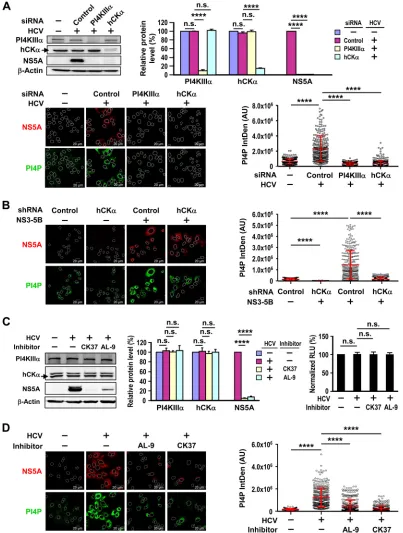
NS5A-mediated PI4P accumulation requires hCK␣activity.To explore whether
hCK␣contributes to PI4P production, we individually silenced hCK␣and PI4KIII␣and
compared the effects on PI4P levels in HCV-infected cells. PI4P levels were quantified as described previously (24, 29). As shown in Fig. 2A (top), HCV infection did not alter
PI4KIII␣and hCK␣levels. Individual transfection with PI4KIII␣and hCK␣siRNAs knocked
down PI4KIII␣and hCK␣levels to approximately 9% and 15% of those detected with
control siRNA transfection; these reductions occurred concomitantly with reduced NS5A expression (Fig. 2A, top). As shown in Fig. 1A, HCV infection augmented
intra-cellular PI4P levels, and HCV-induced PI4P accumulation was ablated by PI4KIII␣ or
hCK␣knockdown (Fig. 2A, bottom). These observations suggest a role for hCK␣in PI4P
production during HCV replication.
Next, we confirmed the inhibitory effects of stable hCK␣ knockdown on PI4P
production in cells with or without NS3–5B expression. In the absence of NS3–5B
expression, PI4P levels were lower in hCK␣ stable knockdown cells than in control
knockdown cells (Fig. 2B). As shown in Fig. 1B, NS3–5B expression upregulated PI4P production in control stable knockdown cells (Fig. 2B). Interestingly, stable depletion of
hCK␣in NS3–5B-expressing cells attenuated the increase in PI4P production induced by
NS3–5B expression (Fig. 2B). Together, these results imply that hCK␣participates in PI4P
production in cells regardless of NS3–5B protein expression, and hCK␣may function
upstream of PI4KIII␣-catalyzed PI4P production.
on November 7, 2019 by guest
http://jvi.asm.org/
FIG 2Effects of hCK␣on HCV-mediated PI4P production. (A) (Top left) Huh7 cells were first transfected with nontargeted siRNA (control) or with PI4KIII␣or hCK␣siRNA. The cells were then either mock infected (⫺) or infected with HCVcc at an MOI of 1. A subset of cells was subjected to Western blot analysis. The arrow marks the migration of hCK␣in the immunoblot. (Top right) PI4KIII␣and hCK␣levels were first normalized to those of-actin, and these levels in different settings were expressed as percentages relative to the levels detected in mock-infected cells, which were arbitrarily assigned the value of 100%. NS5A levels, after normalization to-actin levels, were detected in HCV-infected, siRNA-transfected cells and were compared to levels detected in HCV-infected cells transfected with control siRNA, which were arbitrarily assigned the value of 100%.****, P⬍0.0001; n.s., not statistically significant. (Bottom left) Another set of cells was processed by confocal microscopy using MAb 9E10 and anti-PI4P (IgM) to visualize the intracellular localization of NS5A and PI4P. A representative set of PI4P distribution patterns under different conditions is shown. (Bottom right) PI4P levels were quantitated as described in Materials and Methods. In each group, the PI4P fluorescence intensity was expressed as mean arbitrary units (AU)⫾SD from 300 cells per condition. (B) Paired T7-control shRNA/Huh7 and T7-hCK␣shRNA/Huh7 cells were transfected with the vector plasmid (⫺) or pTM-NS3–5B; the cells were monitored; and PI4P levels were quantitated. (C) (Left) Huh7 cells either remained mock infected or were infected with HCVcc, followed by treatment with DMSO as a vehicle (⫺), 100M CK37, or 5M AL-9. A set of cells
(Continued on next page)
Wong and Chen Journal of Virology
August 2017 Volume 91 Issue 16 e00355-17 jvi.asm.org 6
on November 7, 2019 by guest
http://jvi.asm.org/
[image:6.585.39.442.72.605.2]CK37 is a computationally identified compound that specifically inhibits hCK␣ by targeting its choline-binding site, leading to the inhibition of enzymatic activity (40). We showed previously that CK37 impairs viral RNA replication as well as viral protein expression in a dose-dependent manner (35). We then compared in parallel the inhibitory effects of CK37 and AL-9 on viral protein expression in HCV-infected cells.
AL-9 is a prototypical PI4KIII␣inhibitor derived from 4-aminoquinazoline that targets
PI4KIII␣activity and impairs viral replication by depleting PI4P in the plasma membrane
(41). CK37 at 100M and AL-9 at 5M greatly decreased NS5A expression but did not
affect PI4KIII␣ and hCK␣ levels (Fig. 2C, left and center). Additionally, these two
inhibitors did not influence the viability of HCV-infected cells (Fig. 2C, right).
To determine whether hCK␣activity was critical for PI4P accumulation during HCV
infection, we measured PI4P levels in mock-infected and HCV-infected cells treated with a vehicle, AL-9, or CK37. As expected, AL-9 greatly reduced HCV-induced PI4P
accu-mulation (Fig. 2D), which is consistent with previous findings that PI4KIII␣is primarily
responsible for PI4P production in HCV-expressing cells (24, 41, 42). Similarly, CK37 also
impaired PI4P accumulation in HCV-infected cells (Fig. 2D), suggesting that hCK␣
activity promotes a functional interaction with PI4KIII␣and NS5A to regulate massive
PI4P generation in HCV-expressing cells.
hCK␣activity upregulates NS5A-mediated PI4P production by activating PI4KIII␣.
To demonstrate the direct contribution of hCK␣activity to elevated PI4P production,
we evaluated the abilities of overexpressed wild-type hCK␣-R and an inactive D288A
mutant hCK␣-R (35) to upregulate PI4P production in hCK␣stable knockdown cells
expressing proteins NS3 to NS5B. These two hCK␣-R clones express authentic wild-type
and mutant hCK␣ molecules whose mRNAs are resistant to the restrictive effects
exerted by the hCK␣ short hairpin RNA (shRNA) used to construct hCK␣ stable
knockdown cells (35). Stable knockdown of hCK␣decreased hCK␣levels by 87% from
those detected in control stable knockdown cells (Fig. 3A, top). The levels of both
wild-type and D288A mutant hCK␣-R proteins overexpressed in hCK␣ stable
knock-down cells were approximately 3-fold higher than the endogenous hCK␣ levels
de-tected in control stable knockdown cells (Fig. 3A, top). Interestingly, overexpression of
the wild-type, but not the D288A mutant, hCK␣-R upregulated PI4P levels in hCK␣
stable knockdown cells relative to hCK␣stable knockdown cells transfected with the
vector control plasmid, and these levels were comparable to those detected in control stable knockdown cells (Fig. 3A, bottom).
To determine whether hCK␣-regulated PI4P accumulation occurs via PI4KIII␣
acti-vation in HCV-expressing cells, the inhibitory effect of AL-9 on wild-type hCK␣
-mediated PI4P accumulation in hCK␣ stable knockdown cells was examined. Active
hCK␣-mediated PI4P production was greatly diminished by AL-9 to a level even lower
than that detected in hCK␣ stable knockdown cells overexpressing D288A mutant
hCK␣-R (Fig. 3B). The residual PI4P levels observed in hCK␣ stable knockdown cells
overexpressing mutant hCK␣-R may be attributable to incomplete stable hCK␣
knock-down, resulting in susceptibility to AL-9 inhibition. Therefore, hCK␣upregulates PI4P
production solely via PI4KIII␣activation during HCV replication. This notion supports
the colocalization of hCK␣with NS5A and PI4KIII␣or PI4P (Fig. 1).
Compared with the previous findings that NS5A interacts with PI4KIII␣ and that
NS5A and NS5B activate kinase activity at the viral replication site (24, 28), our results
further emphasize that NS5A alone is insufficient to activate PI4KIII␣ without the
participation of hCK␣. Therefore, hCK␣may function as an indispensable coordinator to
modulate NS5A-stimulated PI4KIII␣activation for massive PI4P production.
FIG 2Legend (Continued)
was then analyzed by immunoblotting. (Center) Relative levels of the indicated cellular and viral proteins were quantified as described for panel A. (Right) Another set of cells was analyzed in a cell viability assay. Cell viability under different conditions was expressed as a percentage relative to that of mock-infected cells treated with the vehicle, which was arbitrarily designated 100%. (D) Huh7 cells either remained mock infected or were infected with HCVcc, followed by treatment with DMSO as a vehicle, AL-9, or CK37. The cells were then monitored, and PI4P levels were quantitated.
on November 7, 2019 by guest
http://jvi.asm.org/
hCK␣activity upregulates PI4KIII␣activity in the absence of viral NS protein expression. Because stable hCK␣ knockdown also decreased PI4P levels in cells
without expression of viral proteins NS3 to NS5B, we next examined whether hCK␣
inactivation inhibits PI4P production and whether PI4KIII␣overexpression compensates
for the defective PI4P production induced by CK37 in the absence of viral NS protein
expression. HA-PI4KIII␣overexpression (Fig. 4A, top) ameliorated the inhibitory effects
of CK37 on basal PI4P production (Fig. 4A, bottom), confirming that hCK␣ activity
modulates basal PI4KIII␣activity, even in the absence of NS3 to NS5B expression.
Next, we performed a rescue analysis to confirm the direct effect of hCK␣activity on
PI4KIII␣ activation. As a positive control, HA-PI4KIII␣ overexpression (Fig. 4B, top)
rescued PI4P accumulation in hCK␣ stable knockdown cells (Fig. 4B, bottom), again
indicating that hCK␣-induced PI4P production is mediated by PI4KIII␣. Similarly,
over-expression of wild-type (but not D288A mutant) hCK␣-R (Fig. 4B, top) restored PI4P in
hCK␣ stable knockdown cells to levels even higher than those detected in control
stable knockdown cells (Fig. 4B, bottom). These findings illustrate that hCK␣activity
directly modulates PI4KIII␣activity, even in the absence of viral NS protein expression.
FIG 3(A) (Top left) T7-control shRNA/Huh7 and T7-hCK␣shRNA/Huh7 cells were cotransfected with pTM-NS3–5B and either a control plasmid (⫺), wild-type hCK␣-R, or D288A mutant hCK␣-R. The cells were then subjected to Western blot analysis. (Top right) Relative protein levels were quantified. Red diamonds mark the levels of overexpressed wild-type or D288A mutant hCK␣-R relative to endogenous hCK␣levels in control stable knockdown cells. Overexpressed and endogenous hCK␣was detected with rabbit anti-hCK␣in an immunoblot analysis. (Bottom) Another set of cells was monitored, and PI4P levels were quantified. (B) (Left) T7-hCK␣shRNA/Huh7 cells were cotransfected with pTM-NS3–5B and wild-type or D288A mutant hCK␣-R and were treated with a vehicle (⫺) or AL-9 as indicated above the images. (Right) The cells were then quantitated to determine PI4P levels.****,P⬍ 0.0001; n.s., nonsignificant.
Wong and Chen Journal of Virology
August 2017 Volume 91 Issue 16 e00355-17 jvi.asm.org 8
on November 7, 2019 by guest
http://jvi.asm.org/
[image:8.585.41.402.72.440.2]hCK␣does not have a role in PI4KIII␣expression.Because hCK␣plays a crucial role in elevated PI4P production, we next investigated whether the inactivation or
depletion of hCK␣affects PI4KIII␣expression and subsequently inhibits PI4P
produc-tion. We showed above that CK37 and AL-9 did not affect PI4KIII␣levels in HCV-infected
cells (Fig. 2C). hCK␣knockdown by specific siRNAs did not exert obvious effects on
FIG 4Effect of hCK␣activity on PI4KIII␣activation without viral NS protein expression. (A) T7/Huh7 cells were transfected with the vector control plasmid or pTM-HA-PI4KIII␣and were then treated with a vehicle or CK37. (Top) The cells were then analyzed by Western blotting (left), and relative protein levels were quantified (right). The red asterisk indicates total levels of overexpressed and endogenous PI4KIII␣, which was detected with rabbit anti-PI4KIII␣by Western blotting, relative to the endogenous PI4KIII␣levels obtained in cells without CK37 treatment and HA-PI4KIII␣overexpression.**,P⬍0.01;***,P⬍ 0.001;****,P⬍0.0001; n.s., nonsignificant. (Bottom) Another set of cells was monitored (left), and PI4P levels were quantitated (right). (B) Paired T7-control shRNA/Huh7 and T7-hCK␣shRNA/Huh7 cells were transfected with either a control vector, a wild-type or D288A mutant hCK␣-R plasmid, or pTM-HA-PI4KIII␣. The cells were then analyzed by Western blotting (top), and PI4P levels were quantitated (bottom). The red asterisk indicates the total levels of overexpressed and endogenous PI4KIII␣ relative to endogenous PI4KIII␣levels obtained in control stable knockdown cells, whereas the red diamonds indicate the levels of overexpressed wild-type or D288A mutant hCK␣-R relative to endogenous hCK␣levels in control stable knockdown cells.
on November 7, 2019 by guest
http://jvi.asm.org/
[image:9.585.42.438.72.543.2]PI4KIII␣ levels in mock- and HCV-infected cells (Fig. 5A). Additionally, comparable
amounts of PI4KIII␣were detected in NS3–5B-expressing cells transfected with
untar-geted or hCK␣ siRNAs (Fig. 5B). Furthermore, a cycloheximide chase experiment
revealed indistinguishable degradation rates for PI4KIII␣in control and hCK␣
siRNA-transfected cells (Fig. 5C). These results collectively show that hCK␣ does not affect
PI4KIII␣expression.
PI4KIII␣inactivation impairs the colocalization of hCK␣with PI4KIII␣and NS5A on the ER membrane.We demonstrated previously that hCK␣activity is required for
the efficient translocation of hCK␣and NS5A to the ER (35) and showed above that the
inactivation of hCK␣or PI4KIII␣by CK37 or AL-9 reduced HCV-mediated PI4P
accumu-lation (Fig. 2D). To provide evidence for the functional link between hCK␣and PI4KIII␣,
we examined whether PI4KIII␣ inactivation exhibited a phenotype similar to that
achieved with hCK␣ knockdown, which impairs NS5A transport to the ER (35), and
whether PI4KIII␣ inactivation also hindered PI4KIII␣ translocation to the ER, because
PI4KIII␣and NS5A localization sites on the ER are indicative of viral replication factories
(24, 42).
Treatment with AL-9 (or CK37 in hCK␣ inactivation) interferes with viral RNA
replication and complicates the detection of NS5A on the ER; therefore, a transient pTM-NS3–5B expression model was employed. T7/Huh7 cells overexpressing NS3–5B
were treated with a vehicle or AL-9, and the subcellular distribution of PI4KIII␣and
NS5A and their localization on the ER, which was immunostained using anti-calreticulin (also known as calregulin [CALR], an ER-resident marker), were examined via confocal microscopy. Quantitative analysis of 20 randomly selected cells indicated that AL-9 treatment not only hindered the distribution of NS5A on the ER but also reduced the
localization of PI4KIII␣on the ER from that achieved with vehicle treatment (Fig. 6A).
FIG 5hCK␣is not involved in PI4KIII␣expression. (A) Huh7 cells were transfected with control or hCK␣siRNA and then either remained mock infected or were infected with HCVcc. (Left) The cells were then harvested and subjected to immunoblot detection. (Right) PI4KIII␣levels under different experimental conditions, relative to the level detected in control siRNA transfection without HCV infection, which was arbitrarily set at 100%, were quantitated. (B) T7/Huh7 cells were transfected first with untargeted or hCK␣ siRNA and then with pTM-NS3–5B. The cells were then subjected to Western blotting. n.s., nonsignificant. (C) Huh7 cells were transfected with control or hCK␣siRNA and were then treated with cycloheximide. (Left) The cells were collected at different time points post-cycloheximide addition for Western blot analysis. (Right) PI4KIII␣levels were first normalized to those of-actin. PI4KIII␣levels at different times, expressed as percentages relative to the level detected at time zero in each siRNA transfection, which was arbitrarily set to 100%, are shown. The data from 5 independent analyses were fit to a linear regression equation in order to assess the degradation rate of PI4KIII␣in control or hCK␣transient knockdown cells.
Wong and Chen Journal of Virology
August 2017 Volume 91 Issue 16 e00355-17 jvi.asm.org 10
on November 7, 2019 by guest
http://jvi.asm.org/
[image:10.585.41.439.71.314.2]FIG 6Effects of PI4KIII␣inactivation on the translocation of hCK␣, NS5A, and PI4KIII␣to the ER. (A) T7/Huh7 cells were first transfected with pTM-NS3–5B and then treated with a vehicle or AL-9. The cells were fixed and stained with MAb 9E10, rabbit anti-PI4KIII␣, and goat anti-CALR, followed by incubation with the appropriate Alexa Fluor-conjugated secondary antibodies. The cells were then examined by confocal microscopy. (B) T7/Huh7 cells were cotransfected with pTM-HA-PI4KIII␣and pTM-NS3–5B and were then treated with a vehicle or AL-9. The cells were immunostained with MAb 9E10, rabbit anti-hCK␣, and goat anti-HA and were examined by confocal microscopy. (C) T7/Huh7 cells were first transfected with pTM-NS3–5B and then treated with a vehicle or AL-9. The cells were immunostained with MAb 9E10, rabbit anti-hCK␣, and goat anti-CALR and were then analyzed by confocal microscopy. In each analysis, a representative image of the fluorescence signals is shown. The boxed area shown in the merged image was enlarged, and the colocalization of the two or three indicated proteins is marked with white arrowheads or yellow arrows, respectively. The colocalization of the indicated proteins was quantitated by determining the colocalization area of the indicated molecules relative to the entire cell area, which is shown in each bar graph.****,P⬍0.0001; n.s., nonsignificant.
on November 7, 2019 by guest
http://jvi.asm.org/
[image:11.585.49.359.70.604.2]In addition, AL-9 treatment hindered the ER colocalization of these two molecules
(Fig. 6A). However, PI4KIII␣ still effectively colocalized with NS5A regardless of AL-9
treatment (Fig. 6A).
We then investigated whether AL-9 affected the colocalization of hCK␣with
over-expressed HA-PI4KIII␣and NS5A. AL-9 decreased the colocalization of hCK␣with NS5A
or with HA-PI4KIII␣, as well as the colocalization of hCK␣ with both HA-PI4KIII␣and
NS5A (Fig. 6B). Because PI4KIII␣ activity was required for the efficient transport of
PI4KIII␣and NS5A to the ER membrane (Fig. 6A) as well as for the colocalization of hCK␣
with HA-PI4KIII␣and NS5A (Fig. 6B), we sought to determine whether PI4KIII␣activity
is also important for the colocalization of hCK␣with NS5A on the ER. As shown in Fig.
6C, treatment with AL-9 not only impeded hCK␣ localization on the ER but also
interfered with the colocalization of hCK␣and NS5A, as well as with the colocalization
of these proteins on the ER.
Collectively, these data show that in addition to the PI4KIII␣-regulated translocation
of PI4KIII␣and NS5A to the ER, the trafficking of hCK␣to the ER is regulated by PI4KIII␣
activity. These results also indicate that PI4KIII␣ activity is required for the effective
colocalization of hCK␣with PI4KIII␣and NS5A on the ER, whereas it is not crucial for
PI4KIII␣and NS5A colocalization.
Inactivation of hCK␣interferes with the translocation of hCK␣, PI4KIII␣, and NS5A to the ER membrane.To further address the functional relationship between
hCK␣and PI4KIII␣, we examined the effect of hCK␣inactivation on the colocalization
of PI4KIII␣ and NS5A on the ER. In agreement with our previous finding that hCK␣
activity is crucial for the transport of NS5A to the ER (35), CK37 treatment hindered the
localization of PI4KIII␣on the ER (Fig. 7A). Additionally, CK37 not only inhibited the
colocalization of PI4KIII␣with NS5A but also interfered with the colocalization of these
proteins on the ER (Fig. 7A). In addition to inhibiting the colocalization of hCK␣with
NS5A, CK37 treatment also interfered with the colocalization of HA-PI4KIII␣ with
hCK␣ and NS5A (Fig. 7B). Moreover, CK37 treatment impaired the colocalization of
hCK␣with NS5A on the ER (Fig. 7C). Therefore, the results shown in Fig. 7 collectively
demonstrate that hCK␣activity plays a critical role in the ER translocation of hCK␣and
NS5A and is also necessary for the effective translocation of PI4KIII␣to the ER.
Inactivation of hCK␣and PI4KIII␣does not affect the localization of NS5B on the ER.We demonstrated previously that the ER localization of NS5A (but not that of
NS5B or NS3) is inhibited by hCK␣knockdown (35). We next investigated whether the
inhibitory effects of hCK␣or PI4KIII␣inactivation on the translocation of NS5A to the
ER are specific by evaluating the effects of hCK␣ or PI4KIII␣ inactivation on the ER
localization of NS5B. In contrast to the inhibitory effects of AL-9 and CK37 on the
localization of HA-PI4KIII␣on the ER, AL-9 or CK37 did not affect the localization of
NS5B on the ER (Fig. 8A). However, the colocalization of NS5B with HA-PI4KIII␣and the
colocalization of these proteins on the ER were inhibited by AL-9 or CK37 (Fig. 8A). As an internal control, Western blot analysis indicated that CK37 or AL-9 treatment did not
affect levels of PI4KIII␣, hCK␣, or NS5B (Fig. 8B). Based on these observations, we
conclude that NS5B localizes on the ER even when PI4KIII␣ is redirected to non-ER
membranes due to the disruption of PI4P synthesis.
hCK␣forms a ternary complex with PI4KIII␣and NS5A.In order to understand
the molecular functions underlying the role of hCK␣in the production of PI4P and the
ER trafficking of hCK␣, PI4KIII␣, and NS5A, we performed coimmunoprecipitation (co-IP)
to examine whether these three functionally interrelated proteins form a complex in
cells. Lysates from cells overexpressing NS3–5B and HA-PI4KIII␣were incubated with a
rabbit isotype IgG or anti-HA, and the precipitated proteins were subjected to
immu-noblot analysis. HA-PI4KIII␣was specifically precipitated by anti-HA but not by control
IgG (Fig. 9A, left). Anti-HA also specifically pulled down hCK␣as well as NS5A (Fig. 9A,
left). Additionally, the precipitation of hCK␣with rabbit anti-hCK␣specifically
cocap-tured HA-PI4KIII␣as well as NS5A (Fig. 9A, right).
Wong and Chen Journal of Virology
August 2017 Volume 91 Issue 16 e00355-17 jvi.asm.org 12
on November 7, 2019 by guest
http://jvi.asm.org/
FIG 7Effects of hCK␣inactivation on the translocation of PI4KIII␣and NS5A to the ER. (A) T7/Huh7 cells were transfected with pTM-NS3–5B, followed by treatment with a vehicle or CK37. The cells were incubated with MAb 9E10, rabbit anti-PI4KIII␣, and goat anti-CALR and were then analyzed by confocal microscopy. (B) T7/Huh7 cells were cotransfected with pTM-HA-PI4KIII␣and pTM-NS3–5B, followed by vehicle or CK37 treatment. The cells were immunostained with MAb 9E10, rabbit anti-hCK␣, and goat anti-HA and were then analyzed by confocal microscopy. (C). T7/Huh7 cells were first transfected with pTM-NS3–5B and then treated with a vehicle or CK37. The cells were immunostained with MAb 9E10, rabbit anti-hCK␣, and goat anti-CALR prior to confocal microscopy. In each study, fluorescence images were analyzed, and colocalization of the indicated proteins was quantified as described in the legend to Fig. 6.****,P⬍0.0001.
on November 7, 2019 by guest
http://jvi.asm.org/
[image:13.585.46.364.71.641.2]To confirm ternary complex formation in infected cells, lysates from
HCV-infected cells were incubated with rabbit isotype IgG or anti-PI4KIII␣, followed by
Western blotting. Precipitation of PI4KIII␣ with anti-PI4KIII␣ also specifically pulled
down NS5A and hCK␣(Fig. 9B, left). Additionally, anti-NS5A not only precipitated NS5A
but also cocaptured PI4KIII␣and hCK␣(Fig. 9B, right). Thus, hCK␣, PI4KIII␣, and NS5A
are interacting partners that form the ternary complex.
hCK␣binds PI4KIII␣independently of NS5A.Because hCK␣was found to
colo-calize with PI4KIII␣in the absence of NS5A, and because NS5A expression augmented
the colocalization of hCK␣and PI4KIII␣(Fig. 1C), we examined whether hCK␣formed
a complex with PI4KIII␣ in the absence of NS5A and whether NS5A expression
en-hanced the binding of hCK␣and PI4KIII␣. The precipitation of HA-PI4KIII␣with anti-HA
from cells coexpressing HA-PI4KIII␣ and hCK␣ tagged with a Flag epitope at its C
terminus, i.e., hCK␣-Flag, with or without NS5A coexpression, pulled down similar
amounts of hCK␣-Flag (Fig. 9C, left). Similarly, the precipitation of hCK␣-Flag with
anti-Flag cocaptured comparable amounts of HA-PI4KIII␣in the presence or absence of
NS5A coexpression (Fig. 9C, right). These results indicate that NS5A does not have a
crucial role in the hCK␣-PI4KIII␣interaction.
PI4KIII␣activity is not necessary for the binding of PI4KIII␣to NS5A or hCK␣ and is not critical for hCK␣-Flag and NS5A interactions.In order to determine the
molecular events underlying the effects of PI4KIII␣ inactivation on the transport of
FIG 8Examination of the effects of PI4KIII␣or hCK␣inactivation on the localization of NS5B on the ER. (A) T7/Huh7 cells were cotransfected with pTM-HA-PI4KIII␣and pTM-NS3–5B, followed by treatment with a vehicle, AL9, or CK37. The cells were immunostained with mouse anti-NS5B, rabbit anti-HA, and goat anti-CALR and were then analyzed by confocal microscopy. Fluorescence images were analyzed to quantify the colocalization of the indicated proteins.****,P⬍0.0001; n.s., nonsignificant. (B) Another set of samples was analyzed by Western blotting (top), and the relative protein levels were quantified (bottom).
Wong and Chen Journal of Virology
August 2017 Volume 91 Issue 16 e00355-17 jvi.asm.org 14
on November 7, 2019 by guest
http://jvi.asm.org/
[image:14.585.47.427.72.419.2]hCK␣, PI4KIII␣, and NS5A to the ER (Fig. 6), phenylarsine oxide (PAO), an inhibitor of
PI4KIII␣(28), was applied to investigate the binding of PI4KIII␣to NS5A or hCK␣. PAO
treatment did not affect the binding of HA-PI4KIII␣to NS5A (Fig. 9D) or hCK␣-Flag (Fig.
9E). In addition, PAO did not affect the binding of hCK␣-Flag to NS5A (Fig. 9F), a finding
consistent with the observation that PI4KIII␣ knockdown did not exert any apparent
effects on the hCK␣-Flag and NS5A interaction (Fig. 9G). These results collectively
indicate that PI4KIII␣ activity is not involved in the interactions between any two
components of the ternary complex.
FIG 9Analysis of the formation of the hCK␣–NS5A–PI4KIII␣ternary complex and the effects of NS5A, PAO, and PI4KIII␣knockdown on protein-protein interactions. (A) T7/Huh7 cells were cotransfected with pTM-HA-PI4KIII␣ and pTM-NS3–5B. The cell lysates were incubated with rabbit isotype-matched control IgG (⫺) or rabbit anti-HA (left) or with rabbit isotype control IgG or rabbit anti-hCK␣(right). The precipitated proteins were subjected to immunoblot analysis. A portion of the cell lysates corresponding to 5% of the total proteins used for co-IP was also loaded as an input control in the Western blot analysis. (B) Lysates obtained from HCV-infected cells were incubated with rabbit control isotype IgG or anti-PI4KIII␣(left) or with mouse control isotype IgG or anti-NS5A (right), and the precipitated proteins were subjected to Western blot detection. (C) T7/Huh7 cells were cotrans-fected with pTM-HA-PI4KIII␣and pCMV6-hCK␣with or without pTM-NS5A. The cell lysates were incubated with rabbit anti-HA (left) or rabbit anti-Flag (right). The isolated proteins were subjected to immunoblot analysis. (D and E) T7/Huh7 cells were cotransfected with pTM-HA-PI4KIII␣ and pTM-NS5A (D) or with pTM-HA-PI4KIII␣ and pCMV6-hCK␣(E). The cells were treated with a vehicle or PAO, and the cell lysates were incubated with rabbit anti-HA prior to Western blot analysis. (F) T7/Huh7 cells were cotransfected with pTM-NS3–5B and pCMV6-hCK␣, followed by vehicle or PAO treatment. Cell lysates were precipitated with rabbit anti-Flag prior to immunoblot analysis. (G) T7/Huh7 cells were first transfected with nontargeted siRNA or PI4KIII␣siRNA, followed by transfection with pCMV6-hCK␣ and pTM-NS5A. The lysates from transfected cells were immunoprecipitated with mouse anti-NS5A prior to Western blot analysis. In panels C to G, the results from three independent analyses did not demonstrate statistically significant differences between the vehicle and PAO treatment or between control and tested conditions. Therefore, representative sets of data are shown.
on November 7, 2019 by guest
http://jvi.asm.org/
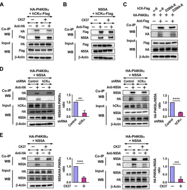
[image:15.585.43.404.70.437.2]Although hCK␣does not have an apparent effect on the interaction of hCK␣ with PI4KIII␣or NS5A, its activity facilitates the binding of PI4KIII␣to NS5A.To
understand the molecular basis of the effects of hCK␣activity on the ER translocation
of hCK␣, PI4KIII␣, and NS5A (Fig. 7), we examined whether CK37 affected the binding
of hCK␣ to PI4KIII␣ or NS5A. According to co-IP analyses, CK37 did not affect the
binding of hCK␣-Flag to HA-PI4KIII␣ (Fig. 10A) or alter the hCK␣-Flag and NS5A
interaction (Fig. 10B). Additionally, wild-type hCK␣-R and the D288A mutant bound
comparably to HA-PI4KIII␣(Fig. 10C), supporting the former observation.
FIG 10Effects of CK37 and hCK␣knockdown on protein-protein interactions. (A and B) T7/Huh7 cells were cotransfected with pTM-HA-PI4KIII␣and pCMV6-hCK␣(A) or with pTM-NS5A and pCMV6-hCK␣(B). The cells were then treated with a vehicle or CK37, and cell lysates were incubated with rabbit isotype IgG or anti-HA (A) or with rabbit isotype IgG or anti-Flag (B). The precipitated proteins were then analyzed by Western blotting. (C) T7/Huh7 cells were cotransfected with pTM-HA-PI4KIII␣and wild-type or D288A mutant hCK␣-R. The cell lysates were then incubated with rabbit anti-Flag, and the precipitated proteins were subjected to Western blot analysis. (D) T7-control shRNA/Huh7 and T7-hCK␣shRNA/Huh7 cells were cotransfected with pTM-HA-PI4KIII␣and pTM-NS5A. The cell lysates were incubated with rabbit anti-HA (left) or an anti-NS5A MAb (right) prior to Western blot analysis. The binding of the two coimmunoprecipitated proteins, as indicated in the quantitation graphs, obtained from hCK␣stable knockdown cells was expressed as a ratio relative to the binding obtained from control stable knockdown cells, which was arbitrarily set at a ratio of 1. (E) T7/Huh7 cells were cotransfected with pTM-HA-PI4KIII␣and pTM-NS5A, followed by treatment with a vehicle or CK37 as indicated. The cell lysates were then incubated with rabbit anti-HA (left) or an anti-NS5A MAb (right), and the precipitated proteins were analyzed by immunoblot detection. The binding of the two coprecipitated proteins, as indicated in each quantitation graph, obtained from vehicle-treated cells was arbitrarily assigned a ratio of 1, and binding obtained from CK37 treatment was expressed as a relative ratio. In panels A to C, the results from three separate studies did not demonstrate statistically significant differences between vehicle and CK37 treatment or between the wild type and D288A mutant hCK␣-R expression. Therefore, representative sets of data are shown.**,P⬍0.01;***,P⬍0.001;****,P⬍0.0001.
Wong and Chen Journal of Virology
August 2017 Volume 91 Issue 16 e00355-17 jvi.asm.org 16
on November 7, 2019 by guest
http://jvi.asm.org/
[image:16.585.42.429.69.461.2]Next, we examined whether hCK␣is involved in the PI4KIII␣-NS5A interaction. The
precipitation of HA-PI4KIII␣from lysates containing HA-PI4KIII␣and NS5A with anti-HA
pulled down a barely detectable NS5A band in hCK␣ stable knockdown cells, in
contrast to the band obtained from control stable knockdown cells (Fig. 10D, left).
Conversely, the precipitation of NS5A with anti-NS5A from hCK␣ stable knockdown
cells cocaptured a barely visible HA-PI4KIII␣band, in contrast to the band observed in
control stable knockdown cells (Fig. 10D, right). Quantitation of the data from three
independent studies indicated that hCK␣stable knockdown reduced the ratios of NS5A
binding to HA-PI4KIII␣and HA-PI4KIII␣binding to NS5A to approximately 0.24 and 0.28,
respectively, relative to that for control stable knockdown cells, which was arbitrarily designated 1 (Fig. 10D).
Because CK37 reduces the colocalization of PI4KIII␣ and NS5A (Fig. 7A), we then
determined whether the binding of PI4KIII␣to NS5A requires hCK␣activity. When cell
lysates containing HA-PI4KIII␣ and NS5A were incubated with anti-HA or anti-NS5A,
smaller amounts of NS5A or HA-PI4KIII␣ coprecipitated with HA-PI4KIII␣ or NS5A,
respectively, in CK37-treated cells than in vehicle-treated cells (Fig. 10E, left and right, respectively). Quantitation of the data from three separate analyses indicated that CK37
treatment reduced the ratios of NS5A binding to HA-PI4KIII␣and HA-PI4KIII␣binding
to NS5A to approximately 0.28 and 0.21 relative to that for vehicle treatment, which was arbitrarily designated 1 (Fig. 10E, left and right, respectively). These results indicate
that hCK␣activity promotes the recruitment of PI4KIII␣to NS5A.
hCK␣upregulates PI4KIII␣activityin vitroin the presence or absence of NS5A.
To further support the role of hCK␣ in PI4KIII␣ activation, the in vitro activity of
recombinant PI4KIII␣was assayed in the presence or absence of recombinant NS5A
and/or hCK␣. Recombinant PI4KIII␣, obtained from the baculovirus expression system,
comprises the amino acid residues between positions 1249 and 2102 in the kinase,
including the kinase catalytic domain. Recombinant NS5A, purified fromEscherichia coli,
comprises amino acids 2061 to 2302 in the HCV polyprotein, i.e., the C-terminal portion of domain 1, low-complexity sequence 1, and the N-terminal portion of domain 2 of
NS5A. Both recombinant proteins are fused with glutathioneS-transferase (GST) at their
N termini, and their expected molecular masses are 124 kDa (for PI4KIII␣) and 53 kDa
(for NS5A). Recombinant hCK␣was purified from 293T cells transfected with
pCMV6-hCK␣. The authenticity of these proteins was confirmed by immunoblotting (Fig. 11A).
As expected, the addition of NS5A to PI4KIII␣ increased PI4KIII␣ activity (Fig. 11B,
compare bars 1 and 2). The addition of hCK␣to PI4KIII␣increased PI4KIII␣basal activity
to levels even higher than those activated by NS5A (Fig. 11B, compare bar 1 with bars
2 and 3). Remarkably, hCK␣, in conjunction with NS5A, greatly augmented PI4KIII␣
activation (Fig. 11B, compare bars 2 and 4). hCK␣-stimulated basal PI4KIII␣activity was
inhibited by both PAO (Fig. 11B, compare bars 3 and 5) and CK37 (Fig. 11B, compare
bars 3 and 6). Likewise, hCK␣-augmented PI4KIII␣ activity triggered by NS5A was
ablated by PAO (Fig. 11B, compare bars 4 and 7) and CK37 (Fig. 11B, compare bars 4
and 8). Based on these results, we conclude that hCK␣ activity directly stimulatesin
vitroPI4KIII␣activity and that ternary complex formation is essential for robust PI4KIII␣ activation.
PI4KIII␣overexpression rescues the impaired viral replication and PI4P produc-tion caused by hCK␣inactivation.Because hCK␣effectively interacted with PI4KIII␣
regardless of PI4KIII␣or hCK␣inactivation (Fig. 9E and 10A and C), we next examined
the functional interaction between these two proteins and tested whether PI4KIII␣
overexpression counteracts the negative effects of CK37 on viral replication in
HCV-infected cells. Total levels of overexpressed HA-PI4KIII␣and endogenous PI4KIII␣were
approximately 4.7-fold greater than the endogenous PI4KIII␣levels detected in
mock-infected cells without CK37 treatment or HA-PI4KIII␣ overexpression (Fig. 12A). The
overexpression of PI4KIII␣in CK37-treated, HCV-infected cells resulted in levels of viral
proteins, such as NS3, NS5A, and Core, higher than those in CK37-treated, HCV-infected
cells without PI4KIII␣overexpression (Fig. 12A). Additionally, the levels of these viral
proteins were comparable to those detected in HCV-infected cells treated with a
on November 7, 2019 by guest
http://jvi.asm.org/
vehicle (Fig. 12A). Based on these results, we conclude that PI4KIII␣ overexpression
compensates for the defects associated with hCK␣inactivation during viral replication.
To determine whether hCK␣modulates viral replication by activating PI4KIII␣for
mass PI4P production, we determined whether PI4KIII␣overexpression could rescue
PI4P production in CK37-treated HCV-infected cells. In agreement with the restoration
of viral replication, the overexpression of PI4KIII␣substantially augmented PI4P
pro-duction in HCV-infected cells treated with CK37 (Fig. 12B). These findings, taken
together, indicate that the deficiency in hCK␣activity observed during suppressed viral
replication proceeds via attenuation of PI4KIII␣-mediated PI4P production.
Expression of active (but not inactive) hCK␣ promotes the translocation of PI4KIII␣and NS5A to the ER in hCK␣stable knockdown cells.According to our results,
hCK␣inactivation interferes with the translocation of PI4KIII␣and NS5A to the ER (Fig.
7); therefore, we next investigated whether hCK␣activity directly upregulates the ER
translocation of PI4KIII␣and NS5A in hCK␣stable knockdown cells. In agreement with
the inhibitory effects of CK37 on the ER translocation of NS5A and PI4KIII␣, stable hCK␣
knockdown also reduced the localization of NS5A and HA-PI4KIII␣on the ER (Fig. 13).
Overexpression of wild-type (but not D288A mutant) hCK␣-R in hCK␣ stable
knock-down cells not only restored NS5A localization on the ER, in agreement with our
previous observations (35), but also rescued the translocation of HA-PI4KIII␣to the ER
(Fig. 13). Moreover, overexpression of wild-type (but not D288A mutant) hCK␣-R
restored the colocalization of NS5A and HA-PI4KIII␣on the ER (Fig. 13). These results,
together with our previous findings (35), demonstrate that hCK␣ activity positively
regulates the translocation of hCK␣as well as NS5A and PI4KIII␣to the ER.
Overexpression of active (but not inactive) PI4KIII␣restores the translocation of PI4KIII␣and NS5A to the ER in CK37-treated cells.Treatment with AL-9 or CK37
attenuated the production of PI4P (Fig. 2D) and the translocation of PI4KIII␣and NS5A
as well as hCK␣ to the ER (Fig. 6 and 7); therefore, we investigated whether PI4P
production is essential for the ER translocation of PI4KIII␣and NS5A. To address this
issue, we investigated the abilities of active and kinase-deficient (KD) (K1792L mutant)
HA-PI4KIII␣to ameliorate the impaired ER translocation of PI4KIII␣and NS5A caused by
FIG 11Upregulation of PI4KIII␣activityin vitroby hCK␣. (A) Thirty nanograms each of recombinant PI4KIII␣, hCK␣, and NS5A proteins was analyzed by Western blotting using rabbit anti-PI4KIII␣, rabbit anti-hCK␣, and an anti-NS5A MAb, respectively. (B) Thein vitroactivity of purified recombinant PI4KIII␣was assayed in the presence or absence of recombinant NS5A, hCK␣, and/or inhibitors in an ADP-Glo kinase assay as indicated. Kinase activity, monitored as the conversion of ATP to ADP under different experimental conditions relative to that detected in PI4KIII␣alone, which was arbitrarily set at a value of 1, was expressed as the fold change in luciferase activity. The dashed red line indicates the threshold for this in vitrokinase assay. The data shown are the quantitated results from four independent experiments.**,P⬍0.01;***,P⬍0.001.
Wong and Chen Journal of Virology
August 2017 Volume 91 Issue 16 e00355-17 jvi.asm.org 18
on November 7, 2019 by guest
http://jvi.asm.org/
[image:18.585.43.401.69.285.2]CK37 in NS3–5B-expressing cells. The PI4KIII␣mutant is unable to produce PI4P or to support viral replication (28). In agreement with the results shown in Fig. 7, CK37
reduced the translocation of PI4KIII␣and NS5A to the ER (Fig. 14A), and overexpression
of wild-type HA-PI4KIII␣, but not its kinase-deficient mutant (Fig. 14B), restored the
translocation of PI4KIII␣ and NS5A to the ER in CK37-treated cells (Fig. 14A). These
results strongly indicate that PI4KIII␣-mediated PI4P production is a prerequisite for the
translocation of PI4KIII␣and NS5A to the ER (also, see below).
DISCUSSION
We previously demonstrated the critical function of hCK␣in promoting HCV
mem-branous RC formation and viral replication (35). In our present study, we further
elucidate the mechanism underlying the regulatory roles of hCK␣in HCV replication.
Like PI4KIII␣, hCK␣ played a crucial role in PI4P production and viral replication, as
shown by siRNA knockdown and inhibitor analyses. Additionally, both hCK␣ and
PI4KIII␣activities were critical for the translocation of hCK␣, PI4KIII␣, and NS5A to the
ER-derived membrane, indicating that these two kinases and NS5A function in concert
to facilitate their own translocation to the ER. Furthermore, hCK␣activity was essential
for the generation of elevated PI4P pools in HCV-expressing cells. The overexpression
of wild-type (but not D288A mutant) hCK␣-R restored PI4P production in hCK␣stable
knockdown cells, which is consistent with our previous finding that the active (but not
FIG 12Rescue of viral replication and PI4P production by PI4KIII␣overexpression in CK37-treated, HCV-infected cells. (A) T7/Huh7 cells remained uninfected or were infected with HCV; they were then transfected with the control vector or pTM-HA-PI4KIII␣and were treated with the vehicle or CK37. (Left) The cells were then analyzed by Western blotting. (Right) PI4KIII␣ and hCK␣levels under the tested conditions relative to those detected in mock-infected cells without HA-PI4KIII␣overexpression or CK37 treatment were determined. The red asterisk marks the total levels of overexpressed and endogenous PI4KIII␣, which were detected using anti-PI4KIII␣in a Western blot, relative to endogenous PI4KIII␣levels in mock-infected cells without overexpression or CK37 treatment, expressed as a percentage. The levels of NS3, NS5A, and core under different conditions, relative to the levels detected in HCV-infected cells without overexpression or CK37 treatment, are also shown. (B) Another set of cells, as described for panel A, was monitored for the intracellular localization of NS5A and PI4P (left), and PI4P levels were quantitated (right).**,P⬍0.01;***,P⬍0.001;****,P⬍0.0001; n.s., nonsignificant.
on November 7, 2019 by guest
http://jvi.asm.org/
[image:19.585.39.401.79.373.2]the D288A mutant) hCK␣-R can rescue viral replication (35). Moreover, wild-type
hCK␣-R-mediated PI4P accumulation was abrogated by AL-9, a PI4KIII␣-specific
inhib-itor. These results collectively indicate that the profound PI4P synthesis mediated by
hCK␣occurs primarily via the upregulation of NS5A-stimulated PI4KIII␣activation and
that PI4P functions as a critical determinant for the effective trafficking of these three interacting partners (see below) to the ER membrane for productive viral replication.
hCK␣ is present predominantly as a cytosolic protein, and HCV infection or the
FIG 13Effects of overexpression of the wild-type and D288A mutant hCK␣-R proteins on the restoration of the ER translocation of PI4KIII␣and NS5A in hCK␣stable knockdown cells. T7-control shRNA/Huh7 and T7-hCK␣shRNA/Huh7 cells were cotransfected with pTM-NS3–5B and pTM-HA-PI4KIII␣along with the vector plasmid, wild-type hCK␣-R, or D288A mutant hCK␣-R. The cells were processed for confocal microscopy using MAb 9E10, goat anti-CALR, and rabbit anti-HA. (Top) Set of representative fluorescence images. (Bottom) Quantitation of the colocalization of the proteins.****,P⬍0.0001; n.s., nonsignificant.
Wong and Chen Journal of Virology
August 2017 Volume 91 Issue 16 e00355-17 jvi.asm.org 20
on November 7, 2019 by guest
http://jvi.asm.org/
[image:20.585.47.364.67.577.2]expression of NS proteins facilitates the localization of hCK␣to the ER (35). Given the multiple intracellular localization sites of NS5A in perinuclear membranes, such as those of the ER, Golgi complex, and lipid droplets (5, 43), as well as the localization sites of
PI4KIII␣in the ER (9, 10), Golgi complex-associated membrane vesicles (13), and the
nucleolus (14), it would be difficult to determine unambiguously where PI4KIII␣and
NS5A localize when PI4P synthesis is interrupted by CK37 or AL-9. Here we used an
ER-residing marker, CALR, to investigate the transport or “translocation” of PI4KIII␣,
NS5A, and hCK␣to the PI4P-enriched, ER-associated membrane, because HCV
replica-tion increases their localizareplica-tion to the CALR-associated membrane. In contrast, AL-9 or CK37 hinders their colocalization with CALR, indicating that interference with PI4P
FIG 14Effects of the overexpression of active and kinase-deficient PI4KIII␣proteins on the rescue of impaired ER translocation of PI4KIII␣and NS5A induced by CK37. (A) T7/Huh7 cells were cotransfected with pTM-NS3–5B and the control plasmid, pEF1A-HA-PI4KIII␣-WT, or pEF1A-HA-PI4KIII␣-KD, followed by vehicle or CK37 treatment. For the positive control for the ER translocation of PI4KIII␣and NS5A, a set of cells cotransfected with pTM-NS3–5B and the control vector was treated with the vehicle. The cells were processed for confocal microscopy using MAb 9E10, goat anti-CAL, and rabbit anti-PI4KIII␣(for cells without HA-PI4KIII␣overexpression) or rabbit anti-HA (for HA-PI4KIII␣overexpression) to quantify the degree of translocation of PI4KIII␣and NS5A to the ER. (B) Another set of samples was analyzed by Western blotting (top), and the relative protein levels were quantified (bottom). The red asterisk marks the levels of overexpressed wild-type or KD mutant HA-PI4KIII␣plus endogenous PI4KIII␣relative to those observed in cells without overexpression or CK37 treatment.**,P⬍0.01;***,P⬍0.001;****,P⬍0.0001; n.s., nonsignificant.
on November 7, 2019 by guest
http://jvi.asm.org/
[image:21.585.45.433.72.502.2]production detours their ER translation to other membranes, which are collectively referred to as “non-ER membranes,” because they are not associated with CALR. Although HCV replication induces the ER transport of only a portion of these three replication components, this process is biologically and virologically relevant, because their “ER translocation” correlates well with elevated PI4P production and viral repli-cation.
In agreement with our previous observation that the ER localization of NS5B is not
affected by hCK␣ knockdown (35), NS5B localization on the ER, as shown by its
colocalization with the ER marker CALR, was not influenced by AL-9 or CK37. However,
the colocalization of NS5B and PI4KIII␣and their colocalization on the ER were inhibited
by AL-9 or CK37. Based on these observations, we conclude that NS5B still localizes on
the ER membrane when PI4P synthesis is impaired by inhibitors, while PI4KIII␣localizes
to non-ER membranes. Therefore, the transport of NS5A and NS5B to the ER is differentially regulated; the route that delivers NS5A to the ER is strictly dependent on PI4P production, whereas the localization of NS5B on the ER is independent of PI4P.
Therefore, HCV replication specifically “redirects” PI4KIII␣, NS5A, and hCK␣to the ER.
Furthermore, we performed a series of co-IP analyses to elucidate the molecular
basis of the function of hCK␣in NS5A-stimulated PI4KIII␣activation and the
translo-cation of these three molecules to the ER. The inactivation of PI4KIII␣by PAO or of hCK␣
by CK37 did not have an apparent effect on PI4KIII␣-hCK␣binding. In support of this
finding, the wild-type and D288A mutant hCK␣-R clones interacted comparably with
PI4KIII␣. Thus, the inability of the D288A mutant to rescue PI4P generation in hCK␣
stable knockdown cells expressing NS3–5B proteins is attributable to its lack of hCK␣
activity rather than to its inability to bind PI4KIII␣. In the present study, as well as in
previous studies (35), we observed that D288A mutant hCK␣still possesses
wild-type-like NS5A-binding ability (35), and PAO or CK37 did not affect the interaction between
these two molecules. In addition, hCK␣interacted effectively with NS5A, even when
PI4KIII␣was knocked down. Collectively, these results imply that PI4P levels do not
influence hCK␣binding to PI4KIII␣ or NS5A. Nevertheless, AL-9 or CK37 reduced the
colocalization of hCK␣with NS5A or PI4KIII␣on the ER membrane. Thus, in the absence
of massive PI4P production, the physical interactions between hCK␣and PI4KIII␣and
between hCK␣and NS5A do not ensure the correct translocation of hCK␣with PI4KIII␣
or NS5A to the same ER membrane; instead, these paired molecules are spatially segregated into different non-ER membrane compartments (Fig. 15A, center and right).
Although AL-9 did not affect PI4KIII␣ and NS5A colocalization, it inhibited the
colocalization of these two molecules on the ER. This observation is consistent with
the finding that the PI4KIII␣-NS5A interaction was not influenced by PAO, as shown in
the present study, or by AL-9, as reported previously (29). Thus, in the absence of
PI4KIII␣activity, PI4KIII␣and NS5A still colocalize and interact with each other on the
same non-ER membrane (Fig. 15A, center). In contrast, CK37 inhibited the colocalization
of these two proteins, a finding compatible with the co-IP results showing that hCK␣
knockdown or inactivation by CK37 impaired PI4KIII␣-NS5A binding. In the absence of
hCK␣ activity, hCK␣, PI4KIII␣, and NS5A appear to segregate into different non-ER
membrane compartments in which hCK␣ still binds to PI4KIII␣ and NS5A (Fig. 15A,
right). Conversely, the paradoxical discrepancy between the differential effects of NS5A
on the colocalization of PI4KIII␣with hCK␣and the binding of PI4KIII␣to hCK␣may be
interpreted as follows. In the absence of NS5A, hCK␣-PI4KIII␣ complexes do not
accumulate on membranes devoid of PI4P, thus increasing the difficulty in visualizing these complexes via microscopy. However, NS5A expression enhances the translocation
of the hCK␣-PI4KIII␣-NS5A complex to the ER, resulting in the formation and
accumu-lation of viral RCs on the PI4P-enriched membrane (Fig. 15B) and increasing the
visibility of these complexes by confocal microscopy over that of the single hCK␣
-PI4KIII␣complex.
The differential effects of CK37 and PAO on the colocalization and interaction of
PI4KIII␣ and NS5A are not attributable to the influence of PI4P production on the
PI4KIII␣-NS5A interaction. Instead, they may be ascribed to the specific function of
Wong and Chen Journal of Virology
August 2017 Volume 91 Issue 16 e00355-17 jvi.asm.org 22