A Novel Method for Studying Nucleated Pathways in
Membranes: Development and Applications for Gene
Delivery
Thesis by
Christina L. Ting
In Partial Fulfillment of the Requirements for the Degree of
Doctor of Philosophy
California Institute of Technology Pasadena, California
2013
c
Acknowledgments
First and foremost, I am grateful to my advisor, Zhen-Gang Wang. His patience, honesty, and encouragement during my time at Caltech has been instrumental in my scientific and personal growth. It has been a pleasure to be the student of such an inspirational person.
I am thankful to Kathy Bubash and Alison Ross for welcoming me to Caltech, and later for always being helpful and positive. I am also thankful to the past and present members of the Wang group for their support, especially to Brad for his mentorship, Issei for taking care of Kiki, and Bilin for organizing the group lunches and hikes.
I wish to thank the members of my committee, William Clemons, Mark Davis and Doug Rees, for helpful discussions on the work done in this thesis.
Of course, my friends and family have made this experience a lot more enjoyable. BG, Steve, Cindy, Aron: thanks for keeping life at and around Caltech fun, I hope part of us never grows up. Special thanks to Aron for not giving up on me during this time. To my cycling friends: thanks for all the adventures in the mountains (asphalt and dirt). These rides have kept me sane. To my family: thanks for your ongoing love and support.
gene therapy. In polymer-based gene delivery systems, biocompatible polymers electrostatically bind and condense the genetic material into protective nanoparticles. These nanoparticles must subsequently overcome several challenges, which remain poorly understood. In particular, once internalized by the cell, the nanoparticles are trapped inside a membrane-bound compartment called the endosome. In the proton sponge hypothesis, the buffering capacity of the polymers leads to an increase in osmotic pressure that eventually ruptures the endosomal membrane and releases the trapped nanoparticles.
To obtain a mechanistic understanding of the endosomal escape, we first develop a coarse-grained model to study the equilibrium interaction between a positively charged nanoparticle and a lipid membrane. Results indicate the existence of a pore with an inserted particle, whose metastability depends on the membrane tension and particle properties (size and charge). These pores are subse-quently shown to lower the critical tension necessary for membrane rupture, thus possibly enhancing the release of the trapped genetic material from the endosome.
by membrane rupture. This suggests a direct role of the nanoparticle in the endosomal escape not previously envisioned in the proton sponge hypothesis, and illustrates the importance of having an induced tension on the membrane.
Acknowledgments iii
Abstract iv
List of Figures ix
List of Tables xi
1 Introduction 1
Bibliography 6
2 Interactions of a Charged Nanoparticle with a Lipid Membrane: Implications for
Gene Delivery 8
2.1 Introduction . . . 8
2.2 Model . . . 10
2.3 Results . . . 17
2.3.1 Rupture of a Uniform Lipid Membrane . . . 17
2.3.2 Thermodynamics of Membrane-Particle Interactions . . . 20
2.3.2.1 Tensionless Membrane . . . 21
2.3.2.2 Membrane Under Tension . . . 23
2.4 Conclusion . . . 25
Appendix A: Gradient Expansion . . . 28
Appendix C: Propagator for a Single Chain in External Fields . . . 32
Bibliography 35 3 The Minimum Free Energy Path to Membrane Pore Formation and Rupture 38 3.1 Introduction . . . 38
3.2 Model and Method . . . 39
3.3 Results . . . 43
3.4 Conclusion . . . 47
Bibliography 48 4 Minimum Free Energy Paths for a Nanoparticle Crossing the Bilayer Membrane 50 4.1 Introduction . . . 50
4.2 Model and Method . . . 52
4.3 Results . . . 55
4.4 Conclusion . . . 60
Bibliography 63 5 Beyond the Endosomal Escape 66 5.1 A Kinetic Model for the Enzymatic Action of Cellulase . . . 66
5.1.1 Introduction . . . 66
5.1.2 Model . . . 69
5.1.2.1 Kinetic Scheme . . . 69
5.1.2.2 Two Random Walkers . . . 70
5.1.2.3 Coupling Potential . . . 73
5.1.3 Results . . . 76
5.1.4 Conclusion . . . 84
5.2.6 Conclusion . . . 98 Appendix D: SCFT Model for the Virus . . . 98
List of Figures
1.1 Schematic of the SCFT . . . 3
1.2 Contour plots showing the SCFT solutions of the membrane pore . . . 3
2.1 Lipid model . . . 11
2.2 1D monomer volume fractions and ion number concentration . . . 18
2.3 Membrane tension as a function of area per lipid . . . 19
2.4 Free energy ∆F(z) for a tensionless membrane: effect of charge density . . . 21
2.5 Contour plots showing membrane particle interactions . . . 22
2.6 Free energy ∆F(z): effect of tension . . . 24
2.7 Free energy ∆F(z): effect of dendrimer generation . . . 25
2.8 Critical rupture tension as a function of particle charge density . . . 25
3.1 Amphiphile and model parameters . . . 41
3.2 Free energy barrierF∗ as a function of the membrane tension: MFEP vs CNT . . . . 44
3.3 Free energy profile and membrane density profiles along the MFEP . . . 45
4.1 Density plots of the transition states for homogeneous rupture . . . 55
4.2 Density plots along the MFEP for rupture with particle whenγ= 0.6. . . 56
4.3 Free energy profile of the MFEP for rupture with particle . . . 57
4.4 Density plots along the MFEP for rupture with particle whenγ= 1.9 . . . 58
4.5 Free energy profile of the MFEP for particle translocation . . . 59
responding coupling potential . . . 83 5.9 The two scenarios for viral self-assembly . . . 87 5.10 The free energy profileg(N) with and without the Donnan potential . . . 91 5.11 The dependence of ∆QR/∆QP on capsid variables (NP, cP, ccap, rcap) with and without
the Donnan potential . . . 92 5.12 Genome density profilesφR corresponding to the optimal genome lengthN∗ . . . 94
List of Tables
4.1 Parameters used for the model membrane . . . 54
Gene therapy is the insertion of a corrective gene into cells to alleviate the symptoms of disease. With advances in the identification of the genetic and molecular origins of diseases, gene therapy holds the potential to treat hereditary diseases by replacing errant genes and nonhereditary diseases by delivering genes to alter the production of naturally occurring or cytotoxic proteins. The latter may be used to treat diseases such as cancer and acquired immunodeficiency virus (AIDS). While simple in principle, several issues remain to be overcome before gene therapy can be successfully applied in the clinic. The primary challenge is developing an efficient method for delivering the gene to the target cells [1–3]. In this thesis we will address the intracellular barriers associated with the gene delivery issue.
and finally the unpackaging of the nucleic acid from the nanoparticle and incorporation into the genome [8]. While many of the above issues involve detailed biochemistry, some are clearly physical in nature can be addressed with theory and simulation, using concepts in polymer and membrane biophysics. In particular, the motivation for this thesis will be to address the endosomal escape mechanism.
Understanding the endosomal escape mechanism is critical for the design of efficient polymer-based gene delivery systems. Once the nanoparticle is internalized by the cell, it becomes trapped inside a membrane-bound compartment called the endosome. At later stages, the endosome fuses with the lysosome, a vesicle specialized for the intracellular digestion of macromolecules. Hence, the nanoparticle containing the polymer-gene complex must escape or face degradation. In the proton sponge hypothesis [9–11], it is believed that the buffering capacity of the polymers leads to an increase in osmotic pressure that translates to increased tension on the endosomal membrane. Eventually, the membrane forms pores and ruptures, thus releasing the trapped nanoparticles into the cytosol.
Two biophysical questions arise naturally from the proton sponge hypothesis: How does a mem-brane form a pore and rupture? And can the nanoparticle interact with the memmem-brane to play a
direct role (beyond simply serving as substrate for protonation) in this process? Though apparently simple, these questions remain unanswered and are fundamental to understanding how to design nanoparticles that can successfully escape the endosome. Our goal is to provide a molecular-level picture of these processes. This has required substantial efforts in developing new computational tools capable of capturing the essential features of these complex biological systems and the time scales of these events.
tech-Figure 1.1: Schematic of the self-consistent (mean) field theory: replacing the interactions among molecules with the interaction between a single molecule and an average external field [12]
[image:14.612.185.468.63.220.2]nique in the polymer community known as the self-consistent field theory (SCFT); see Fig. 1.1. In Chapter 2, we use this model to study the equilibrium interaction between a positively charged nanoparticle and a lipid membrane as a function of the particle size, charge, and membrane tension. For moderately high membrane tensions and for nanoparticles comparable to the membrane thick-ness, we find a metastable pore state (Fig. 1.2) that significantly reduces the critical tension for the mechanical rupture of the membrane, defined as the limit of metastability. This suggests that if the nanoparticle can insert into the membrane, the endosomal escape is enhanced. However, the kinetic pathways for particle insertion and the subsequent membrane rupture cannot be addressed with the equilibrium calculations used in this study. Furthermore, membranes are examples of soft matter systems that can undergo many interesting processes as thermally nucleated(rare)events; particle insertion and rupture are two likely examples.
Figure 1.2: Solutions to the SCFT equations, where the particle position is fixed. The rightmost image corresponds to the metastable pore state. Contour plot shows the lipid head density in cylindrical coordinates.
[image:14.612.155.493.553.630.2]primarily to the following challenges:
1. Long timescales: Conventional molecular dynamics simulations attempt to overcome the long timescales by setting up the system in such an unstable state that the phase transition occurs instantaneously, i.e., via spinodal decomposition, rather than nucleation.
2. High dimensional reaction coordinate: The potential of mean constraint force method artifi-cially selects a reaction coordinate constraint that, in general, does not coincide with the true transition pathway involving the many molecular degrees of freedom.
3. Complex molecules: Simple toy models are developed in an effort to reduce the computational load of the problem. The interpretation of structures comparable to the molecular size is highly problematic for these overly simplified models.
We have developed a novel approach that overcomes all of the aforementioned challenges. The method captures the full minimum free energy path (MFEP) of nucleated events by using a string [13] to identify the pathway for barrier crossing on a known free energy landscape. In SCFT the free energy functional is not known a priori. Thus we traverse the free energy landscape by evaluating the gradients “on the fly” [14–16].
two-molecular picture of the pathways associated with these processes is obtainable for the first time using the method developed in this thesis.
Bibliography
[1] R. C. Mulligan,Science260, 926 (1993).
[2] I. M. Verma and N. Somia,Nature389, 239 (1997).
[3] M. Nishikawa and L. Huang,Human Gene Therapy12, 861 (2001).
[4] A. D. Miller,Nature357, 455 (1992).
[5] M. A. Kay, J. C. Glorioso, and L. NaldiniNat. Med. 7, 33 (2001).
[6] C. E. Thomas, A. Ehrhardt, and M. A. KayNat. Rev. Genet.4, 346 (2003).
[7] M. A. Mintzer and E. A. SimanekChem. Rev.109, 259 (2009).
[8] D. W. Pack, A. S. Hoffman, S. Pun, and P. S. Stayton,Nat. Rev. Drug Discov.4, 581 (2005).
[9] J. Haensler and F. C. Szoka,Bioconjugate Chem. 4, 372 (1993).
[10] J. P. Behr,Chimia.51, 34 (1997).
[11] N. D. Sonawane, F. C. Szoka, and A. S. Verkman,J. Biol. Chem.278, 44826 (2003).
[12] G. H. Fredrickson, The Equilibrium Theory of Inhomogeneous Polymers (Oxford University Press, New York, 2005).
[13] W. N. E, W. Q. Ren, and E. Vanden-Eijnden,Phys. Rev. B66, 052301 (2002).
[14] J. Fraaije,J. Chem. Phys.99, 9202 (1993).
Chapter 2
Interactions of a Charged
Nanoparticle with a Lipid
Membrane: Implications for Gene
Delivery
We use self-consistent field theory to study the thermodynamics of membrane-particle interactions in the context of polymer-based gene delivery systems. Our aim is to guide the design of dendrimers that can overcome the endosomal escape barrier by inserting into membranes and creating pores. We study the interaction between a dendrimer, which we model as a nanoparticle, and a membrane under controlled tension as a function of their separation. In all the cases considered, the lowest free energy state corresponds to the particle absorbing onto the surface of the membrane. However, with moderate tension, we find that a G5 (or larger) generation dendrimer, through thermal fluctu-ation, can insert into membranes to form metastable pores. These pores are subsequently shown to significantly lower the critical tension necessary for membrane rupture, thus possibly enhancing the release of the trapped genetic material from the endosomal vesicle [1].
2.1
Introduction
the endosome to the lysosome, acidification activates hydrolytic enzymes that degrade the trapped genetic material. In the proton sponge hypothesis [3–5], it is believed that cationic polymers are able to avoid this fate by absorbing the protons. This causes the polymer to swell and additional protons to be pumped into the endosome. Together with an attendant influx of counterions, this results in an increase in osmotic pressure that eventually ruptures the membrane and releases the trapped polyplexes into the cytosol.
In addition to the proton sponge effect of the polymers, it has been experimentally shown that nonspecific interactions between free polyamidoamine (PAMAM) dendrimers and lipid membranes enhance membrane permeability and hole formation, depending on dendrimer size (generation, G) and charge (terminal functional groups) [6–9]. Specifically, G7 amine-terminated dendrimers induce the formation of holes, while G5 amine-terminated dendrimers primarily expand pre-exisiting defects, and G5 acetamide-terminated (charge neutral) dendrimers do not cause hole formation. With respect to overcoming the endosomal escape barrier, these observations, together with results indicating that the presence of excess free polymers can increase the transfection efficiency by two orders of magnitude [10, 11], suggest that a molecular understanding of the membrane-particle interactions can provide a foundation for the design of dendrimers as efficient gene delivery vehicles (in what follows, we use the terms dendrimer and particle interchangeably).
MD simulations of G3 to G7 dendrimers on a dipalmitoylphosphatidylcholine (DPPC) bilayer. Their results suggest that the dendrimer is more efficient in inducing membrane permeability, as compared to flexible linear polymers, because its rigid spherical structure prefers to gain favorable electrostatic interactions from both leaflets, thus facilitating the formation of a pore [13].
Here we use self-consistent field theory (SCFT) [14] to explore the structural and energetic properties of metastable and unstable states involved in dendrimer-induced pore formation and membrane rupture. In comparison to direct molecular simulation, SCFT allows access to both true equilibrium and metastable states without limitations due to short simulation times. Furthermore, thermodynamic variables such as the membrane tension are easily and directly controlled. We seek to understand how the effect of the dendrimer is coupled to the effect of the tension in the membrane. Our aim is to find conditions favoring the formation of a metastable pore, which we show can nucleate membrane rupture. As was demonstrated by Lee and Larson [13], this requires properly treating the electrostatics. In particular, we account for the spatially varying dielectric constant and include the Born energy of the ions [15]. Although SCFT has been applied to similar systems, electrostatic effects from charged species were not included [16, 17]. Thus, we begin with a brief description of the model and SCFT derivation.
2.2
Model
Our system consists of a membrane bilayer assembled from double-tailed lipids (L) in solvent (S) with salt ions (±). The lipids are represented as graft copolymers, which we model as discrete Gaussian chains, consisting of a negatively charged head segment ofNH solvophilic head monomers
with volume vH and two identical tail segments, each consisting ofNT solvophobic tail monomers
with volumevT, see Fig. 2.1. The harmonic potential for the connectivity of chainitakes the form
hi=
3kBT
2b2
N−1
X
j=1
z z
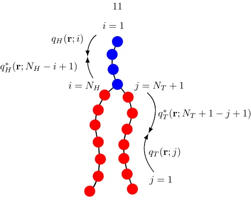
Figure 2.1: A double-tailed lipid model consisting of head (blue) and tail (red) monomers. qI and
q∗I, where I = H, T, are the chain propagator and complementary chain propagator, respectively, used for calculating the single chain statistics; see Appendix C.
Here ri,j is the position of thejth monomer in the ith chain, b is the bond length, N is the total
number of monomers in the chain, kB is the Boltzman constant, and T is the temperature. In
what follows, we set kBT = 1. We note that in using the discrete Gaussian chain as our model,
we have ignored bending rigidity. Indeed, real lipid molecules have bonds with limited flexibility; in particular, unsaturated lipids contain double bonds, or kinks. However, bond order parameters calculated from similar lattice models [18–20] in the same spirit as our discrete Gaussian chain model have shown good qualitative agreement with experimental findings [21–23], and even excellent agreement with molecular dynamics simulations [24–26]. We use the Gaussian model primarily for convenience, as it is the simplest model that captures the conformational degrees of freedom of the lipid molecules. Similar models have been used to study membrane processes such as fusion [27].
In addition to the lipids, there are solvent monomers with volumevS, and salt ions taken as point
charges of elementary electron charge e and valency z±. We work mostly in the grand canonical ensemble, where the number of lipid, solvent, and ion molecules are controlled by their chemical potentials µI (I =L, S,±), which are obtained from the homogeneous bulk phase. For a range of
in the system, whose position and density profile we specify, and solve for the remaining density profiles of the lipids, solvents, and ions; the free energy follows immediately from these solutions.
We begin with a particle-based Hamiltonian for our system, given by
H=
nL X
i=1
hi+
Z
drdr0
( X
J K
ˆ
φJ(r)uJ K(r−r0) ˆφK(r0) +
e2
2ρˆc(r)C(r,r 0) ˆρ
c(r0) +
z2
±e2cˆ±(r) 8πa±(r)
)
. (2.2)
In this expression, the first term accounts for the chain connectivity of the nL lipids. The second
term accounts for the pairwise energetic interaction potentials among species, where the summation is over J K ∈ {HT, T S, SH} and the instantaneous volume fractions of solvent, head, and tail monomers are defined
ˆ
φS(r) =vS nS X
j=1
δ(r−rj),
ˆ
φH(r) =vH nL X i=1 NH X a=1
δ(r−ria),
ˆ
φT(r) = 2vT nL X i=1 NT X b=1
δ(r−rib).
(2.3)
We assume the pairwise interaction potentialuJ K(r−r0) to be short-ranged, so that one may perform
a gradient expansion to quadratic order; see Appendix A. This gives the following approximation:
H ≈
nL X
i=1
hi+
Z
drdr0
( X
J K
h ˆ
φJ(r)χJ KφˆK(r) +
κJ
2 |∇ ˆ
φJ(r)|2
i
+e
2
2ρˆc(r)C(r,r 0) ˆρ
c(r0) +
z2
±e2cˆ±(r) 8πa±(r)
)
.
(2.4)
The Coulomb energy accounts for the long-ranged electrostatic interactions from the total charge density of all charged species:
eρˆc(r) =cFφF(r)−
cH
vH
ˆ
φH(r) +z+eˆc+(r)−z−ecˆ−(r). (2.7)
HerecF is the magnitude of the charge density on the fixed dendrimer andcH is the magnitude of
the charge per head monomer (and is dimensionless). ˆc±(r) is the instantaneous number density of the ions. Note that the density profileφF(r) for the dendrimer is specified by us. Any reasonable
function may be used. We choose a hyperbolic tangent function with a characteristic width for the interface [29] so that the dielectric constant (r), which is spatially varying and depends on the volume fractions of the different species, will be smooth and continuous inr. However, with respect to the lipids, the dendrimer is impenetrable and the density profile of the fixed particle is essentially a step function. Finally, the forth term in Eq. (2.4) is the Born self-energy of the ions in a weakly varying dielectric medium.
The grand canonical partition function Ξ is obtained by summing over all particle degrees of freedom, including the position of each solvent and ion molecule, as well as the position and confor-mation of each lipid chain:
Ξ = ∞ X
n{L,S,±}=0
e(µLnL+µSnS+µ±n±)
nL!nS!n±!vLnLvSnSv±n± Z nL
Y
i=0
Dri
Z nS Y
j=0
drj
Z n± Y
k=0
drk
×Y
r
δh1−φˆH(r)−φˆT(r)−φˆS(r)−φF(r)
i e−H.
Here, the delta functional accounts for the incompressibility at all positions r within the system volume, the notation Dri is shorthand for integrating over all monomers in the chain, i.e., Dri ≡
dri,1dri,2. . . dri,N, and the Boltzmann factorHis given by Eq. (2.4).
In SCFT, the first step is to replace the above (computationally intractable) particle-based model with a field-theoretic model, using a series of techniques related to Hubbard-Stratonovich transformations. This decouples the interactions among particles and replaces them with interactions between single particles and effective fields; for details, see Appendix B. The final result for the field-theoretic partition function can be generically written in the form
Ξ = Z
Dωexp(−F[ω]), (2.9)
where F is an effective Hamiltonian that is complex and depends on the (multidimensional) field variableω. In general, the field-theoretic partition function cannot be evaluated in closed form. The mean-field, or self-consistent field approximation, amounts to assuming that a single field configura-tionω∗ dominates the functional integral, i.e., Ξ≈exp(−F[ω∗]), whereF[ω∗] in our model is given by
F =−e
µL
vL
ZL(ξH, ξT)−
eµS
vS
ZS(ξS)−
eµ±
v±
Z±(ψ) + Z dr ( X J K
(χJ KφJφK−ξJφJ+
κJ
2 [∇φJ]
2) +ψ c
FφF−
cH
vH
φH
−
2(∇ψ)
2
)
.
(2.10)
In Eq. (2.10), and in what follows, we recognize the imaginary nature of the potential field variables at the saddle point and redefine the conjugate chemical potential fieldsiξ→ξ, and the electrostatic potential field−iψ→ψ. Also, the incompressibility constraint is invoked to eliminate φS in favor
ofφH,φT, andφF. Finally, therdependence has been dropped for notational conciseness.
Zl(ξH, ξT) require some explanation. Firstly, we have introduced the Born self-energy of the ions
ub±= z
2
±e2 8πa±
, (2.12)
where is the spatially varying dielectric constant. Although the volume of the salt ions does not enter into the incompressibility, with respect to the self-energy of an ion, we specify a±= 0.3 nm as the radius. ub
± cannot be absorbed into a redefinition of the chemical potential for a spatially varying dielectric medium. The derivation of the expression for the Born energy of a spatially varying dielectric medium is rather involved and we refer the interested reader to a complete derivation by Wang [15]. Secondly, we have introduced the chain propagator qI(r;i) forI=H, T, whereiis the
monomer index, to obtain the single-chain statistics of the lipid. The propagator accounts for the chain connectivity and the Boltzmann weight due to the self-consistent potential field. Calculation of the propagator for our discrete Gaussian chain is described in Appendix C, with the initial condition for placing the end monomers:
qH(r; 1) = exp{−vHξH},
qT(r; 1) = exp{−vTξT}.
(2.13)
Finally, the mean-field configuration that gives F[ω∗] is obtained by requiring that Eq. (2.10) is stationary with respect to variations in the fields. Variation with respect to the volume fraction fieldsφH and φT gives,
ξH =ξS+χSH(1−2φH−φT −φF) + (χHT−χST)φT
−κH∆φH−
cH
vH
ψ−(H−S)
(∇ψ)2
2 +
z±2e2c± 8πa±2
!
, ξT =ξS+χST(1−φH−2φT −φF) + (χHT−χSH)φH
−κT∆φT −(T −S)
(∇ψ)2
2 +
z2
±e2c± 8πa±2
!
.
(2.14)
Here,c±= eµ±v−±1exp
∓z±eψ−ub± is the ion distribution. Variation with respect toψgives,
−∇(∇ψ) =cFφF−
cH
vH
φH±(z±ec±) ; (2.15)
with respect toξS gives,
1−φH−φT−φF = eµSexp{−vSξS}; (2.16)
and with respect toξH andξT gives,
φH(r) =
vHeµL
vL NH X
i=1
qH(r;i)evHξHq∗H(r;NH−i+ 1),
φT(r) = 2
vTeµL
vL NT X
i=1
qT(r;i)evTξTq∗T(r;NT + 1−i+ 1).
(2.17)
Here we have introduced the complementary chain propagatorq∗I(r;i), which also satisfies a similar equation to that forqI(r;i), but with the initial conditions (for beginning at the branch point):
q∗H(r; 1) = evHξHq
T(r;NT + 1)qT(r;NT + 1),
q∗T(r; 1) = evHξHq
T(r;NT + 1)qH(r;NH).
In this section, we study the thermodynamics of membrane-particle interactions and consider the implications to the endosomal escape, where, in addition to the proton sponge effect, another possible effect of the dendrimers is to insert into the endosomal membrane and form pores that can nucleate rupture at significantly lower tensions. As a reference, we first study the tension required to rupture a uniform membrane in the absence of the dendrimer.
2.3.1
Rupture of a Uniform Lipid Membrane
Lipid membranes such as DPPC are often used as models for understanding the more complex and diverse biological membranes that define the boundaries of (and within) a cell. We begin by developing a coarse-grained model of a lipid membrane in a volume containing explicit solvent and mobile ion species at physiological salt concentration, 150 mM. For the lipid we choose NH = 2
and NT = 8 (per tail), with the monomer volumes vH =vT = 0.05 nm3. With these values, the
total volume of our lipid is vl = 0.9 nm3, which is on the order of the value for a lipid in a fully
hydrated bilayer (vl= 1.232 nm3) as determined by Nagle [30]. The desired surface charge density
is experimentally obtained by specifying the composition of lipid species in the membrane [31]. For simplicity, we choose to distribute the charge evenly among the head monomers and setcH = 0.25
for the (dimensionless) charge magnitude per head monomer.
The Flory and square gradient interaction parameters are chosen so that the model captures real-istic features of a lipid membrane. SettingχHT = 75,χT S = 25,χSH = 0 andκH=κT = 2, κS = 0,
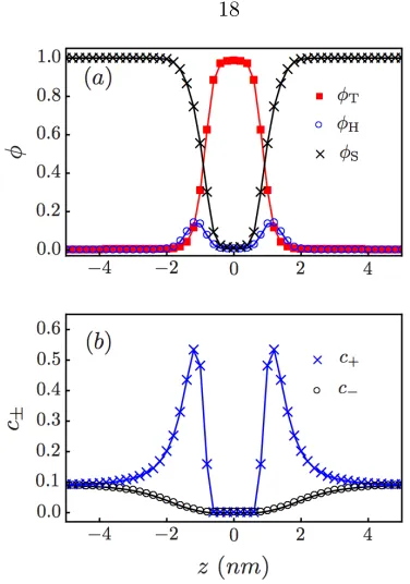
Figure 2.2: (a) Volume fractions and (b) ion number concentrations (nm−3) across the axis
perpen-dicular to the membrane
see Fig. 2.2(a). Both values are similar to those obtained from experiments and molecular dynamic simulations [32]. We choose T = 2 and H = 50 for the dielectric constants of the tail and head
regions [33], respectively, and for the solvent we useS = 80. Figure 2.2(b) shows an accumulation
of cations in the head region due to the negatively charged head monomers, and a depletion of ions in the tail region due to the low value of the dielectric constant,T.
The above parameters are chosen based on experimental results for membranes in the tensionless state. The tension is defined by γ=∂F/∂A|n
l, where F is the appropriate free energy and A is the area. To study the limit of stability for a membrane under increasing tensions, we work in a semi-open system (open with respect to solvent and small ions but closed with respect to the number of lipids so that the number of lipids in the membrane is fixed) and obtain the free energy per unit areaf as a function of the area per lipidσ. The tension is then evaluated according to
γ=f+σ ∂f ∂σ
n
l
Figure 2.3: Membrane tension (kBT /nm2) as a function of the area per lipid (nm2). The limit of
metastability occurs where∂γ/∂σ= 0.
For a mechanically stable membrane, (∂γ/∂σ)>0. Therefore, the condition (∂γ/∂σ) = 0 signals the onset of mechanical instability, which we identify with the point of rupture and term the value of tension at this point the critical tension. In Fig. 2.3, the rupture corresponds to the maximum of the tension-area curve, with a critical value of the areal expansion of∼0.45, and critical tension
γc∼4.5 kBT/nm2. The same calculation is repeated in the grand canonical ensemble, which is open
to all species. In this system, the excess grand potential (given by Eq. (2.10) relative to the uniform bulk solution) per unit area directly gives the tension up to the rupture value. The results from these two ensembles are identical. For convenience, particularly when a dendrimer is present, in what follows we work in the grand canonical ensemble.
We note that the rupture captured by this one-dimensional calculation is the limit of metasta-bility for a uniform static membrane. In reality, thermal fluctuations and lipid rearrangements are responsible for regions of membrane thinning [34, 35] that lead to rupture under much lower ten-sions. Therefore, it is not surprising that our value for the critical tension is higher than the values determined from micropipette aspiration experiments (0.75−2.5 kBT/nm2, depending on degree
event. In what follows, we explore the thermodynamics of membrane-particle interactions.
2.3.2
Thermodynamics of Membrane-Particle Interactions
Properties of the fixed particle are chosen to capture the size and charge of different generations of PAMAM dendrimers [39]. Because the outer surface of a high-generation dendrimer is highly congested and unlikely to be penetrated by lipids (see Zhang [6] and references within), we define a radiusRF, which excludes any lipids from within the volume of the particle. The total charge of the
particle is given by the number of terminal amine groups, which are essentially all protonanted at neutral pH [40]. Due to significant backfolding of these amines [41], the charge can be assumed to be distributed evenly in the volume of the particle (see Fig. 2.7 for the dendrimer sizes and charge densities used in this work).
We find the results insensitive to the dielectric constant chosen for the particle, and assign
F =S = 80. Because the system is axially symmetric, we work in cylindrical coordinates, with the
center of the particle defined asrF = (rF, zF) = (0, zF) and the membrane positioned atz= 0. The
vertical position of the particle zF then becomes a natural reaction coordinate for the system. In
what follows, we constrain the position of the particle and numerically solve the set of SCF equations at each position. The solutions to the uniform membrane case are used as the boundary condition, holding the membrane at fixed tension and its outer edges at fixed position.
In general, the SCF equations can have multiple solutions, corresponding to different free energy minima, with one being the global minimum and the rest being the metastable minima. Capturing all the free energy minima is a nontrivial task. However, symmetry and simple physical intuition can often be used to limit the search. Here, we take advantage of the different ways of initializing the SCF equations as a means to access the stable and metastable minima. We consider two methods:
1. We begin with a noninteracting membrane-particle system andslowlymove the particle along the path of decreasingzF. At each step, the previous solutions are used to initialize the new
equations.
Figure 2.4: Free energy profile (kBT) for the tensionless membrane. ∆F is relative to a
nonin-teracting membrane-particle system, where zF → ∞. The particle is a G5 dendrimer with radius
RF = 2.7 nm and charge density cF = 1.55 nm−3 (all 128 surface amines are protonated). For
comparison, we have also plotted the results for a hypothetical G5 dendrimer withcF = 5.00 nm−3.
Empty symbols correspond to method 1; + and×correspond to method 2 (see text for description).
hole in the membrane. This hole can be interpreted as a temporary defect caused by fluctua-tions.
2.3.2.1 Tensionless Membrane
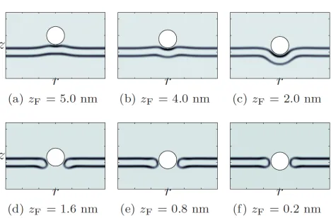
We first apply method 1 to a tensionless membrane and a fully protonated G5 dendrimer. While the specific interactions are local, the electrostatic interactions are long-ranged and rearrangements of the lipid molecules allow the membrane to reach out and meet the particle, as shown in Fig. 2.5(a). At this point, the particle is within the attractive range of the membrane. This is depicted in Fig. 2.4, where the potential well in the free energy profile is due to a competition between satisfying the favorable electrostatic interactions and deforming the membrane. From the free energy profile, we see that the particle will continue towardszF ∼0 nm, corresponding to a metastable, partially wrapped
state.
Figure 2.5: φH in cylindrical coordinates for a tensionless membrane and a fully protonated G5
dendrimer withcF = 1.55 nm−3. (a–c) are solutions to both methods 1 and 2, while (d–f) are only
solutions to the latter.
of pushing the particle through the membrane could provide information as to the energetics and forces required by the cell (in particular, the proteins recruited) to enforce shape transitions of the membrane involved in endocytosis.
We next solve the same SCF equations by method 2. Depending on the extent of the hole created by the particle and the parameters of the system (membrane tension, dendrimer charge and dendrimer size), the system will fall into one of two solutions: either the membrane reseals the hole and partially wraps the particle, or the membrane forms a head-lined pore around the particle. This is observed from the density plots (Fig. 2.5) and from the free energy profile (Fig. 2.4), where the SCF solutions correspond to a partially wrapped particle untilzF ∼2.0 nm. Beyond this, if thermal
fluctuations initiate a hole (as simulated by method 2), then the system will find the solutions to the higher energy path corresponding to a membrane pore. However, the particle must squeeze its way into the pore, as can be seen in Fig. 2.5(d) and the free energy increases with decreasing zF.
This indicates that any pore induced into a tensionless membrane is highly unstable and will be short-lived. In all of our calculations, it is important to remember that the particle position is fixed. In other words, we have constrained the SCF equations so that the membrane is forced to respond to a particle at fixed zF. If we lift this constraint, the membrane will expel the particle and reseal
groups.
In Fig. 2.4 we compare the result for a hypothetical, highly charged G5 dendrimer (cF = 5.00 nm−3)
to the result for a fully protonated G5 dendrimer (cF = 1.55 nm−3). Due to screening by the salt
ions, increasing the charge density does not increase the range of electrostatic attraction. It increases the extent and stability of the wrapped state. If the system manages to reach the unstable pore state, it will be short-lived, according to the same explanation given in the preceding paragraph for
cF = 1.55 nm−3. From method 1, we find that electrostatic interactions alone are insufficient for a
dendrimer to induce holes in a tensionless membrane. From method 2, even if thermal fluctuations assist by initiating temporary holes, we find that the dendrimer is unable to stabilize pores in a tensionless membrane.
2.3.2.2 Membrane Under Tension
In the proton sponge hypothesis, an increase in osmotic pressure is believed to rupture the endosome. We account for the osmotic pressure by applying a tension to the membrane, and explore the combined effects of the applied tension and the electrostatic interactions between the membrane and the particle. In Fig. 2.6, we continue with a fully protonated G5 dendrimer and plot the free energy profile forγ= 0.74 kBT/nm2. The cost of deforming a membrane under tension has shifted
the metastable state from partially wrapped (zF ∼0 nm) to surface-absorbed (zF ∼2.8 nm). The
Figure 2.6: Free energy profile (kBT) for a membrane withγ= 0.74kBT /nm2, shown together with
the tensionless membrane from Fig. 2.4. The particle is once again a fully protonated G5 dendrimer. Empty symbols correspond to method 1; + and×correspond to method 2 (see text for description). Note that we are well below the limit of metastability for a homogeneous membrane, where rupture occurs atγc∼4.5kBT /nm2.
membrane permeability and hole formation are dependent on dendrimer generation and terminal functional groups [6–9]. Therefore, we also consider G3–G7 dendrimers. Our results agree with experimental trends, where we find that G3 dendrimers do not stabilize pores, while G5 and G7 dendrimers do, see Fig. 2.7.
However, with respect to the endosomal escape, our interest lies in dendrimers that can nucleate rupture, not stabilize pores. Recall that rupture of a uniform membrane occurs atγc∼4.5 kBT/nm2.
Consider the membrane under tension with an inserted G5 dendrimer (cF = 1.55 nm−3) shown in
Fig. 2.6, where the pore state is metastable. If we slowly increase the tension from this state, we find that the limit of metastability occurs at γc∼0.84 kBT/nm2, see Fig. 2.8. For tensions exceeding
this value, the pore radius expands indefinitely and the membrane ruptures. Interestingly, even a highly charged particle, which should have strong adhesion to the head-lined pore periphery, lowers the rupture tension, whereγc levels off at∼1.3 kBT/nm2.
Figure 2.7: Free energy profile (kBT) for a membrane withγ= 0.74kBT /nm2 interacting with G3
(RF = 1.8 nm, cF = 1.30 nm−3), G5 (RF = 2.7 nm, cF = 1.55 nm−3), and G7 (RF = 4.0 nm,
cF = 2.0 nm−3) dendrimers. Empty symbols correspond to method 1; +, ×, andNcorrespond to
method 2 (see text for description).
Figure 2.8: Rupture tension (kBT /nm2) for a membrane containing a pore, plotted as a function of
the charge density (nm−3) of the G5 dendrimer stabilizing the pore
rupture. The study of the actual nucleation barriers and pathways to pore formation and rupture are beyond the scope of this work. Nevertheless, these results show that highly charged dendrimers can greatly lower the rupture tensions. In terms of the endosomal escape, this dendrimer-induced rupture at significantly lower tensions is precisely what we seek.
2.4
Conclusion
[image:36.612.201.429.302.454.2]mechanism, where our main goal is to understand the role of the dendrimer in nucleating membrane rupture (with the eventual release of the genetic material from the endosome). For different mem-brane tensions, dendrimer sizes and charge densities, we have explored the structural and energetic properties of the system. In what follows, we summarize our main findings and the implications for designing polymer-based gene delivery vectors.
For tensionless membranes, we find that the hydrophilic pore with an inserted dendrimer is unstable; the tensionless membrane prefers to satisfy the favorable electrostatic interactions by partially wrapping the dendrimer. Our results indicate that increasing the charge density of the dendrimer only increases the extent and stability of the wrapped state, while the pore state, if somehow created, remains unstable and short-lived. In other words, the tensionless membrane is so robust that even a highly charged particle is unable to insert into the membrane and stabilize a pore. This suggests that the osmotic pressure contribution from the proton sponge hypothesis is a necessary component if the endosomal escape is to be enhanced by particle insertion into the membrane.
To study the effect of the osmotic pressure, we apply a tension γ = 0.74 kBT/nm2 to the
membrane. This value is well below the limit of metastability for a uniform membrane, for which
γc∼4.5 kBT/nm2. For G5 dendrimers and higher, we find that the pore corresponds to a metastable
state, while for G3 dendrimers the pore is unstable. This agrees with experimental trends on the ability of the dendrimer to cause membrane permeability and hole formation [6–9]. Lee and Larson [13], using MD simulations on similar membrane-particle systems, found that dendrimer-induced membrane disruption was dependent on generation and concentration. We believe that the apparent concentration dependence is related to local membrane tensions that are induced by nearby particles due to the fixed membrane area. This is in agreement with the effect of tension in our findings, where metastable pores exist only in combination with an applied tension. Finally, our results indicate that these same metastable pores can act as nucleation sites for rupture.
The free energy profile in this sense yields the potential of mean force between the particle and the membrane. The multiple unstable and metastable states are obtained from the behavior of the free energy as a function ofzF. However, the reaction coordinate for nucleating rupture most likely will
not bezF, but will involve the rearrangement of the lipids in the pre-pore state. The current study
does not address how the particle becomes inserted into the membrane from the partially wrapped state, nor does it address the subsequent nucleation event leading to rupture.
To study the pathways associated with these activated processes will require minimum energy path (MFEP) calculations using, e.g., the nudged elastic band [42, 43] or the string method [44]. We note that simpler calculations, where physical insight is used to impose one or more reaction coordinate constraints (in a similar spirit to our particle position zF) have sometimes been used
to study nucleation processes in membrane fusion [27]. However, the structures associated with dendrimer-induced pore formation and membrane rupture are more complicated and it is not obvious that some simple constraints can be identified. In Chapter 3, we apply MFEP calculations to dynamic SCF theory to address the actual nucleation events involved in the dendrimer-induced pore formation and membrane rupture.
Appendix A: Gradient Expansion
For the second term in Eq. (2.2), we focus on one term in the sum and perform a change of variables ¯
r=r−r0 and expand aroundr0
Z
drdr0φˆJ(r)uJ K(r−r0) ˆφK(r0) =
Z
d¯rdr0φˆJ(¯r+r0)uJ K(¯r) ˆφK(r0)≈
Z
d¯rdr0
ˆ
φJ(r0) + ¯rT∇φˆJ(r0) +
1 2¯r
T∇∇φˆ J(r0)¯r
uJ K(¯r) ˆφK(r0).
(A-1)
The zeroth-order term in Eq. (A-1) defines the Flory interaction parameter
Z
d¯ruJ K(¯r)
Z
dr0φˆJ(r0) ˆφK(r0)≡χJ K
Z
dr0φˆJ(r0) ˆφK(r0). (A-2)
AssuminguJ K(¯r) is symmetric, the first-order term vanishes; performing an integration by parts,
the second-order term defines the phenomenological square gradient parameterκJ K
−1
2 Z
d¯r¯rT¯ruJ K(¯r)
Z
dr0∇φˆJ(r0)T∇φˆK(r0)≡
κJ K
2 Z
dr0∇φˆJ(r0)T∇φˆK(r0). (A-3)
Neglecting the cross square gradient terms and definingκJ J ≡κJ gives the following approximation
to Eq. (2.2):
H ≈
nL X
i=1
hi+
Z
drdr0
( X
J K
h ˆ
φJ(r)χJ KφˆK(r) +
κJ
2 |∇ ˆ
φJ(r)|2
i +e
2
2ρˆc(r)C(r,r 0) ˆρ
c(r0)
)
2
where J is the instantaneous density,Ais the operator andf is the auxiliary field.
2. Alternatively, one can introduce the field variables φI through a series of formal functional
analysis identities for the Dirac distribution. First the instantaneous densities (expressed as a unitless volume fractions) are converted into smooth scalar density fields ˆφI =φI:
Z
DφIδ
h
φI(r)−φˆI(r)
i
F[φI(r)] =F[ ˆφI(r)]. (B-2)
Then the associated (conjugated) fieldsξI are introduces via the formal Fourier representation
ofδ[f(r)−g(r)]:
δhf(r)−g(r)i= Z
DξIexp
i
Z
drξI(r)
h
f(r)−g(r)i
. (B-3)
Using these two identities, we can now write the partition function in Eq. (2.8) as
Ξ = ∞ X
n{L,S,±}=0
e(µLnL+µSnS+µ±n±)
nL!nS!n±!vLnLvSnSv±n± Z nL
Y
i=0
Dri
Z nS Y
j=0
drj
Z n± Y
k=0
drk
Z
DφI
Z
DξIexp
i
Z
drξI(r)[φI(r)−φˆI(r)]
×exp − nL X i=1
hi−
Z dr X J K h
φJ(r)χJ KφK(r) +
κJ
2 |∇φJ(r)|
2i+z 2
±e2cˆ±(r) 8πa±(r)
×
Z
Dψ exp
i
Z
drψ(r)
cFφF(r)−
cH
vH
φH(r)±z±ecˆ±(r)
−
Z
dr(r)
2 (∇ψ(r))
2
.
(B-4)
Where we have used Eq. (B-1), together with Eq. (2.6), to decouple the quadratic terms in the Coulomb energy. Note, also, that incompressibility condition is imposed directly by definingφS =
1−φH−φT−φF. Separating the terms containing the microscopic densities from those containing
the fields, we write
Ξ = Z
DφI
Z
DξI
Z
Dψ
×
∞ X
nL=0
eµLnL
vLnLnL!
Z nL Y
i=0
Driexp
−
nL X
i=1
hi−
Z
drξH,TφˆH,T
×
∞ X
nS=0
eµSnS
vnS
S nS!
Z nS Y
j=0
drjexp
−
Z
drξSφˆS
×
∞ X
n±=0
eµ±n±
vn±
± n±! Z n±
Y
k=0
drkexp
−
Z
drez+ψˆc±− Z
drz
2
±e2cˆ± 8πa±
×exp
−
Z
drψ
cFφF −
cH
vH
φH
−
2(∇ψ)
2 ×exp − Z
drX
J K
χJ KφJφK−ξJφJ−
κJ
2 [∇φJ]
2
.
(B-5)
In anticipation of the imaginary nature for the field variablesξI andψat the saddle point, we have
Fitting this expression into Eq. (2.9), it is evident that the field theoretic partition function is given by Eq. (2.10) with the single molecule partition functions given by Eq. (2.11). For the lipids, the Boltzmann factor contains the term hfor the chain connectivity. This is further weighted by the external fieldsξH andξT. To obtain the single chain statistics for the lipids, our approach will be to
compute the chain propagatorsqH andqT, as described in Appendix C. Finally, we comment that
the Boltzmann factor for the ions contains the Born-self energyub
Appendix C: Propagator for a Single Chain in External Fields
We introduce the chain propagator by analogy to a Markovian process, where the monomer index
ican be thought of as the analogue of a discrete time variable in a stochastic process. In this way we seek to build up the configuration of the graft copolymer, from the free end of each arm to the branch point, beginning with the initial condition that
qI(r,1) = e−vIξI(r) (C-1)
forI=H, T. Moving to subsequent monomersicorresponds to propagating forward in the “time” index:
qI(r;i) = e−vIξI(r)
Z
dr0Γ(|r−r0|)qI(r0;i−1). (C-2)
Here i∈(2, NI) and Γ denotes the transition probability from one monomer to the next, assumed
to be Gaussian. For notational simplicity, in what follows we drop the subscript I and denote
q(r, i) as simplyqi(r). The above expression is essentially a reduced partition function for a chain
beginning at r and ending anywhere. Rather than attempt to evaluate Eq. (C-2) directly by an integration scheme, we recognize that for a reasonably smooth external potential, the contributions are dominated by the transitions for which|r−r0|is on the order of the range of (or less than) the Gaussian. Thus, following the usual practice in deriving a differential equation from the Chapman-Kolmogoroff equation, we expandr0 aroundr to quadratic order. Further representing the spatial derivatives by differences in cylindrical coordinates for our axially-symmetric membrane-particle system, we get
qi+1(rj, zk) = e−vξ(rj,zk)
1−4γqi(rj, zk) +γqi(rj, zk+1) +γqi(rj, zk−1)
+γ(1 + 1 2j)q
i(r
j+1, zk) +γ(1−
1 2j)q
i(r j−1, zk)
.
Γ(rj, zk−1→rj, zk) =γ,
Γ(rj+1, zk →rj, zk) =γ(1 +
1 2j),
Γ(rj−1, zk →rj, zk) =γ(1−
1 2j),
Γ(rj, zk →rj, zk) = 1−4γ,
(C-5)
where the last expression can be thought of as the “survival probability”. Eq. (C-3) applies toj 6= 0. Forj= 0, we have
qi+1(r0, zk) =e−vξ(r0,zk)
1−6γ
qi(r0, zk) +γqi(r0, zk+1) +γqi(r0, zk−1) + 4γqi(r1, zk)
, (C-6)
from which we similarly obtain the transition probabilities. Note that the positivity of the transition probabilities requires that γ <1/6. Continuing with the analogy to a Markovian process, we can define an equivalent “master equation” for the chain propagator on our discrete grid, and Eq. (C-2) becomes
qi(rj, zk) = e−vξ(rj,zk)
X
r0
j,z
0
k
Γ(r0j, zk0 →rj, zk)qi−1(r0j, z
0
k), (C-7)
where it is understood that the summation is restricted to the nearest neighbors of (rj, zk). The
[2] D. W. Pack, A. S. Hoffman, S. Pun, and P. S. Stayton,Nat. Rev. Drug Discov.4, 581 (2005).
[3] J. Haensler and F. C. Szoka,Bioconjugate Chem. 4, 372 (1993).
[4] J. P. Behr,Chimia.51, 34 (1997).
[5] N. D. Sonawane, F. C. Szoka, and A. S. Verkman,J. Biol. Chem.278, 44826 (2003).
[6] Z.-Y. Zhang and B. D. Smith,Bioconjugate Chem.11, 805 (2000).
[7] S. Hong, A. U. Bielinska, A. Mecke, B. Keszler, J. L. Beals, X. Shi, L. Balogh, B. G. Orr, J. R. Baker, and M. M. Banaszak Holl,Bioconjugate Chem.15, 774 (2004).
[8] A. Mecke, I. J. Majoros, A. K. Patri, J. R. Baker, M. M. Banaszak Holl, and B. G. Orr,Langmuir 21, 10348 (2005).
[9] J. Chen, J. A. Hessler, K. Putchakayala, B. K. Panama, D. P. Khan, S. Hong, D. G. Mullen, S. C. DiMaggio, A. Som, G. N. Tew, A. N. Lopatin, J. R. Baker, M. M. Banaszak Holl and B. G. Orr, J. Phys. Chem. B113, 11179 (2009).
[10] S. Boeckle, K. von Gersdorff, S. van der Piepen, C. Culmsee, E. Wagner, and M. Orgis, J. Gene. Med. 6, 1102 (2004).
[11] Y. Yue, F. Jin, R. Deng, J. Cai, Y. Chen, M. Lin, H.-F. Kung, and C. Wu,J. Control. Release 155, 67 (2011).
[13] H. Lee and R. G. Larson,J. Phys. Chem. B112, 12279 (2008).
[14] G. H. Fredrickson, The Equilibrium Theory of Inhomogeneous Polymers (Oxford University Press, New York, 2005).
[15] Z.-G. Wang,Phys. Rev. E81, 021501 (2010).
[16] Q. Zhang and Y. Ma,J. Chem. Phys.125, 164710 (2006).
[17] V. V. Ginzburg and S. Balijepalli,Nano Lett.7, 3716 (2007).
[18] A. Ben-Shaul, I. Szleifer, and W. M. Gelbart,Proc. Natl. Acad. Sci. USA81, 4601 (1984).
[19] I. Szleifer and A. Ben-Shaul,J. Chem. Phys.83, 3597 (1985).
[20] I. Szleifer, A. Ben-Shaul, and W. M. Gelbart,J. Chem. Phys.83, 3612 (1985).
[21] T. Zemb and C. Chachaty,Chem. Phys. Lett. 88, 68 (1982).
[22] J. Charvolin,J. Chim. Phys. Phys. Chim. Biol.80, 15 (1983).
[23] B. Lindman, “Physics of Amphiphiles: Micelles, Vesicles and Microemulsions,” Proceedings of the International School of Physics “Enrico Fermi”(North-Holland, 2005).
[24] P. van der Ploeg and H. Berendsen,J. Chem. Phys.76, 3271 (1982).
[25] P. van der Ploeg and H. Berendsen,Mol. Phys.49, 233 (1983).
[26] O. Edholm, H. Berendsen, and P. van der Ploeg,Mol. Phys.48, 379 (1983).
[27] K. Katsov, M. M¨uller, and M. Schick,Biophys. J.87, 3277 (2004).
[28] K. Hong and J. Noolandi,Macromolecules13, 964 (1980).
[29] M. W. Matsen and R. B. Thompson,Macromolecules41, 1853 (2008).
[30] J. F. Nagle and M. C. Wiener,Biochem. Biophys. Acta942, 1 (1988).
[37] E. A. Evans, R. Waugh, and L. Melnik,Biophys. J.16, 585 (1976).
[38] B. M. Discher, Y. Y. Won, D. S. Ege, J. C. M. Lee, F. S. Bates, D. E. Discher, and D. A. Hammer,Science 284, 1143 (1999).
[39] P. K. Maiti, T. Cagin, G. F. Wang, and W. A. Goddard,Macromolecules37, 6236 (2004).
[40] M. F. Ottaviani, F. Montalti, M. Romanelli, N. J. Turro, and D. A. Tomalia, J. Phys. Chem. 100, 11033 (1996).
[41] P. K. Maiti and W. A. Goddard,J. Phys. Chem. B110, 25628 (2006).
[42] G. Henkelman, B. P. Uberuaga, and H. Jonsson,J. Chem. Phys.113, 9901 (2000).
[43] G. Henkelman and H. Jonsson,J. Chem. Phys.113, 9978 (2000).
Chapter 3
The Minimum Free Energy Path to
Membrane Pore Formation and
Rupture
We combine dynamic self-consistent field theory with the string method to calculate the minimum energy path to membrane pore formation and rupture. In the regime where nucleation can occur on experimentally relevant time scales, the structure of the critical nucleus is between a solvophilic stalk and a locally thinned membrane. Classical nucleation theory fails to capture these molecular details and significantly overestimates the free energy barrier. Our results suggest that thermally nucleated rupture may be an important factor for the low rupture strains observed in lipid membranes [1].
3.1
Introduction
ments. Furthermore, computer simulations are limited by the number of amphiphiles and are usually performed under constant area. A pore opening under such conditions simultaneously relaxes the surface tension and can either expand, reseal, or stabilize, depending in a nontrivial manner on the system size [15]. In solvent-free models consisting of two- or three-bead “lipids”, there is disagree-ment between density functional predictions [10] and Monte Carlo simulations [12] with regard to the existence of small metastable pores. The interpretation of structures comparable to the lipid molecular size is highly problematic for these overly simplified models.
3.2
Model and Method
In this chapter, we study the fullminimum free energy path(MFEP) to pore formation and rupture by combining the string method [16] with dynamic self-consistent field (DSCF) theory [17]. As opposed to calculations that require physical insight to impose one or more constraints on the system [18], the string method automatically determines the reaction coordinate of the MFEP connecting two stable states on a given free energy landscape, while DSCF theory provides a full description (at the mean-field level) of the lipid conformation changes. We begin with an arbitrary set of states between two free energy minima. The states are connected on the free energy landscape by a string and relax towards the MFEP by an iterative procedure. First, all states are evolved independently for some time ∆taccording to
∂φI
∂t =−D δF δφI
where φI is the monomer volume fraction, D is the mobility coefficient, and F is the free energy
functional of the system. Note that for a system not at equilibrium, the gradient cannot be computed using the usual SCF theory. Instead, we solve for a hypothetical external potentialV(r), which makes thegivennonequilibrium densityφI(r) an “equilibrium” one; see [19] for details. Briefly,V(r) must
satisfy
φI(r) =eµI N
X
i=1
qI(r, i)eV(r)qI∗(r, N−i), (3.2)
where the chain propagator qI(r, i) is again obtained using the discrete version of Eq. (C-2), i.e.,
Eq. (C-7). The equation of motion then becomes
∂φI
∂t =−D[ξI(r)−VI(r)], (3.3)
whereξI is the usual field obtained by taking the variation of the free energy functional, i.e., δφδF
I. Then, to prevent the states from falling into one of the two end states (the trivial equilibrium solutions), we make use of the connectivity imposed by the string. Precisely, we compute the total arc lengthlof the string according to its Euclidian distance:
l=
Nstates−1
X
i=1
||φi+1−φi||, (3.4)
and interpolate the new states so that they are equidistantly spaced along the string:
s=l/(Nstates−1). (3.5)
The procedure is repeated until the dynamics balance the reparameterization, i.e., the evolution of the string has reached a steady state. At this point the string coincides with the MFEP [16] and the free energy and density profiles of all states along the MFEP are immediately known without addi-tional calculations. We note that a similar strategy was recently employed to study the nucleation of order-order transitions in diblock copolymer melts [20].
theory (CNT) [21, 22]. For a membrane under tensionγ >0, CNT defines the free energy of a pore of radiusras
F = 2πrσ−πr2γ. (3.6)
Hereσ >0 is the line energy. The first term is the cost of forming the rim of a pore and the second term is the relief in elastic energy. The above expression leads to a free energy barrierF∗=πσ2/γ
at a critical radiusr∗=σ/γ, beyond which the pore grows indefinitely (ruptures).
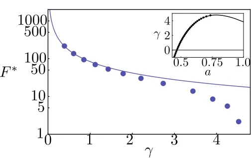
For membranes with different tensions γ, we obtain properties of the nucleation pathway, in-cluding the structure and activation energyF∗ of the critical nucleus. Comparing the values of our calculatedF∗ with those predicted by CNT, we find that CNT is valid only for small γ, where the free energy barrier is nearly insurmountable on experimentally realistic time scales. For the phys-ically relevant regime, where F∗ . O(10 kT), CNT significantly overestimates the barrier height. Furthermore, in this regime the critical nuclei are not well-defined pores, as assumed by CNT, but rather “stalks” of amphiphile head groups or, in the case of large γ, merely a local thinning of the membrane.
[image:52.612.234.414.56.155.2]where the numbers of amphiphile and solvent molecules are controlled by their chemical potentials.
kT is used as the energy unit. The particle-based Hamiltonian for the system, accounting for the chain connectivity of thenamphiphiles and the pairwise energetic interactions among species, is
H=
n
X
i=1
hi({r}) +
X
J K
∈{AB,BS,SA} Z
drdr0φˆJ(r)uJ K(r,r0) ˆφK(r0). (3.7)
The particle to field transformation follows the same derivation given in Chapter 2. Briefly, the interactions among particles are decoupled and replaced with interactions between particles and effective fields; see also [23]. The resulting field-theoretic partition function can be generically written Ξ =RDωexp(−F[ω]), whereF is an effective, complex-valued free energy that depends on the field variableω. Here, we make the mean-field approximation, which amounts to assuming that a single field configurationω∗ dominates the functional integral so that Ξ≈exp(−F[ω∗]). In our modelF[ω∗] is given by
F =−e
µP
vP
ZP(ξA, ξB)−
eµS
vS
ZS(ξS) +
X
J K
∈{AB,BS,SA} Z
dr
χJKφJφK−ξJφJ+
κJ
2 (∇φJ)
2
. (3.8)
The Floryχparameters and the square-gradient coefficients capture, respectively, the local and non-local part of the short-ranged interactions [24]. Their values are chosen to reproduce some known experimental properties of lipid membranes. The incompressibility condition φS +φA+φB = 1 is
used to eliminateφS and we have used the imaginary nature of the potential field variables at the
saddle point to redefine the conjugate potential fieldsiξ→ξ[23]. The partition functions that arise in Eq. (3.8) are for a single molecule in its respective field(s) and are given byZS =
R
dre−vSξS for the solvents and ZP =
R
drqA(r;NA)e2vAξAq2B(r;NB+ 1) for the amphiphiles. The chain propagators
qA andqB account for the chain connectivity and the Boltzmann weight due to the self-consistent
critical nucleus, we may regard pore formation and rupture as occurring at constant tension, which we implement as the boundary condition. In what follows, we work in the grand canonical ensemble, as it is most convenient for studying the MFEP. In this open system, the excess grand potential (Eq. (3.8) relative the bulk solution) per unit area directly gives the tension up to the rupture value, identified as the point of vanishing slope in Fig. 3.2 inset. This corresponds to a critical tensionγc∼4.77kT /nm2 and an areal strain of∼0.6. The linear stretching modulus is found to
be 170 mN/m, which falls within the range for lipid membranes, as determined from micropipette aspiration experiments [26]. Finally, to confirm that the results are independent of the ensemble choice, we repeat the same calculation in the canonical ensemble. In this closed system, the tension is evaluated according to
γ=f+a ∂f ∂a
n
. (3.9)
Heref is the Helmholtz free energy per unit area andais the area per lipid. The results from these two ensembles are identical.
The rupture captured above corresponds to the limit of metastability for a uniform membrane. In reality, thermal fluctuations and lipid rearrangements can nucleate pore formation and rupture when the membrane is subjected to a positive tension γ. If the timescale for nucleation is suffi-ciently long relative to the timescale for molecular relaxation, then the nucleation rate is of the form ν = ν0exp(−F∗/kT), where ν0 is some transition frequency associated with the molecular
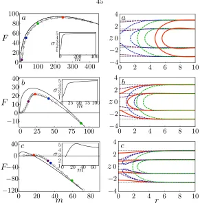
DSCF-Figure 3.2: The free energy barrier as a function of the surface tension: DSCF-MFEP (markers) and CNT (line). Inset: the surface tension as a function of the area per lipid: grand canonical (markers) and canonical (line) ensemble.
MFEP calculations provide an exact description (within the mean-field framework) of the nucleation pathway. In Fig. 3.2, we plot the free energy barrier F∗ as a function of the membrane tension γ
on a log-linear plot. Also shown is the result from CNT [Eq. (3.6)], where the line energy σ is a phenomenological parameter that is assumed constant and used to describe the excess free energy cost associated with forming the rim of a pore. An equilibrium line energy is only well-defined for a pore with zero curvature in a tensionless membrane. Our SCF method determines this value (σeq = 5.16kT /nm) as input into the CNT. For smallγ, the predictions from CNT agree well with
the DSCF-MFEP calculations. Indeed, we expect CNT to become exact in the limitγ= 0. However, in this regime, the barrier is too high, and the rate vanishingly small, for nucleation to be a relevant mechanism. Asγincreases, the free energy barrier decreases (reflecting the fact that the metastable intact membrane becomes less stable) and vanishes at the critical tension (γc), corresponding to the
spinodal. From Fig. 3.2, CNT severely over-predicts the free energy barrier in the important regime whereγ∼3−4kT /nm2, and completely fails to capture the spinodal. To understand the source of
discrepancy between CNT and our results, we examine the MFEP for three representative values of
γ.
[image:55.612.198.451.64.223.2]Figure 3.3: Left: Free energy as a function of the deficiency in the number of lipids: MFEP (dashed lines, states given by the colored markers) and CNT (solid lines). Right: density profiles for images along the respective MFEPs. The contour lines correspond to 25% of the maximum solvophilic (A) density of the initial intact membrane. In all cases, the critical nucleus is shown in red and the intact membrane in grey. From top to bottom: γa, γb, γc= 0.85,2.73,4.52.
well-defined for macroscopic pores. The free energies of the states along the MFEP are given by the markers and the prediction from CNT is given by the solid line. Here the line energy used for the CNT result is obtained from the respective membrane containing a pore with zero curvature. On the right panel, we plot the density profiles of the amphiphiles for selected states along the MFEP. For γ = 0.85 kT /nm2 [Fig. 3.3(a)], except at the very initial stages, the MFEP closely follows
the prediction from CNT, with the nucleation process largely involving the expansion of a well-defined solvophilic pore with negligible penetration of solvents. The density profile is nearly invariant (but shifted in radial direction) once the pore forms. For this low γ, the free energy barrier is
[Fig. 3.3(b)] and observe that CNT over-predicts the free energy barrier and under-predicts the size of the critical nucleus. From the density profiles obtained from the DSCF-MFEP calculations, we see that the critical nucleus is not even a well-defined pore, but rather a “stalk” of solvophilic monomers that contains finer molecular structure than can be captured by CNT. Therefore, CNT is not a good model for the MFEP in the intermediate regime whereF∗= 21.59 kT. We note that a similar structure has been observed as a transition state [18] for the fusion of two bilayers. Finally, consider a membrane approaching the spinodal: γ= 4.52kT /nm2 [Fig. 3.3(c)]. Here CNT grossly
over-predicts the nucleation barrier and under-predicts the size of the critical nucleus. In fact, the nucleation pathways predicted by the two methods qualitatively differ, with the CNT prediction crossing the MFEP and approaching it from below at large pore sizes. From the density profiles [Fig. 3.3(c), right], we find, not surprisingly, that a small perturbation involving local membrane thinning is enough to nucleate pore formation and rupture. Interestingly, rupture occurs even before the membrane is able to fully prepare for pore formation and asolvophobichole that is penetrated by solvents forms in the membrane. Only afterwards do the amphiphiles rearrange to line the pore with solvophilic monomers and seal off the hole. This can be seen from the contour lines given in blue and green. The free energy barrier in this case is only twice the thermal energy, and hence we do not expect the picture of nucleation as a rare event to hold. However, with longer-chain amphiphiles, such as in the case of polymersomes [6], we expect a higher barrier height for the same amount of strain. Thus the scenario presented in this near-spinodal case can still be relevant.
be significantly lower than the strain at the limit of mechanical stability of the membrane. Thus, thermally nucleated rupture may be an important factor for the low rupture strains observed in lipid membranes [7].
3.4
Conclusion
In conclusion, we have combined the string method with DSCF theory to obtain the MFEP to pore formation and rupture for a range of membrane tensions. For the experimentally relevant regime whereF∗.O(10kT), the critical nucleus is somewhere between a stalk-like structure [Fig. 3.3(b)] and a thinned membrane leading to a hole that is partially exposed to solvents [Fig. 3.3(c)]. In this regime, CNT fails to capture the important local rearrangements of the lipids and significantly over-predicts the nucleation barrier. Within the framework of mean-field theory for describing spatially localized fluctuation phenomena, the present work (and that by Cheng et al. [20]) represents the most advanced methodology in treating nucleation in soft condensed matter, including membranes. The combination of the string method and DSCF theory opens the way to studying a wide range of related membrane nucleation phenomena beyond pore formation and rupture, such as membrane fusion and fission [28, 29], and particle insertion and penetration [30].
Bibliography
[1] C. L. Ting and Z.-G. Wang,Phys. Rev. Lett.106, 168101 (2011).
[2] B. Alberts, A. Johnson, J. Lewis, M. Raff, K. Roberts, and P. Walter,Molecular Biology of the Cell(Garland Science, New York and Abingdon, 2007).
[3] L. Yang, T. M. Weiss, R. I. Lehrer, and H. W. Huang,Biophys. J.79, 2002 (2000).
[4] D. Zhelev and D. Needham,Biochim. Biophys. Acta 1147, 89 (1993).
[5] J. Ghel,Acta Physiol. Scand.177, 437 (2003).
[6] B. M. Discher, Y.-Y. Won, D. S. Ege, J. C.-M. Lee, F. S. Bates, D. E. Discher, and D. A. Hammer,Science 284, 1143 (1999).
[7] K. Olbrich, W. Rawicz, D. Needham, and E. Evans,Biophys. J.79, 321 (2000).
[8] O. Sandre, L. Moreaux, and F. Brochard-Wyart,Proc. Natl. Acad. Sci. USA96,10591 (1999).
[9] R. Netz and M. Schick,Phys. Rev. E.53, 3875 (1996).
[10] V. Talanquer and D. Oxtoby,J. Chem. Phys.118, 872 (2003).
[11] M. Muller and
![Figure 1.1: Schematic of the self-consistent (mean) field theory: replacing the interactions amongmolecules with the interaction between a single molecule and an average external field [12]](https://thumb-us.123doks.com/thumbv2/123dok_us/15687.1132/14.612.155.493.553.630/schematic-consistent-replacing-interactions-amongmolecules-interaction-molecule-external.webp)