ITCH E3 Ubiquitin Ligase Interacts with Ebola Virus VP40 To
Regulate Budding
Ziying Han,aCari A. Sagum,bMark T. Bedford,bSachdev S. Sidhu,cMarius Sudol,dRonald N. Hartya
Department of Pathobiology, School of Veterinary Medicine, University of Pennsylvania, Philadelphia, Pennsylvania, USAa; Department of Epigenetics and Molecular Carcinogenesis, M. D. Anderson Cancer Center, University of Texas, Smithville, Texas, USAb; Department of Molecular Genetics, University of Toronto, Toronto, Ontario, Canadac; Department of Physiology, Institute for Molecular and Cell Biology (IMCB, AStar), National University of Singapore, Singapored
ABSTRACT
Ebola virus (EBOV) and Marburg virus (MARV) belong to the
Filoviridae
family and can cause outbreaks of severe hemorrhagic
fever, with high mortality rates in humans. The EBOV VP40 (eVP40) and MARV VP40 (mVP40) matrix proteins play a central
role in virion assembly and egress, such that independent expression of VP40 leads to the production and egress of virus-like
particles (VLPs) that accurately mimic the budding of infectious virus. Late (L) budding domains of eVP40 recruit host proteins
(e.g., Tsg101, Nedd4, and Alix) that are important for efficient virus egress and spread. For example, the PPxY-type L domain of
eVP40 and mVP40 recruits the host Nedd4 E3 ubiquitin ligase via its WW domains to facilitate budding. Here we sought to
iden-tify additional WW domain host interactors and demonstrate that the PPxY L domain motif of eVP40 interacts specifically with
the WW domain of the host E3 ubiquitin ligase ITCH. ITCH, like Nedd4, is a member of the HECT class of E3 ubiquitin ligases,
and the resultant physical and functional interaction with eVP40 facilitates VLP and virus budding. Identification of this novel
eVP40 interactor highlights the functional interplay between cellular E3 ligases, ubiquitination, and regulation of
VP40-medi-ated egress.
IMPORTANCE
The unprecedented magnitude and scope of the recent 2014-2015 EBOV outbreak in West Africa and its emergence here in the
United States and other countries underscore the critical need for a better understanding of the biology and pathogenesis of this
emerging pathogen. We have identified a novel and functional EBOV VP40 interactor, ITCH, that regulates VP40-mediated
egress. This virus-host interaction may represent a new target for our previously identified small-molecule inhibitors of virus
egress.
F
iloviruses continue to cause severe outbreaks of hemorrhagic
fever in humans, and there are currently no approved vaccines
or therapeutics to combat Ebola virus (EBOV) and Marburg virus
(MARV) infections. A better understanding of the interplay
be-tween EBOV and host cells will provide new insights into EBOV
pathogenesis and identify novel targets for antiviral intervention.
EBOV VP40 (eVP40) is the major virion structural protein that
plays a crucial role in the assembly and budding of both virus-like
particles (VLPs) and infectious virions. Indeed, eVP40 recruits
multiple host proteins to facilitate late stages of virion assembly
and egress (
1–9
). For example, the well-described late (L) budding
domain motifs (PTAP and PPxY) of eVP40 mediate the
recruit-ment of ESCRT and ESCRT-associated proteins that facilitate
complete and efficient virus-cell separation (
2
,
4
,
10–16
).
The PPxY core motif recruits WW domain-bearing proteins
with diverse functions (
17–25
). In previous work, we and others
demonstrated that the viral PPxY motif within eVP40, MARV
VP40 (mVP40), and other viral matrix proteins interacts
specifi-cally with WW domains of host Nedd4, a HECT family E3
ubiq-uitin (Ub) ligase that is linked with the cellular ESCRT machinery
(
1
,
3
,
5
,
8
,
9
,
26–43
). In general, viral PPxY/Nedd4 WW domain
interactions promote the ubiquitinylation of viral matrix proteins,
which is beneficial for efficient virus production (
3
,
8
,
9
,
28–31
,
35–38
,
40–50
).
Although the PPxY core motif is important for this interaction,
there is a built-in degree of specificity of PPxY/WW domain
bind-ing such that specific PPxY-containbind-ing proteins will interact
phys-ically and functionally with only select WW domain partners (
51
).
In addition, although it is clear that Nedd4 is important for
bud-ding, the importance of other WW domain-containing host
pro-teins for virus budding remains to be determined.
To further identify the complement of WW domain proteins
capable of binding to the eVP40 PPxY motif, we used an unbiased
approach in which we assessed the ability of an EBOV
PPxY-con-taining peptide to bind to a glutathione
S
-transferase (GST) array
of 115 mammalian proteins known to contain one or more WW
domain modules (
52
). Using this technique, we identified ITCH, a
HECT family E3 ubiquitin ligase. We used
coimmunoprecipita-tion (co-IP) to confirm the previously undescribed
PPxY-de-pendent physical interaction between eVP40 and ITCH and,
importantly, demonstrated a functional role for ITCH in
eVP40 budding. Indeed, the expression of ITCH not only led to
the ubiquitination of eVP40 but also was required for efficient
PPxY-mediated egress of eVP40 VLPs and live recombinant
vesic-Received2 June 2016Accepted25 July 2016
Accepted manuscript posted online3 August 2016
CitationHan Z, Sagum CA, Bedford MT, Sidhu SS, Sudol M, Harty RN. 2016. ITCH E3 ubiquitin ligase interacts with Ebola virus VP40 to regulate budding. J Virol 90:9163–9171.doi:10.1128/JVI.01078-16.
Editor:D. S. Lyles, Wake Forest University
Address correspondence to Ronald N. Harty, rharty@vet.upenn.edu.
Copyright © 2016, American Society for Microbiology. All Rights Reserved.
on November 7, 2019 by guest
http://jvi.asm.org/
ular stomatitis virus (VSV) expressing the EBOV VP40 PPxY L
domain motif (VSV-M40) (
14
).
MATERIALS AND METHODS
Cell lines, plasmids, reagents, and viruses.HEK293T and BHK-21 cells were maintained in Dulbecco’s modified Eagle’s medium (DMEM) sup-plemented with 10% fetal calf serum (FCS) and penicillin (100 U/ml)-streptomycin (100g/ml) at 37°C in a humidified 5% CO2incubator.
Human wild-type (WT) HAP1 cells (kindly provided by K. Chandran, Albert Einstein College of Medicine, New York, NY) and HAP1-ITCH⫺/⫺
cells (Horizon Discovery) were maintained in Iscove’s modified Dulbec-co’s medium (IMDM) supplemented with 10% FCS and penicillin (100 U/ml)-streptomycin (100g/ml) at 37°C in a humidified 5% CO2
incu-bator.
Plasmids expressing wild-type eVP40 (WT) and eVP40-⌬PT/PY (PTAPPEY deletion mutant) were described previously (3,32,
53). Plasmids expressing c-myc–ITCH-WT or the enzymatically inactive ITCH-C830A mutant were kindly provided by G. Melino (Leicester Uni-versity, UK) and were described previously (54). ITCH-specific or ran-dom small interfering RNAs (siRNAs) were purchased from Dharmacon. the Jun N-terminal kinase 1 (JNK1) inhibitor SP600125 was obtained from Sigma-Aldrich. Mouse anti-c-Myc monoclonal antibody (MAb) clone 4A6 (catalog number 05-724) was purchased from EMD Millipore. Rabbit anti-ITCH (catalog number SAB4200036), mouse anti-GST (cat-alog number SAB4200237), mouse antihemagglutinin (anti-HA) (cat(cat-alog number H9658), and mouse anti--actin (catalog number A1978) anti-sera were obtained from Sigma-Aldrich. Mouse anti-glyceraldehyde-3-phosphate dehydrogenase (GAPDH) (6C5) was obtained from Abcam (catalog number ab8245). Anti-VSV-M MAb 23H12 was kindly provided by D. Lyles (Wake Forest School of Medicine, NC). Recombinant VSV-M40 was propagated in BHK-21 cells and was described previously (14). Protein array experiments.To generate the “proline-rich” reading array, the WW and SH3 domains were codon optimized for bacterial expression and cloned into a pGex vector. All WW and SH3 domains were expressed as GST fusions inEscherichia coliand purified on glutathione-Sepharose beads. The recombinant domains were arrayed onto nitrocel-lulose-coated glass slides (OncyteAvid slides; Grace Bio-Labs, Bend, OR), using an Aushon 2470 arrayer with solid pins, as described previously (52). Fluorescence labeling of the biotinylated peptide probe and slide binding were also described previously (52). Two peptides were tested on the array: eVP40-WT (MRRVILPTAPPEYMEAI[Lys-biotin]) and eVP40 mutant (MRRVILPTAAAEAMEAI[Lys-biotin]) peptides. The fluores-cent signal was detected by using a GeneTac LSIV scanner (Genomic Solutions).
Expression and purification of GST fusion proteins.GST-WW do-main fusion proteins were purified fromE. coliBL21(DE3) cells grown in LB broth with appropriate antibiotics at 37°C. GST-WW domain fusion proteins were induced with isopropyl--D-thiogalactopyranoside (IPTG) (0.2 mM) for 3 h at 30°C. Bacterial cultures were centrifuged at 5,000 rpm for 10 min at 4°C, and lysates were extracted by using B-PER bacterial protein extract reagent according to the protocol supplied by the manu-facturer (Pierce). GST-WW domain fusion proteins were purified with glutathione-Sepharose 4B and eluted with elution buffer (100 mM Tris-Cl [pH 8.0], 120 mM NaCl, 30 mM reduced glutathione). Purified proteins were analyzed on SDS-PAGE gels and stained with Coomassie blue.
GST peptide pulldown.Streptavidin-agarose beads (25l) (Milli-pore) were prewashed once with 1⫻mild buffer (50 mM Tris-HCl [pH 7.5], 150 mM NaCl, 0.1% Nonidet P-40 [NP-40], 5 mM EDTA, 5 mM EGTA, 15 mM MgCl2), and 15g of biotinylated peptide was incubated
with prewashed streptavidin beads in 500l of 1⫻mild buffer for 1 h at 4°C with rocking. The beads were then washed three times with mild buffer. Two to three micrograms of the indicated GST-WW domain fu-sion protein was incubated with the bound peptides in 500l of 1⫻mild buffer for 1 h at 4°C with rocking, and the beads were then washed three times with 1⫻mild buffer. The beads were suspended with 30l of 2⫻
loading buffer with boiling. Ten microliters of supernatants was analyzed by SDS-PAGE and Western blotting with mouse anti-GST antiserum fol-lowed by horseradish peroxidase (HRP)-conjugated anti-mouse IgG.
siRNA analysis. HEK293T cells in Opti-MEM in collagen-coated 6-well plates were transfected twice with either control siRNAs or ITCH-specific siRNAs at a final concentration of 200 nM by using Lipofectamine (Invitrogen) at 2-day intervals. A total of 0.5g of eVP40 plasmid DNA was transfected with the second round of siRNAs. Cell extracts and VLPs were harvested at 24 h posttransfection, and the indicated proteins were detected in cell and VLP samples by Western blotting using specific anti-sera.
IP/Western analysis.Human HEK293T cells were transfected with the indicated plasmids by using Lipofectamine reagent (Invitrogen) ac-cording to the supplier’s protocol. Cells were harvested and lysed in non-denaturing buffer (20 mM Tris-HCl [pH 8.0], 137 mM NaCl, 1.0% NP-40, 2.0 mM EDTA, 2.0 mM EGTA, and 10% glycerol) at 18 to 20 h posttransfection. Cell lysates were clarified for 10 min at 3,000 rpm. Su-pernatants were incubated with anti-eVP40 or normal IgG (Cell Signal-ing) for 5 h at 4°C. Protein A-agarose beads (Invitrogen) were added to the samples and incubated with agitation overnight at 4°C. The beads were washed five times in nondenaturing lysis buffer, suspended in loading buffer with boiling, and then fractionated by SDS-PAGE. The indicated proteins were detected in precipitates by Western blotting using specific antisera.
VLP budding assays.Filovirus VLP budding assays using HEK293T cells and eVP40 only were described previously (3,6,29,32,55).
Virus infection and titration.ITCH-WT or HAP1 ITCH knockout (KO) cells were infected with recombinant VSV-M40 at a multiplicity of infection (MOI) of 0.1 for 1 h. The inoculum was removed, cells were washed three times with phosphate-buffered saline (PBS), and cells were then incubated in serum-free Opti-MEM for an additional 7 h. At 8 h postinfection, virions were harvested from the medium, and titers were determined by standard plaque assays on BHK-21 cells. Briefly, BHK-21 cells in 6-well plates were washed once with PBS, inoculated with 200l of 10-fold serial dilutions of virus in serum-free DMEM in triplicate, and incubated for 1 h. The inoculum was removed, and cells were washed three times with PBS and then incubated with 2 ml of Eagle’s MEM con-taining 5% fetal bovine serum (FBS) and 1% methylcellulose at 37°C for 36 to 48 h until plaques were observed. Cells were washed twice with PBS, fixed with methanol, and stained with a crystal violet solution. Infected cells were lysed in radioimmunoprecipitation assay (RIPA) buffer (50 mM Tris-HCl [pH 8], 150 mM NaCl, 1% NP-40, 0.5% sodium deoxy-cholate, 0.1% SDS, and protease inhibitors). VSV M protein was detected by SDS-PAGE and Western blotting using anti-VSV-M monoclonal an-tibody 23H12.
RESULTS
Binding of eVP40 PPxY to host WW domain arrays.
To identify
WW domain-bearing host proteins capable of interacting with the
proline-rich PPxY motif of eVP40, we prepared biotinylated
pep-tides harboring either the WT PPxY motif (MRRVILPTA
PPEY
M
EAI) or a mutated PPxY motif (MRRVILPTA
AAEA
MEAI)
(mo-tifs are indicated in boldface type). The biotinylated peptides were
fluorescently labeled and used to screen a specially prepared
pro-line-rich reading array composed of almost all known WW
main-containing (115 domains) and a large number of SH3
do-main-containing (40 domains) proteins. The WT eVP40 PPxY
motif-containing peptide bound robustly to select WW domains,
including, but not limited to, Nedd4 (WW domain 3 [WW3], as
expected), Nedd4L (WW3), and ITCH (WW1) (
Fig. 1A
). As
ex-pected, no interactions were observed between the WT eVP40
peptide and any of the SH3 domains (data not shown). In
addition, no interaction was observed between the PPxY
mu-tant peptide and any of the WW or SH3 domains (data not
Han et al.on November 7, 2019 by guest
http://jvi.asm.org/
shown). We confirmed these three specific interactions using
pu-rified GST-WW domain fusion proteins (
Fig. 1B
) and a GST
pull-down assay to demonstrate that the WT eVP40 peptide (
Fig. 1C
,
lanes 1), but not the PPxY mutant peptide (
Fig. 1C
, lanes 2),
in-teracted with WW1 from ITCH, WW3 from Nedd4, and WW3
from Nedd4L. These findings underscore the high degree of
spec-ificity possessed by the eVP40 PPxY motif, suggesting that the
newly identified eVP40 interactor ITCH is likely to play an
impor-tant biological role in the EBOV life cycle, as the PPxY motif
tar-gets and binds to only select host WW domains.
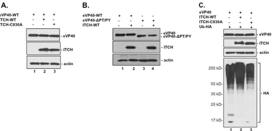
Co-IP of eVP40 and ITCH.
We next sought to confirm the
above-described eVP40-ITCH interaction in mammalian cells.
Briefly, HEK293T cells were transfected with eVP40-WT,
ITCH-WT, and/or an enzymatically inactive ITCH-C830A mutant
(
Fig. 2A
). Cell extracts were immunoprecipitated with either
rab-bit preimmune IgG (
Fig. 2A
, lanes 1 to 3) or polyclonal
anti-eVP40 antiserum (
Fig. 2A
, lanes 4 to 6), and c-
myc
-tagged ITCH
was detected in precipitated samples by Western blotting using
anti-c-
myc
antiserum. Both ITCH-WT and ITCH-C830A were
detected in eVP40 precipitates (
Fig. 2A
, lanes 5 and 6) but not in
preimmune IgG precipitates of identical samples (
Fig. 2A
, lanes 2
and 3). These results indicated that eVP40-WT interacts with both
WT and enzymatically inactive forms of ITCH in transiently
transfected HEK293T cells, albeit the interaction between eVP40
and ITCH-C830A was reduced slightly compared to that between
eVP40 and ITCH-WT (
Fig. 2A
).
We next sought to determine whether this interaction was
depen-dent on the eVP40 PPxY motif. To this end, HEK293T cells were
transfected with eVP40-WT, eVP40-
⌬
PT/PY, and/or ITCH-WT
(
Fig. 2B
). Cell extracts were immunoprecipitated with either rabbit
preimmune IgG (
Fig. 2B
, lanes 1 to 4) or polyclonal anti-eVP40
antiserum (
Fig. 2B
, lanes 5 to 8), and c-
myc
-tagged ITCH was
detected in precipitated samples by Western blotting using
anti-c-
myc
antiserum. ITCH was detected in eVP40 precipitates from
cells expressing eVP40-WT and ITCH-WT (
Fig. 2B
, lane 6) but
FIG 1GST-WW domain pulldown of the eVP40 PPxY peptide. (A)Identifi-cation of eVP40 PPxY motif readers using fluorescently labeled biotinylated peptides to screen a proline-rich reading array. The GST-WW domain fusion proteins are arrayed in duplicate, at different angles. Robust interactions with multiple WW domains were observed, including those of Nedd4, Nedd4L, and ITCH (arrows). (B) The indicated GST-WW domain fusion proteins were purified fromE. coliand analyzed in Coomassie-stained SDS-PAGE gels. MW, molecular weight (in thousands). (C) Biotinylated eVP40-WT (MRRVILPTA PPEYMEAI[Lys-biotin]) (lanes 1) or eVP40 mutant (MRRVILPTAAAEAME AI[Lys-biotin]) (lanes 2) peptides were used to pull down the indicated GST-WW domain fusion proteins. GST-WW domain fusion proteins were detected by Western blotting with mouse anti-GST antibody.
FIG 2ITCH interacts with eVP40 in a PPxY-dependent manner. (A and B) Extracts from HEK293T cells transfected with the indicated plasmids were first immunoprecipitated (IP) with either rabbit preimmune serum (IgG) or poly-clonal anti-eVP40 antiserum, and c-myc-tagged ITCH was then detected in precipitated samples by Western blotting (WB) using anti-c-mycantiserum. (C) Extracts from HEK293T cells transfected with the indicated plasmids were first immunoprecipitated with either rabbit preimmune serum (IgG) (lanes 1 to 3) or polyclonal anti-eVP40 antiserum (lanes 4 to 6), and proteins modified by HA-tagged ubiquitin (Ub) were then detected in precipitated samples by Western blotting using anti-HA antiserum.
on November 7, 2019 by guest
http://jvi.asm.org/
[image:3.585.50.349.65.419.2] [image:3.585.278.533.67.384.2]not in those from cells expressing eVP40-
⌬
PT/PY and ITCH-WT
(
Fig. 2B
, lane 8). As expected, ITCH was not detected in
preim-mune IgG precipitates of identical samples (
Fig. 2B
, lanes 2 and 4).
These results indicate that the eVP40-ITCH interaction is PPxY
dependent, which correlates well with GST pulldown results
(
Fig. 1
).
Since ITCH interacts with eVP40, we asked whether eVP40 was
ubiquitinated by ITCH. To test this, HEK293T cells were
trans-fected with combinations of plasmids encoding eVP40,
ITCH-WT, ITCH-C830A (inactive mutant), and/or Ub-HA (
Fig. 2C
).
Cell extracts were immunoprecipitated with either rabbit
preim-mune IgG (
Fig. 2C
, lanes 1 to 3) or polyclonal anti-eVP40
antise-rum (
Fig. 2C
, lanes 4 to 6), and HA-tagged ubiquitin was detected
in precipitated samples by Western analysis. Ubiquitinated species
of eVP40 that corresponded in size to mono- and diubiquitinated
forms were detected in cells expressing eVP40, ITCH-WT, and
Ub-HA (
Fig. 2C
, lane 5) but not in cells expressing either eVP40
and Ub-HA (lane 4) or eVP40, ITCH-C830A, and Ub-HA (lane
6). Together, these results demonstrate a physical and functional
interaction between ITCH and eVP40 leading to the
ubiquitina-tion of eVP40. Western analysis of cell extracts corresponding to
those shown
Fig. 2A
to
C
revealed appropriate expression levels of
eVP40, eVP40-
⌬
PT/PY, ITCH-WT, ITCH-C830A, and actin (
Fig.
3A
to
C
).
Enzymatically active ITCH ligase enhances eVP40 VLP
bud-ding.
Next, we sought to confirm that overexpression of
enzymat-ically active ITCH enhances the egress of eVP40 VLPs. To this end,
HEK293T cells were transfected with constant amounts of eVP40
and increasing amounts of either ITCH-WT or ITCH-C830A
plasmid DNA (
Fig. 4A
). Cell extracts and VLPs were harvested,
and the indicated proteins were detected by Western analysis (
Fig.
4A
). Interestingly, we observed a consistent 2- to 3-fold increase
(
Fig. 4B
) in eVP40 VLP budding in the presence of exogenous
ITCH-WT (
Fig. 4A
, compare lane 2 with lanes 3 and 4) but not in
the presence of exogenous ITCH-C830A (compare lane 2 with
lanes 5 and 6) compared to the eVP40-alone control. Indeed,
overexpression of ITCH-C830A appeared to have a dominant
negative effect on the budding of eVP40 VLPs, as levels of eVP40
in VLP samples expressing ITCH-C830A were reduced compared
to those in samples expressing eVP40 alone (
Fig. 4A
). We also
detected ITCH-WT, but not ITCH-C830A, in budding VLPs (
Fig.
4A
), which supports a functional role for ITCH in budding, as
other host proteins important for budding (e.g., Nedd4 and
Tsg101) are also packaged into VLPs (
14
,
32
).
We used a pharmacological approach to demonstrate that the
activation of ITCH ligase was critical for the enhanced egress of
eVP40 VLPs. Activation of ITCH ligase depends on
phosphoryla-tion by Jun amino-terminal kinase (JNK) (
56
). As SP600125 is a
well-described, selective inhibitor of JNK activity (
57
,
58
), we
reasoned that SP600125 may inhibit the egress of eVP40 VLPs
by blocking JNK-mediated activation of ITCH. To test this,
HEK293T cells were transfected with eVP40 in the absence or
presence of increasing amounts of SP600125, and eVP40 in cell
extracts and VLPs was detected by Western analysis (
Fig. 4C
).
Indeed, we found that budding of eVP40 VLPs was reduced by
⬃
10- and 20-fold in the presence of 5 and 10
M SP600125,
respectively (
Fig. 4C
, compare lane 2 with lanes 3 and 4).
SP600125 had little (
⬍
2-fold) to no effect on the cellular
expres-sion of eVP40 and endogenous ITCH (
Fig. 4C
). Together, these
data not only confirm that enzymatically active ITCH is required
for the efficient egress of eVP40 VLPs but also imply that a
phar-macological approach targeting this host pathway may represent a
viable and novel strategy to target and inhibit virus egress.
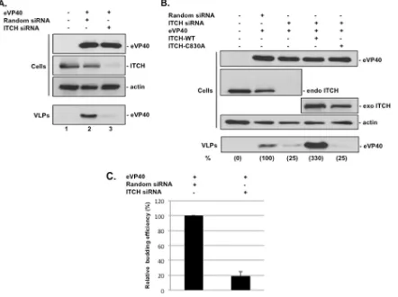
ITCH is required for efficient eVP40 VLP egress.
To further
confirm that the interaction between eVP40 and ITCH was
func-tionally relevant for eVP40 VLP budding, we used an siRNA
knockdown/rescue approach. Briefly, HEK293T cells were mock
transfected or transfected with eVP40-WT plus random or
ITCH-specific siRNAs (
Fig. 5A
). Expression of eVP40-WT and efficient
knockdown of endogenous ITCH (
⬎
90%) were confirmed by
Western analysis (
Fig. 5A
). We found that the knockdown of
en-FIG 3Western analysis of expression controls for the indicted proteins from cell extracts shown inFig. 2. Extracts from HEK293T cells were transfected with the indicated plasmids.Han et al.
on November 7, 2019 by guest
http://jvi.asm.org/
[image:4.585.65.518.63.279.2]dogenous ITCH resulted in a 75 to 80% decrease in the egress of
eVP40 VLPs compared to random siRNA controls (
Fig. 5A
).
To validate these siRNA results, we attempted to rescue eVP40
VLP budding in ITCH siRNA knockdown cells by transiently
ex-pressing exogenous ITCH-WT or ITCH-C830A (
Fig. 5B
).
West-ern analysis was used to detect and confirm the expression of
eVP40-WT and endogenous and exogenous ITCH in cell extracts
(
Fig. 5B
). As described above, cells receiving ITCH-specific
siRNAs showed an
⬃
75% decrease in eVP40 VLP egress
com-pared to random siRNA controls (
Fig. 5B
, compare lanes 2 and 3).
Interestingly, budding of eVP40 VLPs was enhanced by
⬃
3-fold in
cells expressing exogenous ITCH-WT compared to control
sam-ples (
Fig. 5B
, compare lanes 2 and 4), whereas no enhancement of
eVP40 VLP egress was observed in cells expressing exogenous
ITCH-C830A (
Fig. 5B
, compare lanes 2 and 5). These results
con-firm that knockdown of endogenous ITCH in HEK293T cells
re-sulted in a 4- to 5-fold decrease in eVP40 VLP egress in multiple
experiments (
Fig. 5C
) and, importantly, that the enzymatic
activ-ity of ITCH ligase was required for the efficient egress of eVP40
VLPs.
Reduced budding of VLPs and virus from ITCH knockout
cells.
We next took advantage of the HAP1-ITCH
⫺/⫺(KO) cell
line as a genetic approach to determine whether the expression of
endogenous ITCH is critical for the efficient egress of both eVP40
VLPs and live recombinant VSV expressing the eVP40 PPxY L
domain motif and flanking residues. Briefly, parental HAP1 cells
expressing endogenous ITCH (ITCH-WT) and HAP1 ITCH-KO
cells were transfected with eVP40, and eVP40 was detected in cell
extracts and VLPs at 24 h posttransfection by Western analysis
(
Fig. 6A
). We found that budding of eVP40 VLPs was reduced in
multiple independent experiments by up to 20-fold in ITCH KO
cells compared to that in ITCH-WT cells (
Fig. 6A
and
B
). Equal
expression of eVP40 and the absence of ITCH in the KO cell line
were confirmed by Western analysis (
Fig. 6A
).
To confirm that the reduced levels of eVP40 VLP egress were
due to the lack of ITCH expression in KO cells, ITCH KO cells
were transfected with eVP40 alone or in combination with
ITCH-WT, and eVP40 was detected in cell extracts and VLPs by
Western analysis (
Fig. 6C
). Indeed, we observed an
⬃
8-fold
in-crease in eVP40 VLP budding from KO cells receiving exogenous
ITCH-WT compared to that from KO cells expressing eVP40
alone (
Fig. 6C
and
D
). Appropriate levels of eVP40 and ITCH-WT
in cell extracts were confirmed by Western analysis (
Fig. 6C
).
We next asked whether the decrease in eVP40 VLP budding
from ITCH KO cells would also be observed for live-virus
bud-ding. To test this, we utilized our previously described
recombi-nant VSV (VSV-M40) that was engineered to express the PPxY L
domain motif and flanking residues from eVP40 in place of the
PPxY L domain and flanking residues of VSV-M (
14
). Parental
and ITCH KO cells were infected with VSV-M40 at an MOI of 0.1,
and infected-cell extracts and supernatants were harvested at the
peak budding time of 8 h postinfection (
Fig. 7
). Infectious virions
released into the medium were quantified by standard plaque
as-says on BHK-21 cells, and these data revealed a
⬎
1-log decrease in
FIG 4Enzymatically active ITCH enhances eVP40 VLP budding. (A) Western analysis of cell extracts and VLPs from HEK293T cells transfected with the indicated plasmids. (B) Budding efficiency of eVP40 VLPs in the presence of exogenous ITCH-WT relative to that with eVP40 alone. Error bars represent the standard deviations of the means from three independent experiments. C) Western analysis of cell extracts and VLPs from HEK293T cells mock transfected or transfected with eVP40 in the absence (0) or presence of SP600125 at a concentration of 5 or 10M. The budding efficiencies of eVP40 in VLPs relative to the control sample (lane 2) (100%) are shown in parentheses.on November 7, 2019 by guest
http://jvi.asm.org/
[image:5.585.139.451.65.349.2]the average titer of VSV-M40 in ITCH KO cells compared to that
in parental ITCH-WT cells (
Fig. 7
). Western analysis of
infected-cell extracts confirmed the lack of ITCH expression in KO infected-cells
and that the expression levels of the VSV M protein in WT and KO
cells were equivalent and unaffected by the lack of ITCH
expres-sion (
Fig. 7
). It should be noted that a similar degree of budding
inhibition was observed for WT VSV, whose M protein contains a
PPxY L domain motif (data not shown). These data suggest that,
like Nedd4, ITCH plays a role in facilitating the budding of
mul-tiple negative-strand RNA viruses that depend on a PPxY-type L
domain for efficient egress.
DISCUSSION
EBOV VP40 is the major viral structural protein that orchestrates
virion assembly and egress. EBOV VP40 accomplishes this, in
part, by hijacking or recruiting host proteins/pathways to facilitate
efficient virus separation from the site of budding at the plasma
membrane. Here we have identified ITCH as a novel eVP40
inter-actor that regulates the budding process.
A role for cellular ubiquitination and particularly the E3
ubiq-uitin ligase Nedd4 in promoting the efficient budding of many
enveloped RNA viruses, including EBOV, has been well
docu-mented (
3
,
8
,
9
,
26–30
,
32–43
,
59
,
60
). ITCH is one of nine
mem-bers of the Nedd4 family of HECT-type E3 ligases with canonical
C2, WW, and HECT domains as their main functional
compo-nents (
61
). Of all the mammalian WW domains tested here, we
identified WW domain 1 (WW1) of ITCH as one of the strongest,
specific interactors with the PPxY motif of eVP40 (
Fig. 1
). Indeed,
the high degree of specificity of the eVP40 PPxY motif for binding
to only select mammalian WW domains in our WW and SH3
domain array strongly suggests that the interacting host proteins
likely play biologically meaningful roles in the life cycle of EBOV.
As both Nedd4 and ITCH appear to be recruited to the eVP40
PPxY motif, our results raised the intriguing possibility that these
related HECT-type E3 ligases may be recruited by eVP40 to
pro-vide analogous or perhaps cell type-specific functions to promote
the late stages of virus assembly and egress. Indeed, other
Nedd4-like E3 ligases have been implicated in the assembly and egress of
several RNA viruses (
28
,
39
,
41
,
62
,
63
). It is possible that WW
domain selectivity depends in part on the sequence of the WW
domain itself and/or modifications of the PPxY ligand, such as
tyrosine phosphorylation (
64
).
We confirmed both the physical and functional nature of the
ITCH WW domain-eVP40 PPxY interaction using both VLP and
live-virus budding assays (
Fig. 5
to
7
). Although a role for the
ITCH E3 ubiquitin ligase as a novel host contributor to
eVP40-FIG 5siRNA knockdown/rescue of ITCH regulates eVP40 VLP budding. (A) Western analysis of eVP40, endogenous ITCH, and actin in extracts and VLPs from HEK293T cells mock treated or treated with the indicated siRNAs. (B) HEK293T cells were transfected as indicated, and Western analysis was used to detect eVP40 and endogenous (endo) and exogenous (exo) ITCH in cell extracts and eVP40 in VLPs. The budding efficiencies of eVP40 in VLPs relative to the control sample (lane 2) (100%) are shown in parentheses. (C) Average budding efficiency of eVP40 VLPs in cells treated with ITCH-specific siRNA relative to that in cells treated with the random siRNA control. Error bars represent the standard deviations of the means from three independent experiments.Han et al.
on November 7, 2019 by guest
http://jvi.asm.org/
[image:6.585.72.514.68.398.2]mediated budding has not been reported previously, ITCH was
iden-tified previously as a functional contributor to the budding process of
some retroviruses (
63
,
65–67
). ITCH has been more widely studied
for its role in immune regulation and inflammatory signaling, such as
its ability to regulate lymphocyte activation, differentiation, and
im-mune tolerance (for a review, see reference
68
). Whether there is any
link between the immune-regulatory activities of ITCH and its role in
promoting virus egress remains to be determined, as does a role for
ITCH in facilitating egress and modulating the pathogenesis of
infec-tious EBOV. Nonetheless, we demonstrated that (i) the eVP40-ITCH
interaction is PPxY dependent, (ii) enzymatically active ITCH
ubiq-uitinated eVP40, (iii) siRNA or genetic knockdown of endogenous
ITCH in HAP1 cells resulted in a decrease in eVP40 VLP and
live-VSV-M40 egress, and (iv) the addition of enzymatically active ITCH
rescued this budding defect.
While evidence for the “probudding” role of ubiquitination
and E3 ligases such as Nedd4 and ITCH continues to accumulate,
a counteracting and antagonistic “antibudding” role for host
in-terferon-stimulated gene 15 (ISG15) and ISGylation (covalent
modification of a target protein with ISG15) is emerging. ISG15 is
an interferon (IFN)-induced ubiquitin-like protein that plays a
key role as a central regulator of the innate immune response to a
plethora of viral pathogens (
69
,
70
; for reviews, see references
71
and
72
). Indeed, we and others found that free/unconjugated
ISG15 interferes with Nedd4 ligase activity, thereby preventing its
functional interaction with eVP40 and indirectly preventing the
budding of eVP40 VLPs (
53
,
72
,
73
). It will be of interest to
deter-mine whether this mechanistically novel function for ISG15 (
53
,
73
) and the functional interplay between ubiquitination and
ISGylation pathways extend to ITCH ligase as well.
In sum, our results provide new insights into the range of host
proteins that regulate EBOV VP40-mediated egress. While we
predict that ITCH should also interact with the PPxY motif of
MARV VP40, this remains to be determined. A better
understand-ing of this ongounderstand-ing battle and interplay between the virus and early
host innate immune defenses will be critical for our overall
under-standing of the biology and pathogenesis of the virus as well as for
the future development of effective antiviral therapies. Indeed, we
have identified small-molecule inhibitors of virus-host
interac-tions, including those that target PPxY-WW domain interacinterac-tions,
with promising potential as broad-spectrum antiviral
therapeu-tics (
29
,
55
,
74
).
ACKNOWLEDGMENTS
We thank G. Melino, K. Chandran, T. Brummelkamp, and D. Lyles for kindly providing reagents; C. Berry for help with statistical analysis; L. King for critical reading of the manuscript; and B. D. Freedman, J. Madara, G. Ruthel, X. Liu, J. Li, and J. Liang for helpful comments.
This work was supported in part by NIH grants AI102104, AI113952, and AI103785 to R.N.H.; by Cancer Prevention and Research Institute of Texas grant RP13042 to M.T.B. for the protein array analysis; and by seed grants from the National University of Singapore Medical School and Mechanobiology Institute in Singapore to M.S.
FUNDING INFORMATION
This work, including the efforts of Marius Sudol, was funded by NUS-Medical School and Mechanobiology Institute, Singapore (seed grants). This work, including the efforts of Ronald N. Harty, was funded by HHS | National Institutes of Health (NIH) (AI102104, AI113952, and AI103785). This work, including the efforts of Mark T. Bedford, was funded by Cancer Prevention and Research Institute of Texas (CPRIT) (RP13042).
REFERENCES
1.Han Z, Madara JJ, Liu Y, Liu W, Ruthel G, Freedman BD, Harty RN.
2015. ALIX rescues budding of a double PTAP/PPEY L-domain deletion
FIG 6eVP40 VLP budding from ITCH WT and KO cells. (A) HAP1 ITCH-WT (parental) or HAP1 ITCH KO cells were transfected with an eVP40 expression plasmid, and the indicated proteins were detected in cell extracts and VLPs by Western analysis. (B) Budding of eVP40 VLPs in ITCH KO cells relative to eVP40 VLPs from control ITCH-WT cells. Error bars represent the standard deviations of the means from four independent experiments. (C) HAP1 ITCH KO cells were transfected as indicated, and the indicated proteins were detected in cell extracts and VLPs by Western analysis. (D) Budding of eVP40 VLPs from ITCH KO cells transfected with ITCH-WT relative to bud-ding of eVP40 VLPs from control ITCH KO cells (set at 1). Error bars represent the standard deviations of the means from three independent experiments.
FIG 7Decreased budding of live recombinant VSV-M40 in ITCH KO cells. HAP1 ITCH-WT (parental) or HAP1 ITCH KO cells were infected with re-combinant VSV-M40, and virus titers were quantified by plaque assays at 8 h postinfection. Virus titers from ITCH KO cells were reduced⬎10-fold com-pared to those from ITCH-WT cells. ITCH, actin, and VSV M proteins were detected in the indicated infected-cell extracts by Western analysis. *** indi-cates aPvalue of⬍0.001, as determined by a Studentttest.
on November 7, 2019 by guest
http://jvi.asm.org/
[image:7.585.300.543.65.213.2] [image:7.585.41.285.67.333.2]mutant of Ebola VP40: a role for ALIX in Ebola virus egress. J Infect Dis
212(Suppl 2):S138 –S145.http://dx.doi.org/10.1093/infdis/jiu838. 2.Hartlieb B, Weissenhorn W. 2006. Filovirus assembly and budding.
Virology344:64 –70.http://dx.doi.org/10.1016/j.virol.2005.09.018. 3.Harty RN, Brown ME, Wang G, Huibregtse J, Hayes FP.2000. A PPxY
motif within the VP40 protein of Ebola virus interacts physically and functionally with a ubiquitin ligase: implications for filovirus budding. Proc Natl Acad Sci U S A97:13871–13876.http://dx.doi.org/10.1073/pnas .250277297.
4.Jasenosky LD, Kawaoka Y.2004. Filovirus budding. Virus Res106:181– 188.http://dx.doi.org/10.1016/j.virusres.2004.08.014.
5.Liu Y, Lee MS, Olson MA, Harty RN.2011. Bimolecular complemen-tation to visualize filovirus VP40-host complexes in live mammalian cells: toward the identification of budding inhibitors. Adv Virol2011:341816. 6.Lu J, Qu Y, Liu Y, Jambusaria R, Han Z, Ruthel G, Freedman BD, Harty
RN.2013. Host IQGAP1 and Ebola virus VP40 interactions facilitate vi-rus-like particle egress. J Virol87:7777–7780.http://dx.doi.org/10.1128 /JVI.00470-13.
7.Noda T, Ebihara H, Muramoto Y, Fujii K, Takada A, Sagara H, Kim JH, Kida H, Feldmann H, Kawaoka Y. 2006. Assembly and budding of ebolavirus. PLoS Pathog 2:e99. http://dx.doi.org/10.1371/journal.ppat .0020099.
8.Timmins J, Schoehn G, Ricard-Blum S, Scianimanico S, Vernet T, Ruigrok RW, Weissenhorn W.2003. Ebola virus matrix protein VP40 interaction with human cellular factors Tsg101 and Nedd4. J Mol Biol
326:493–502.http://dx.doi.org/10.1016/S0022-2836(02)01406-7. 9.Yasuda J, Nakao M, Kawaoka Y, Shida H.2003. Nedd4 regulates egress
of Ebola virus-like particles from host cells. J Virol77:9987–9992.http: //dx.doi.org/10.1128/JVI.77.18.9987-9992.2003.
10. Bieniasz PD.2006. Late budding domains and host proteins in enveloped virus release. Virology344:55– 63.http://dx.doi.org/10.1016/j.virol.2005 .09.044.
11. Calistri A, Salata C, Parolin C, Palu G. 2009. Role of multivesicular bodies and their components in the egress of enveloped RNA viruses. Rev Med Virol19:31– 45.http://dx.doi.org/10.1002/rmv.588.
12. Chen BJ, Lamb RA.2008. Mechanisms for enveloped virus budding: can some viruses do without an ESCRT? Virology372:221–232.http://dx.doi .org/10.1016/j.virol.2007.11.008.
13. Harty RN.2009. No exit: targeting the budding process to inhibit filovirus replication. Antiviral Res 81:189 –197.http://dx.doi.org/10 .1016/j.antiviral.2008.12.003.
14. Irie T, Licata JM, Harty RN.2005. Functional characterization of Ebola virus L-domains using VSV recombinants. Virology336:291–298.http: //dx.doi.org/10.1016/j.virol.2005.03.027.
15. Liu Y, Harty RN.2010. Viral and host proteins that modulate filovirus budding. Future Virol5:481– 491.http://dx.doi.org/10.2217/fvl.10.33. 16. Urata S, de la Torre JC.2011. Arenavirus budding. Adv Virol 2011:
180326.
17. Bork P, Sudol M.1994. The WW domain: a signalling site in dystro-phin? Trends Biochem Sci19:531–533.http://dx.doi.org/10.1016/0968 -0004(94)90053-1.
18. Chen HI, Sudol M.1995. The WW domain of Yes-associated protein binds a proline-rich ligand that differs from the consensus established for Src homology 3-binding modules. Proc Natl Acad Sci U S A92:7819 – 7823.http://dx.doi.org/10.1073/pnas.92.17.7819.
19. Hu H, Columbus J, Zhang Y, Wu D, Lian L, Yang S, Goodwin J, Luczak C, Carter M, Chen L, James M, Davis R, Sudol M, Rodwell J, Herrero JJ.2004. A map of WW domain family interactions. Proteomics4:643– 655.http://dx.doi.org/10.1002/pmic.200300632.
20. Ilsley JL, Sudol M, Winder SJ.2002. The WW domain: linking cell signalling to the membrane cytoskeleton. Cell Signal14:183–189.http: //dx.doi.org/10.1016/S0898-6568(01)00236-4.
21. Sudol M.1996. Structure and function of the WW domain. Prog Biophys Mol Biol65:113–132.
22. Sudol M.1996. The WW module competes with the SH3 domain? Trends Biochem Sci 21:161–163. http://dx.doi.org/10.1016/S0968 -0004(96)30018-2.
23. Sudol M, Chen HI, Bougeret C, Einbond A, Bork P.1995. Charac-terization of a novel protein-binding module—the WW domain. FEBS Lett369:67–71.http://dx.doi.org/10.1016/0014-5793(95)00550-S. 24. Sudol M, Hunter T.2000. NeW wrinkles for an old domain. Cell103:
1001–1004.http://dx.doi.org/10.1016/S0092-8674(00)00203-8. 25. Sudol M, Sliwa K, Russo T.2001. Functions of WW domains in the
nucleus. FEBS Lett 490:190 –195.http://dx.doi.org/10.1016/S0014 -5793(01)02122-6.
26. Blot V, Perugi F, Gay B, Prevost MC, Briant L, Tangy F, Abriel H, Staub O, Dokhelar MC, Pique C.2004. Nedd4.1-mediated ubiquitination and subsequent recruitment of Tsg101 ensure HTLV-1 Gag trafficking to-wards the multivesicular body pathway prior to virus budding. J Cell Sci
117:2357–2367.http://dx.doi.org/10.1242/jcs.01095.
27. Bouamr F, Melillo JA, Wang MQ, Nagashima K, de Los Santos M, Rein A, Goff SP.2003. PPPYEPTAP motif is the late domain of human T-cell leukemia virus type 1 Gag and mediates its functional interaction with cellular proteins Nedd4 and Tsg101. J Virol77:11882–11895.http://dx.doi .org/10.1128/JVI.77.22.11882-11895.2003. (Author Correction,78:4383, 2004,http://dx.doi.org/10.1128/JVI.78.8.4383.2004.)
28. Chung HY, Morita E, von Schwedler U, Muller B, Krausslich HG, Sundquist WI.2008. NEDD4L overexpression rescues the release and infectivity of human immunodeficiency virus type 1 constructs lacking PTAP and YPXL late domains. J Virol82:4884 – 4897.http://dx.doi.org/10 .1128/JVI.02667-07.
29. Han Z, Lu J, Liu Y, Davis B, Lee MS, Olson MA, Ruthel G, Freedman BD, Schnell MJ, Wrobel JE, Reitz AB, Harty RN.2014. Small-molecule probes targeting the viral PPxY-host Nedd4 interface block egress of a broad range of RNA viruses. J Virol88:7294 –7306.http://dx.doi.org/10 .1128/JVI.00591-14.
30. Harty RN, Brown ME, McGettigan JP, Wang G, Jayakar HR, Hu-ibregtse JM, Whitt MA, Schnell MJ.2001. Rhabdoviruses and the cellular ubiquitin-proteasome system: a budding interaction. J Virol75:10623– 10629.http://dx.doi.org/10.1128/JVI.75.22.10623-10629.2001. 31. Kikonyogo A, Bouamr F, Vana ML, Xiang Y, Aiyar A, Carter C, Leis J.
2001. Proteins related to the Nedd4 family of ubiquitin protein ligases interact with the L domain of Rous sarcoma virus and are required for gag budding from cells. Proc Natl Acad Sci U S A98:11199 –11204.http://dx .doi.org/10.1073/pnas.201268998.
32. Licata JM, Simpson-Holley M, Wright NT, Han Z, Paragas J, Harty RN.
2003. Overlapping motifs (PTAP and PPEY) within the Ebola virus VP40 protein function independently as late budding domains: involvement of host proteins TSG101 and VPS-4. J Virol77:1812–1819.http://dx.doi.org /10.1128/JVI.77.3.1812-1819.2003.
33. Martin-Serrano J, Perez-Caballero D, Bieniasz PD.2004. Context-dependent effects of L domains and ubiquitination on viral budding. J Virol 78:5554 –5563. http://dx.doi.org/10.1128/JVI.78.11.5554-5563 .2004.
34. Medina G, Pincetic A, Ehrlich LS, Zhang Y, Tang Y, Leis J, Carter CA.
2008. Tsg101 can replace Nedd4 function in ASV Gag release but not membrane targeting. Virology 377:30 –38. http://dx.doi.org/10.1016/j .virol.2008.04.024.
35. Sakurai A, Yasuda J, Takano H, Tanaka Y, Hatakeyama M, Shida H.
2004. Regulation of human T-cell leukemia virus type 1 (HTLV-1) bud-ding by ubiquitin ligase Nedd4. Microbes Infect6:150 –156.http://dx.doi .org/10.1016/j.micinf.2003.10.011.
36. Sette P, Jadwin JA, Dussupt V, Bello NF, Bouamr F.2010. The ESCRT-associated protein Alix recruits the ubiquitin ligase Nedd4-1 to facilitate HIV-1 release through the LYPXnL L domain motif. J Virol84:8181– 8192.http://dx.doi.org/10.1128/JVI.00634-10.
37. Sette P, Nagashima K, Piper RC, Bouamr F.2013. Ubiquitin conjuga-tion to Gag is essential for ESCRT-mediated HIV-1 budding. Retrovirol-ogy10:79.http://dx.doi.org/10.1186/1742-4690-10-79.
38. Urata S, Yasuda J.2010. Regulation of Marburg virus (MARV) budding by Nedd4.1: a different WW domain of Nedd4.1 is critical for binding to MARV and Ebola virus VP40. J Gen Virol91:228 –234.http://dx.doi.org /10.1099/vir.0.015495-0.
39. Usami Y, Popov S, Popova E, Gottlinger HG.2008. Efficient and specific rescue of human immunodeficiency virus type 1 budding defects by a Nedd4-like ubiquitin ligase. J Virol82:4898 – 4907.http://dx.doi.org/10 .1128/JVI.02675-07.
40. Vana ML, Tang Y, Chen A, Medina G, Carter C, Leis J.2004. Role of Nedd4 and ubiquitination of Rous sarcoma virus Gag in budding of virus-like particles from cells. J Virol78:13943–13953.http://dx.doi.org/10.1128 /JVI.78.24.13943-13953.2004.
41. Weiss ER, Popova E, Yamanaka H, Kim HC, Huibregtse JM, Gottlinger H.2010. Rescue of HIV-1 release by targeting widely divergent NEDD4-type ubiquitin ligases and isolated catalytic HECT domains to Gag. PLoS Pathog6:e1001107.http://dx.doi.org/10.1371/journal.ppat.1001107. 42. Yasuda J, Hunter E, Nakao M, Shida H.2002. Functional involvement
Han et al.
on November 7, 2019 by guest
http://jvi.asm.org/
of a novel Nedd4-like ubiquitin ligase on retrovirus budding. EMBO Rep
3:636 – 640.http://dx.doi.org/10.1093/embo-reports/kvf132.
43. Zhadina M, Bieniasz PD. 2010. Functional interchangeability of late domains, late domain cofactors and ubiquitin in viral budding. PLoS Pat-hog6:e1001153.http://dx.doi.org/10.1371/journal.ppat.1001153. 44. Calistri A, Del Vecchio C, Salata C, Celestino M, Celegato M, Gottlinger
H, Palu G, Parolin C.2009. Role of the feline immunodeficiency virus L-domain in the presence or absence of Gag processing: involvement of ubiquitin and Nedd4-2s ligase in viral egress. J Cell Physiol218:175–182.
http://dx.doi.org/10.1002/jcp.21587.
45. Klinger PP, Schubert U.2005. The ubiquitin-proteasome system in HIV replication: potential targets for antiretroviral therapy. Expert Rev Anti Infect Ther3:61–79.http://dx.doi.org/10.1586/14787210.3.1.61. 46. Lewis B, Whitney S, Hudacik L, Galmin L, Huaman MC, Cristillo AD.
2014. Nedd4-mediated increase in HIV-1 Gag and Env proteins and im-munity following DNA-vaccination of BALB/c mice. PLoS One9:e91267.
http://dx.doi.org/10.1371/journal.pone.0091267.
47. Pincetic A, Medina G, Carter C, Leis J.2008. Avian sarcoma virus and human immunodeficiency virus, type 1 use different subsets of ESCRT proteins to facilitate the budding process. J Biol Chem283:29822–29830.
http://dx.doi.org/10.1074/jbc.M804157200.
48. Urata S, Noda T, Kawaoka Y, Yokosawa H, Yasuda J.2006. Cellular factors required for Lassa virus budding. J Virol80:4191– 4195.http://dx .doi.org/10.1128/JVI.80.8.4191-4195.2006.
49. Usami Y, Popov S, Popova E, Inoue M, Weissenhorn W, Gottlinger GH.2009. The ESCRT pathway and HIV-1 budding. Biochem Soc Trans
37:181–184.http://dx.doi.org/10.1042/BST0370181.
50. Zhadina M, McClure MO, Johnson MC, Bieniasz PD.2007. Ubiquitin-dependent virus particle budding without viral protein ubiquitination. Proc Natl Acad Sci U S A104:20031–20036.http://dx.doi.org/10.1073 /pnas.0708002104.
51. Einbond A, Sudol M.1996. Towards prediction of cognate complexes between the WW domain and proline-rich ligands. FEBS Lett384:1– 8.
http://dx.doi.org/10.1016/0014-5793(96)00263-3.
52. Espejo A, Cote J, Bednarek A, Richard S, Bedford MT.2002. A protein-domain microarray identifies novel protein-protein interactions. Biochem J367:697–702.http://dx.doi.org/10.1042/bj20020860. 53. Okumura A, Pitha PM, Harty RN.2008. ISG15 inhibits Ebola VP40 VLP
budding in an L-domain-dependent manner by blocking Nedd4 ligase activity. Proc Natl Acad Sci U S A105:3974 –3979.http://dx.doi.org/10 .1073/pnas.0710629105.
54. Rossi M, Rotblat B, Ansell K, Amelio I, Caraglia M, Misso G, Bernas-sola F, Cavasotto CN, Knight RA, Ciechanover A, Melino G.2014. High throughput screening for inhibitors of the HECT ubiquitin E3 ligase ITCH identifies antidepressant drugs as regulators of autophagy. Cell Death Dis5:e1203.http://dx.doi.org/10.1038/cddis.2014.113.
55. Lu J, Han Z, Liu Y, Liu W, Lee MS, Olson MA, Ruthel G, Freedman BD, Harty RN.2014. A host-oriented inhibitor of Junin Argentine hemor-rhagic fever virus egress. J Virol88:4736 – 4743.http://dx.doi.org/10.1128 /JVI.03757-13.
56. Gao M, Labuda T, Xia Y, Gallagher E, Fang D, Liu YC, Karin M.2004. Jun turnover is controlled through JNK-dependent phosphorylation of the E3 ligase Itch. Science306:271–275.http://dx.doi.org/10.1126/science .1099414.
57. Cheng J, Fan YH, Xu X, Zhang H, Dou J, Tang Y, Zhong X, Rojas Y, Yu Y, Zhao Y, Vasudevan SA, Zhang H, Nuchtern JG, Kim ES, Chen X, Lu F, Yang J.2014. A small-molecule inhibitor of UBE2N induces neu-roblastoma cell death via activation of p53 and JNK pathways. Cell Death Dis5:e1079.http://dx.doi.org/10.1038/cddis.2014.54.
58. Diao L, Zhang B, Xuan C, Sun S, Yang K, Tang Y, Qiao W, Chen Q, Geng Y, Wang C.2005. Activation of c-Jun N-terminal kinase (JNK) pathway by HSV-1 immediate early protein ICP0. Exp Cell Res308:196 – 210.http://dx.doi.org/10.1016/j.yexcr.2005.04.016.
59. Gottwein E, Bodem J, Muller B, Schmechel A, Zentgraf H, Krausslich HG.2003. The Mason-Pfizer monkey virus PPPY and PSAP motifs both contribute to virus release. J Virol77:9474 –9485.http://dx.doi.org/10 .1128/JVI.77.17.9474-9485.2003.
60. Segura-Morales C, Pescia C, Chatellard-Causse C, Sadoul R, Bertrand E, Basyuk E.2005. Tsg101 and Alix interact with murine leukemia virus Gag and cooperate with Nedd4 ubiquitin ligases during budding. J Biol Chem280:27004 –27012.http://dx.doi.org/10.1074/jbc.M413735200. 61. Rotin D, Kumar S.2009. Physiological functions of the HECT family of
ubiquitin ligases. Nat Rev Mol Cell Biol10:398 – 409.http://dx.doi.org/10 .1038/nrm2690.
62. Heidecker G, Lloyd PA, Soheilian F, Nagashima K, Derse D.2007. The role of WWP1-Gag interaction and Gag ubiquitination in assembly and release of human T-cell leukemia virus type 1. J Virol81:9769 –9777.http: //dx.doi.org/10.1128/JVI.00642-07.
63. Martin-Serrano J, Eastman SW, Chung W, Bieniasz PD.2005. HECT ubiquitin ligases link viral and cellular PPXY motifs to the vacuolar pro-tein-sorting pathway. J Cell Biol168:89 –101.http://dx.doi.org/10.1083 /jcb.200408155.
64. Garcia M, Cooper A, Shi W, Bornmann W, Carrion R, Kalman D, Nabel GJ.2012. Productive replication of Ebola virus is regulated by the c-Abl1 tyrosine kinase. Sci Transl Med4:123ra24.http://dx.doi.org/10 .1126/scitranslmed.3003500.
65. Dorjbal B, Derse D, Lloyd P, Soheilian F, Nagashima K, Heidecker G.
2011. The role of ITCH protein in human T-cell leukemia virus type 1 release. J Biol Chem 286:31092–31104. http://dx.doi.org/10.1074/jbc .M111.259945.
66. Jadwin JA, Rudd V, Sette P, Challa S, Bouamr F.2010. Late domain-independent rescue of a release-deficient Moloney murine leukemia virus by the ubiquitin ligase Itch. J Virol84:704 –715.http://dx.doi.org/10.1128 /JVI.01319-09.
67. Rauch S, Martin-Serrano J. 2011. Multiple interactions between the ESCRT machinery and arrestin-related proteins: implications for PPXY-dependent budding. J Virol85:3546 –3556.http://dx.doi.org/10.1128/JVI .02045-10.
68. Venuprasad K, Zeng M, Baughan SL, Massoumi R.2015. Multifaceted role of the ubiquitin ligase Itch in immune regulation. Immunol Cell Biol
93:452– 460.http://dx.doi.org/10.1038/icb.2014.118.
69. Morales DJ, Lenschow DJ.2013. The antiviral activities of ISG15. J Mol Biol425:4995–5008.http://dx.doi.org/10.1016/j.jmb.2013.09.041. 70. Zhu ZX, Wei JC, Shi ZX, Wu KB, Ma ZY.2012. Advances in
ubiquitin-like protein ISG15-mediated anti-viral response. Bing Du Xue Bao28:78 – 83. (In Chinese.)
71. Campbell JA, Lenschow DJ.2013. Emerging roles for immunomodula-tory functions of free ISG15. J Interferon Cytokine Res33:728 –738.http: //dx.doi.org/10.1089/jir.2013.0064.
72. Harty RN, Pitha PM, Okumura A. 2009. Antiviral activity of innate immune protein ISG15. J Innate Immun1:397– 404.http://dx.doi.org/10 .1159/000226245.
73. Malakhova OA, Zhang DE.2008. ISG15 inhibits Nedd4 ubiquitin E3 activity and enhances the innate antiviral response. J Biol Chem283:8783– 8787.http://dx.doi.org/10.1074/jbc.C800030200.
74. Madara JJ, Han Z, Ruthel G, Freedman BD, Harty RN. 2015. The multifunctional Ebola virus VP40 matrix protein is a promising therapeu-tic target. Future Virol10:537–546.http://dx.doi.org/10.2217/fvl.15.6.
on November 7, 2019 by guest
http://jvi.asm.org/