Herpes Simplex Virus 1 DNA Polymerase RNase H Activity Acts
in a 3
=
-to-5
=
Direction and Is Dependent on the 3
=
-to-5
=
Exonuclease Active Site
Jessica L. Lawler,a,bPurba Mukherjee,a*Donald M. Coena,b
aDepartment of Biological Chemistry and Molecular Pharmacology, Harvard Medical School, Boston,
Massachusetts, USA
bPhD Program in Virology, Harvard Medical School, Boston, Massachusetts, USA
ABSTRACT The catalytic subunit (Pol) of herpes simplex virus 1 (HSV-1) DNA merase has been extensively studied both as a model for other family B DNA poly-merases and for its differences from these enzymes as an antiviral target. Among the activities of HSV-1 Pol is an intrinsic RNase H activity that cleaves RNA from RNA-DNA hybrids. There has long been a controversy regarding whether this activity
is due to the 3=-to-5=exonuclease of Pol or whether it is a separate activity, possibly
acting on 5= RNA termini. To investigate this issue, we compared wild-type HSV-1
Pol and a 3=-to-5= exonuclease-deficient mutant, D368A Pol, for DNA polymerase
ac-tivity, 3=-to-5=exonuclease activity, and RNase H activityin vitro. Additionally, we
as-sessed the RNase H activity using differentially end-labeled templates with 5= or 3=
RNA termini. The mutant enzyme was at most modestly impaired for DNA
polymer-ase activity but was drastically impaired for 3=-to-5=exonuclease activity, with no
ac-tivity detected even at high enzyme-to-DNA substrate ratios. Importantly, the
mu-tant showed no detectable ability to excise RNA with either a 3= or 5= terminus,
while the wild-type HSV-1 Pol was able to cleave RNA from the annealed RNA-DNA
hairpin template, but only detectably with a 3=RNA terminus in a 3=-to-5=direction
and at a rate lower than that of the exonuclease activity. These results suggest that
HSV-1 Pol does not have an RNase H separable from its 3=-to-5=exonuclease activity
and that this activity prefers DNA degradation over degradation of RNA from RNA-DNA hybrids.
IMPORTANCE Herpes simplex virus 1 (HSV-1) is a member of theHerpesviridae fam-ily of DNA viruses, several of which cause morbidity and mortality in humans. Al-though the HSV-1 DNA polymerase has been studied for decades and is a crucial target for antivirals against HSV-1 infection, several of its functions remain to be
elu-cidated. A hypothesis suggesting the existence of a 5=-to-3=RNase H activity intrinsic
to this enzyme that could remove RNA primers from Okazaki fragments has been particularly controversial. In this study, we were unable to identify RNase H activity
of HSV-1 DNA polymerase on RNA-DNA hybrids with 5= RNA termini. We detected
RNase H activity on hybrids with 3=termini, but this was due to the 3=-to-5=
exonu-clease. Thus, HSV-1 is unlikely to use this method to remove RNA primers during DNA replication but may use pathways similar to those used in eukaryotic Okazaki fragment maturation.
KEYWORDS 3=-to-5=exonuclease, DNA polymerase, DNA replication, RNase H, herpes simplex virus
H
erpesviruses, including herpes simplex virus 1 (HSV-1), encode their own DNAreplication machinery, which is indispensable for copying their double-stranded DNA (dsDNA) genomes and for lytic replication within the host. Specifically, HSV-1
Received15 October 2017Accepted11 December 2017
Accepted manuscript posted online13 December 2017
CitationLawler JL, Mukherjee P, Coen DM. 2018. Herpes simplex virus 1 DNA polymerase RNase H activity acts in a 3=-to-5=direction and is dependent on the 3=-to-5=exonuclease active site. J Virol 92:e01813-17.https://doi.org/ 10.1128/JVI.01813-17.
EditorRozanne M. Sandri-Goldin, University of California, Irvine
Copyright© 2018 American Society for Microbiology.All Rights Reserved. Address correspondence to Donald M. Coen, don_coen@hms.harvard.edu.
*Present address: Purba Mukherjee, Skaggs Institute for Chemical Biology, The Scripps Research Institute, La Jolla, California, USA.
OF VIRAL GENE EXPRESSION
crossm
on November 6, 2019 by guest
http://jvi.asm.org/
requires seven virally encoded proteins to effect DNA replication: the origin-binding protein (UL9), the single-stranded DNA binding protein (ICP8), the helicase-primase complex (UL5, UL8, and UL52), and the DNA polymerase holoenzyme (UL30 and UL42) (1, 2).
HSV-1 DNA replication has been shown to utilize both leading and lagging strand
DNA replicationin vitro(3, 4). Leading strand synthesis proceeds continuously following
separation of the DNA strands at the replication fork. Lagging strand synthesis, in contrast, requires the synthesis by the helicase-primase complex of RNA primers annealed to template DNA, forming Okazaki fragments. In order to complete lagging strand synthesis, removal of the RNA primers, replacement of these gaps with DNA, and a ligation reaction are required (5). So far, it is unclear how HSV-1 removes RNA primers for completion of lagging strand DNA replication. Two methods for removing RNA primers during HSV-1 DNA replication have been proposed (6–8): (i) the HSV-1 DNA polymerase catalytic subunit (HSV-1 Pol) displaces the RNA primer strand during synthesis and a viral or cellular nuclease is recruited for “flap” removal (as for eukaryotic
Okazaki fragment maturation) (7, 8); and (ii) HSV-1 Pol has a 5=-to-3=RNase H activity
that removes the RNA prior to the gap-filling reaction (as for Escherichia coli DNA
polymerase I) (6). Since HSV-1 Pol has an associated RNase H activity (6, 9–12; and see below), the second proposal has been attractive.
HSV-1 Pol contains four domains: the pre-NH2-terminal domain, the NH2-terminal
domain, the 3=-to-5=exonuclease (Exo) domain, and the polymerase domain, composed
of the palm, finger, and thumb subdomains (13). Like many other family B (Pol␣family)
DNA polymerases (9, 13, 14), HSV Pol contains an intrinsic 3=-to-5=exonuclease activity
important for proofreading during DNA replication (9, 12, 15). The Exo domain contains three highly conserved motifs (named Exo I, Exo II, and Exo III) that are required for this
3=-to-5= exonuclease function (9, 12, 13, 16). For example, the Exo I D368A mutant
exhibits severely impaired exonuclease activity while maintaining high levels of poly-merase activity (17, 18). Additionally, HSV-1 Pol exhibits intrinsic apurinic/apyrimidinic
and 5=deoxyribose phosphate lyase activity specific to the C-terminal portion of the
enzyme (19).
HSV-1 Pol preparations have also been found to contain an RNase H activity (6,
9–12). Originally, an RNase H activity said to have 5=-to-3=directionality copurified with
HSV-1 Pol (6), but a contaminant, predicted to be the viral alkaline nuclease UL12, was present following a similar protein purification protocol (20, 21). Subsequently, RNase H activity intrinsic to HSV-1 Pol was confirmed and mapped to the N-terminal portion
of the enzyme containing the 3=-to-5=exonuclease, NH2-terminal, and pre-NH2terminal
domains (10, 12). However, it is unclear whether this RNase H acts on 5=RNA termini,
is instead attributable to the 3=-to-5= exonuclease, or is due to a different function
altogether. Hall et al. showed that the D368A mutation ablated cleavage of both single-stranded DNA (ssDNA) and dsDNA substrates, and they were unable to detect
5=-to-3=DNase activity, but they did not test RNA-DNA hybrid templates (21).
More recently, Liu et al. (13) identified a structural motif in the NH2-terminal domain
that strongly resembles motifs found in certain other RNA-binding proteins, with nearby negatively charged residues that might coordinate divalent cations, reviving the
hypothesis (6) that HSV-1 Pol has a 5=-to-3=RNase H activity to remove RNA primers.
However, the specificity of HSV-1 Pol RNase H activity for either 5=or 3= termini and
whether it depends on the 3=-to-5=exonuclease active site have remained in question.
To address these questions, we carried outin vitroassays utilizing purified wild-type
(WT) Pol and the D368A exonuclease-deficient mutant, testing the ability of these enzymes to extend a fluorescently labeled DNA hairpin primer-template and degrade dsDNA and RNA-DNA hybrid hairpin substrates over time.
RESULTS
Purified His-WT Pol and His-D368A exonuclease-deficient Pol retain similar polymerase activities.To test whether the RNase H activity of HSV-1 Pol requires an
active 3=-to-5=exonuclease catalytic site, we analyzed thein vitroactivities of purified
Lawler et al. Journal of Virology
on November 6, 2019 by guest
http://jvi.asm.org/
WT Pol and the previously characterized exonuclease-deficient single mutant, D368A
Pol (17, 18). We expressed 6⫻His-tagged full-length WT Pol and 6⫻His-tagged D368A
Pol separately using baculovirus expression (22) and purified these enzymes forin vitro
characterization. Next, we assessed the 5=-to-3= polymerization function of both
en-zymes by testing the ability of these Pols and control enen-zymes to extend a hairpin
template (S1) with a 5=overhang and a 5=fluorescent label (Fig. 1A and Fig. 2A). (Black
specks that appear on the gel images are due to background fluorescence from the tray on which the images were taken, and multiple images have been compared to reduce errors in quantitation.) Under the conditions tested, the positive-control enzyme,
FIG 1Primer-templates and substrates used in the study, their structure, and the activities assayed using those oligonucleotides. Black letters denote deoxyribonucleotides, and red letters denote ribonucleotides.
FIG 2WT and D368A Pol exhibit similar polymerase activities. (A) Cartoon of the 6-FAM-labeled DNA hairpin primer-template (S1; see Fig. 1A) used for the polymerase assays showing the direction of extension of the primer and the addition of dNTPs and MgCl2to start the reaction. (B) Fluorescent image of a
denaturing polyacrylamide gel loaded with equal amounts of each polymerase reaction mixture over increasing time points (0, 10 s, 30 s, 1 min, 2 min, 4 min, 8 min, 16 min, 32 min). The assays shown here contained a 2-fold molar excess of primer-template relative to Pol. The 0- and 32-min control reactions (Klenow fragment and RNase H) are shown. Arrows on the left side of the gel denote the starting and fully extended primer-template (P/T).
on November 6, 2019 by guest
http://jvi.asm.org/
[image:3.585.46.368.74.282.2] [image:3.585.45.378.455.661.2]Klenow fragment ofE. coliDNA Pol I, could extend the primer to the end of the dsDNA hairpin primer-template, producing a larger product that comigrates with a synthesized hairpin template of the correct size (data not shown), while the negative-control
enzyme,E. coliRNase H, did not detectably extend the primer (Fig. 2B). Both WT and
D368A Pol exhibited 5=-to-3=polymerase activity, as evidenced by the accumulation of
products that comigrate with the fully extended product produced by Klenow frag-ment. D368A Pol also produced a slightly slower migrating species (doublet), which is
likely due to the addition of a nontemplated nucleotide to the 3= terminus of the
extended primer, which cannot be removed in the absence of an exonuclease activity (23). The production of a doublet in a similar assay has also been reported for another
3=-to-5=exonuclease-deficient herpesvirus polymerase (24).
Previous comparisons of the polymerase activities of D368A and WT Pols utilized a high ratio of polymerase to primer-template and did not discern any defect in the D368A Pol compared to the WT (17, 18, 21). However, it was unclear whether the authors were unable to detect a difference in polymerase function due to the high enzyme concentrations and long reaction times tested or whether the activities were truly the same. We therefore performed the assays at a range of concentrations so that the ratios of Pol to primer-template varied from 1:10 to 10:1. The extension reactions
using Pol-to-primer-template ratios of ⬎1:1 were completed too quickly for us to
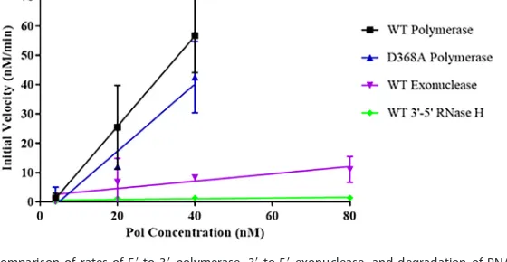
measure the initial velocities. When we compared the slopes of the initial rates obtained over ratios of 1:10 to 1:1 (40 nM polymerase), using multiple protein prepa-rations, we observed a minor decrease in polymerase activity of the D368A Pol relative to that of the WT Pol (Table 1 and Fig. 3), suggesting that this mutation within the
exonuclease domain at most slightly impairs the 5=-to-3= polymerase activity of the
[image:4.585.39.545.92.161.2]enzyme.
TABLE 1Comparison of the slopes of the initial velocities of WT and D368A HSV Pol activities plotted against the concentration of enzymea
Parameter
Value for:
Polymerase 3=-to-5=exonuclease 3=-to-5=RNase H
WT Pol activity (min⫺1) 1.54⫾0.02 0.12⫾0.05 0.013⫾0.008
D368A Pol activity (min⫺1) 1.1⫾0.3 ⬍0.0012⫾0.0006 ⬍0.001⫾0.003 D368A Pol activity relative to WT Pol activity (%) 74 ⬍0.96 ⬍8
WT Pol activity/WT other activity (fold difference) 1.0 13 120
aValues derived from Fig. 3 are presented in the top two rows and were used to calculate the fold difference in activity between the WT and D368A Pol and to
compare the rate of each nuclease activity of WT Pol to that of the polymerase activity in the bottom two rows.
FIG 3Comparison of rates of 5=-to-3=polymerase, 3=-to-5=exonuclease, and degradation of RNA-DNA with 3=termini over a range of enzyme concentrations. The initial rates of the WT 5=-to-3=polymerase, D368A 5=-to-3=polymerase, WT 3=-to-5=exonuclease, and WT degradation of RNA with 3=termini in RNA-DNA hybrids were determined over a range of polymerase concentrations in three separate experiments, and these data were compiled and graphed using GraphPad Prism software. Slopes were calculated to generate data contained in Table 1. Error bars represent standard deviations.
Lawler et al. Journal of Virology
on November 6, 2019 by guest
http://jvi.asm.org/
[image:4.585.66.346.535.680.2]D368A Pol does not exhibit detectable 3=-to-5=exonuclease activity.We next
wanted to test whether the D368A mutant enzyme retained any detectable 3=-to-5=
exonuclease activity on our hairpin substrate. To that end, we performed assays like the polymerase assays but in the absence of deoxynucleoside triphosphate (dNTPs) (Fig. 4), again using DNA hairpin S1, which minimizes the background degradation of any unannealed ssDNA (Fig. 1A and 4A). The positive control, Klenow fragment, could degrade the starting substrate, producing smaller products (Fig. 4B). In contrast, the
negative control,E. coli RNase H, was unable to degrade the substrate (Fig. 4B). We
detected 3=-to-5= exonuclease activity associated with the WT Pol, as expected,
pro-ducing a “ladder” of smaller products (Fig. 4B, black arrows indicate very short prod-ucts). Upon comparing the initial rate of the exonuclease activity (quantified by the decrease in the amount of the substrate band over time) to the polymerase activity (measured as the amount of primer extended by at least 1 nucleotide compared to the input primer in each lane) across various concentrations of WT polymerase, we
deter-mined that the 3=-to-5=exonuclease activity of HSV Pol is 13-fold slower than its 5=-to-3=
polymerase activity (Fig. 3 and Table 1). We were unable, however, to detect any degradation associated with the D368A mutant at all concentrations tested up to a 10-fold molar excess of Pol relative to the substrate (2:1 Pol-to-substrate ratio shown in Fig. 4B; data not shown for 10:1 Pol-to-substrate ratio). These data confirm previous
reports on the D368A mutant (7, 8), which exhibits at least a 50-fold defect in 3=-to-5=
exonuclease activity relative to that of the WT (Table 1).
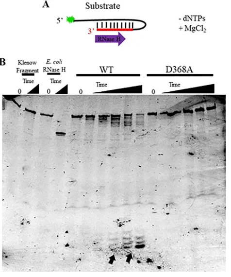
WT Pol, but not D368A Pol, has substantial 3=-to-5=RNase H activity.To investigate
the RNase H activity of HSV Pol and its dependence on the 3=-to-5= exonuclease
FIG 4D368A Pol shows no detectable 3=-to-5=exonuclease activity. (A) Cartoon of the 6-FAM-labeled DNA hairpin substrate used for the exonuclease assays showing the direction of degradation of the substrate (S1; see Fig. 1A) and the addition of MgCl2without dNTPs to start the reaction. (B) Fluorescent image of a
denaturing polyacrylamide gel loaded with equal amounts of each exonuclease reaction over increasing time points (0, 1 min, 2 min, 4 min, 8 min, 16 min, 32 min, 60 min). The assays shown here contained a 2-fold molar excess of Pol relative to the substrate. The 0- and 60-min control reactions (Klenow fragment and RNase H) are shown. Arrows (bottom of gel) denote smaller products formed upon longer incubation of WT Pol with the substrate.
on November 6, 2019 by guest
http://jvi.asm.org/
[image:5.585.43.378.73.365.2]domain, we used fluorescently labeled synthetic RNA-DNA hairpins as the substrates with sequences comparable to those of the dsDNA oligonucleotides (Fig. 1B and C) and
assayed the degradation of the substrates in the absence of dNTPs, as in the 3=-to-5=
exonuclease assays. First, we analyzed the degradation of a 5= fluorescently labeled
substrate, S2, from which we could detect RNase H activity on RNA-DNA with 3=RNA
termini (Fig. 1B and 5A). HSV Pol was reported to not have an associated 5=-to-3=DNase
activity (21), so we did not expect to observe any removal of the fluorescent label on
the 5= end of the substrate. Klenow fragment served as a negative control and was
unable to degrade the RNA-DNA substrate (Fig. 5B). As a positive control for RNase H
activity, E. coli RNase H specifically removed the RNA portion of the hybrid hairpin
substrate, leaving intact the single-stranded DNA portion of the oligonucleotide (Fig. 5B). WT HSV Pol exhibited readily detectable RNase H activity on this substrate in
the 3=-to-5=direction, as evidenced by the decrease in the full-length substrate band,
the production of a “ladder” of smaller products, and, at longer times of incubation, the accumulation of much smaller products (Fig. 5B, black arrows). These smaller products were shorter than 5 nucleotides based on a comparison to a 6-carboxyfluorescein (6-FAM)-labeled oligonucleotide of that size (data not shown). While accumulation of
the “ladder” is visible at both larger (⬎35 nucleotides) and much smaller sizes over the
time course, we believe the relative lack of visible products between these sizes is
caused by the slow 3=-to-5=RNase H activity, followed by more rapid ssDNA
degrada-tion by the 3=-to-5=exonuclease (see below).
By calculating the slope of the initial velocities of HSV Pol RNase H cleavage over differing polymerase concentrations (calculated using the same methods as those for
FIG 5WT Pol, but not D368A Pol, shows detectable 3=-to-5=RNase H activity. (A) Cartoon of the 6-FAM-labeled hairpin RNA-DNA substrate with a 3=RNA terminus (S2; see Fig. 1B) used for RNase H assays showing degradation of the substrate expected for a 3=-to-5=RNase H activity and the addition of MgCl2without dNTPs to start the reaction. (B) Fluorescent image of a denaturing polyacrylamide gel
loaded with equal amounts of each RNase H reaction mixture over increasing time points (0, 8 min, 16 min, 32 min, 60 min, 120 min). Assays shown contained a 10-fold molar excess of Pol relative to the substrate. The 0- and 60-min control reactions (Klenow fragment and RNase H) are displayed. Arrows (bottom of gel) denote the smaller products accumulating upon longer incubation of WT Pol and the substrate.
Lawler et al. Journal of Virology
on November 6, 2019 by guest
http://jvi.asm.org/
[image:6.585.86.324.72.357.2]the 3=-to-5= exonuclease), we compared the polymerase, 3=-to-5= exonuclease, and RNase H activities. WT HSV Pol-mediated degradation of the RNA-DNA hairpin substrate
containing a 3= RNA terminus was slower than both the cleavage of the 3= DNA
terminus-containing substrate and the 5=-to-3=polymerase activity, by 13- and 120-fold,
respectively (Fig. 3 and Table 1). These data suggest that HSV Pol degrades DNA with
3=termini from dsDNA more rapidly than RNA with 3=termini from DNA-RNA hybrids
in the 3=-to-5=direction.
D368A Pol, however, showed no reduction of the starting substrate or accumulation of smaller products over time, even at polymerase concentrations of up to 10-fold molar excess (Fig. 5B). Therefore, we conclude that any detectable HSV Pol RNase H
activity on RNA-DNA hybrids with 3= RNA termini is dependent upon the 3=-to-5=
exonuclease active site.
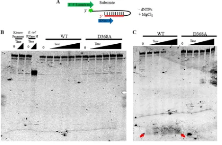
Neither WT nor D368A Pol exhibits detectable RNase H activity on a substrate with a 5= RNA terminus. We next analyzed the degradation of a 3= fluorescently labeled substrate, S3, from which we could analyze RNase H activity upon a hairpin
RNA-DNA substrate with a 5= RNA terminus and 3=DNA overhang, as occurs during
Okazaki fragment maturation. Since HSV Pol contains a 3=-to-5=exonuclease, we utilized
a bulky, fluorescent 6-FAM label both to detect the changes in the size of our substrate
and to inhibit 3=-to-5=exonuclease cleavage of this label from the substrate (Fig. 1C and
6A). The negative control, Klenow fragment, was again unable to degrade the RNA-DNA
substrate, and the 3=-to-5=exonuclease activity was likely inhibited by the 6-FAM label
(Fig. 6B). The positive control, E. coliRNase H, again cleaved only the RNA from the
RNA-DNA hairpin template, leaving intact the ssDNA (Fig. 6B). In contrast to its activity
FIG 6Neither WT nor D368A Pol exhibits detectable 5=-to-3=RNase H activity. (A) Cartoon of the 6-FAM-labeled hairpin RNA-DNA substrate with a 5=RNA terminus (S3; see Fig. 1C) used for RNase H assays showing the degradation of the substrate expected for a 5=-to-3=
RNase H activity and the addition of MgCl2without dNTPs to start the reaction. Additionally, the direction of 3=-to-5=exonuclease removal
of the fluorescent tag from the 3=end of the substrate is indicated. (B) Fluorescent image of a denaturing polyacrylamide gel loaded with equal amounts of each RNase H reaction over increasing time points (0, 8 min, 16 min, 32 min, 60 min, 120 min). These assays contained a 2-fold molar excess of Pol relative to the substrate. The 0- and 60-min control reactions (Klenow fragment and RNase H) are shown. (C) Fluorescent image of the same reaction as that in panel B but containing a 10-fold molar excess of Pol relative to the substrate. Black arrows (top of gel) denote the disappearance of the substrate over longer incubations with WT Pol. Red arrows (bottom of gel) denote the appearance of a band over longer incubations with WT Pol.
on November 6, 2019 by guest
http://jvi.asm.org/
[image:7.585.40.475.73.358.2]on substrate S2 (Fig. 5), at concentrations up to and including 2-fold excess polymerase, WT HSV Pol exhibited no detectable RNase H cleavage of this substrate, as evidenced by the absence of the production of smaller products, a lack of decrease over time in the intensity of the band corresponding to the starting substrate, and a lack of any smaller species (Fig. 6B). D368A Pol similarly showed no activity on this substrate at this concentration (Fig. 6B). At a 10-fold molar excess of WT Pol over oligonucleotide, which was the highest concentration of Pol tested, we could see a small amount of activity at longer incubation times signified by a decrease in the signal of the starting substrate and the accumulation of very short products (Fig. 6C, arrows) with no “laddering” of different sized products visible. Neither a decrease in the starting substrate nor the accumulation of smaller species was detected in the presence of the D368A mutant,
indicating that this activity is due to 3=-to-5=exonuclease removal of the 3=fluorescent
label and not a product of an RNase H activity on an RNA-DNA substrate with a 5=RNA
terminus.
DISCUSSION
By comparing the in vitro activities of WT HSV Pol and a 3=-to-5=
exonuclease-deficient mutant, D368A Pol, we found that any detectable RNase H activity proceeds
exclusively in the 3=-to-5=direction and is dependent on an active 3=-to-5=Exo active
site. Thus, the RNase H activity intrinsic to HSV-1 Pol (6, 9–12) appears to be due to the
3=-to-5=exonuclease. Though no RNase H activity was detectable by our methods on
the substrate with a 5=RNA terminus by the Exo mutant Pol, the possibility that there
is some small amount of activity below the sensitivity of our assay does exist. However, concentrations up to a 10-fold molar excess of polymerase relative to the substrate were tested, and either no activity was visible or, for WT Pol, this activity was
over-shadowed by the 3=-to-5=exonuclease, even in the presence of a bulky 6-FAM label on
the 3= end of the substrate. These results suggest that if a 5=-to-3= exonuclease or
endonuclease RNase H activity of HSV-1 Pol does exist, it is miniscule. Additionally, the polymerase activity was no more than minimally affected in the purified mutant polymerase, ruling out the possibility that it was misfolded or otherwise inhibited by the purification process. These results are consistent with the reported absence of an
HSV-1 Pol 5=-to-3= DNase activity (20, 21). However, we cannot exclude the formal
possibility that HSV-1 Pol actually has an RNase H activity that acts on RNA-DNA hybrids
containing a 5=terminus, and we simply have not yet identified the proper substrate
(e.g., one that might mimic what HSV-1 Pol encounters during Okazaki fragment maturation) and conditions to enable its detection. Nevertheless, since we did not
visualize any digestion of RNA from RNA-DNA containing a 5=RNA end, even with a vast
molar excess of HSV Pol, we view this as an unlikely scenario.
These data show that an active 3=-to-5=exonuclease is required not only for HSV Pol
degradation of ssDNA and dsDNA, as previously shown (21), but also for RNA
degra-dation from RNA-DNA hybrids containing 3=RNA ends. However, the rate of RNA-DNA
degradation is much lower than the 3=-to-5=exonuclease activity on either ssDNA or
dsDNA. So, this activity may be a nonspecific activity of the 3=-to-5=exonuclease rather
than something that serves a biological purpose during infection.
The absence of any detectable RNase H degradation by the 3=-to-5=
exonuclease-deficient D368A Pol on either of the substrates tested argues against the hypothesis of
a separate activity and/or domain from the 3=-to-5= exonuclease responsible for this
activity (6, 10, 12, 13, 25), such as the hypothesized RNA-binding motif in the NH2
-terminal domain (13). While these data alone do not rule out the possibility that the
NH2-terminal domain contributes to the RNase H activity due to the 3=-to-5=
exonu-clease, the large distance (42 Å) between the 3=-to-5=exonuclease active site and the
putative RNA-binding motif makes any connection between these domains for this activity highly unlikely. Rather, we favor the possibility that the putative RNA-binding
motif within the NH2-terminal domain is responsible for a different, still undetermined
activity associated with HSV-1 replication. One hypothesis for such an activity is autoregulation of Pol translation via mRNA binding, as suggested by Liu et al. (13).
Lawler et al. Journal of Virology
on November 6, 2019 by guest
http://jvi.asm.org/
Another hypothesis, which was raised by Swan et al. upon discovering a similar motif
in yeast Pol␦, is an interaction with RNA primers to abet strand displacement synthesis
or to recruit a 5=flap endonuclease for removal of the RNA primer (26). We are currently
investigating the role of this motif in HSV-1 replication.
Due to the absence of a detectable RNase H activity on RNA-DNA substrates with a
5=RNA terminus, we consider it unlikely that HSV Pol can remove RNA primers during
lagging strand synthesis on its own. Perhaps HSV-1 uses a mechanism similar to that of eukaryotes for the removal of RNA primers in which the polymerase displaces the RNA primer and a separate nuclease is recruited for removal of the displaced RNA strand (5).
Studies have found thatin vitro, HSV Pol has a limited strand displacement activity that
is improved using the D368A Exo mutant enzyme (likely because the mutation elimi-nates idling) and that HSV Pol can coordinate with nuclease Fen-1 for the removal of both annealed DNA and RNA and to produce ligatable nicks (7, 8). However, the
applicability of thesein vitroactivitiesin vivois unknown. Alternatively, a viral protein,
such as the viral alkaline nuclease (UL12), could be recruited to cleave the primers. While our study contains evidence against an RNase H activity inherent to HSV Pol that
acts on RNA-DNA hybrids with 5=termini, more work is needed to determine the exact
mechanism of RNA primer removal during HSV-1 lagging strand DNA replication.
MATERIALS AND METHODS
Protein expression and purification. 6⫻His-tagged full-length WT HSV Pol and a previously characterized 3=-to-5=exonuclease-deficient mutant, D368A Pol (17, 18), were cloned into the pFastBac HTC vector and expressed in a baculovirus system as previously described (22) and then purified as follows. Cells were harvested at 65 h postinfection and centrifuged at 2,800⫻gfor 30 min. The resulting pellets were washed in Dulbecco’s phosphate-buffered saline (DPBS) with 10% glycerol, centrifuged at 2,800⫻gfor 10 min, and frozen at⫺80°C. For enzyme purification, cell pellets were resuspended in lysis buffer (25 mM HEPES [pH 7.5], 500 mM NaCl, 10% [wt/vol] sucrose, 5 mM imidazole, and 2 Roche complete protease inhibitor tablets per 100 ml), and the cells were lysed on ice in the presence of 1 mg/ml lysozyme by sonication using a Branson Ultrasonics sonifier model S-450 (5-s pulses with 9-s pauses at 20% amplitude for 15 min). All subsequent steps were performed at 4°C. The suspension was centrifuged at 30,000⫻gfor 1 h, and the supernatant was then passed through a 0.45-m filter. The clarified supernatant was loaded onto a preequilibrated 10-ml GE HiTrap Talon column, washed with 20 column volumes of lysis buffer, and eluted using lysis buffer containing a gradient of 5 to 150 mM imidazole. Fractions determined to contain Pol by Coomassie blue staining were diluted 10-fold with column buffer (25 mM HEPES [pH 7.5], 1 mM dithiothreitol [DTT], 10% [wt/vol] sucrose) and loaded onto a preequilibrated 5-ml GE HiTrap heparin HP column. The column was washed with 20 column volumes of column buffer containing 0.1 M NaCl and eluted with column buffer containing a gradient of 0.1 to 1 M NaCl. Fractions shown to contain Pol by Coomassie blue staining were concentrated with an Amicon Ultra-15 centrifugal unit (Millipore) and dialyzed in storage buffer [25 mM HEPES (pH 7.5), 150 mM NaCl, 20% (vol/vol) sucrose, 2 mM tris(2-carboxyethyl)phosphine (TCEP)]. Proteins were estimated to be at least 90% pure by Coomassie blue staining, and the major species present were all detected by Western blotting with the 1051c mouse monoclonal antibody against HSV Pol (generously provided by C. Knopf) (27), suggesting that any visible contaminants were cleavage products of Pol. Samples were quantified using theA280value detected by NanoDrop (Thermo Scientific) and the extinction coefficient calculated
using the amino acid sequence (28), flash frozen, and stored at⫺80°C.
Enzyme assays. 6-Carboxyfluorescein (6-FAM)-labeled dsDNA and RNA-DNA hairpin primer-template substrates used in thein vitroassays were synthesized by IDT (Fig. 1). Master mixes designed to give final concentrations of 25 mM HEPES (pH 7.5), 1 mM DTT, 10% glycerol, and 25 mM NaCl were combined on ice with 40 nM primer-template and differing concentrations of Pol corresponding to the molar ratios of Pol to DNA indicated (1:10, 1:2, 1:1, 2:1, and 10:1). Twenty-microliter DNA polymerase reactions were initiated with the addition of MgCl2to 8 mM and all four dNTPs to 1 mM, and the contents
were mixed and incubated at 37°C. Aliquots were quenched in loading buffer containing 80% formamide and 100 mM EDTA at the following time points: 0, 10 s, 30 s, 1 min, 2 min, 4 min, 8 min, 16 min, and 32 min (polymerase assays). Klenow fragment andE. coliRNase H (NEB) were used as controls under the same conditions and quenched at the 32-min time point. Samples were separated on a 7 M urea–15% denaturing polyacrylamide gel, and the FAM-labeled substrate was detected at 520 nm following excitation at 495 nm using an Amersham Imager 600. Exonuclease and RNase H assays were carried out under the same conditions, but in the absence of dNTPs, and samples containing HSV-1 Pol were taken at the following time points: 0, 1 min, 2 min, 4 min, 8 min, 16 min, 32 min, and 60 min for exonuclease assays; 0, 8 min, 16 min, 32 min, 60 min, and 120 min for RNase H assays; and, for Klenow fragment and E. coliRNase H, 60 min for both assays.
Rate comparisons. ImageQuant TL software was used to quantify the percentage of primer-template extended to longer product for the polymerase assays or the decrease in starting substrate for the exonuclease and RNase H activities across the different time points. The values were plotted and the slopes of the activities within the initial linear range (initial velocities) were compared across different
on November 6, 2019 by guest
http://jvi.asm.org/
concentrations of enzyme using GraphPad Prism software. These rates were subsequently plotted against the protein concentration using Prism, and the slopes of the subsequent fitting were used to quantify activity rates for comparison.
ACKNOWLEDGMENTS
We thank the members of the Coen and Hogle labs for their support and input, especially Han Chen for her mentorship in learning different techniques, former lab member Shariya Terrell for optimization of the enzyme purification and use of some of her protein, and Jean Pesola for her help in analyzing the data. Additionally, we thank Charles Knopf for his generous donation of the 1051c antibody.
We acknowledge NIH for the funding to support this project (R01AI019838).
REFERENCES
1. McGeoch DJ, Dalrymple MA, Dolan A, McNab D, Perry LJ, Taylor P, Challberg MD. 1988. Structures of herpes simplex virus type 1 genes required for replication of virus DNA. J Virol 62:444 – 453.
2. Wu CA, Nelson NJ, McGeoch DJ, Challberg MD. 1988. Identification of herpes simplex virus type 1 genes required for origin-dependent DNA synthesis. J Virol 62:435– 443.
3. Falkenberg M, Lehman IR, Elias P. 2000. Leading and lagging strand DNA synthesis in vitro by a reconstituted herpes simplex virus type 1 repli-some. Proc Natl Acad Sci U S A 97:3896 –3900.https://doi.org/10.1073/ pnas.97.8.3896.
4. Stengel G, Kuchta RD. 2011. Coordinated leading and lagging strand DNA synthesis by using the herpes simplex virus 1 replication complex and minicircle DNA templates. J Virol 85:957–967. https://doi.org/10 .1128/JVI.01688-10.
5. Balakrishnan L, Bambara RA. 2013. Okazaki fragment metabolism. Cold Spring Harb Perspect Biol 5:a010173.https://doi.org/10.1101/cshperspect .a010173.
6. Crute JJ, Lehman IR. 1989. Herpes simplex-1 DNA polymerase. Identifi-cation of an intrinsic 5=-3=exonuclease with ribonuclease H activity. J Biol Chem 264:19266 –19270.
7. Zhu Y, Trego KS, Song L, Parris DS. 2003. 3=to 5=exonuclease activity of herpes simplex virus type 1 DNA polymerase modulates its strand displacement activity. J Virol 77:10147–10153.https://doi.org/10.1128/ JVI.77.18.10147-10153.2003.
8. Zhu Y, Wu Z, Cardoso MC, Parris DS. 2010. Processing of lagging-strand intermediates in vitro by herpes simplex virus type 1 DNA polymerase. J Virol 84:7459 –7472.https://doi.org/10.1128/JVI.01875-09.
9. Knopf CW, Weisshart K. 1988. The herpes simplex virus DNA polymerase: analysis of the functional domains. Biochim Biophys Acta 951:298 –314.
https://doi.org/10.1016/0167-4781(88)90100-5.
10. Marcy AI, Olivo PD, Challberg MD, Coen DM. 1990. Enzymatic activities of overexpressed herpes simplex virus DNA polymerase purified from recombinant baculovirus-infected insect cells. Nucleic Acids Res 18: 1207–1215.https://doi.org/10.1093/nar/18.5.1207.
11. Knopf KW. 1979. Properties of herpes simplex virus DNA polymerase and characterization of its associated exonuclease activity. Eur J Biochem 98:231–244.https://doi.org/10.1111/j.1432-1033.1979.tb13181.x. 12. Weisshart K, Kuo AA, Hwang CB, Kumura K, Coen DM. 1994. Structural
and functional organization of herpes simplex virus DNA polymerase investigated by limited proteolysis. J Biol Chem 269:22788 –22796. 13. Liu S, Knafels JD, Chang JS, Waszak GA, Baldwin ET, Deibel MR, Jr,
Thomsen DR, Homa FL, Wells PA, Tory MC, Poorman RA, Gao H, Qiu X, Seddon AP. 2006. Crystal structure of the herpes simplex virus 1 DNA polymerase. J Biol Chem 281:18193–18200.https://doi.org/10.1074/jbc .M602414200.
14. Wang TS, Wong SW, Korn D. 1989. Human DNA polymerase alpha: predicted functional domains and relationships with viral DNA poly-merases. FASEB J 3:14 –21.
15. Hwang YT, Liu BY, Coen DM, Hwang CB. 1997. Effects of mutations in the Exo III motif of the herpes simplex virus DNA polymerase gene on enzyme activities, viral replication, and replication fidelity. J Virol 71: 7791–7798.
16. Bernad A, Blanco L, Lazaro JM, Martin G, Salas M. 1989. A conserved 3=-5=
exonuclease active site in prokaryotic and eukaryotic DNA polymerases. Cell 59:219 –228.https://doi.org/10.1016/0092-8674(89)90883-0. 17. Hall JD, Orth KL, Sander KL, Swihart BM, Senese RA. 1995. Mutations
within conserved motifs in the 3=-5=exonuclease domain of herpes simplex virus DNA polymerase. J Gen Virol 76:2999 –3008.https://doi .org/10.1099/0022-1317-76-12-2999.
18. Kuhn FJ, Knopf CW. 1996. Herpes simplex virus type 1 DNA polymerase. Mutational analysis of the 3=-5=-exonuclease domain. J Biol Chem 271: 29245–29254.
19. Bogani F, Boehmer PE. 2008. The replicative DNA polymerase of herpes simplex virus 1 exhibits apurinic/apyrimidinic and 5=-deoxyribose phos-phate lyase activities. Proc Natl Acad Sci U S A 105:11709 –11714.
https://doi.org/10.1073/pnas.0806375105.
20. Knopf CW, Weisshart K. 1990. Comparison of exonucleolytic activities of herpes simplex virus type-1 DNA polymerase and DNase. Eur J Biochem 191:263–273.https://doi.org/10.1111/j.1432-1033.1990.tb19119.x. 21. Hall JD, Orth KL, Claus-Walker D. 1996. Evidence that the nuclease
activities associated with the herpes simplex type 1 DNA polymerase are due to the 3=-5=exonuclease. J Virol 70:4816 – 4818.
22. Terrell SL, Coen DM. 2012. The pre-NH2-terminal domain of the
herpes simplex virus 1 DNA polymerase catalytic subunit is required for efficient viral replication. J Virol 86:11057–11065.https://doi.org/ 10.1128/JVI.01034-12.
23. Clark JM, Joyce CM, Beardsley GP. 1987. Novel blunt-end addition reac-tions catalyzed by DNA polymerase I of Escherichia coli. J Mol Biol 198:123–127.https://doi.org/10.1016/0022-2836(87)90462-1.
24. Chen H, Beardsley GP, Coen DM. 2014. Mechanism of ganciclovir-induced chain termination revealed by resistant viral polymerase mu-tants with reduced exonuclease activity. Proc Natl Acad Sci U S A 111:17462–17467.https://doi.org/10.1073/pnas.1405981111.
25. Haffey ML, Novotny J, Bruccoleri RE, Carroll RD, Stevens JT, Matthews JT. 1990. Structure-function studies of the herpes simplex virus type 1 DNA polymerase. J Virol 64:5008 –5018.
26. Swan MK, Johnson RE, Prakash L, Prakash S, Aggarwal AK. 2009. Struc-tural basis of high-fidelity DNA synthesis by yeast DNA polymerase delta. Nat Struct Mol Biol 16:979 –986.https://doi.org/10.1038/nsmb.1663. 27. Strick R, Hansen J, Bracht R, Komitowski D, Knopf CW. 1997. Epitope
mapping and functional characterization of monoclonal antibodies spe-cific for herpes simplex virus type I DNA polymerase. Intervirology 40:41– 49.https://doi.org/10.1159/000150519.
28. Wilkins MR, Gasteiger E, Bairoch A, Sanchez JC, Williams KL, Appel RD, Hochstrasser DF. 1999. Protein identification and analysis tools in the ExPASy server. Methods Mol Biol 112:531–552.
Lawler et al. Journal of Virology