HIV-1 Adapts To Replicate in Cells Expressing Common Marmoset
APOBEC3G and BST2
Alberto Fernández-Oliva,aAndrés Finzi,b*Hillel Haim,b*Luis Menéndez-Arias,aJoseph Sodroski,b,cBeatriz Pachecoa,b
Centro de Biología Molecular Severo Ochoa, Consejo Superior de Investigaciones Científicas and Universidad Autónoma de Madrid, Madrid, Spaina; Department of
Cancer Immunology and Virology, Dana-Farber Cancer Institute, and Department of Microbiology and Immunobiology, Harvard Medical School, Boston, Massachusetts, USAb; Department of Immunology and Infectious Diseases, Harvard School of Public Health, Boston, Massachusetts, USAc
ABSTRACT
Previous studies have shown that a major block to HIV-1 replication in common marmosets operates at the level of viral entry and that this block can be overcome by adaptation of the virus in tissue-cultured cells. However, our current studies indicate that HIV-1 encounters additional postentry blocks in common marmoset peripheral blood mononuclear cells. Here, we show that the common marmoset APOBEC3G (A3G) and BST2 proteins block HIV-1 in cell cultures. Using a directed-evolution method that takes advantage of the natural ability of HIV-1 to mutate during replication, we have been able to overcome these blocks in tissue-cultured cells. In the adapted viruses, specific changes were observed ingag,vif,env, andnef. The contribution of these changes to virus replication in the presence of the A3G and BST2 restriction factors was studied. We found that certain amino acid changes in Vif and Env that arise during adaptation to marmoset A3G and BST2 allow the virus to replicate in the presence of these restriction factors. The changes in Vif reduce expression levels and encapsidation of marmoset APOBEC3G, while the changes in Env increase viral fitness and discretely favor cell-to-cell transmission of the virus, allowing viral escape from these restriction factors.
IMPORTANCE
HIV-1 can infect only humans and chimpanzees. The main reason for this narrow tropism is the presence in many species of dominant-acting factors, known as restriction factors, that block viral replication in a species-specific way. We have been explor-ing the blocks to HIV-1 in common marmosets, with the ultimate goal of developexplor-ing a new animal model of HIV-1 infection in these monkeys. In this study, we observed that common marmoset APOBEC3G and BST2, two known restriction factors, are able to block HIV-1 in cell cultures. We have adapted HIV-1 to replicate in the presence of these restriction factors and have characterized the mechanisms of escape. These studies can help in the development of a novel animal model forin vivoinfection of marmosets with HIV-1-like viruses.
T
he main cause of AIDS is chronic infection by human immu-nodeficiency virus type 1 (HIV-1). Without treatment, HIV-1 infection results in a progressive depletion of CD4⫹T cells that leads to severe immunodeficiency, characterized by opportunistic infections and certain types of cancer that are the leading causes of death in HIV-1-positive patients.The presence of several barriers to HIV-1 replication in cells of many species narrows the viral tropism to humans and chimpan-zees. The limited species tropism of HIV-1 is due to two types of host factors: (i) factors that are required for HIV-1 replication but that exhibit species-specific changes that do not allow efficient use by HIV-1 and (ii) dominant-acting factors that block replication in many species. The latter, also known as restriction factors, are part of so-called intrinsic antiviral immunity. Altogether, intracel-lular restriction factors can act as powerful barriers to viral repli-cation. However, viruses have developed mechanisms that can antagonize restriction factors in an equally successful way. These viral countermeasures are often proteins encoded by accessory genes that are not needed for viral replication in the absence of restriction factors. The main restriction factors that block HIV-1 and other lentivirus infections at different stages of the viral life cycle are TRIM5␣ (1), APOBEC3G (A3G) (2), BST2 (3, 4), SAMHD1 (5,6), and the recently discovered Mx2 (7–9).
BST2, also known as tetherin, CD317, or HM1.24, tethers viral particles to the plasma membrane of the cell, blocking their release
(3,4). BST2 is able to block the release of a broad range of envel-oped viruses (10,11). To escape from the action of BST2, viruses have developed a variety of strategies. In HIV-1, the accessory protein Vpu suppresses the activity of human BST2; in HIV-2, Env is the protein responsible for counteracting the activity of BST2, whereas Nef overcomes the restriction imposed by BST2 in most simian immunodeficiency viruses (SIVs) (12–16). A3G and APOBEC3F (A3F) are cellular cytidine deaminases that can be incorporated into virions in a species-specific way, blocking virus
Received25 September 2015Accepted16 October 2015
Accepted manuscript posted online28 October 2015
CitationFernández-Oliva A, Finzi A, Haim H, Menéndez-Arias L, Sodroski J, Pacheco B. 2016. HIV-1 adapts to replicate in cells expressing common marmoset APOBEC3G and BST2. J Virol 90:725–740.doi:10.1128/JVI.02431-15.
Editor:S. R. Ross
Address correspondence to Beatriz Pacheco, b.pacheco.phd@hotmail.com.
*Present address: Andrés Finzi, Centre de Recherche du CHUM and Department of Microbiology, Infectiology and Immunology, Université de Montréal, Montreal, Quebec, Canada; Hillel Haim, Department of Microbiology, Carver College of Medicine, University of Iowa, Iowa City, Iowa, USA.
Supplemental material for this article may be found athttp://dx.doi.org/10.1128 /JVI.02431-15.
Copyright © 2015, American Society for Microbiology. All Rights Reserved.
crossmark
on November 7, 2019 by guest
http://jvi.asm.org/
replication by various mechanisms. These mechanisms include hypermutation of the viral genome during reverse transcription, which leads to degradation of the replication intermediates or generation of noninfectious virions, inhibition of elongation of HIV-1 DNA by reverse transcriptase (RT), and reduction of the efficiency of plus-strand DNA transfer and inhibition of integra-tion (17–22). The viral infectivity factor (Vif) can inhibit incorpo-ration of the A3G/A3F proteins in a species-specific manner by promoting their degradation (23–25).
Due to the limited tropism of HIV-1, the development of an animal model of HIV-1 infection has been challenging. The in-creased knowledge and understanding of the host restriction fac-tors that block replication of HIV-1 in the last few years has al-lowed the construction of some macaque-tropic HIV-1 variants that contain about 90% HIV-1 sequences and 10% SIV sequences (26,27) and that are able to replicate efficiently in macaque pe-ripheral blood lymphocytes (PBLs). Some of these adapted viruses have been shown to cause AIDS in pigtail macaques that have been treated with anti-CD8 antibodies to transiently deplete CD8⫹T cells (28).
To date, lentiviruses able to infect New World monkeys have not been described. Our knowledge of the host restriction factors that block replication of lentiviruses in New World monkeys is fragmentary. However, some of these monkeys, like common marmosets, have been frequently used in animal models in other fields and are an attractive prospect for the development of a new animal model of HIV-1 infection. Previous studies have suggested that one major blockade to HIV-1 infection in New World mon-keys occurs at the level of viral entry, because HIV-1 envelope glycoproteins cannot effectively bind the CD4 and CCR5 recep-tors of common marmosets (29). Using a directed-evolution method that takes advantage of the natural ability of the virus to mutate during replication, we were able to generate HIV-1 vari-ants able to replicate in cells expressing the common marmoset receptors CD4 and CXCR4 (30). The adapted viruses, however, were unable to replicate in common marmoset PBLs, suggesting the presence of additional postentry blocks. In this study, we ob-served that common marmoset A3G (marA3G) and BST2 (marBST2) proteins block HIV-1 in cell cultures, and we adapted HIV-1 to replicate in the presence of these restriction factors. The basis for the observed escape of the adapted viruses was studied.
MATERIALS AND METHODS
Cell lines and reagents.293T and Cf2Th cells were obtained from the American Type Culture Collection and maintained in Dulbecco’s modi-fied Eagle’s medium (DMEM) supplemented with 10% fetal bovine se-rum (DMEM-10). The 174⫻CEM cells were obtained from the NIH AIDS Research and Reference Reagent Program (catalog number 272) from Peter Cresswell (31) and maintained in RPMI 1640 supplemented with 10% fetal bovine serum (RPMI-10).
Cf2Th cell lines stably expressing marmoset or human CD4 and CXCR4 have been described previously (30). Cf2Th cells stably expressing marmoset A3G or BST2 and marmoset CD4 and CXCR4 receptors were prepared by transfecting the Cf2Th-CD4-CXCR4 cell lines with a pcDNA3.1(⫹) plasmid expressing marmoset A3G or BST2 with a C-ter-minal or N-terC-ter-minal hemagglutinin (HA) tag, respectively. The trans-fected cells were selected with antibiotic to obtain stable cell lines express-ing the proteins. Then, sexpress-ingle clones expressexpress-ing marmoset CD4, CXCR4, and A3G or BST2 were obtained by limited dilution. Alignments of the
marmoset A3G and BST2 proteins with ortholog proteins of other species are shown in Fig. S1 and S2, respectively, in the supplemental material.
The following reagents were obtained through the NIH AIDS Re-search and Reference Reagent Program, Division of AIDS (DAIDS), NIAID, NIH: HIV-1 SF2 p24 antiserum (catalog number 4250) from DAIDS, NIAID, produced by BioMolecular Technologies; anti-human BST2 (catalog number 11721) from Klaus Strebel and Amy Andrew (32); pcDNA-HVif (catalog number 10077) and pcDNA-Vphu (catalog num-ber 10076), both from Stephan Bour and Klaus Strebel (33); pNL-U35
(catalog number 968) from Klaus Strebel (34); and Nevirapine (catalog number 4666).
Virus replication.Replication-competent HIV-1 variants were gener-ated by transfecting 20g of the pNL4-KBCJ1.2 plasmid, which contains an infectious HIV-1 NL4.3 provirus, with the KB9 Env sequence contain-ing the changes E151K, E172K, and A561T, which allow the use of mar-moset CD4 and CXCR4 (30), into 2⫻106293T cells using the calcium phosphate transfection method as previously reported (30).
For the replication kinetics assays, cells were infected with 30,000 RT units (cpm) of HIV-1 NL4-KBCJ1.2 variants for 14 h and then washed once with phosphate-buffered saline (PBS). Every 3 or 4 days, the cell supernatants were removed and used for RT assays. The cells were trypsinized, diluted 1/5 in fresh medium, and replated.
Analysis of the sequences of adapted viruses.The genomes of the adapted viruses were amplified by PCR from genomic DNA isolated from infected cells with the QIAamp DNA blood minikit (Qiagen). Five PCR fragments containing long terminal repeat (LTR)-gag, pol, Vif-Vpu, Env, or Nef-LTR sequences were generated by PCR with PfuUltra High-Fidel-ity DNA polymerase (Stratagene) and overlapping primers designed to cover the entire genome. These fragments were either cloned into the pCR4blunt-TOPO plasmid (Invitrogen) and sequenced or directly se-quenced to obtain the consensus sequence of the adapted viruses.
Site-directed mutagenesis.DNA sequences of consensus changes in matrix, Vif, Env, and Nef that were associated with viral adaptation to marA3G or marBST2 were introduced by site-directed mutagenesis using the QuikChange II XL site-directed mutagenesis protocol (Stratagene) into the pNL4-KBCJ1.2 plasmid; the pCMV⌬P1⌬envpA plasmid con-taining the gag, pol, andvifregions of NL4-3 HIV-1; or the pSVIII-KBCJ1.2 plasmid expressing the full-length HIV-1(pSVIII-KBCJ1.2) envelope glycoprotein. The presence of the desired mutations was verified by auto-mated DNA sequencing.
Single-cycle infectivity assay.The efficiency of a single cycle of HIV-1 infection was measured by using recombinant reporter viruses expressing firefly luciferase and pseudotyped with the vesicular stomatitis virus G glycoprotein (VSV-G) or HIV-1 Env. Recombinant luciferase-expressing HIV-1 variants (35) were generated by transfecting 293T cells using the cal-cium phosphate transfection method with 4g of the pCMV⌬P1⌬envpA packaging plasmid, 4g of an HIV-1-derived vector plasmid that ex-presses the firefly luciferase, and 2g of the pSVIII-Env plasmid express-ing HIV-1 Env or 1g of the pHCMV-G plasmid expressing the VSV envelope glycoprotein G and 1g of a Rev-expressing plasmid. In certain experiments, viruses were produced in the presence of A3G or BST2, including different amounts of pcDNA3.1(⫹) plasmid expressing these proteins in the transfection mixture. Forty-eight hours after transfection, supernatants containing reporter viruses were harvested and cleared by low-speed centrifugation. The amounts of virus in the supernatants were quantified by measuring the RT activity.
Target cells were seeded at a density of 6,000 cells/well in 96-well lu-minometer-compatible tissue culture plates. Twenty-four hours later, the medium was changed and different amounts of viruses were added to the cells. Forty-eight hours later, the medium was removed and cells were lysed with 30l of passive lysis buffer (Promega). Luciferase activity was measured using a Berthold Centro LB 960 microplate luminometer or a BMG Labtech Fluostar Optima microplate reader.
Encapsidation of APOBEC3G.Recombinant viruses with different Vif variants were generated in the presence or absence of A3G. For this
on November 7, 2019 by guest
http://jvi.asm.org/
purpose, 293T cells were seeded in 6-well plates at a density of 3⫻105per
well and cotransfected using the calcium phosphate transfection method with the pHCMV-G plasmid expressing VSV envelope glycoprotein G; a Rev-expressing plasmid; the pCMV⌬P1⌬envpA packaging plasmid con-taining the different Vif variants; a plasmid expressing the reporter firefly luciferase gene; and a pcDNA3.1 vector expressing marA3G or huA3G, both containing a C-terminal HA tag for easy detection by Western blot-ting (WB); or an empty pcDNA3.1 vector. Forty-eight hours after trans-fection, supernatants containing viruses were harvested, cleared by low-speed centrifugation, and filtered with a 0.45-m polyethersulfone (PES) filter. The viruses were pelleted though a 20% sucrose cushion by centrif-ugation at 4°C in a fixed-angle rotor for 1 h at 30,000⫻g. The viruses were analyzed by SDS-PAGE and WB probing with anti-p24 and anti-HA (clone 3F10; Roche) antibodies. The blots were visualized with an Im-ageQuant LAS 4000 mini Imager, and the amounts of A3G and p24 were quantified using Quantity One software (Bio-Rad).
Coexpression of APOBEC3G and Vif mutants. 293T cell were cotransfected with a pcDNA3.1 vector expressing marA3G, human A3G (huA3G), African green monkey A3G (agmA3G), or rhesus macaque A3G (rhA3G) containing a C-terminal HA tag or an empty pcDNA3.1 vector and the pcDNA-HVif plasmid, which contains a partially codon-opti-mized Vif. The cells were incubated for 24 h in the presence or absence of 10M MG132 and then were harvested and lysed with 40l of lysis buffer (1% NP-40 in PBS plus 2 mM EDTA, 1 mM benzamidine, and 2 mM phenylmethylsulfonyl fluoride [PMSF] as protease inhibitors). The cell lysates were analyzed by SDS-PAGE and WB probing with an anti-HA antibody. The blots were stripped and reprobed with an anti-Vif antibody; then, the membranes were stripped again and probed with an anti-␣ -tubulin antibody. The blots were visualized with an ImageQuant LAS 4000 mini Imager, and the amounts of A3G, Vif, and tubulin were quan-tified using Quantity One software (Bio-Rad).
Cell-to-cell transmission assay.293T cells (producer or donor cells) were plated in 12-well plates at a final concentration of 2⫻105/ml and
cotransfected with a pcDNA3.1 plasmid, empty or expressing marBST2; the pSVIIIenv plasmid, which contains the different Env variants; a plas-mid expressing green fluorescent protein (pHIV-1-GFP); and the pCMV⌬P1⌬envpA packaging plasmid. One day later, 174⫻CEM cells (target cells) were labeled with CellTrace Far Red DDAO-SE dye (Invit-rogen) at a concentration of 1M diluted in PBS for 15 min at 37°C. The labeled target cells were added to donor cells at a final concentration of 106/ml and cocultured for 48 h. The reverse transcriptase inhibitor nevi-rapine (NVP) was added at a concentration of 20M to a control sample when the coculture was started and maintained during the assay. After 48 h of coculture, cells were harvested, washed twice with PBS, and fixed with 1% formaldehyde in PBS. The cells were analyzed in a FACSCanto A flow cytometer, and the percentage of double-positive cells was calculated.
Particle release assay.293T cells were cotransfected with the pNL4-KBCJ1.2 plasmid containing the full-length HIV-1 proviral genome with wild-type (WT) or mutant Env or the plasmid pNLU35, which contains an
HIV-1 proviral genome with thevpugene deleted, and either an empty pcDNA3.1 vector or different amounts of a pcDNA3.1 vector expressing marBST2 or huBST2 with an N-terminal HA tag. In some cases, the cells were also cotransfected with a plasmid (pcDNA-Vphu) expressing the Vpu protein or a plasmid expressing the Nef protein from SIVmac.
Twen-ty-four hours posttransfection, the cells were harvested and lysed with lysis buffer containing protease inhibitors. Cell culture supernatants containing the released viruses were filtered with a 0.45-m PES filter and pelleted through a 20% sucrose-PBS cushion by centrifugation at 30,000⫻gfor 1 h. Pelleted viruses and cell lysates were analyzed by WB with an anti-p24 polyclonal antibody and a monoclonal anti-HA antibody. The blots were visualized with an ImageQuant LAS 4000 mini Imager, and the percentage of particle release was calculated as the ratio between the p24 in the cell culture supernatants and the total p24 (cells plus virions).
Statistics.All experiments were done at least twice (in most cases three or more times). The means and standard deviations were calculated with the Excel program. When appropriate, two-tailedPvalues were calculated using the Sigma Plot program’s Studentttest.
Nonhuman primate blood samples and ethics statement.Blood samples from common marmosets and rhesus macaques were used in this study to isolate mRNA needed for cloning A3G and BST2. The animals were housed at the New England Primate Research Center and were cared for according to the standards of the Association for Assessment and Accreditation of Laboratory Animal Care and the Harvard Medical School Animal Care and Use Committee.
Collection of nonhuman primate blood samples for this study was approved by the Harvard Medical Area (HMA) Standing Committee on Animals (protocol number 04789) and conducted in accordance with the Guide for the Care and Use of Laboratory Animals by experienced veter-inarians at the New England Primate Research Center.
The HMA Standing Committee on Animals has an approved animal welfare assurance on file with the Office for Laboratory Animal Welfare. The assurance number on file is A3431-01.
For this study, only nonsurgical collection of blood was needed. No other procedures were carried out on the animals for the study. For phle-botomy, the animals were sedated with ketamine HCl (10 to 50 mg/kg of body weight intramuscularly [i.m.]) or with telazol (4 to 10 mg/kg i.m.) to reduce pain and discomfort. The phlebotomy site was prepped with alco-hol. Blood samples were obtained from a peripheral vein. The amounts of blood collected from any animal as a single sample did not exceed 10% of the circulating blood volume every 2 weeks.
The common marmosets utilized in this study were socially housed. The rhesus macaques were socially housed unless they were being condi-tioned for other studies or were scheduled for return to the breeding colony. Compensatory enrichment was provided to animals that were not socially housed. The enrichment provided include manipulable devices, foraging opportunities, food items, structural and environmental en-hancements, and positive human interaction. Enrichment devices were rotated on a weekly basis and included toys, mirrors, radios, TVs/VCRs, foraging boards, and a variety of complex foraging devices.
RESULTS
Common marmoset APOBEC3G and BST2 block HIV-1 infec-tion.We were intrigued by the lack of infectivity in marmoset PBLs of the HIV-1 variants adapted to replicate in the presence of marmoset CD4 and CXCR4. We assessed the ability of marA3G and marBST2 proteins to block HIV-1.
Previous studies had shown that marA3G was able to block HIV-1 (36). To assess the anti-HIV-1 activity of marA3G, we pre-pared single-round luciferase reporter viruses in 293T cells in the presence or absence of marA3G and tested their infectivity in Cf2Th target cells. Using this system, we observed that the infec-tivity of viruses prepared in the presence of marA3G was about 10-fold lower than that of the viruses prepared in the absence of A3G (Fig. 1A), indicating that marA3G has an inhibitory effect on the infectivity of HIV-1, in agreement with previous work (36).
Several studies have shown that certain regions in the N-termi-nal part of Vif are important for A3G and A3F binding (37–40). We selected a few residues in these regions that we observed to be conserved within the same viral species but not between different species of primate lentiviruses and generated a small library of single and double Vif mutants containing specific changes at res-idues 34, 36, and 41. In this library, we also included a triple mu-tant of Vif containing the amino acid changes D14S, R15E, and R17Q (SEMQ); this mutant was previously shown to be able to interact with rhA3G and to productively replicate in human cells that express rhA3G (41). To test the ability of these Vif mutants to HIV-1 Variants That Overcome Marmoset A3G and BST2
on November 7, 2019 by guest
http://jvi.asm.org/
counteract marA3G, we prepared viruses containing these Vif changes in the presence or absence of marA3G and tested their infectivity in target cells. Viruses containing the SEMQ Vif mutant exhibited an approximately 2-fold increase in infectivity com-pared to the WT virus when precom-pared in the presence of marA3G, indicative of a partial escape from the restriction factor (Fig. 1A). All the other Vif mutants tested did not show a significant increase in their infectivity in the presence of marA3G (data not shown).
To test the ability of marBST2 to block the release of HIV-1, we coexpressed BST2, which contained an N-terminal HA tag, with HIV-1 NL4.3 lacking Vpu (⌬Vpu) in 293T cells. As previously reported (3,4), in the absence of Vpu, huBST2 drastically reduced the amount of viral particles released into the medium, and the
addition of HIV-1 Vpu, but not SIVmacNef, was able to recover
the particle release (Fig. 1B). Similarly, marBST2 also decreased the amount of viral particles released; however, the addition of HIV-1 Vpu or SIVmacNef was not able to counteract the effect of
marBST2 or induce degradation of marBST2 (Fig. 1B). Of note, the effect of marBST2 on HIV-1 particle release was not as strong as the block imposed by huBST2. This might be related to lower levels of expression or anti-HIV-1 activity of marBST2 compared with those of huBST2.
To evaluate if the two marmoset restriction factors efficiently inhibited HIV-1 replication, we infected Cf2Th cell lines express-ing marA3G or marBST2, as well as common marmoset CD4 and CXCR4, with the NL4-KBCJ1.2 virus. The NL4-KBCJ1.2 virus is a FIG 1Restriction of HIV-1 by common marmoset APOBEC3G and BST2. (A) Cf2Th cells were infected with VSV-G-pseudotyped HIV-1 luciferase reporter viruses prepared in the absence or presence of marA3G. The infectivity of the viruses was determined by measuring the relative luciferase activity in the target cells. RLU, relative luciferase units. Similar results were obtained in three independent experiments. The chart shows the results of one representative experiment. (B) 293T cells were cotransfected with a NL4-3⌬Vpu proviral plasmid and different amounts of plasmids expressing marmoset or human BST2. In some cases, cells were also cotransfected with plasmids expressing HIV-1 Vpu or SIVmacNef. Twenty-four hours later, the medium was collected and the viruses were pelleted by
centrifugation in a 20% sucrose cushion. The cell lysates and viruses were analyzed by immunoblot probing with an anti-p24 antibody. The relative particle release was calculated by dividing the amount of p24 in the supernatant by the total p24 (cells plus supernatant). The membranes were stripped and reprobed with an anti-HA antibody (to detect BST2HA); then, the membranes were stripped again and probed with an anti-tubulin (␣tubulin) antibody as a loading control. The
WBs of one representative experiment out of four are shown. (C) Cf2Th cells stably expressing marmoset CD4 and CXCR4, with or without marA3G or marBST2, were infected with WT (NL4-KBCJ1.2) or Vif SEMQ mutant HIV-1. The replication of the viruses is represented as RT activity in the culture medium versus time after virus inoculation. The threshold of the RT assay is indicated by the horizontal lines. The infection kinetics of one representative experiment out of two are shown.
on November 7, 2019 by guest
http://jvi.asm.org/
[image:4.585.41.542.67.441.2]derivative of the HIV-1 NL4.3 strain containing KB9 Env with the changes E151K, E172K, and A561T, which allow use of marmoset CD4 and CXCR4 (30). Throughout this work, we refer to NL4-KBCJ1.2 as the WT virus. As shown inFig. 1C, HIV-1 WT and SEMQ replicated efficiently in the parental cells, which do not express marA3G or marBST2. However, we did not observe sig-nificant replication of either virus in cells expressing marA3G or marBST2, which indicates that marA3G and marBST2 are able to block HIV-1 replication in cell cultures.
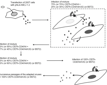
Adaptation of HIV-1 to common marmoset APOBEC3G and BST2. Using a directed-evolution method similar to that em-ployed in previous virus adaptations (30, 42, 43), we adapted HIV-1 to replicate in Cf2Th cells expressing marmoset CD4 and CXCR4 receptors and marA3G or marBST2. The adaptation pro-tocol is depicted inFig. 2. We wanted to carry out the adaptation to marA3G and marBST2 in cells expressing marmoset CD4 and CXCR4 to ensure that any changes that might appear in the virus during adaptation would be compatible with the use of these re-ceptors. The parental Cf2Th cell line used is a dog cell line that was selected to carry out the adaptation because HIV-1 replicates effi-ciently in these cells when complemented with an appropriate CD4 receptor and a CXCR4 or CCR5 coreceptor (42,44). In ad-dition, some factors, like A3G or TRIM5␣, that could potentially interfere with the adaptations have not been found in the dog genome (45,46). The parental HIV-1 strain used for the adapta-tions was NL4-KBCJ1.2, which can use marmoset CD4 and
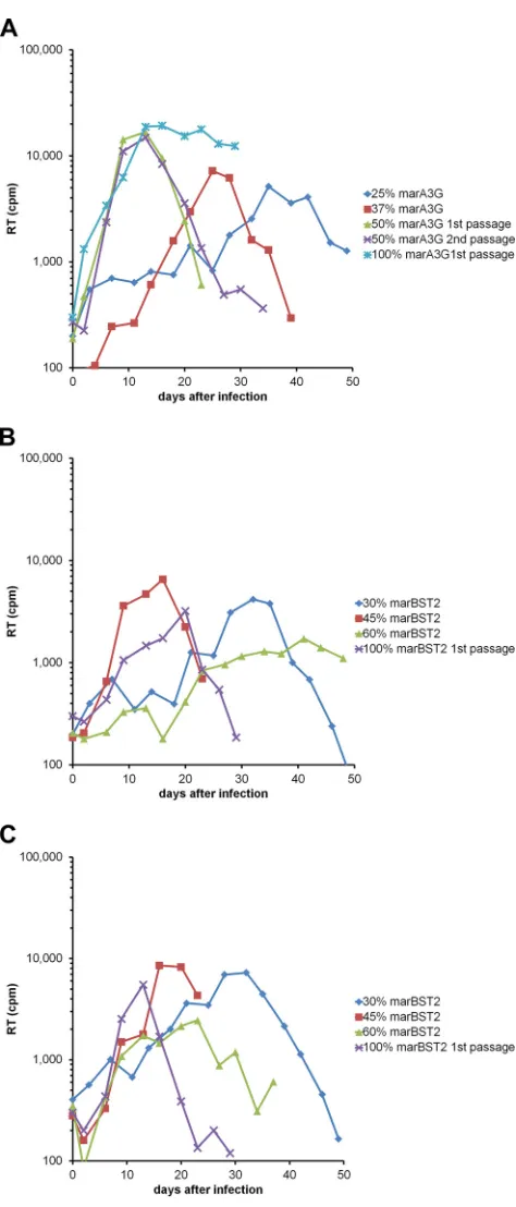
CXCR4. In addition to the WT virus, we also studied a mutant containing the SEMQ changes in Vif, as we had seen that the mutant was able to partially overcome the block imposed by marA3G. After several passages of the viruses in cell cultures with increasing percentages of cells expressing either marA3G or marBST2, we obtained viruses that were able to replicate in cul-tures in which 100% of the cells expressed the corresponding re-striction factor (Fig. 3). Both HIV-1 variants, WT and SEMQ, adapted to replicate in cells expressing marBST2; however, only the SEMQ variant adapted to replicate in cell lines expressing marA3G. These adapted viruses were passaged a few more times in the marA3G- or marBST2-expressing cell lines to increase viral fitness (data not shown).
After the adaptation, the genomic DNA of the infected cells was isolated and the sequences of the integrated HIV-1 proviruses were amplified and sequenced. We observed changes in several regions of the viral genome; in the viruses adapted to marA3G, some G-to-A changes suggestive of the cytidine deaminase activity of marA3G were observed (Fig. S3 in the supplemental material shows the LTR region of the viruses passaged in cells expressing marA3G [Fig. S3A] or marBST2 [Fig. S3B]). We centered our attention on the changes that appeared in regions that are ex-pected to be relevant to A3G and BST2 escape—Vif in the case of marA3G adaptation and Vpu, Env, and Nef in the case of marBST2 adaptation—and selected those that were maintained FIG 2Schematic representation of the direct-evolution strategy. Replication-competent HIV-1 WT or Vif SEMQ mutants were prepared in 293T cells by transfection with the pNL4-KBCJ1.2 plasmid. The viruses produced in the 293T cells were used to infect cell cultures of Cf2Th-marCD4/CXCR4 where only 25% or 30% of the cells expressed marmoset A3G or BST2, respectively. The cell culture supernatants from the day when the RT activity reached a peak were used to start a new round of infection in a cell culture mixture where 37% or 45% of the cells expressed marmoset A3G or BST2, respectively. The process was repeated in cell cultures with increasing percentages of cells expressing the restriction factors until we were able to infect cell cultures where all the cells expressed the restriction factor. After obtaining viruses able to replicate in cell cultures where all the cells expressed marmoset A3G or BST2, the viruses were passaged a few times in the cell lines.
HIV-1 Variants That Overcome Marmoset A3G and BST2
on November 7, 2019 by guest
http://jvi.asm.org/
[image:5.585.116.472.65.349.2]during virus passage for further study.Figure 4shows a schematic representation of the selected changes in these regions.
The marA3G adaptation gave rise to an initial change in Vif from Asp99 to Gly. Further passaging of the SEMQ/D99G virus
gave rise to an additional change at residue 71 of Vif (G71D). In the two adaptations (WT and SEMQ) to marBST2, changes in Env and Nef, but not in Vpu, were observed. In addition, we observed in all three adaptations the appearance of a change in the matrix protein of Glu52 to Lys (E52K), which was surprising. Two similar changes in this region of the matrix protein appeared during the adaptation of NL4-3 and NL4-3(KB9 Env) to replicate in Cf2Th cells expressing marmoset CD4 and CXCR4 (30).
To study the contributions of these changes to the replication of HIV-1 in cells expressing marA3G or marBST2, we prepared viruses containing the changes alone or in certain combinations and infected target cells.
The adaptation of the WT virus to marBST2 resulted in the appearance of two single-residue changes, N656S and R742K, within the sequence of Env that together allowed the replication of the virus in cells expressing marBST2 (Fig. 5A, right). The Nef residue change Y202C alone did not permit replication in cells expressing marBST2, and a combination of this change with the Env changes did not improve replication efficiency in these cells (Fig. 5A, right). Although the matrix change E52K alone did not allow replication of the virus in the presence of marBST2, when combined with the Env changes, it improved the replication effi-ciency of the virus (Fig. 5A, right). All these changes in Env, Nef, and matrix also increased the replication efficiency in the parental cell line, which does not express marBST2 (Fig. 5A, left).
In the adaptation of the SEMQ virus to marBST2, two different compensatory changes arose in the Env sequence, P722L and R740K, that allowed low levels of replication in cells expressing marBST2 (Fig. 5B, right). Viruses containing these Env changes also had enhanced replication in the parental cell line that does not express marBST2 compared with the WT virus, and as in the pre-vious case, addition of the matrix change E52K improved replica-tion in the presence of marBST2. We did not observe replicareplica-tion in the cells expressing marBST2 of viruses containing the Nef changes A15T, C55R, and D150N, which appeared during adap-tation to marBST2; moreover, a combination of these changes with the above-mentioned Env changes did not improve replica-tion in the cells expressing marBST2 (Fig. 5B, right).
The abilities of the different Vif variants to replicate in the presence of marA3G were studied. The SEMQ mutant had very low levels of replication in cells expressing marA3G (Fig. 6, top right). The viruses containing the SEMQ and D99G changes also initially replicated at low levels in the cells expressing marA3G; however, after several days in culture, the SEMQ/D99G virus started to replicate more efficiently, reaching replication levels close to those attained in the parental cell line (Fig. 6, top). When all the Vif changes that appeared during adaptation to marA3G were combined, we also observed low levels of replication in cells expressing marA3G (Fig. 6, top right). Although these viruses rep-licated poorly in cells expressing marA3G, surprisingly, when the Vif changes were combined with the Env changes N656S and R742K or P722L and R740K, we obtained viruses that efficiently replicated in cells expressing marA3G (Fig. 6, middle and bot-tom). The Env changes in the absence of the Vif changes did not allow replication in the marA3G cell lines, either alone or com-bined with the matrix change E52K (Fig. 6, middle and bottom). Effect of marA3G on the infectivity of Vif mutant viruses.To determine the contributions of the adaptation-associated Vif changes to the infectivity of the virus, we used a single-cycle infec-tivity assay. For this purpose, we produced single-cycle viruses FIG 3Replication kinetics of the virus at different passages of adaptation. The
charts represent the replication kinetics of the viruses at different steps of the adaptation process shown inFig. 2. The RT activity in 0.5 ml of the culture supernatant was measured at the indicated time points. (A) Adaptation of WT virus to marA3G. (B) Adaptation of WT virus to marBST2. (C) Adaptation of SEMQ virus to marBST2.
on November 7, 2019 by guest
http://jvi.asm.org/
[image:6.585.45.282.60.614.2]containing WT or mutant Vif proteins and a luciferase reporter gene in the presence or absence of marA3G. These viruses were used to infect Cf2Th cells, and the viral infectivity was determined by measuring the relative luciferase activities in the target cells. We studied the contribution of each mutant individually and in dif-ferent combinations (Fig. 7A). The SEMQ mutant showed a 2-fold increase in infectivity, as had been observed in our results shown inFig. 1A. When all the adaptation-associated changes were combined with this mutant (SEMQ/G71D/D99G), an addi-tional increase in infectivity was observed, indicating that the G71D and D99G changes might also contribute to escape from marA3G restriction. The other mutants, either G71D or D99G alone or combined, did not show significant differences in infec-tivity with respect to WT viruses. No differences in infecinfec-tivity were observed between WT and⌬Vif viruses, which corroborates the idea that WT Vif cannot counteract the restriction of marA3G.
Incorporation of marA3G in virions.Vif binds A3G in a spe-cies-specific way, excluding A3G from incorporating into virions. To assess the effects of the Vif mutants on the incorporation of marA3G into virions, we prepared WT or mutant viruses in the presence of marA3G or huA3G as a control. The viruses were pelleted, and the amount of A3G incorporated into the viruses was quantified by WB. The relative amount of A3G incorporated into virions was calculated as the ratio of A3G (anti-HA antibody) to p24 in the pelleted viruses (Fig. 7B). As shown inFig. 7B, the WT viruses exhibited a significant (about 3-fold) reduction in huA3G
incorporation compared to⌬Vif viruses, in agreement with pre-viously published results (23–25). With respect to marA3G, the WT viruses did not show significant differences in A3G incorpo-ration relative to⌬Vif viruses (Fig. 7B), in agreement with our previous result showing that HIV-1 Vif cannot overcome marA3G restriction (Fig. 7A). The single- and double-residue mutants ex-hibited only slight variations in A3G incorporation with respect to the WT and⌬Vif viruses, while the SEMQ mutant showed an approximately 2-fold reduction in A3G incorporation relative to the⌬Vif viruses. The SEMQ/G71D/D99G mutant exhibited the lowest incorporation of marA3G into nascent viral particles. These results are consistent with data from the infectivity assays described above (i.e., viruses that incorporate the smallest amount of marA3G exhibit the highest infectivity rates) and indicate that the Vif changes contribute to escape from marA3G restriction by reducing its encapsidation into virions.
Degradation of marA3G by Vif mutants.After binding to A3G, Vif targets A3G for proteasomal degradation by recruiting the E3 ubiquitin ligase complex Cul5-RBX2-ELOB-ELOC (24, 25). We wanted to know whether our Vif mutants were able to induce degradation of marA3G. For this purpose, we cotrans-fected 293T cells with a vector expressing WT or mutant Vif and marA3G (or, as a control, huA3G, agmA3G, or rhA3G) and mea-sured the steady-state levels of A3G by WB. Supporting our pre-vious results, we observed that coexpression with Vif proteins containing the SEMQ or SEMQ plus G71D and D99G changes FIG 4Schematic representation of the HIV-1 genome and the encoded Vif, Env, and Nef proteins showing domains and relevant changes that arose during adaptation. The horizontal line with tick marks represents the full genome of the virus. Each tick corresponds to 1,000 nucleotides. The genomes of the adapted viruses were amplified by PCR from genomic DNA isolated from the infected cells. The PCR fragments were either directly sequenced or cloned in a vector and then sequenced. The changes that appeared in regions that were expected to be relevant to A3G or BST2 escape (Vif, Env, and Nef) and that were maintained in successive passages are shown as vertical bars. V1 to V5, variable regions; FP, fusion peptide; HR1/2, helical regions 1 and 2; TM, transmembrane domain; CT, cytoplasmic tail.
HIV-1 Variants That Overcome Marmoset A3G and BST2
on November 7, 2019 by guest
http://jvi.asm.org/
[image:7.585.113.472.70.354.2]resulted in lower levels of marA3G in the cells than with WT Vif (Fig. 7C). The addition of a proteasome inhibitor, MG132, re-duced the ability of the Vif mutants to degrade marA3G compared with WT Vif, suggesting that, as previously reported for huA3G (25), these Vif variants induce degradation of marA3G though a proteasome-mediated pathway (Fig. 7C, bottom).
As expected, WT Vif was able to reduce the expression levels of huA3G, but not agmA3G or rhA3G, while the SEMQ mutant had a reduced ability to induce degradation of huA3G and an in-creased ability to induce degradation of agmA3G, in agreement with the respective loss and gain of anti-huA3G and anti-agmA3G activity of the mutant (41).
Effect of marBST2 in particle release of Env mutants.BST2 is a type II transmembrane protein that also has a C-terminal glyco-sylphosphatidylinositol (GPI) anchor, allowing it to interact with lipid membranes at both ends. Thanks to this unusual topology, BST2 prevents the release of mature virions by keeping them
chored to the cell surface. The HIV-1 Vpu accessory protein an-tagonizes the effect of human BST2 and restores normal virus budding and release during infection. To establish if the Env changes in the viruses adapted to marBST2-expressing cells exert a similar effect, we carried out a particle release assay. Viruses con-taining the Env mutants were produced in 293T cells in the presence or absence of marBST2. The amount of p24 in the intracellular (pro-cessing and anchored viruses) and extracellular (released free virions) fractions was determined by immunoblotting with a polyclonal anti-body elicited against p24; the percentage of released viral particles was calculated as the ratio of p24 in the virions (extracellular) to the total p24 (intracellular plus extracellular) (Fig. 8A). Even though we had seen that the N656S/R742K and P722L/R740K Env mutants were able to replicate in cells that express marBST2 (Fig. 5), when we tested the effects of the Env mutations on particle release, we did not observe significant differences in particle release with respect to WT viruses (Fig. 8A) or the control⌬Vpu viruses (Fig. 1B).
FIG 5Replication kinetics of HIV-1 mutants derived from adaptation to marmoset BST2. Cf2Th cells expressing marmoset CD4 and CXCR4, with (right) or without (left) marBST2, were infected with different HIV-1 variants. The RT activity in 0.5 ml of the culture supernatant was measured at the indicated time points. (A) Replication of viruses containing the Env changes that appeared during the adaptation of the WT virus to marBST2. (B) Replication of viruses containing the Env changes that appeared during the adaptation of the SEMQ virus to marBST2. The replication of the WT virus is shown in all the charts as a reference. The threshold of the RT assay is indicated by the horizontal lines. The results of one infection experiment out of two are shown.
on November 7, 2019 by guest
http://jvi.asm.org/
[image:8.585.43.536.64.461.2]Effect of Env mutants on the internalization and degradation of BST2.The interaction between HIV-1 Vpu and huBST2 leads to the endocytosis of BST2, endosome sequestration, and partial proteasome degradation of BST2 (47–54). The ability of the Env mutants to downregulate/degrade BST2 was studied by WB and flow cytometry.
First, we studied the effect of the expression of WT or mutant Env on the steady-state expression levels of BST2 in 293T cells by
WB. While we had observed that the addition of Vpu decreased the expression levels of huBST2 (Fig. 1B), we did not observe a reduction in the expression levels of marBST2 either in the pres-ence of the different Env variants (Fig. 8B) or in the prespres-ence of HIV-1 Vpu or SIVmacNef (Fig. 1B).
One possibility is that the Env mutants decrease the amount of BST2 on the cell surface, inducing the internalization of the pro-tein without an effect on the total levels of propro-tein expression, as FIG 6Replication kinetics of HIV-1 mutants derived from adaptation to marmoset A3G. Cf2Th cells expressing marmoset CD4 and CXCR4, with (right) or without (left) marA3G, were infected with different HIV-1 variants. The RT activity in 0.5 ml of the culture supernatant was measured at the indicated time points. The replication of the WT virus is shown in all the charts as a reference. The threshold of the RT assay is indicated by the horizontal lines. The results of one infection experiment out of two are shown.
HIV-1 Variants That Overcome Marmoset A3G and BST2
on November 7, 2019 by guest
http://jvi.asm.org/
[image:9.585.42.534.63.564.2]FIG 7Adaptation-induced changes in Vif increase viral infectivity in the presence of marA3G and reduce the packaging of marA3G into virions and the total expression level of marA3G. (A) Effects of marA3G on the infectivity of Vif mutant viruses. Luciferase reporter viruses pseudotyped with VSV-G and containing WT or mutant Vif proteins were produced in 293T cells in the presence or absence of marA3G. The viruses were used to infect Cf2Th target cells, and 2 days later, the infectivity of the viruses was determined by measuring the relative luciferase activities in the target cells. The relative infectivity of each variant was calculated by dividing the infectivity of the viruses prepared in cells expressing marA3G by the infectivity of the viruses prepared in cells that did not express A3G. The mean relative infectivities⫾standard deviations of four independent experiments (with each point tested in duplicate) are represented. Two-tailedPvalues were calculated using Student’sttest. (B) Incorporation of marA3G into virions. WT or mutant Vif viruses were produced in 293T cells in the presence or absence of marA3G. The viruses were pelleted by centrifugation in a 20% sucrose cushion and analyzed by immunoblot probing with an anti-HA antibody (A3GHA) and an
anti-p24 antibody. A representative WB out of 5 independent experiments is shown. The relative amount of A3G incorporated into virions was calculated as the ratio of HA (A3G) to p24 and normalized to 1, relative to⌬Vif. The averages of the relative A3G incorporated for the five experiments⫾standard deviations are shown above the WB. (C) A3G degradation by Vif mutants. 293T cells were cotransfected with a vector expressing marA3G, huA3G, agmA3G, or rhA3G
on November 7, 2019 by guest
http://jvi.asm.org/
[image:10.585.40.540.72.617.2]has been reported for HIV-2 Env (12). Using two different anti-bodies raised against huBST2, we studied the cell surface expres-sion levels of marBST2 in the presence of WT or mutant Env. However, the antibodies used were raised against huBST2 and exhibited only very low affinity for marBST2; using this approach, we did not observe a significant difference in surface expression levels of marBST2 in the presence or absence of Env variants or Vpu (data not shown). Using a marBST2 with an internal HA tag (which should be exposed on the cell surface), we did not observe differences in cell surface expression levels of the protein in the presence or absence of the WT or Env mutants. However, the total expression levels of the construct were much lower than the expression levels of marBST2 with an N-terminal tag, which had been used in all the other experiments. Furthermore, this marBST2 with an internal HA tag was unable to block the re-lease of HIV-1. Although these results are not fully conclusive due to the lack of an anti-BST2 antibody that efficiently recog-nizes marBST2, the Env mutants do not appear to downregu-late marBST2 from the cell surface.
Infectivity of the Env mutant viruses. The adaptations of HIV-1 to marBST2 led to the appearance of a few changes in Env that allow viral replication in cells that express this restriction factor. These changes also increase replication efficiency in the parental cell lines that do not express marBST2 (Fig. 5). However, our data indicated that the Env mutants did not increase the effi-ciency of particle release or induce the degradation/internaliza-tion of marBST2. We decided to study the infectivity of Env mu-tant viruses using single-cycle luciferase reporter viruses. Higher infectivity of the Env mutants relative to WT viruses could indi-cate that HIV-1 had adapted to overcome marBST2 by introduc-ing changes that improve viral fusion and entry into the host cell; in this way, the Env mutants might compensate for the effect of
marBST2 instead of adapting the envelope proteins to directly counteract marBST2 with a Vpu-like activity.
Single-round viruses containing the WT or mutant Env and a luciferase reporter gene were produced in the absence of marBST2 and were used to infect Cf2Th cells expressing marmoset CD4 and CXCR4 receptors. Viruses used to infect target cells were isolated from donor cells, so this system measures cell-free viral infectivity (Fig. 9A). Viral infectivity was determined by measuring the rela-tive luciferase activity in the target cells 48 h postinfection (Fig. 9B). In this assay, Env mutants exhibited infectivity similar to that of WT viruses. These results argue against a direct effect of the Env changes on viral entryper se.
Cell-to-cell transmission of Env mutants.HIV and many other viruses are transmitted not only as cell-free viral particles diffusing in the extracellular environment, but also directly and more efficiently by cell-to-cell transfer through virological syn-apses formed between the infected cell and a target cell (55,56). Since we did not observe an improvement in cell-free infectiv-ity between WT and mutant Env viruses, we wanted to deter-mine if Env mutants could overcome the marBST2 restriction by favoring cell-to-cell transmission. For this purpose, HIV-1 pseudoviral particles containing WT or Env mutants, as well as the GFP reporter gene, were produced in 293T cells (donor cells). Twenty-four hours posttransfection, 174⫻CEM cells previously labeled with DDAO-SE (target cells) were added and cocultured for 2 days. Viral transmission was assessed by flow cytometry measuring the percentage of GFP- and DDAO-SE-double-positive cells. We focused on target cells that had been infected, which should be DDAO⫹GFP⫹double-positive cells, and we compared the percentages of target cells infected in the presence and absence of marBST2 to calculate the rela-tive viral transmission (Fig. 9C). The presence of marBST2
containing a C-terminal HA tag and a plasmid expressing a partially codon-optimized Vif with different changes. Twenty-four hours later, the cells were harvested and lysed. The cell lysates were analyzed by WB with an anti-HA antibody, and the amount of A3G was quantified. The membranes were stripped and reprobed with an anti-Vif antibody; then, the membranes were stripped again and probed with an anti-tubulin antibody as a loading control. A representative WB out of two independent experiments is shown. The relative amount of A3G in the cells was calculated as the ratio of HA (A3G) to tubulin and normalized to 1 relative to WT Vif. The averages of the relative A3G in the cells for the two experiments⫾standard deviations are shown above the WB.
FIG 8No differences in particle release and in total expression of marBST2 were observed in the presence of Env mutants. (A) Particle release of Env mutant viruses in the presence of marBST2. Viruses containing WT or mutant Env were produced in 293T cells in the presence or absence of marBST2 or huBST2. Twenty-four hours later, the medium was collected and the viruses were pelleted by centrifugation in a 20% sucrose cushion. The cell lysates and viruses were analyzed by immunoblot probing with an anti-p24 antibody. The percentage of particle release was calculated as the ratio of p24 in the released virions to the total p24 (virions plus cell lysates). (B) Total expression of marBST2. The same experiments shown in panel A were performed. The membranes were stripped and reprobed with an anti-HA antibody (to detect BST2HA); then, the membranes were stripped again and probed with an anti-tubulin antibody as a loading control
(bottom). The experiments were repeated 3 times with similar results. The WBs of one representative experiment are shown.
HIV-1 Variants That Overcome Marmoset A3G and BST2
on November 7, 2019 by guest
http://jvi.asm.org/
[image:11.585.41.545.70.204.2]greatly reduced viral transmission of WT viruses compared to the viral transmission in the absence of the restriction factor. The Env mutants improved viral transmission in the presence of marBST2 at different levels. The compensatory changes N656S/R742K were associated with the highest viral transmis-sion ratio. As expected, the control infection adding NVP, a potent inhibitor of reverse transcriptase, drastically reduced viral transmission in both the presence and absence of marBST2. These results suggest that the N656S/R742K and P722L/R740K Env mutants escape marBST2 al least in part by increasing cell-to-cell viral transmission. Together, our results suggest that the mechanism that the virus uses to escape marBST2 might be different from that reported previously for HIV-1 Vpu and huBST2 (3,4).
DISCUSSION
Here, we have studied the blockade of HIV-1 by common mar-moset A3G and BST2 and adapted HIV-1 to replicate in the pres-ence of these restriction factors. We also started to explore the escape mechanisms that HIV-1 develops to evade marA3G and marBST2 in cell cultures. Just a few mutations that emerged dur-ing the adaptation within the Vif and Env sequences of HIV-1 were sufficient to partially overcome marA3G and marBST2 and to allow replication of these viruses in cell lines that express the restriction factors.
The analysis of mutants adapted to replicate in the presence of marA3G revealed that two single changes within the sequence of HIV-1 Vif, in addition to the SEMQ changes, appeared to partially FIG 9Adapted HIV-1 Env variants do not show differences in cell-free infectivity but increase cell-to-cell transmission. (A) Schematic representation of the cell-free viral-infectivity and cell-to-cell transmission assays. FACS, fluorescence-activated cell sorting. (B) Cell-free virus infectivity assay. Luciferase reporter viruses expressing WT or mutant Env were produced in 293T cells in the absence of marBST2. After 48 h, viruses were harvested and isolated. Serial dilutions of each virus were used to infect Cf2Th cells expressing marmoset CD4 and CXCR4 receptors. Two days later, the infectivity of the viruses was determined by measuring the relative luciferase activity in the target cells. Similar results were obtained in two independent experiments. The results of one representative experiment are shown. (C) Cell-to-cell transmission assay. 293T donor cells were transfected with constructs for expression of HIV-1 GFP reporter viruses containing WT or mutant Env in the presence or absence of marBST2. Twenty-four hours later, the cells were mixed with 174⫻CEM target cells prelabeled with DDAO-SE and cocultured. (Left) Two days later, the target cells were analyzed by flow cytometry for DDAO and GFP expression, and the percentage of DDAO⫹ GFP⫹double-positive cells (infected target cells) was determined. (Right) The relative viral transmission was calculated as the ratio of infected target cells in the presence of marBST2 to infected target cells in the absence of marBST2. The results shown in the charts represent the means⫾standard deviations of four independent experiments, each done in duplicate. Two-tailedPvalues were calculated using Student’sttest.
on November 7, 2019 by guest
http://jvi.asm.org/
[image:12.585.79.510.61.453.2]overcome the marA3G block. Previous studies have shown that sev-eral residues in the N terminus of Vif are part of a nonlinear binding site for human A3G and A3F (Fig. 4) (37–40). Furthermore, residues 14 to 17 (DRMR), which are necessary for interaction with huA3F but not critical for Vif binding to huA3G, have also been observed to facilitate species-specific recognition of A3G (40,41). All our ob-served changes, SEMQ, G71D, and D99G, are located within the Vif N-terminal region. Moreover, these residues seem to be well con-served in a species-specific manner; thus,a priori, one might expect that they have emerged to allow HIV-1 Vif protein to specifically recognize marA3G, a model that our experimental results seem to corroborate. Previous studies had already demonstrated that the in-teraction between Vif and A3G is highly species specific; even a single-amino-acid change could modify the specificity of Vif. For example, the D128K substitution in human A3G prevents the interaction with HIV-1 Vif but allows the interaction with SIVagmVif (57).
The set of changes that emerged at the end of the adaptation (SEMQ/G71D/D99G) partially counteracts marA3G by reducing the incorporation of marA3G into nascent virions more efficiently than the single mutations (Fig. 7B). This reduced incorporation of marA3G into virions seems to be due to a decrease in the steady-state levels of marA3G in the presence of these Vif mutants. A3G incorporation into virions, as well as the ability of HIV and SIV Vif proteins to block its packaging, has been previously studied (23, 58). HIV-1 Vif has been successfully modified to overcome the A3G restriction factor of rhesus macaques and African green monkeys (41). In each case, there was a direct correlation between the decrease of A3G packaging into virions and the increase in the infectivity of these virions, which is consistent with the results obtained in our study. We hypothesize that our Vif mutants main-tain the same escape mechanism that HIV-1 uses to counteract huA3G but have adapted their structures to favor the interaction with marA3G.
In the case of HIV-1 adaptation to replicate in the presence of marBST2, the analysis of mutants revealed that compensatory changes emerged within the sequence of the envelope glycopro-teins instead of appearing in Vpu, which is the protein evolved by HIV-1 to counteract human BST2 in nature. In each adaptation, two Env changes, N656S/R742K or P722L/R740K, were shown to be sufficient to overcome the marBST2 block in cell cultures.
Vpu is one of the accessory proteins of HIV-1, although it is absent in other, related primate lentiviruses, such as HIV-2 and most SIVs. In these viruses, the block imposed by BST2 is coun-teracted by the envelope glycoproteins of HIV-2 and the Nef pro-tein of SIVs (12–16). The ability to counteract BST2 by adapting different viral proteins suggests that overcoming the restriction of BST2 is a key factor for improving viral pathogenesis. Antagonism of BST2 by HIV-1 Env has not been previously described. How-ever, a previous study found that compensatory changes in the cytoplasmic tail of gp41 restored resistance to BST2 in a strain of SIV withnefdeleted, which regained a pathogenic phenotype in rhesus macaques (59). These studies suggest that when Nef is not an alternative as a BST2 antagonist, Env can evolve to counteract its effects. Some of the Nef changes that arose in our adaptation increased the replication efficiency of the virus in the parental cell lines; however, they were not able to counteract marBST2 (Fig. 5). A recent publication has shown that canine BST2 (dogBST2) is able to block HIV-1 particle release (60). HIV-1 is able to replicate efficiently in Cf2Th cells if they are engineered to express a func-tional CD4 and CXCR4 or CCR5. However, the titer of the virus in
these cell lines is not as high as in human cell lines. Thus, it is possible that an endogenous dogBST2 expressed in the Cf2Th cells exerts mild activity against HIV-1 and that some Nef changes, like Y202C, counteract dogBST2. During the adaptation of SEMQ to marBST2, some changes in Nef also evolved (data not shown). Although their contributions to viral replication were not tested, this supports the idea that dogBST2 expressed in Cf2Th cells might have some mild activity against HIV-1. If Nef is used to counteract dogBST2, then Env would be a reasonable alternative for marBST2 counteraction.
In contrast to many previous studies that showed that over-coming BST2 restriction results in an increase in the amount of released virions, we observed no differences in particle release in-duced by Env mutants. Furthermore, no differences in the total marBST2 expression levels or signals of degradation induced by Env mutants were observed. These results together suggest that adapted Env mutants do not use the same molecular escape mech-anism as Vpu, which mediates ubiquitination and proteasomal degradation of BST2 (49). Since BST2 is a membrane protein, HIV-1 variants adapted to marBST2 could operate by removing marBST2 from the plasma membrane without affecting the total amount of BST2; such a situation occurs with HIV-2 Env, whose escape mechanism consists of endosomal sequestration of BST2 within thetrans-Golgi network, with no concomitant BST2 degra-dation (12). However, our preliminary results studying marBST2 downregulation from the plasma membrane by flow cytometry in-dicate that the Env mutants do not decrease the total amount of marBST2 at the plasma membrane.
HIV-1 Env variants adapted to replicate in the presence of marBST2 do not show significant differences in cell-free viral in-fectivity but moderately increase cell-to-cell transmission. BST2 has been observed to restrict both cell-free and direct cell-to-cell transmission (61–63). Cell-free transmission involves the release of viruses in the extracellular medium and their free diffusion until they reach a new host cell, which means that virions spend more time in the extracellular medium. One possible explanation for not observing differences in cell-free viral infectivity between WT and mutant viruses is that HIV-1 Env mutants could be more unstable than the WT Env, becoming inactivated more rapidly under the conditions used in the cell-free viral-infectivity assay. However, cell-to-cell transmission allows more rapid and direct transfer of the virus through virological synapses. Our results in-dicate that Env mutants can overcome the marBST2 restriction in part by increasing cell-to-cell transmission, although the exact mechanism is still not clear and does not seem to directly target marBST2. In a recent study, Durham and Chen (64) have shown that HIV-1 cell-free and cell-to-cell infections are differentially regulated by distinct elements in the cytoplasmic tail of gp41. They propose that during cell-to-cell infection there are differences in the function of Env during virological synapse formation com-pared to cell-free infection and that the formation of the virolog-ical synapse can overcome packaging or other defects. It is possible that our Env mutants compensate for the defect in particle release imposed by marBST2, at least in part, through a mechanism that favors virological synapse formation.
The Env changes that allow replication in cells that express marBST2 also increase the replication infectivity of the virus in the parental cells that do not express marBST2 (Fig. 5and6). Al-though these envelope changes do not allow efficient replication in cells that express marA3G, when combined with the Vif changes HIV-1 Variants That Overcome Marmoset A3G and BST2
on November 7, 2019 by guest
http://jvi.asm.org/
that allow partial escape from marA3G, they increase viral repli-cation in the marA3G-expressing cells (Fig. 6). These results to-gether indicate that escape from these restriction factors is linked to an increase in viral fitness, at least in the Cf2Th cell lines. Pre-vious studies have shown that an increase in the viral fitness of hepatitis C virus passaged in tissue culture cells is associated with increased resistance to inhibitors of viral replication that target viral or cellular factors without the appearance of mutations spe-cifically associated with resistance to the inhibitors (65). In our case, we found that adaptation to restriction factors is associated with the appearance of mutations that also increase viral fitness and that the increase in fitness allows replication in the presence of the restriction factors. In the case of marBST2, it appears that an increase in viral fitness is sufficient to allow escape from the block-ade; in contrast, in the case of A3G, the combination of some changes in Vif that partially overcome the marA3G block plus some changes that increase viral fitness is needed. The main dif-ference between the situations might be related, on one hand, to the different strengths of marBST2 and marA3G restriction and, on the other hand, to the different mechanisms of action of the restriction factors. Our results show that the blockade imposed by marBST2 is not as strong as the blockade imposed by huBST2 (Fig. 1B). This situation might favor a mechanism of escape that in-volves an increase in viral fitness in the target cells. We believe that the blockade imposed by marA3G is stronger, and thus, some A3G-specific Vif changes are needed to overcome the block.
In summary, we have identified compensatory genetic changes that provide resistance to common marmoset APOBEC3G and BST2 in HIV-1 strains adapted to infect cells expressing these restriction factors, with the ultimate goal of developing a new animal model for AIDS research involving infection with more complete HIV-1-like viruses. Preliminary studies combining the Env and Vif changes shown in this work with changes that allow use of marmoset CD4 and CXCR4 (30) did not allow replication of the virus in marmoset PBLs (data not shown). One reasonable explanation for this lack of replication in marmoset PBLs is that the viruses have evolved to overcome marmoset A3G and BST2 in the context of canine cells. It is also possible that in marmoset PBLs different or additional changes may be needed to efficiently overcome A3G and BST2. However, we have obtained evidence for the existence of at least two early postentry blocks to HIV-1 that are not mediated by TRIM5␣in marmoset lymphocytes (B. Pacheco, L. Menéndez-Arias, and J. Sodroski, unpublished data and data presented at the 40th Meeting on Retroviruses, Cold Spring Harbor, NY, 18 to 23 May 2015). These early postentry blocks are strong enough to prevent replication of the viruses in the marmoset lymphocytes, even if the viruses were perfectly adapted to marmoset A3G and BST2. Currently, we are investi-gating the nature of the factors involved in these early postentry blocks and adapting HIV-1 to overcome them. The Env and Vif mutants obtained in this work are a good starting point for further adaptation in marmoset PBLs.
ACKNOWLEDGMENTS
We acknowledge the personnel at the flow cytometry facility at Centro de Biología Molecular Severo Ochoa for their technical support. We thank the New England Primate Research Center for providing the blood needed to clone common marmoset A3G and BST2.
The study was conceived and designed by J.S. and B.P. A.F.-O., A.F., H.H., and B.P. performed experiments. A.F.-O., A.F., L.M.-A., J.S., and
B.P. analyzed data. J.S. and B.P. wrote the manuscript. We all read and approved the manuscript.
FUNDING INFORMATION
HHS | NIH | National Institute of Allergy and Infectious Diseases (NIAID) provided funding to Joseph G. Sodroski under grant numbers AI63987 and AI67854.
This research was supported by the European Commission under the 7th Framework Programme through a Marie Curie Career Integration Grant (332623) and a by Scholar Award from the Harvard University Center for AIDS Research (CFAR), an NIH-funded program (P30AI060354), to B.P.; by a Spanish Ministry of Economy and Competitiveness grant (BIO2013-48788-C2-1-R) to L.M.-A.; and by an institutional grant from the Fundación Ramón Areces to the Centro de Biología Molecular Severo Ochoa. B.P. is a recipient of a CSIC JAE-Doc contract, a program cofi-nanced by the European Social Fund. The funders had no role in study design, data collection and interpretation, decision to publish, or prepa-ration of the manuscript.
REFERENCES
1.Stremlau M, Owens CM, Perron MJ, Kiessling M, Autissier P, Sodroski J.2004. The cytoplasmic body component TRIM5alpha restricts HIV-1 infection in Old World monkeys. Nature427:848 – 853.http://dx.doi.org /10.1038/nature02343.
2.Sheehy AM, Gaddis NC, Choi JD, Malim MH.2002. Isolation of a human gene that inhibits HIV-1 infection and is suppressed by the viral Vif protein. Nature 418:646 – 650. http://dx.doi.org/10.1038 /nature00939.
3.Neil SJ, Zang T, Bieniasz PD.2008. Tetherin inhibits retrovirus release and is antagonized by HIV-1 Vpu. Nature451:425– 430.http://dx.doi.org /10.1038/nature06553.
4.Van Damme N, Goff D, Katsura C, Jorgenson RL, Mitchell R, Johnson MC, Stephens EB, Guatelli J. 2008. The interferon-induced protein BST-2 restricts HIV-1 release and is downregulated from the cell surface by the viral Vpu protein. Cell Host Microbe3:245–252.http://dx.doi.org /10.1016/j.chom.2008.03.001.
5.Hrecka K, Hao C, Gierszewska M, Swanson SK, Kesik-Brodacka M, Srivastava S, Florens L, Washburn MP, Skowronski J.2011. Vpx relieves inhibition of HIV-1 infection of macrophages mediated by the SAMHD1 protein. Nature474:658 – 661.http://dx.doi.org/10.1038/nature10195. 6.Laguette N, Sobhian B, Casartelli N, Ringeard M, Chable-Bessia C,
Segeral E, Yatim A, Emiliani S, Schwartz O, Benkirane M. 2011. SAMHD1 is the dendritic- and myeloid-cell-specific HIV-1 restriction factor counteracted by Vpx. Nature474:654 – 657.http://dx.doi.org/10 .1038/nature10117.
7.Kane M, Yadav SS, Bitzegeio J, Kutluay SB, Zang T, Wilson SJ, Schog-gins JW, Rice CM, Yamashita M, Hatziioannou T, Bieniasz PD.2013. MX2 is an interferon-induced inhibitor of HIV-1 infection. Nature502: 563–566.http://dx.doi.org/10.1038/nature12653.
8.Goujon C, Moncorge O, Bauby H, Doyle T, Ward CC, Schaller T, Hue S, Barclay WS, Schulz R, Malim MH.2013. Human MX2 is an interfer-on-induced post-entry inhibitor of HIV-1 infection. Nature502:559 –562.
http://dx.doi.org/10.1038/nature12542.
9.Liu Z, Pan Q, Ding S, Qian J, Xu F, Zhou J, Cen S, Guo F, Liang C. 2013. The interferon-inducible MxB protein inhibits HIV-1 infection. Cell Host Microbe14:398 – 410.http://dx.doi.org/10.1016/j.chom.2013 .08.015.
10. Jouvenet N, Neil SJ, Zhadina M, Zang T, Kratovac Z, Lee Y, McNatt M, Hatziioannou T, Bieniasz PD.2009. Broad-spectrum inhibition of ret-roviral and filoviral particle release by tetherin. J Virol83:1837–1844.http: //dx.doi.org/10.1128/JVI.02211-08.
11. Sakuma T, Noda T, Urata S, Kawaoka Y, Yasuda J.2009. Inhibition of Lassa and Marburg virus production by tetherin. J Virol83:2382–2385.
http://dx.doi.org/10.1128/JVI.01607-08.
12. Le Tortorec A, Neil SJ.2009. Antagonism to and intracellular sequestra-tion of human tetherin by the human immunodeficiency virus type 2 envelope glycoprotein. J Virol83:11966 –11978.http://dx.doi.org/10.1128 /JVI.01515-09.
13. Zhang F, Wilson SJ, Landford WC, Virgen B, Gregory D, Johnson MC, Munch J, Kirchhoff F, Bieniasz PD, Hatziioannou T.2009. Nef proteins