Crystal Structure of Marburg Virus VP40 Reveals a Broad, Basic Patch
for Matrix Assembly and a Requirement of the N-Terminal Domain
for Immunosuppression
Shun-ichiro Oda,aTakeshi Noda,bKaveesha J. Wijesinghe,cPeter Halfmann,dZachary A. Bornholdt,aDafna M. Abelson,a Tammy Armbrust,dRobert V. Stahelin,c,eYoshihiro Kawaoka,b,d,fErica Ollmann Saphirea,g
Department of Immunology and Microbial Science, The Scripps Research Institute, La Jolla, California, USAa; International Research Center for Infectious Diseases, Institute of Medical Science, The University of Tokyo, Tokyo, Japanb; Department of Chemistry and Biochemistry, the Eck Institute for Global Health, and the Boler-Parsegh Center for Rare and Neglected Diseases, University of Notre Dame, Notre Dame, Indiana, USAc; Department of Pathobiological Sciences, School of Veterinary Medicine, University of Wisconsin—Madison, Madison, Wisconsin, USAd; Department of Biochemistry & Molecular Biology, Indiana University School of Medicine—South Bend, South Bend, Indiana, USAe; Division of Virology, Department of Microbiology and Immunology, Institute of Medical Science, The University of Tokyo, Tokyo, Japanf; Skaggs Institute for Chemical Biology, The Scripps Research Institute, La Jolla, California, USAg
ABSTRACT
Marburg virus (MARV), a member of the filovirus family, causes severe hemorrhagic fever with up to 90% lethality. MARV
ma-trix protein VP40 is essential for assembly and release of newly copied viruses and also suppresses immune signaling in the
in-fected cell. Here we report the crystal structure of MARV VP40. We found that MARV VP40 forms a dimer in solution, mediated
by N-terminal domains, and that formation of this dimer is essential for budding of virus-like particles. We also found the
N-ter-minal domain to be necessary and sufficient for immune antagonism. The C-terN-ter-minal domains of MARV VP40 are dispensable
for immunosuppression but are required for virus assembly. The C-terminal domains are only 16% identical to those of Ebola
virus, differ in structure from those of Ebola virus, and form a distinct broad and flat cationic surface that likely interacts with
the cell membrane during virus assembly.
IMPORTANCE
Marburg virus, a cousin of Ebola virus, causes severe hemorrhagic fever, with up to 90% lethality seen in recent outbreaks.
Mo-lecular structures and visual images of the proteins of Marburg virus are essential for the development of antiviral drugs. One
key protein in the Marburg virus life cycle is VP40, which both assembles the virus and suppresses the immune system. Here we
provide the molecular structure of Marburg virus VP40, illustrate differences from VP40 of Ebola virus, and reveal surfaces by
which Marburg VP40 assembles progeny and suppresses immune function.
M
arburg virus (MARV) and the related Ebola virus (EBOV)
both belong to the
Filoviridae
family. These are enveloped
viruses with a nonsegmented single-stranded and negative-sense
RNA genome. Genomes of Marburg viruses differ from those of
ebolaviruses by more than 50% at the nucleotide level (
1
). MARV
causes severe and rapidly progressing hemorrhagic fever in
hu-mans and nonhuman primates, with lethality ranging from 25%
to over 90% depending on the geographic location and viral strain
(
2
). The 19-kb MARV genome encodes seven structural proteins.
Each of these proteins is of critical importance, and most are
known to perform multiple functions during the viral life cycle.
VP40 is the Marburg virus matrix protein, which builds the
protein shell underneath the viral envelope and confers the
hall-mark filamentous morphology to the Marburg virion. VP40 alone
is able to induce the assembly and budding of filamentous
virus-like particles (VLPs), which resemble authentic virions (
3
,
4
). The
intracellular distribution of filovirus VP40 varies during the
pro-gression of the viral life cycle (
5
,
6
), as it orchestrates the
distribu-tion of the other viral components and viral assembly (
6
,
7
). The
majority of it, however, traffics to and associates with the cellular
membrane (
5
,
7
,
8
). Within the virion, VP40 interacts with both
the lipid envelope and the core nucleocapsid complex, which
con-tains the NP, VP35, L, VP30, and VP24 proteins (
9
). VP40 also
interacts with the GP viral surface protein. Indeed, coexpression
of VP40 and GP alters the distribution pattern of GP, leading to
GP accumulation at the VP40-positive basolateral clusters at
which budding occurs (
7
). MARV VP40 (mVP40) and EBOV
VP40 (eVP40) are 34% identical (49% homologous) in amino
acid sequence, with differences concentrated in the C-terminal
domains (CTDs).
mVP40 has been shown to be immunosuppressive,
antagoniz-ing signal transduction from the interferon (IFN) receptor. A
dys-regulation of the host immune response is characteristic of
pri-mate MARV infections (
10
). In EBOV, VP24 antagonizes signal
transduction from the interferon receptor by interacting with
karyopherin
␣
(
11–14
). In MARV, instead of VP24, the VP40
protein antagonizes signal transduction by inhibiting the
phos-Received24 June 2015 Accepted9 November 2015
Accepted manuscript posted online9 December 2015
CitationOda S, Noda T, Wijesinghe KJ, Halfmann P, Bornholdt ZA, Abelson DM,
Armbrust T, Stahelin RV, Kawaoka Y, Saphire EO. 2016. Crystal structure of Marburg virus VP40 reveals a broad, basic patch for matrix assembly and a requirement of the N-terminal domain for immunosuppression. J Virol 90:1839 –1848.
doi:10.1128/JVI.01597-15.
Editor:W. I. Sundquist
Address correspondence to Erica Ollmann Saphire, [email protected].
This is manuscript 29065 from The Scripps Research Institute.
Copyright © 2016, American Society for Microbiology. All Rights Reserved.
on November 7, 2019 by guest
http://jvi.asm.org/
phorylation of JAK1, TYK1, STAT1, and STAT2 under the stimuli
of both type I and type II IFNs (
15
). However, the mechanism by
which MARV VP40 exerts this immunosuppressive effect is not
yet understood.
Here we describe the crystal structure and biochemical analysis
of MARV VP40. We found that MARV VP40 (mVP40) exists as a
dimer in solution and, like eVP40, is folded into N- and
C-termi-nal domains. The N-termiC-termi-nal domain of mVP40 is similar in
structure to that of eVP40, and the dimer interface it assembles is
critical for matrix assembly and budding. The C-terminal domain,
however, is more loosely folded than that of eVP40 and features an
extended, highly basic patch covering one side. This basic patch is
larger and flatter than that of eVP40, and residues within it are
essential for matrix assembly and budding. In our analysis of the
separate immunosuppressive role of MARV VP40, we found that
this function can be attributed to the N-terminal domain (NTD)
of mVP40 and is likely a function of the oligomeric ring structure
that the NTD assembles.
MATERIALS AND METHODS
Expression of wild-type and mutant MARV VP40.mVP40 was expressed with an N-terminal 6⫻His tag in the pET46 Ek/LIC expression vector
(Novagen). Site-directed mutagenesis of mVP40 was performed by over-lap extension (16), using a plasmid encoding wild-type VP40 as the tem-plate.
Rosetta 2Escherichia colicells were transformed with the expression vector harboring the gene encoding mVP40. Liter-volume cultures of cells were inoculated with an overnight culture and grown in LB medium (RPI). Cells were grown to mid-log phase (up to an optical density at 600 nm of 0.4) at 37°C, and the culture was cooled at 25°C for 30 min. Protein expression was subsequently induced by addition of 0.3 mM IPTG (iso-propyl--D-thiogalactopyranoside) (final concentration) with vigorous shaking at 25°C. Following induction for 18 h, cells were harvested by centrifugation.
Expression of selenomethionine-derived MARV VP40. Hexahisti-dine-tagged mVP40 contains 10 methionine residues among 317 amino acids. Selenomethionine-labeled mVP40 protein was prepared for phas-ing by expression of mVP40 in Rosetta 2 (DE3) cells carryphas-ingpET 46-MARV_VP40. Cells were grown in M9 medium containing 2 mM MgSO4, 0.1 mM CaCl2, 0.2% glucose, 1g/ml riboflavin, 1g/ml thiamine, 1
[image:2.585.40.552.77.404.2]g/ml nicotinamide, 1g/ml pyridoxine, and 0.04 mg/mlL-amino acids (except Met), plus 100g/ml of ampicillin. Cells were grown to mid-log phase at 37°C and cooled at 25°C for 30 min. At mid-log phase, immedi-ately prior to induction, the medium was supplemented with amino acids as follows: 0.1 mg/mlL-Thr,L-Lys, andL-Phe; 0.05 mg/mlL-Leu,L-Ile, and
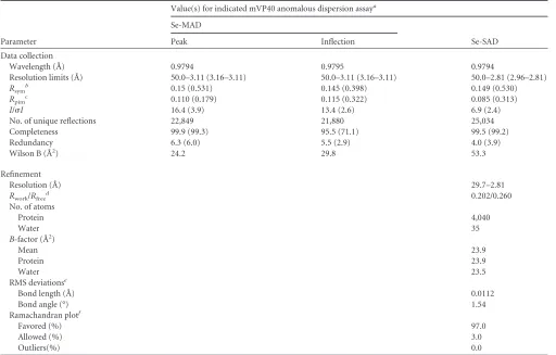
TABLE 1Crystallographic data collection and refinement statistics
Parameter
Value(s) for indicated mVP40 anomalous dispersion assaya
Se-MAD
Se-SAD
Peak Inflection
Data collection
Wavelength (Å) 0.9794 0.9795 0.9794
Resolution limits (Å) 50.0–3.11 (3.16–3.11) 50.0–3.11 (3.16–3.11) 50.0–2.81 (2.96–2.81)
Rsymb 0.15 (0.531) 0.145 (0.398) 0.149 (0.530)
Rpimc 0.110 (0.179) 0.115 (0.322) 0.085 (0.313)
I/I 16.4 (3.9) 13.4 (2.6) 6.9 (2.4)
No. of unique reflections 22,849 21,880 25,034
Completeness 99.9 (99.3) 95.5 (71.1) 99.5 (99.2)
Redundancy 6.3 (6.0) 5.5 (2.9) 4.0 (3.9)
Wilson B (Å2) 24.2 29.8 53.3
Refinement
Resolution (Å) 29.7–2.81
Rwork/Rfree
d 0.202/0.260
No. of atoms
Protein 4,040
Water 35
B-factor (Å2)
Mean 23.9
Protein 23.9
Water 23.5
RMS deviationse
Bond length (Å) 0.0112
Bond angle (°) 1.54
Ramachandran plotf
Favored (%) 97.0
Allowed (%) 3.0
Outliers(%) 0.0
a
Se-MAD space group,I222; Se-SAD space group,I222; Se-MAD cell dimensions (Å),a⫽96.0,b⫽106.8, andc⫽128.4; Se-SAD cell dimensions,a⫽95.8,b⫽107.3, andc⫽ 127.7. Data for the highest-resolution shells are shown in parentheses.
b
Rsym⫽ ⌺|Ii⫺ ⬍Ii⬎|/⌺Ii, whereIiis the observed intensity and⬍Ii⬎is the average intensity obtained from multiple observations of symmetry-related reflections. cR
pim, precision-indicating and multiplicity-weighted Rmerge.
d
Rwork⫽ ⌺hkl||Fobs|⫺|Fcalc||/⌺hkl|Fobs|. Five percent of the reflections were excluded for theRfreecalculation. obs, observed; calc, calculated.
eRMS, root mean square. f
Data are based on the criteria of MolProbity.
on November 7, 2019 by guest
http://jvi.asm.org/
L-Val; and 0.04 mg/ml seleno-L(⫹)-methionine. Protein expression was induced by adding 0.3 mM IPTG, and growth of the culture was contin-ued overnight. Cells were harvested and frozen at⫺20°C.
Purification of MARV VP40 protein.Cells were thawed and sus-pended in lysis buffer (20 mM Tris-HCl, 500 mM NaCl, 20 mM imida-zole, pH 8.0) supplemented with a protease inhibitor cocktail (Roche) and lysed by microfluidization (Microfluidics). The cell extract was centri-fuged at 15,000⫻gfor 1 h, and the supernatant was incubated with Ni2⫹
resin (Qiagen), preequilibrated with lysis buffer, at 4°C for 30 min. The Ni2⫹resin was washed with more than 15 column volumes of lysis buffer,
and the 6⫻His-tagged mVP40 was eluted with elution buffer (20 mM Tris-HCl, 500 mM NaCl, 300 mM imidazole, pH 8.0). CHAPS {3-[(3-cholamidopropyl)-dimethylammonio]-1-propanesulfonate} (10 mM) was added to the protein solution after elution, the protein was loaded onto a size exclusion column equilibrated with buffer (20 mM Tris-HCl, 500 mM NaCl, 10 mM CHAPS, pH 8.0), and fractions containing mVP40 were collected. After size exclusion chromatography was performed, mVP40 was dialyzed against an ion exchange buffer (10 mM Tris-HCl, 200 mM NaCl, 10 mM CHAPS, pH 8.0) at 4°C for 3 h and loaded onto a Mono S ion exchange column (GE Healthcare). mVP40 was eluted using a 200 mM to 500 mM linear gradient of NaCl. The eluted protein solution was dialyzed against size exclusion buffer, concentrated by centrifugal filtration (Amicon) to 3 mg/ml, and used for crystallization. mVP40 sam-ples used for biophysical experiments were equilibrated to 20 mM MES buffer at pH 6.0, after elution from Ni resin.
Size exclusion chromatography with inline multiangle laser light scattering (SEC-MALS).Purified proteins (typically 0.2 to 0.4 mg total) were loaded onto a Superdex 75 10/300 gel filtration column (GE Health-care) preequilibrated with buffer (20 mM HEPES-NaOH [pH 8.0], sup-plemented with 300 mM NaCl). Following elution from the column, the fast protein liquid chromatography (FPLC) system was connected in-line with a miniDAWN TREOS followed by an Optilab T-rEX refractometer (Wyatt Technologies). Data processing and absolute molecular mass cal-culations were performed using ASTRA software (Wyatt Technologies).
Crystallization of MARV VP40.Initial screening of crystallization was carried out using an Oryx8 crystallization robot (Douglas Instru-ments) and commercially available crystal screening kits on MRC vapor diffusion crystallization plates. Minuscule crystals were observed under diverse conditions, including the presence of polyethylene glycol (PEG), salt, alcohol, and other organic precipitants. Microcrystals grown under
ethanol conditions were optimized, and crystals that were suitable for diffraction studies were ultimately obtained in 17.5% ethanol–75 mM NaCl–10 mM CHAPS–100 mM Tris-HCl (pH 7.7) at 296 K by hanging drop vapor diffusion.
Diffraction and data collection.Once crystals grew to equilibrium, the crystal tray was moved to 4°C so that crystals could be harvested at 4°C to avoid damage caused by the evaporation of alcohol. After harvesting, crystals were soaked in a solution containing optimized reservoir solution supplemented with 20% MPD for a few seconds and transferred to liquid nitrogen for freezing.
Multiwavelength anomalous dispersion (MAD) data were collected to 3.11-Å resolution at beamline 23-ID-B of the Advanced Photon Source (APS) (Table 1), Argonne National Laboratory. Crystals were mounted in a cryo-stream of nitrogen at 100 K, and diffraction data were collected through the long axis of rod-shaped crystals using a 20-m-by-20- m-diameter mini-beam. Diffraction data were recorded on a Mar Mosaic 300 charge-coupled-device (CCD) detector (Rayonix). The peak wavelength of a Se-Met crystal was based on an X-ray absorption fine structure (XAFS) scan. Complete data sets at the peak (0.9794 Å) and reflection edge (0.9795 Å) were collected from one crystal. Single-wavelength anom-alous dispersion (SAD) data were collected to 2.81 Å from another crystal using the same protocol (Table 1). The diffraction data were indexed, integrated, and scaled using HKL2000 (17).
Crystal structure determination.The structure was solved using the 2W-MAD protocol of Auto-Rickshaw (18), and input diffraction data were prepared using programs of the CCP4 suite (19). Fluorescent anti-body (FA) values were calculated using the program SHELXC (20). Based on an initial analysis of the data, the maximum resolution for substructure determination and initial phase calculation was set to 3.6 Å.
All of the 17 heavy atoms requested were found using the program SHELXD (21). The correct hand orientation for the substructure was determined using the programs ABS (22) and SHELXE (20), and the occupancy of all substructure atoms was refined and initial phases were calculated using the program MLPHARE (19). The 2-fold non-crystallo-graphic-symmetry (NCS) operator was found using RESOLVE (23). Den-sity modification, phase extension, and NCS averaging were performed using the program DM (42). A partial alpha-helical model was produced using the program HELICAP (43), and 525 of 622 residues in the asym-metric unit were built using BUCCANEER (24), starting from the HELI-CAP model. Model refinement was performed using Refmac5 at 2.8-Å
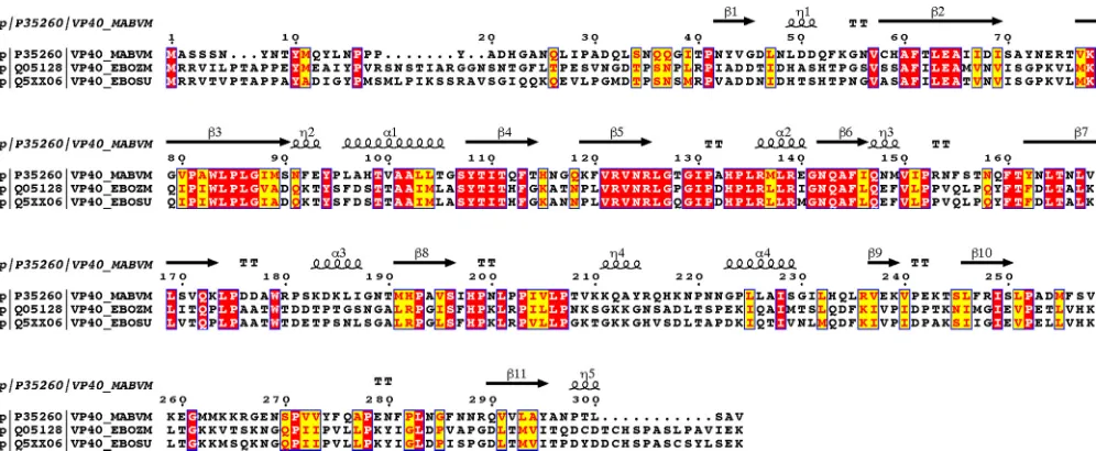
FIG 1Sequence alignment. Amino acid sequences of mVP40, Zaire ebolavirus (also known as Ebola virus) VP40, and Sudan ebolavirus (also known as Sudan virus) VP40 were aligned using T-COFFEE (39). The figure was generated using ESPript (40). Secondary-structure elements are indicated as follows:␣,␣-helix; ,-strand;, 310helix.
on November 7, 2019 by guest
http://jvi.asm.org/
[image:3.585.45.543.65.270.2]resolution and SAD data. Manual model correction was performed using COOT (25), and refinement procedures were iterated until convergence. VLP budding assay.Budding of virus-like particles (VLPs) into cell supernatants was detected by Western blot analyses. Wild-type mVP40 and mutant mVP40 bearing a Strep-Tag were cloned into pTriEx-5 (No-vagen) and transfected into cells using polyethyleneimine (PEI). VLPs were harvested 48 h posttransfection. Cell culture medium was spun down at 3,000⫻gfor 5 min to pellet any cells out of the media. Superna-tants were layered over 20% sucrose–20 mM HEPES (pH 7.4). VLPs were pelleted at approximately 150,000⫻gfor 1 h at 277 K and then resus-pended in phosphate-buffered saline (PBS) buffer supplemented with 1% NP-40, 1% Triton X-100, and 1 mM EDTA. Cell lysates were collected by washing cells twice with PBS followed by lysis in CytoBuster (EMD Bio-sciences). VLPs and cell lysates were then run on SDS denaturing gels, transferred onto polyvinylidene difluoride (PVDF) Immobilon transfer membranes (Millipore), and probed with an anti-Strep-Tag antibody (AbD Serotec).
Multilamellar vesicle (MLV) sedimentation assay.POPC [1-hexa-decanoyl-2-(9Z-octadecenoyl)-sn-glycero-3-phosphocholine], POPE
[1-hexadecanoyl-2-(9Z-octadecenoyl)-sn-glycero-3-phosphoethanol-amine], and POPS [1-hexadecanoyl-2–(9Z-octadecenoyl)-sn-glycero-3-phospho-L-serine] were mixed in the molar ratios noted below and dried under N2gas for 10 min. In all sedimentation assays, 30% POPE and 30%
POPS were used to maintain consistency (unless otherwise stated) and POPC levels were scaled accordingly. Lipids were hydrated with resuspen-sion buffer (25 mM HEPES [pH 7.4] containing 150 mM KCl) and incu-bated at 37°C for 15 min to facilitate hydration of lipids to form MLVs (26–30). These lipid solutions were then incubated at room temperature with 5M mVP40 proteins for 20 min. The total assay volume was 100l, and the final lipid concentration was 1 mM. Samples were centrifuged at 48,000⫻gfor 20 min. During centrifugation, mVP40 bound to MLV sediments, while unbound proteins remained in the supernatant. Super-natant was carefully removed from the pellet, and the pellet was resus-pended in an amount of buffer equal to the volume that was removed as the supernatant. Volumes (40l) of each supernatant and pellet sample were separated by SDS-PAGE. The protein band was resolved with Coo-massie brilliant blue staining, and gel band intensities were analyzed using Image J software. For the quantification, the gel images are first converted to 8-bit images using Image J software. Using the rectangular selection tool, a rectangular region around the gel bands is selected and marked as
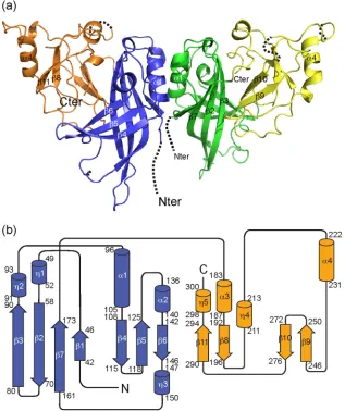
FIG 2Crystal structure of Marburg virus VP40. (a) The N-terminal domain-mediated dimer of mVP40 is illustrated. In monomer 1, the N-terminal domain (Nter; residues 38 to 176) is shown in blue and the C-terminal domain (Cter; residues 177 to 303) in orange. In monomer 2, the NTD is green and the CTD yellow. The dimer of MARV VP40 forms a butterfly shape, with the wings twisted relative to each other in and out of the plane of the page. The N termini and some residues in surface loops are not observed in the electron density. Connectivity in missing sections is illustrated by dashed lines. (b) Topology diagram of a single protomer of Marburg virus VP40, colored with the NTD (N) in blue and the CTD (C) in orange. Secondary-structure elements are indicated as follows:␣, ␣-helix;,-strand;, 310helix.
on November 7, 2019 by guest
http://jvi.asm.org/
[image:4.585.134.451.68.446.2]the first lane. By selecting plot lanes, a graphical representation of the band intensities as peaks is created by the software, and by using the wand tool, the area under the peak for each gel band is obtained. The fraction of proteins associated with MLVs of each lipid composition was calculated by the following ratio: (area under the peak for pellet gel band)/(area under the peak for supernatant⫹pellet gel bands). Each experiment was performed at least three times to calculate the mean, the standard devia-tion (or the standard error of the mean where shown), and thePvalue.
ISRE reporter gene assay.293T cells (1⫻105) (ATCC) were
trans-fected with 0.25g of interferon-stimulated response element (ISRE) firefly luciferase reporter plasmid, 0.005g of Renilla luciferase plasmid, and 1.0g of the VP40 pTriEx-5 expression plasmids, encoding N-termi-nal Strep-Tag II. All transfections were performed using polyethylenimine (31), and cells were treated with 1,000 units of beta IFN (IFN-; PBL Assay Science) at 40 h posttransfection. At 18 h post-IFN treatment, cells were harvested using reporter lysis buffer and analyzed using a dual-luciferase reporter assay system (Promega), performed according to the manufac-turer’s instructions. Protein expression was detected by Western blot analysis.
Immunofluorescence (IF) and confocal microscopy.Hemagglutinin (HA)-fusion WT and mutant (T105R, W83R/N148A) mVP40s were cloned into the pCAGGS plasmid. Intact mVP40 K210E mutant was cloned into pCAGGS plasmid. 293 cells were transfected with plasmids expressing wild-type or mutant mVP40. At 24 h posttransfection, cells were fixed with 4% paraformaldehyde, permeabilized with phosphate-buffered saline (PBS) containing 0.1% Triton X-100 for 5 min at room
temperature, and incubated with rabbit anti-VP40 antibody (K210E) or anti-HA antibody (WT, T105R, W83R/N148A). Alexa Fluor 488 goat an-ti-mouse IgG (Molecular Probes) was used to visualize the primary anti-VP40 and anti-HA antibodies. Cells were observed under an LSM780 microscope (Carl Zeiss).
Protein structure accession number.Coordinates and structure factors have been deposited into the Protein Data Bank under accession code 5B0V.
RESULTS
Crystal structure of MARV VP40.
mVP40 crystallized in space
group
I222
with two protomers in the crystallographic
asymmet-ric unit (ASU) and diffracted to a resolution of 2.8 Å (
Table 1
). The
construct crystallized was the complete mVP40, containing
resi-dues 1 to 303 (
Fig. 1
and
2
). Of these, the N terminus (residues 1 to
37 in molecule 1 and 1 to 35 in molecule 2) and a few short loops
(residues 71 to 72, 156 to 157, and 264 to 267 in molecule 1 and
residues 216 to 218, 259 to 260, 264 to 269, and 302 to 303 in
molecule 2) were disordered.
mVP40 consists of two distinct domains, an N-terminal
do-main (NTD; residues 36 to 173) and a C-terminal dodo-main (CTD;
residues 183 to 303), which are connected by a linker region
(res-idues 174 to 182) (
Fig. 1
and
2b
). The mVP40 NTD consists of
seven

-strands, two
␣
-helices, and three 3
10helices and adopts a

-sandwich-like structure. The CTD consists of six strands and six
helices and adopts a small

-sandwich-like structure as well.
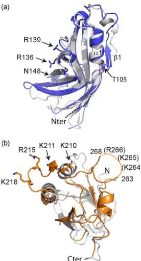
The NTDs of mVP40 and eVP40 are 42% identical in sequence
and adopt comparable folds, with an root mean square deviation
(RMSD) between the C-alpha atoms of 2.4 Å (
Fig. 3a
). The CTDs
of mVP40 and eVP40, however, are only 16% identical in
se-quence. The CTDs differ more extensively in structure and align
with a 5.6-Å RMSD between C-alpha atoms (
Fig. 3b
). In
particu-lar, residues 208 to 221 of the CTD trace a different pathway in
mVP40 than in eVP40 and form an extended, flat, basic patch.
mVP40 forms a dimer, and the dimer interface is essential to
matrix assembly.
To date, the only oligomer defined for mVP40 is
an oligomeric ring (
32
). In order to determine if mVP40, like
eVP40, also forms a dimer in solution (
33
), we analyzed purified
mVP40 using size exclusion chromatography (SEC) coupled with
multiangle light scattering (MALS) (SEC-MALS). In these
exper-iments, wild-type mVP40 was determined to be approximately 63
kDa in solution, while a point mutant, T105R, yielded a 34-kDa,
likely monomeric VP40 (see
Fig. 5c
).
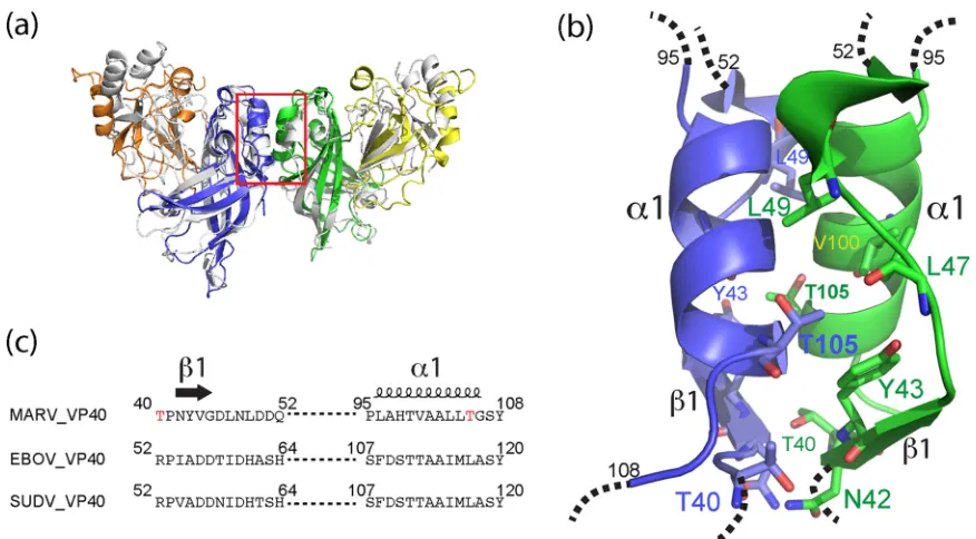
A potential NTD-to-NTD dimer interface was revealed in the
crystal packing of mVP40 that is similar to the dimer interface of
eVP40 (
Fig. 4a
). This interface, however, is smaller than that of
eVP40, being approximately 670 Å
2. This interface includes strand

1 (residues 40 to 52) and helix
␣
1 (residues 97 to 105) and is
assembled mostly by hydrophobic contacts involving residues
T40, P41, N42, Y43, L49, D50, Q52, A97, H98, A101, A102, L104,
and T105. Three direct hydrogen bonds across the dimeric
inter-face between T40 and N42, between T40 and Y43, and between
N42 to N42 of the opposite monomer were noted (
Fig. 4b
).
Resi-due T105 is central to this interface and forms a hydrophobic
contact to Y43. Mutation of threonine at this position to arginine
(T105R) resulted in primarily monomeric VP40 in solution, as
mentioned earlier (
Fig. 5c
).
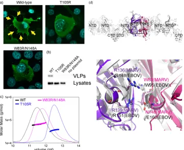
Immunofluorescence assays (IFA) illustrated that, while
wild-type mVP40 successfully assembled and budded VLPs from the
cell, T105R mVP40 failed to assemble or bud VLPs. Moreover,
T105R mVP40 failed to translocate to the plasma membrane and,
FIG 3Comparison of mVP40 and eVP40 in NTD and CTD structures. (a) NTDs (Nter) of mVP40 (blue) and eVP40 (white) were superimposed using least-squares (LSQ) superposition by Coot (25). Overall, thesandwich struc-ture is conserved, with some differences in1 and loops that connect the -strands. (b) Superimposed CTDs (Cter) of mVP40 (orange) and Sudan ebolavirus VP40 (white). Note that the CTDs differ more extensively in struc-ture than the NTDs, particularly the two loops containing residues 209 to 222 (left) and 256 to 271 (right), which trace paths in mVP40 different from those traced by the corresponding residues of eVP40. Residues K210, K211, R215, and K218, which contribute to the basic patch of the mVP40 CTD, are illus-trated in the stick model. Residues K264, K265, and R266, which contribute to the basic patch but are on the disordered loop, are indicated in parentheses.
on November 7, 2019 by guest
http://jvi.asm.org/
[image:5.585.90.234.64.330.2]instead, remained diffuse throughout the cytoplasm (
Fig. 5a
and
b
), as did a mutation at the equivalent position in eVP40, L117R
(
33
). Formation of an NTD-mediated dimer appears critical to
assembly of Marburg virus-like particles.
For Ebola virus, dimers are thought to assemble into the viral
matrix by a conformational change that exposes a separate surface
of the NTD involving W95 and E160 (
33
,
34
). These residues
aligned in sequence and structure to W83 and N148 of mVP40
(
Fig. 5d
). A double W83R/N148A mutant was constructed in
or-der to determine if dimers of mVP40 further assemble via an
in-terface involving a similar surface. SEC-MALS revealed that both
wild-type mVP40 and W83R/N148A mVP40 formed dimers in
solution as expected (
Fig. 5c
). However, only WT mVP40
bled and budded VLPs; the W83R/N148A mutant failed to
assem-ble and to bud VLPs (
Fig. 5c
).
The CTD bears a basic patch essential for membrane
target-ing and matrix assembly.
mVP40 interacts with the acidic lipid
bilayer surface to maintain the structural integrity of the virus and
to organize the matrix structure essential for virus budding (
35
,
36
). A significant basic patch was observed on the surface of the
CTD, with the basic patch of each mVP40 monomer exposed
along a single side of the dimer (
Fig. 6a
). This patch was primarily
derived from two loops that extended in opposite directions. The
first basic loop, spanning residues 208 to 221, contained the basic
amino acids K210, K211, R215, and K218 (
Fig. 3b
and
6a
). The
corresponding loop of eVP40, spanning residues 210 to 233, was
not observed in the eVP40 structure. Its sequence, however, was
less basic in charge than that of mVP40 and likely pointed in the
opposite direction (
Fig. 3b
). The second loop in the mVP40 basic
patch spanning residues 251 to 271, between

9 and

10,
con-tained basic amino acids K259, K264, K265, and R266 and was
similar in sequence composition to that of eVP40. The combined
basic patch of mVP40 formed an essentially flat surface, while that
of eVP40 involved more extensively projecting, flexible, and
dis-ordered loop structures.
To investigate the role of this basic surface in mVP40, we
in-troduced mutations at three different clusters of basic residues.
K210E single, R215A/K218A double, and K264A/K265A/R266A
triple mutations were made. Each mutant expressed to a level
equivalent to the level seen with wild-type VP40, but each failed to
assemble and to bud VLPs (
Fig. 6c
). Additional substitutions at
position 210 confirmed the importance of the basic charge. The
acidic K210E mutant failed to bud VLPs, and the neutral/polar
K210S mutant was significantly reduced in its ability to bud VLPs,
but the basic K210R mutant and the wild-type mVP40 mutant
were able to assemble and to bud VLPs from transfected cells (
Fig.
6c
and
d
).
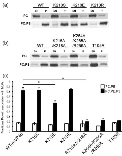
A multilamellar vesicle (MLV) sedimentation assay was used to
assess the effect of mutations on the CTD basic patch and of the
dimer interface on the membrane binding ability of mVP40.
mVP40 associates with the plasma membrane by interacting with
anionic phospholipids in a nonspecific manner, dependent on the
anionic charge density (K. J. Wijesinghe and R. V. Stahelin,
un-published data). We tested K210S, K210E, K210R, R215A/K218A,
and K264A/K265A/R266A mutant mVP40 in order to investigate
the importance of these residues in mediating electrostatic
inter-actions with the plasma membrane. In this assay, POPC:POPE
(70:30) vesicles composed of zwitterionic phospholipids were
used as control vesicles. Wild-type mVP40 exhibited only a
mar-ginal association with MLVs lacking anionic phospholipids but an
FIG 4The dimer interface. (a) Marburg VP40 is colored with NTDs in blue and green and CTDs in orange and yellow. Sudan ebolavirus VP40 is illustrated in white and is aligned on the blue mVP40 NTD. The dimer interface (magnified in panel b) is outlined in red. Structures were superimposed by secondary-structure matching (SSM) using Coot (25). (b) Closeup view of the Marburg virus VP40 dimeric interface, in which one monomer is colored blue and the opposing monomer green. The main chains are illustrated in ribbons, with key residues that contribute to this interface illustrated in stick models. (c) Sequence alignment of the corresponding regions is shown below the model. Residue T105 resides in the middle of the hydrophobic pocket and T40 at the base. SUDV, Sudan ebolavirus.
on November 7, 2019 by guest
http://jvi.asm.org/
[image:6.585.73.511.63.305.2]85% association with MLVs containing 30% of the anionic lipid
POPS (
Fig. 7
).
mVP40 bearing the K210E mutation exhibited a 35%
reduc-tion in its associareduc-tion with anionic phospholipid,
phosphatidyl-serine (POPS)-containing MLVs. mVP40 bearing neutral/polar
K210S associated with these MLVs at near wild-type levels, and
mVP40 bearing basic K210R achieved a wild-type-level
associa-tion with MLVs (
Fig. 7a
and
c
).
mVP40 bearing either the R215A/K218A double mutation at
the second site or the K264A/K265A/R266A triple mutation at the
third site exhibited a dramatic decrease in association with
POPS-containing MLVs (
Fig. 7b
and
c
). These results suggest that
ala-nine replacement in the basic patches of the C-terminal domain
disrupts critical electrostatic interactions necessary for mVP40
as-sociation with POPS-containing membranes (
Fig. 7b
and
c
).
T105 is at the center of the dimeric interface of the mVP40.
A T105R point mutation resulted in mVP40 that failed to
dimerize and was instead monomeric in solution.
T105R-bear-ing mVP40 also failed to associate with anionic
phospholipid-containing MLVs (
Fig. 7b
and
c
), suggesting that dimerization,
in addition to the basic patch, is also important for membrane
association. Dimerization may be important for formation of
the extended basic surface formed by the pair of VP40 CTDs.
What structure of mVP40 antagonizes interferon?
In
addi-tion to its role in matrix assembly and viral budding, MARV VP40
has an additional role in interferon suppression, antagonizing the
host innate immune response by blocking the JAK/STAT
signal-ing pathway (
15
). Valmas et al. (
15
) recently demonstrated that
mVP40 blocks induction of both ISG54 and
gamma-activated-sequence (GAS) reporter genes after the stimulation by type I and
type II IFN, respectively. We analyzed various truncations of
mVP40 in order to determine which domain or structure of
mVP40 antagonizes interferon signaling.
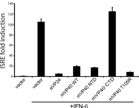
Wild-type mVP40 successfully antagonized IFN-

-stimulated
enhancement of ISRE reporter gene expression (
Fig. 8
), as
previ-ously reported (
15
). The CTD of mVP40, when expressed alone,
failed to antagonize IFN signaling. In contrast, the NTD of
mVP40, when expressed alone, did antagonize IFN signaling, and
FIG 5Cellular localization of wild-type and mutant mVP40s. In each set of three images, the top left panel illustrates nuclear staining with DAPI. Alexa 488-conjugated anti-mouse IgG was used to visualize the primary antibodies. (a) Wild-type (WT) Marburg VP40 localizes to the cell membrane and buds VLPs (indicated with yellow arrows). T105R mVP40 fails to localize to and concentrate at the membrane and fails to assemble and bud VLPs. T105R mVP40 instead stains more diffusely in the cytoplasm. W83R/N148A mVP40 fails to assemble and bud VLPs, does not accumulate at the membrane, and also stains more diffusely in the cytoplasm. (b) Western blot of cell lysates and purified VLPs from transfected cells. The WT strain buds VLPs. Both the T105R and W83R/N148A mutants failed to be released from the cell into the supernatant as VLPs. (c) Multiangle light scattering analysis of the wild type (WT) and mVP40 mutants analyzed by size exclusion chromatography is illustrated. The absolute molecular mass distributions are shown as horizontal lines (WT, 63 kDa; W83R/N148A, 68 kDa; T105R, 36 kDa), with the corresponding light-scattering curves shown as solid lines. WT, W83R/N148A-containing, and T105R-containing mVP40s are shown in black, pink, and blue, respectively. (d) NTD-mediated oligomerization interface model. Two copies of the NTD of mVP40 (pink and purple) are superimposed onto two NTDs of eVP40 in the crystal structure of rearranged, hexameric eVP40 (white). In the lower part of the panel, eVP40 residues W95, E160, R148, and R151, each critical to the hexameric assembly, are shown in white in the stick model. mVP40 residues W83 and N148, which align with eVP40 residues W95 and E160, are shown in magenta. mVP40 residues R136 and R139, which align with eVP40 residues R148 and R151, are shown in purple. The zoomed-in view illustrates the interaction that could be made by mVP40 in this interface.
on November 7, 2019 by guest
http://jvi.asm.org/
[image:7.585.114.475.66.362.2]it did so as well as wild-type, full-length VP40. Thus, we found that
the mVP40 NTD is necessary and sufficient for this function (
Fig.
8
). Interestingly, the NTD, when expressed alone, forms a ring
oligomer (
32
). Hence, the ring oligomer could be linked to IFN
antagonism. For eVP40, the ring is assembled by surfaces of the
NTD different from the dimer interface and dimerization is not
required for ring formation (
33
,
37
). Similarly, we found that
T105R-bearing mVP40, which was unable to dimerize in solution,
was fully functional in IFN antagonism. Hence, the formation of
the dimer, although critical for matrix assembly, is not critical for
the IFN antagonism function of mVP40.
DISCUSSION
The crystal structure of Marburg virus VP40 presented here
illus-trates that the N-terminal domain is relatively conserved but that
the C-terminal domain varies more extensively in structure
among the filoviruses. The structure and accompanying
mutagen-esis reveal that mVP40 forms an NTD-mediated dimer, which is
essential for assembly and budding of virus-like particles. A T105R
point mutation at the dimer interface yields monomeric mVP40
and disrupts and assembly and budding of mVP40 VLPs. Previous
work demonstrated that a T40A point mutation in mVP40 also
disrupts budding (
38
). This crystal structure reveals that T40
re-sides on the edge of the dimer interface and makes a hydrogen
bond across the interface to the main chain of Y43 of the opposing
monomer (
Fig. 4b
).
For eVP40, dimers are thought to further assemble into the
matrix via an “oligomerization interface” containing W95 and
E160, which is exposed upon electrostatically driven
rearrange-ment of the NTD and CTD at the membrane (
33
). Mutagenesis of
the equivalent residues in mVP40 (W83 and N148) similarly
abol-ishes the budding of VLPs. Rearrangement of the mVP40 CTD
away from the NTD would be necessary to expose this W83- and
N148-containing surface of the NTD. Hence, mVP40 probably
undergoes structural rearrangement to mediate matrix assembly
at the membrane similarly to that of eVP40.
The crystal structure of mVP40 also reveals a single basic patch
on the upper surface of the CTDs in the dimer. This basic patch is
larger and flatter in mVP40 than in eVP40. Further, the
polypep-FIG 6Characterization of the CTD basic patch in mVP40. (a) Top view of the electrostatic surface potential of the mVP40 dimer. (b) Side view. The dimer interface is indicated by a dashed line. The electrostatic surface potential was generated by APBS (41) within CueMol (http://cuemol.sourceforge.jp/en/). Basic residues are labeled. (c) Western blot of cell lysates and purified VLPs from transfected cells. The gel was spliced on the solid line. WT and K210R bud VLPs. VLPs budding of K210S is notably reduced. All mutant proteins were expressed in cells, but those mutants which removed the basic charge failed to be released from the cell into the supernatant as VLPs. (d) IFA demonstrates that WT mVP40 translocates to the cell membrane and buds VLPs (indicated with yellow arrows). In contrast, K210E is impaired in membrane trafficking and does not bud VLPs.
FIG 7mVP40 association with lipid vesicles containing neutral phospholipids and anionic phospholipids. (a and b) SDS-PAGE of supernatant (SN) and pellet (P) fractions collected from MLV sedimentation assays on lipid compo-sitions appearing above the images. In all liposomes, 30 mol% of POPE (PE) was used, and, where shown, 30 mol% POPS (PS) was used to maintain con-sistency. Thus, control vesicles consisted of 70 mol% POPC and 30% POPE and, for test vesicles, 30% POPS was added at the expense of POPC. (See Results for clarification of the distinction between panels a and b.) (c) Quan-tification of MLV sedimentation assay results. The data represent averages of the results of three sedimentation assays, with error bars representing the stan-dard errors of the means. *,P⬍0.05.
on November 7, 2019 by guest
http://jvi.asm.org/
[image:8.585.296.538.62.371.2] [image:8.585.39.292.64.383.2]tides that form this surface in MARV individually trace outward in
opposite directions, while those of eVP40 point upward and are
disordered. As a result, mVP40 forms a broader, flatter basic patch
than eVP40. This patch contains three clusters of basic residues,
each of which is essential to matrix assembly and budding.
We have further used an MLV sedimentation assay to
deter-mine if the basic patch and dimeric interface of mVP40 have an
effect on its membrane binding ability. mVP40 associates with
membranes containing anionic phospholipids in a highly
charge-dependent manner, behaving as a nonspecific anionic charge
sen-sor (Wijesinghe and Stahelin, unpublished). The relatively flat
basic patch of dimeric mVP40 provides an ideal surface to mediate
electrostatic interactions between the protein and the anionic
phospholipids, facilitating recruitment of mVP40 to the plasma
membrane. All basic patch mutants in which a cationic charge has
been reversed (K210E) or replaced with hydrophobic residues
(R215A/K218A and K264A/K265A/R266A) failed to bud VLPs. In
membrane association assays, the K210E single-point mutant
ex-hibits a reduction of about 30% in association with
POPS-con-taining MLVs, while double and triple mutants exhibit a more
dramatic (
⬃
70%) reduction in association. We suspect that the
failure of these mutants to form VLPs occurred because these
mutations reduce critical electrostatic interactions necessary to
recruit mVP40 to the anionic inner leaflet of the plasma membrane.
The mutant behavior in MLV association assays is consistent with the
behavior seen in VLP budding assays and strongly supports the idea
of the necessity of electrostatic interactions for mVP40 to associate
with anionic lipids of the cytoplasmic leaflet of the plasma
mem-brane.
According to immunofluorescence assays (IFA), the
mono-meric T105R mutant exhibits largely cytoplasmic and nuclear
rather than membrane localization. As shown by the MLV assay,
T105R-bearing mVP40 cannot robustly associate with anionic
phospholipid-containing MLVs. This poor association suggests
that mVP40 requires its dimeric structure in order to associate
with anionic inner leaflet of the plasma membrane. It is possible
that the basic patch of each mVP40 monomer in the dimeric
struc-ture has a synergistic effect on membrane association.
Marburg virus VP40 has also been shown to antagonize JAK/
STAT signaling, but the molecular mechanism by which this
oc-curs and the structural form of mVP40 responsible for this
func-tion were unknown. Here we demonstrate that the NTD of
mVP40 is necessary and sufficient for JAK/STAT signaling
antag-onism. When the NTD of mVP40 is expressed alone, in the
ab-sence of the CTD, it assembles into ring-like structures (
32
).
Hence, the ring assembly of mVP40 may be linked to interferon
antagonism.
In summary, the 2.8-Å crystal structure of Marburg virus VP40
and additional results of biochemical analysis presented here
illus-trate the dimeric interface required for virus assembly, assign the
domain and, likely, the oligomer of Marburg virus VP40 required
for its role in JAK/STAT signal antagonism, and provide a
tem-plate for biological exploration of the many roles of mVP40 in the
Marburg virus life cycle.
ACKNOWLEDGMENTS
We thank Javad Aman and Kelly Warfield of Integrated BioTherapeutics, Inc., for the gift of MARV VP40 cDNA and monoclonal antibodies. We thank Craig Ogata for advice and help during data collection.
FUNDING INFORMATION
[image:9.585.41.285.64.253.2]Japan Society for Promotion of Science provided funding to Takeshi Noda. Health and Labor Sciences Research Grants, Japan provided fund-ing to Yoshihiro Kawaoka. HHS | National Institutes of Health (NIH) provided funding to Robert V. Stahelin under grant number AI081077. HHS | NIH | National Institute of Allergy and Infectious Diseases (NIAID) provided funding to Erica Ollmann Saphire under grant number R44 AI08843. Ministry of Education, Culture, Sports, Science, and Technol-ogy (MEXT) provided funding to Takeshi Noda. HHS | NIH | National Institute of General Medical Sciences (NIGMS) provided funding to Ka-veesha J. Wijesinghe under grant number T32GM075762. Burroughs Wellcome Fund (BWF) provided funding to Erica Ollmann Saphire.
This research was supported by NIH/NIAID grant R44 AI08843 and an Investigators in the Pathogenesis of Infectious Disease award from the Burroughs Wellcome Fund (E.O.S.), a CBBI fellowship supported by NIH T32GM075762 (K.J.W.), NIH grant AI081077 (R.V.S.), Health and Labor Sciences research grants, Japan (Y.K.), and a Grant-in-Aid for Young Scien-tists from the Japan Society for the Promotion of Science, a Grant-in-Aid for Scientific Research on Priority Areas from the Ministry of Education, Culture, Sports, Science, and Technology, and PRESTO from Japan Science and Technology Agency (T.N.) and used resources of the Advanced Photon Source, a U.S. Department of Energy (DOE) Office of Science User Facil-ity operated for the DOE Office of Science by Argonne National Labora-tory under contract DE-AC02-06CH11357.
REFERENCES
1.King AMQ, Adams MJ, Lefkowitz EJ, Carstens EB.2012. Virus taxon-omy: classification and nomenclature of viruses. 9th Report of the Inter-national Committee on Taxonomy of Viruses, vol 9. Elsevier, Philadel-phia, PA.
2.Brauburger K, Hume A, Mühlberger E, Olejnik J. 1 October 2012. Forty-five years of Marburg virus research. Viruseshttp://dx.doi.org/10 .3390/v4101878.
3.Jasenosky LD, Neumann G, Lukashevich I, Kawaoka Y.2001. Ebola virus VP40-induced particle formation and association with the lipid bilayer. J Vi-rol75:5205–5214.http://dx.doi.org/10.1128/JVI.75.11.5205-5214.2001. 4.Swenson DL, Warfield KL, Kuehl K, Larsen T, Hevey MC, Schmal-FIG 8VP40 inhibits ISRE-induced gene expression. In the ISRE reporter
assay, 293T cells were cotransfected with wild-type or mutant VP40s and firefly luciferase driven by an ISRE promoter, treated with IFN-40 h posttransfec-tion, and assayed for luciferase activity. Data are shown as the mean fold induction⫾standard deviation relative to the untreated negative control, for an individual experiment performed in triplicate. The leftmost control repre-sents empty vector in the absence of IFN-. Ebola virus VP24 (eVP24) is used as a positive control for interferon antagonism (11).
on November 7, 2019 by guest
http://jvi.asm.org/
john A, Bavari S, Aman MJ.2004. Generation of Marburg virus-like particles by co-expression of glycoprotein and matrix protein. FEMS Immunol Med Microbiol 40:27–31.http://dx.doi.org/10.1016/S0928 -8244(03)00273-6.
5.Kolesnikova L, Bamberg S, Berghöfer B, Becker S.2004. The matrix protein of Marburg virus is transported to the plasma membrane along cellular membranes: exploiting the retrograde late endosomal pathway. J Virol78:2382–2393.http://dx.doi.org/10.1128/JVI.78.5.2382-2393.2004. 6.Nanbo A, Watanabe S, Halfmann P, Kawaoka Y. 2013. The spatio-temporal distribution dynamics of Ebola virus proteins and RNA in in-fected cells. Sci Rep3:1206.
7.Mittler E, Kolesnikova L, Herwig A, Dolnik O, Becker S.2013. Assembly of the Marburg virus envelope. Cell Microbiol15:270 –284.http://dx.doi .org/10.1111/cmi.12076.
8.Kolesnikova L, Bugany H, Klenk HD, Becker S.2002. VP40, the matrix protein of Marburg virus, is associated with membranes of the late endo-somal compartment. J Virol76:1825–1838.http://dx.doi.org/10.1128/JVI .76.4.1825-1838.2002.
9.Johnson RF, Bell P, Harty RN.2006. Effect of Ebola virus proteins GP, NP and VP35 on VP40 VLP morphology. Virol J3:31.http://dx.doi.org /10.1186/1743-422X-3-31.
10. Hensley LE, Alves DA, Geisbert JB, Fritz EA, Reed C, Larsen T, Geisbert TW.2011. Pathogenesis of Marburg hemorrhagic fever in cynomolgus macaques. J Infect Dis204(Suppl 3):S1021–S1031.http://dx.doi.org/10 .1093/infdis/jir339.
11. Reid SP, Leung LW, Hartman AL, Martinez O, Shaw ML, Carbonnelle C, Volchkov VE, Nichol ST, Basler CF.2006. Ebola virus VP24 binds karyopherin alpha1 and blocks STAT1 nuclear accumulation. J Virol80:
5156 –5167.http://dx.doi.org/10.1128/JVI.02349-05.
12. Mateo M, Reid SP, Leung LW, Basler CF, Volchkov VE.2010. Ebola-virus VP24 binding to karyopherins is required for inhibition of interferon signaling. J Virol84:1169 –1175.http://dx.doi.org/10.1128/JVI.01372-09. 13. Reid SP, Valmas C, Martinez O, Sanchez FM, Basler CF.2007. Ebola virus VP24 proteins inhibit the interaction of NPI-1 subfamily karyopherin alpha proteins with activated STAT1. J Virol81:13469 – 13477.http://dx.doi.org/10.1128/JVI.01097-07.
14. Xu W, Edwards MR, Borek DM, Feagins AR, Mittal A, Alinger JB, Berry KN, Yen B, Hamilton J, Brett TJ, Pappu RV, Leung DW, Basler CF, Amarasinghe GK.2014. Ebola virus VP24 targets a unique NLS binding site on karyopherin alpha 5 to selectively compete with nuclear import of phosphorylated STAT1. Cell Host Microbe16:187–200.http: //dx.doi.org/10.1016/j.chom.2014.07.008.
15. Valmas C, Grosch M, Schümann M, Olejnik J, Martinez O, Best S, Krähling V, Basler C, Mühlberger E.2010. Marburg virus evades inter-feron responses by a mechanism distinct from ebola virus. PLoS Pathog
6:e1000721.http://dx.doi.org/10.1371/journal.ppat.1000721.
16. Sambrook J, Russell DW, Russell DW.2001. Molecular cloning: a lab-oratory manual, 3rd ed. Cold Spring Harbor Lablab-oratory Press, Cold Spring Harbor, NY.
17. Otwinowski Z, Minor W.1997. Processing of X-ray diffraction data collected in oscillation mode. Methods Enzymol276:307–326.http://dx .doi.org/10.1016/S0076-6879(97)76066-X.
18. Panjikar S, Parthasarathy V, Lamzin VS, Weiss MS, Tucker PA.
2005. Auto-rickshaw: an automated crystal structure determination platform as an efficient tool for the validation of an X-ray diffraction experiment. Acta Crystallogr D Biol Crystallogr61:449 – 457.http://dx .doi.org/10.1107/S0907444905001307.
19. Collaborative Computational Project, Number 41994. The CCP4 suite: programs for protein crystallography. Acta Crystallogr D Biol Crystallogr
50:760 –763.http://dx.doi.org/10.1107/S0907444994003112.
20. Sheldrick GM.2010. Experimental phasing with SHELXC/D/E: combin-ing chain traccombin-ing with density modification. Acta Crystallogr D Biol Crys-tallogr66(Pt 4):479 – 485.http://dx.doi.org/10.1107/S0907444909038360. 21. Schneider TR, Sheldrick GM.2002. Substructure solution with SHELXD. Acta Crystallogr D Biol Crystallogr58:1772–1779.http://dx.doi.org/10 .1107/S0907444902011678.
22. Hao Q.2004. ABS: a program to determine absolute configuration and evaluate anomalous scatterer substructure. J Appl Crystallogr37:498 – 499.http://dx.doi.org/10.1107/S0021889804008696.
23.Terwilliger TC. 2000. Maximum-likelihood density modification. Acta Crystallogr D Biol Crystallogr56:965–972.http://dx.doi.org/10 .1107/S0907444900005072.
24. Cowtan K.2006. The Buccaneer software for automated model building.
1. Tracing protein chains. Acta Crystallogr D Biol Crystallogr62:1002– 1011.http://dx.doi.org/10.1107/S0907444906022116.
25. Emsley P, Cowtan K.2004. Coot: model-building tools for molecular graphics. Acta Crystallogr D Biol Crystallogr60:2126 –2132.http://dx.doi .org/10.1107/S0907444904019158.
26. Bangham AD, Horne RW.1964. Negative staining of phospholipids and their structural modification by surface-active agents as observed in the electron microscope. J Mol Biol8:660.http://dx.doi.org/10.1016/S0022 -2836(64)80115-7.
27. Papahadjopoulos D, Watkins J.1967. Phospholipid model membranes. II. Permeability properties of hydrated liquid crystals. Biochim Biophys Acta135:639 – 652.http://dx.doi.org/10.1016/0005-2736(67)90095-8. 28. Saarikangas J, Zhao H, Pykalainen A, Laurinmaki P, Mattila PK,
Kin-nunen PK, Butcher SJ, Lappalainen P.2009. Molecular mechanisms of membrane deformation by I-BAR domain proteins. Curr Biol19:95–107.
http://dx.doi.org/10.1016/j.cub.2008.12.029.
29. Patil YP, Jadhav S.2014. Novel methods for liposome preparation. Chem Phys Lipids177:8 –18.http://dx.doi.org/10.1016/j.chemphyslip.2013.10 .011.
30. Mattila PK, Pykalainen A, Saarikangas J, Paavilainen VO, Vihinen H, Jokitalo E, Lappalainen P.2007. Missing-in-metastasis and IRSp53 de-form PI(4,5)P2-rich membranes by an inverse BAR domain-like mecha-nism. J Cell Biol176:953–964.http://dx.doi.org/10.1083/jcb.200609176. 31. Longo PA, Kavran JM, Kim MS, Leahy DJ.2013. Transient mammalian
cell transfection with polyethylenimine (PEI). Methods Enzymol529:
227–240.http://dx.doi.org/10.1016/B978-0-12-418687-3.00018-5. 32. Timmins J, Schoehn G, Kohlhaas C, Klenk HD, Ruigrok RW,
Weis-senhorn W.2003. Oligomerization and polymerization of the filovirus matrix protein VP40. Virology312:359 –368.http://dx.doi.org/10.1016 /S0042-6822(03)00260-5.
33. Bornholdt ZA, Noda T, Abelson DM, Halfmann P, Wood MR, Kawaoka Y, Saphire EO.2013. Structural rearrangement of ebola virus VP40 begets multiple functions in the virus life cycle. Cell154:763–774.
http://dx.doi.org/10.1016/j.cell.2013.07.015.
34. Hoenen T, Biedenkopf N, Zielecki F, Jung S, Groseth A, Feldmann H, Becker S.2010. Oligomerization of Ebola virus VP40 is essential for par-ticle morphogenesis and regulation of viral transcription. J Virol84:7053– 7063.http://dx.doi.org/10.1128/JVI.00737-10.
35. Neumann P, Lieber D, Meyer S, Dautel P, Kerth A, Kraus I, Garten W, Stubbs MT.2009. Crystal structure of the Borna disease virus matrix protein (BDV-M) reveals ssRNA binding properties. Proc Natl Acad Sci U S A106:3710 –3715.http://dx.doi.org/10.1073/pnas.0808101106. 36. Money VA, McPhee HK, Mosely JA, Sanderson JM, Yeo RP. 2009.
Surface features of a Mononegavirales matrix protein indicate sites of membrane interaction. Proc Natl Acad Sci U S A106:4441– 4446.http: //dx.doi.org/10.1073/pnas.0805740106.
37. Gomis-Rüth FX, Dessen A, Timmins J, Bracher A, Kolesnikowa L, Becker S, Klenk HD, Weissenhorn W.2003. The matrix protein VP40 from Ebola virus octamerizes into pore-like structures with specific RNA binding properties. Structure11:423– 433.http://dx.doi.org/10 .1016/S0969-2126(03)00050-9.
38. Makino A, Yamayoshi S, Shinya K, Noda T, Kawaoka Y.2011. Identi-fication of amino acids in Marburg virus VP40 that are important for virus-like particle budding. J Infect Dis204(Suppl 3):S871–S877.http://dx .doi.org/10.1093/infdis/jir309.
39. Notredame C, Higgins DG, Heringa J.2000. T-Coffee: a novel method for fast and accurate multiple sequence alignment. J Mol Biol302:205– 217.http://dx.doi.org/10.1006/jmbi.2000.4042.
40. Gouet P, Robert X, Courcelle E.2003. ESPript/ENDscript: extracting and rendering sequence and 3D information from atomic structures of proteins. Nucleic Acids Res31:3320 –3323.http://dx.doi.org/10.1093/nar /gkg556.
41. Baker NA, Sept D, Joseph S, Holst MJ, McCammon JA.2001. Electro-statics of nanosystems: application to microtubules and the ribosome. Proc Natl Acad Sci U S A98:10037–10041.http://dx.doi.org/10.1073/pnas .181342398.
42. Cowtan KD.1994. ‘dm’: an automated procedure for phase improvement by density modification. Joint CCP4 and ESF-EACBM Newsl Prot Crys-tallogr31:34 –38.
43. Morris RJ, Zwart PH, Cohen S, Fernandez FJ, Kakaris M, Kirillova O, Vonrhein C, Perrakis A, Lamzin VS.2004. Breaking good resolutions with ARP/wARP. J Synchrotron Radiat11:56 –59.http://dx.doi.org/10 .1107/S090904950302394X.
on November 7, 2019 by guest
http://jvi.asm.org/