Isolates of Herpes Simplex Virus 2
Ruchi M. Newman,a*Susanna L. Lamers,bBrian Weiner,aStuart C. Ray,cRobert C. Colgrove,d,eFernando Diaz,dLichen Jing,f Kening Wang,hSakina Saif,aSarah Young,aMatthew Henn,aOliver Laeyendecker,gAaron A. R. Tobian,cJeffrey I. Cohen,h David M. Koelle,f,i,jThomas C. Quinn,c,gDavid M. Kniped
Broad Institute of Harvard and MIT, Cambridge, Massachusetts, USAa
; Bioinfoexperts, LLC, Thibodaux, Louisiana, USAb
; Departments of Medicine and Pathology, Johns Hopkins University, Baltimore, Maryland, USAc
; Department of Microbiology and Immunobiology, Harvard Medical School, Boston, Massachusetts, USAd
; Department of Medicine, Mount Auburn Hospital, Cambridge, Massachusetts, USAe
; Department of Medicine, University of Washington, Seattle, Washington, USAf
; Laboratory of Immunoregulation, National Institute of Allergy and Infectious Diseases, National Institutes of Health, Baltimore, Maryland, USAg
; Laboratory of Infectious Diseases, National Institute of Allergy and Infectious Diseases, National Institutes of Health, Bethesda, Maryland, USAh
; Department of Laboratory Medicine and Global Health, University of Washington, Seattle, Washington, USAi
; Vaccine and Infectious Diseases Divisions, Fred Hutchinson Cancer Research Center, Seattle, Washington, USAj
ABSTRACT
Herpes simplex virus 2 (HSV-2), the principal causative agent of recurrent genital herpes, is a highly prevalent viral infection
worldwide. Limited information is available on the amount of genomic DNA variation between HSV-2 strains because only two
genomes have been determined, the HG52 laboratory strain and the newly sequenced SD90e low-passage-number clinical isolate
strain, each from a different geographical area. In this study, we report the nearly complete genome sequences of 34 HSV-2
low-passage-number and laboratory strains, 14 of which were collected in Uganda, 1 in South Africa, 11 in the United States, and 8 in
Japan. Our analyses of these genomes demonstrated remarkable sequence conservation, regardless of geographic origin, with the
maximum nucleotide divergence between strains being 0.4% across the genome. In contrast, prior studies indicated that HSV-1
genomes exhibit more sequence diversity, as well as geographical clustering. Additionally, unlike HSV-1, little viral
recombina-tion between HSV-2 strains could be substantiated. These results are interpreted in light of HSV-2 evolurecombina-tion, epidemiology, and
pathogenesis. Finally, the newly generated sequences more closely resemble the low-passage-number SD90e than HG52,
sup-porting the use of the former as the new reference genome of HSV-2.
IMPORTANCE
Herpes simplex virus 2 (HSV-2) is a causative agent of genital and neonatal herpes. Therefore, knowledge of its DNA genome and
genetic variability is central to preventing and treating genital herpes. However, only two full-length HSV-2 genomes have been
reported. In this study, we sequenced 34 additional HSV-2 low-passage-number and laboratory viral genomes and initiated
anal-ysis of the genetic diversity of HSV-2 strains from around the world. The analanal-ysis of these genomes will facilitate research aimed
at vaccine development, diagnosis, and the evaluation of clinical manifestations and transmission of HSV-2. This information
will also contribute to our understanding of HSV evolution.
H
erpes simplex virus 1 (HSV-1) and herpes simplex virus 2
(HSV-2) are two closely related human species of
herpesvi-ruses in the genus
Simplexvirus
of the family
Herpesviridae
(
1
).
HSV-1 is mostly associated with orofacial infections, while HSV-2
is generally associated with genital herpes. Both viruses cause
sig-nificant human disease, so knowledge of the structure of their
DNA genomes and the extent of their genetic variation is very
important. A high overall GC content and the presence of highly
reiterated repeat regions in both noncoding and coding portions
of the genome complicate sequence determination (
2
).
The HSV linear double-stranded DNA genomes consist of two
covalent linked components, the long (L) and short (S)
compo-nents, which invert relative to each other by intramolecular
re-combination (
1
). The L component consists of unique sequences
(U
L) bounded by inverted repeats (R
Land R
L=
), and the S
compo-nent consists of unique sequences (U
S) bounded by inverted
re-peats (R
Sand R
S=
) (
3
). The termini contain direct repeats of a
sequence called the “
a
” sequence, and copies of this sequence are
present in an inverted form, designated the
a=
sequence, at the L-S
junction (
4
). The genomic structure can therefore be diagrammed
as
a
n-R
L-U
L- R
L=
-
a=
m-R
S=
-U
S-R
S-
a
(
1
). The inverted copies of the
“
a
” sequences promote recombination between the termini and
the internal repeats, resulting in the inversion of the L and S
com-ponents. This results in four isomers of the viral genome, which
are all packaged in virions (
4
). There are 84 recognized unique
protein-coding open reading frames (ORFs) and several RNA
transcripts that are not proven to encode proteins (
1
). They
in-clude the latency-associated transcripts (LATs) and several
Received18 May 2015Accepted19 May 2015
Accepted manuscript posted online27 May 2015
CitationNewman RM, Lamers SL, Weiner B, Ray SC, Colgrove RC, Diaz F, Jing L, Wang K, Saif S, Young S, Henn M, Laeyendecker O, Tobian AAR, Cohen JI, Koelle DM, Quinn TC, Knipe DM. 2015. Genome sequencing and analysis of geographically diverse clinical isolates of herpes simplex virus 2. J Virol 89:8219 – 8232.doi:10.1128/JVI.01303-15.
Editor:R. M. Sandri-Goldin
Address correspondence to Ruchi M. Newman, [email protected].
*Present address: Ragon Institute of MGH, MIT and Harvard, Cambridge, Massachusetts, USA.
T. C. Quinn and D. M. Knipe contributed equally to this article.
Copyright © 2015, American Society for Microbiology. All Rights Reserved.
doi:10.1128/JVI.01303-15
on November 7, 2019 by guest
http://jvi.asm.org/
microRNAs. Five genes are located within the R
Land R
Ssequences
and are therefore diploid.
The complete genome of the HSV-1 laboratory strain 17 was
determined in 1988 (
5
), and a large panel of HSV-1 genomes was
recently sequenced (
6
,
7
). Analysis of this large panel of HSV-1
genomes from several geographically distinct regions (
6
) has
shown that despite high levels of sequence conservation, HSV-1
strains exhibit interstrain diversity, as well as geographic
cluster-ing (
6
). Furthermore, these whole-genome studies confirm that
HSV-1 strains undergo recombination with high frequency across
the entire genome (
6
).
The complete genome sequence of the HSV-2 HG52
labora-tory strain was published in 1988, based on Sanger sequencing (
8
),
and it has served as the reference genome for HSV-2. The original
Sanger sequence of HSV-2 HG52 contains some errors, but these
were corrected by Illumina sequencing (
2
) (GenBank accession
number
JN561323
). The complete genome of the first
low-pas-sage-number HSV-2 isolate, SD90e, was published in 2014 (
2
).
Currently, these are the only complete HSV-2 genomes that have
been determined.
There is, however, some limited information about the
evolu-tion and diversity of HSV-2 genomes based on analysis of
individ-ual HSV-2 genes. Previous analysis of HSV-2 glycoprotein genes
from 47 HSV-2 isolates from Europe and Africa has shown
evi-dence of less genetic variation than HSV-1 and a high probability
of recombination in HSV-2 (
9
). There is also evidence that the
HSV-2 strains from the United States/Western Europe and Africa
have diverged from each other (
9
) and have differences in
immu-nological and pathogenic properties (
10
). Therefore, there has
been a need to generate additional HSV-2 genome sequences for
comparative purposes.
Based on the analysis of glycoprotein B (
UL27
) gene sequences,
HSV-2 was originally reported to have diverged 6.6 million years
ago from the closely related species HSV-1 (
11
), while analysis of 8
well-conserved genes led to a revised date of 8.4 to 8.5 million
years (
12
). Analysis of the genome of a chimpanzee herpesvirus
(ChHV) isolated in 2004 showed that HSV-2 was more closely
related to ChHV than to HSV-1 (
13
,
14
). Phylogenetic analysis
suggested that HSV-2 might be the original human herpes simplex
virus (
14
). However, using molecular dating, a recent study
con-cluded that HSV-1 diverged from ChHV about 6 million years ago
but that HSV-2 diverged from ChHV only 1.6 million years ago.
The authors hypothesized that the latter occurred in a second,
independent transmission to humans (
15
).
To facilitate comparative studies of HSV-2 evolution and
pathogenesis, we present nearly full-length HSV-2 sequence data
from 34 new strains, including low-passage-number isolates from
diverse geographic locations throughout the world, and the initial
comparative analysis of these genomes. These provide genomic
information for the study of phenotypic differences, including
antigenic diversity, among global isolates. This information will
also assist in the development of therapeutic strategies, including
accurate diagnostics, identification of naturally occurring drug
resistance mutations, and vaccine design.
MATERIALS AND METHODS
Viruses.The genomes of 34 HSV-2 strains were determined in this study. Fourteen of the viral isolates were obtained from individuals in the Rakai district in Uganda. These Ugandan isolates were cultured using cell monolayers of human foreskin fibroblasts (Hs27; Diagnostic Hybrids,
Athens, OH). The cultures were monitored every 24 h for 4 days until a cytopathic effect (CPE) of at least 80% was reached, and the virus was harvested. These isolates underwent two additional passages in Vero cells prior to DNA isolation. Three viral isolates obtained at Johns Hopkins Hospital in Baltimore, MD, were cultured and identified using the ELVIS-HSV system (Diagnostic Hybrids, Athens, OH), which utilizes a geneti-cally engineered baby hamster kidney cell line to indicate the presence of HSV. The isolates then underwent two additional passages in Vero cells prior to DNA preparation. Five samples from four subjects in Seattle, WA, were collected between 1996 and 2007. They were initially isolated in human diploid fibroblasts and then passaged twice in Vero cells prior to DNA preparation. The U.S. laboratory strain 333-R519 was propagated as described previously (16). The U.S. strain BethesdaP5 is a fresh human isolate that has been passaged only 4 times and only in human diploid fibroblasts (MRC5 cells). HSV-2 strain SD66 was isolated in Carletonville, South Africa, and propagated as described previously (10,17). HSV-2 strain 89-390 was isolated in Boston, MA, and propagated as described previously (10,18). The SD66 and 89-390 primary isolates were passaged 3 times on Vero cells to prepare stocks for these experiments. Eight HSV-2 strains obtained in a clinic in Tokyo, Japan, and provided by T. Kawana were isolated on R66 cells (19), passaged once in BJ-1 cells (human fibro-blasts), and then passaged twice in Vero cells before viral DNA was iso-lated.
Preparation of viral DNA.HSV-2 DNA from the Ugandan and Seattle strains was isolated from cytoplasmic and supernatant virions as de-scribed previously, with slight modifications (20). Viral DNAs from Vero cells infected with SD66 and 89-390 were prepared by double banding in NaI gradients, as described previously (2). Virion DNA was prepared from the Japanese isolates as described previously (21).
Genome sequencing and assembly. Library construction and se-quencing on the Illumina platform were performed at the Broad Institute as described previously (22). Consensus genome assembly was performed as described previously (2). Briefly, Illumina fragment pair data were first processed using ALLPATHS-LG (version R44182) to find overlaps be-tween fragment pairs and to fill gaps where no overlap was present. This generated a set of sequencing fragments that consist of the complete se-quence between two ends of a paired read set. These unpaired filled frag-ments were then analyzed using Roche’s runMapping (version vMapAsmResearch-10/14/2011) program with default parameters and a reference genome, the original HSV-2 HG52 sequence. This reference consisted of unique segments (ULand US) and single copies of the repeat segments (RLand RS) of the HG52 genome flanked by a small amount of additional repetitive sequence at each terminus. The runMapping tool produced consensus sequences built from the placements of the filled fragment reads from each sample to the HSV-2 HG52 reference genome. HSV-2 sequence alignments.Alignments were generated to compare full-length HSV-2 sequence populations with FSA v1.15.7 using default parameters and the anchor-annealing technique for very long sequences (23). One alignment contained the 34 HSV-2 sequences generated in this study, along with four sequences from the GenBank database: the original Sanger sequence for HG52 (RefSeq; accession no.NC_001798.1), the up-dated Illumina sequence for HSV-2 strain HG52 (HG52 ILMN; accession no.JN561323), the HSV-2 SD90e sequence (2) (accession no.KF781518), and the ChHV genome sequence (accession no.NC_023677.1).
An additional HSV-2 alignment was generated containing the 34 newly sequenced genomes, the published SD90e genome, and the two genome sequences of the HG52 reference strains described above. Small repeat regions between and sometimes within HSV-2 coding domains and within the long and short terminal repeats characterize HSV-2. Therefore, to increase the quality of the alignments used for subsequent analyses, the full-length HSV-2 genome alignment was manually edited with MEGA5 software (24). This approach also allowed the localization of regions where sequence amplification was not efficient. Problematic regions were removed prior to phylogenetic analysis. This resulted in the exclusion of ⬃3,000 bp out of a total of⬃152,000 bp, or approximately 2% of the
on November 7, 2019 by guest
http://jvi.asm.org/
genome sequence. Identity plots of this alignment were generated using Geneious version 6.0.5 (25).
Diversity and divergence calculations.Diversity and divergence cal-culations were performed using MEGA5 software with all positions in alignments containing gaps and missing data eliminated. For the calcula-tion of divergence between the 34 full-length HSV-2 genomes, a pairwise distance (p-distance) was calculated. Estimates of diversity within open reading frames were calculated using the Tamura-Nei molecular model (identified as the best-fitting model using the hierarchical test based on the Bayesian information criterion), and standard errors were calculated using a bootstrap procedure (1,000 replicates). Amino acid diversity was similarly calculated using the Poisson correction method. The ratio of nonsynonymous (dN) to synonymous (dS) substitutions for each site (dN/dS) was calculated by averaging over all sequence pairs using the Nei-Gojobori model. Divergence between the NCBI HSV-2 reference se-quence HG52, the HG52 Illumina sese-quence, and the SD90e sese-quence and
all other HSV-2 genomes was calculated using the Tamura-Nei substitu-tion model. Addisubstitu-tional analysis of the diversity and divergence of 7 HSV-2 ORF sequences available in GenBank (UL23,UL27,UL30,UL49,US4,US7,
andUS8) was performed as described above.
Construction of phylogenetic trees.The randomized accelerated maximum-likelihood program (RAxML [26]) was run with 1,000 boot-strap replicates to construct phylogenies for the ChHV-1 and HSV-2 full-genome alignment and the HSV-2-only alignment. The single most likely tree from the 1,000 replicates is shown, along with the total percentage (0% to 100%) of bootstrap support for each branch. Bootstrap support values for branching of ChHV from the HSV-2 clade were robust (100%). Bootstrap support values within the HSV-2 clade were all below 10% and are not shown.
Analysis of recombination.The recombination detection program (RDP) (27) was run on all full-length genome sequences representing all available genotypes and subtypes. Any sequences that produced
consis-TABLE 1Genomes, information on cell passage, and accession numbers
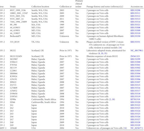
Virus Strain Collection location Collection yr
Primary clinical
isolate Passage history and notes (reference[s]) Accession no.
HSV-2 8937_1999_3336 Seattle, WA, USA 2003 Yes 2 passages on Vero cells KR135298
HSV-2 10883_2001_13347 Seattle, WA, USA 2005 Yes 2 passages on Vero cells KR135311
HSV-2 9335_2005_576 Seattle, WA, USA 2009 Yes 2 passages on Vero cells KR135312
HSV-2 9335_2007_14 Seattle, WA, USA 2011 Yes 2 passages on Vero cells KR135313
HSV-2 7444_1996_25809 Seattle, WA, USA 1996 Yes 2 passages on Vero cells KR135314
HSV-2 89_390 MA, USA 1989 Yes 3 passages on Vero cells KR135321
HSV-2 44_619833 MD, USA 2007 Yes 2 passages on Vero cells KR135308
HSV-2 44_419851 MD, USA 2007 Yes 2 passages on Vero cells KR135309
HSV-2 44_319857 MD, USA 2007 Yes 2 passages on Vero cells KR135310
HSV-2 BethesdaP5 MD, USA Unknown Yes 4 passages on human diploid fibroblasts
(MRC5 cells)
KR135330
HSV-2 333_R519 TX, USA Unknown No Plaque-purified version of HSV-2 strain
333; unknown no. of passages on Vero cells; virulent in animal models (16)
KR135331
HSV-2 HG52 Scotland, UK Prior to 1971 No Laboratory-adapted strain, attentuated for
virulence (8,10,35)
NC_001798.1
HSV-2 HG52 ILMN Scotland, UK Prior to 1971 No Illumina sequence of strain HG52 JN561323
HSV-2 M22987 Rakai, Uganda 2007 Yes 2 passages on Vero cells KR135299
HSV-2 D30613 Rakai, Uganda 2007 Yes 2 passages on Vero cells KR135300
HSV-2 F70764 Rakai, Uganda 2007 Yes 2 passages on Vero cells KR135301
HSV-2 M1119 Rakai, Uganda 2007 Yes 2 passages on Vero cells KR135302
HSV-2 L22861 Rakai, Uganda 2007 Yes 2 passages on Vero cells KR135303
HSV-2 H00066 Rakai, Uganda 2007 Yes 2 passages on Vero cells KR135304
HSV-2 K39924 Rakai, Uganda 2007 Yes 2 passages on Vero cells KR135305
HSV-2 A76191 Rakai, Uganda 2007 Yes 2 passages on Vero cells KR135306
HSV-2 J09622 Rakai, Uganda 2008 Yes 2 passages on Vero cells KR135307
HSV-2 J32715 Rakai, Uganda 2007 Yes 2 passages on Vero cells KR135315
HSV-2 G75809 Rakai, Uganda 2007 Yes 2 passages on Vero cells KR135316
HSV-2 A76832 Rakai, Uganda 2007 Yes 2 passages on Vero cells KR135317
HSV-2 D39650 Rakai, Uganda 2008 Yes 2 passages on Vero cells KR135318
HSV-2 D39765 Rakai, Uganda 2008 Yes 2 passages on Vero cells KR135319
HSV-2 SD90e Carletonville, South Africa 1994 Yes 3 passages on Vero cells (2,10) KF781518
HSV-2 SD66 Carletonville, South Africa 1994 Yes 3 passages on Vero cells KR135320
HSV-2 JA1 Japan 2009 Yes 2 passages on Vero cells KR135322
HSV-2 JA2 Japan 2010 Yes 2 passages on Vero cells KR135323
HSV-2 JA3 Japan 2008 Yes 2 passages on Vero cells KR135324
HSV-2 JA5 Japan 2009 Yes 2 passages on Vero cells KR135325
HSV-2 JA6 Japan 2009 Yes 2 passages on Vero cells KR135326
HSV-2 JA7 Japan 2010 Yes 2 passages on Vero cells KR135327
HSV-2 JA8 Japan 2009 Yes 2 passages on Vero cells KR135328
HSV-2 JA9 Japan 2010 Yes 2 passages on Vero cells KR135329
ChHV-1 105640 USA 2004 Yes Unknown no. of passages on Vero cells (14) NC_023677.1
on November 7, 2019 by guest
http://jvi.asm.org/
[image:3.585.46.546.82.538.2]tently lowPvalues among the RDP’s multiple tests for recombination were subjected to further analysis. Simplot (28) was used to apply a boot-scanning approach to full-length sequences using the following parame-ters: 1,000-bp window, 1,000-bp step size, GapStrip:on, 100 repetitions, and F84 (maximum likelihood)T/tof 2.0. Highly related sequences were grouped to reduce phylogenetic noise during boot scanning. Groups were defined by phylogenetic analysis and a significant bootstrap of⬎90%. The groups included 13 genomes from Uganda (Uganda clade; strains M22987, D30613, F70764, M1119, L22861, H00066, A76191, J09622, J32715, G75809, A76832, D39650, and D39765), 4 genomes from Japan (Japan clade; strains JA2, JA3, JA6, and JA9), 2 genomes from the United States (US clade; 9335_2005_576 and 9335_2007_14), 2 genomes from Uganda and the United States (UG_US clade; K39924 and 44_619833), and 5 genomes from the United States and South Africa (US_ZA clade; 89_390, 44_419851, 333_9519, SD90e, and SD66). Recombination signal in Simplot was considered positive at a cutoff of 70% (29).
Nucleotide sequence accession numbers.The sequences of the 34 HSV-2 isolates described were submitted to GenBank under the accession numbers given inTable 1.
RESULTS
Genomic sequencing and assembly.
We performed
high-throughput, paired-end Illumina sequencing of purified,
ran-domly fragmented viral DNA with read lengths of 101 bp.
Refer-ence-assisted assembly of the genomes of 34 HSV-2 isolates
gathered for this study (
Table 1
) generated contig sequence
span-ning the U
Land U
Sregions of the HSV-2 genome, as well as single
copies of each of the long and short inverted-repeat regions (R
Land R
S). Average read coverage for these genomes ranged from
3,100- to 9,300-fold. The contigs were aligned and combined into
single genomes using the HG52 reference genome (
NC_001798
)
as a scaffold. As has been reported for other recent HSV-1 and
HSV-2 genome sequences (
2
,
6
), Illumina sequencing was unable
to distinguish individual copies of the inverted-repeat regions and
could not efficiently resolve all of the small repeat regions between
HSV-2 coding domains and within the R
Land R
Sterminal repeats
that characterize HSV-2. Generation of a second copy of the R
Land R
Sinverted repeats bounding their respective unique
se-quences was therefore accomplished by inverting a copy of each
sequence in the final assemblies. The repeat structure of several of
the regions flanking R
Land R
Sresulted in low read depth in these
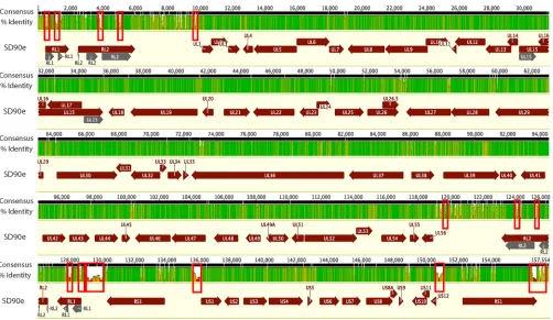
regions and led to gaps in the genome assemblies. Because of the
inability of automatic alignment algorithms to consistently handle
small insertions and deletions in these problematic regions and to
increase the quality of the alignments used for subsequent
analy-ses, these regions (
Fig. 1
, red boxes, and data not shown) were
removed from the assemblies, and trimmed versions of the
ge-nomes were used for alignment and phylogenetic analysis. As with
previous HSV-2 genomes (
2
), numerous base substitutions and
insertions/deletions (indels) were detected in the aligned
se-quences. No large indels were observed, however.
Alignment and genomic diversity.
The generation of 34
addi-tional nearly full-length HSV-2 genome sequences provided us
with the opportunity to assess the relatedness of HSV-2 strains
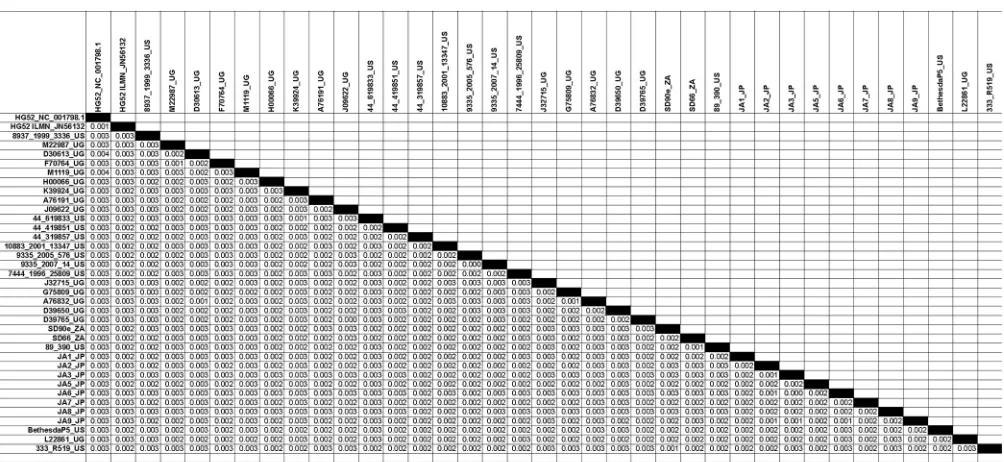
circulating in the United States, Africa, and Asia. Pairwise distance
measurements of the 34 new genomes and the 2 previously
re-FIG 1Overview of the features of the HSV-2 genome and sequence diversity in a genome alignment of 34 full genome sequences. The black bar at the top represents a consensus sequence drawn from the alignment of the genomes of 34 HSV-2 strains. The identity plot derived from this alignment, shown below, is colored as follows: green, 100% identity; green-brown, 30 to⬍100% identity; red,⬍30% identity. Below the identity plot, HSV-2 coding regions are shown, with arrows indicating the direction of the reading frame. The red boxes indicate regions of the multigenome alignment that either contained gaps or failed to align properly. These regions were deleted from the genome sequences in subsequent analyses.
on November 7, 2019 by guest
http://jvi.asm.org/
[image:4.585.43.545.63.354.2]ported genomes (
Table 1
) indicated that all the genomes were
closely related to each other, as well as to HSV-2 HG52 and HSV-2
SD90e, with the maximum nucleotide divergence between strains
being 0.4% across the genome (
Table 2
).
To compare these geographically diverse HSV-2 genomes, we
first assessed the levels of DNA diversity of our sequenced strains,
along with the two existing HG52 sequences, across the genome
compared to the low-passage-number strain SD90e (
Fig. 1
). We
noted that the genomes were largely conserved within the U
Land
U
S, with the highest variation observed in the intergenic regions.
Regions with the highest levels of variation (
⬎
70%) were localized
to known repetitive regions flanking the large internal and
termi-nal repeat regions (
Fig. 1
). This clustering of variation could be
attributed to the inherent variation in these regions, as well as to
difficulties in sequencing and assembling these problematic
re-gions with current deep-sequencing technologies.
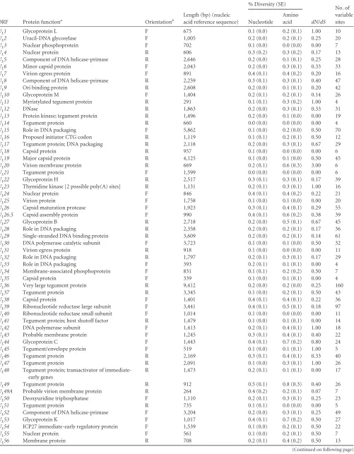
Analysis of nucleotide and amino acid diversity among these
HSV-2 genomes and within 70 U
Land U
SORFs confirmed
high-level sequence conservation, with no pair of ORFs exceeding 0.5%
diversity at the nucleotide level and 0.8% diversity at the amino
acid level (
Table 3
). R
Land R
SORFs were incomplete in a number
of genomes, so they were not included in this analysis. Only one
ORF exhibited nucleotide diversity levels over 0.4% (
UL49
), and
ten ORFs showed amino acid diversity greater than 0.4% (
U
L20
,
UL26.5
,
UL27
,
UL39
,
UL44
,
UL49
,
UL53
,
US7
,
US4
, and
US11
).
We also examined the ratio of nonsynonymous to
synony-mous substitutions (
dN
/
dS
) to detect evidence of selection
pres-sure within HSV-2 ORFs (
Table 3
). We found that while the
ma-jority of ORFs appeared to be under negative, purifying selection
(
dN
/
dS
⬍
1), several ORFs (
U
L1
,
U
L11
,
U
L23
,
U
L42
,
U
L45
,
U
L47
,
US4
,
US8
, and
US11
) showed more evidence of neutral selection
(
dN
/
dS
⫽
1) (
Table 3
). One ORF (
U
L20
) appeared to show
evi-dence of positive selection (
dN
/
dS
⬎
1), although the relatively
small number of variable sites present in the ORF makes
interpre-tation difficult.
Because nearly all of the 34 new genomes were from
low-pas-sage-number isolates, we compared the nucleotide and amino
acid divergences of their ORFs from those of the HG52 laboratory
strain (both the Sanger RefSeq [
NC_001798.1
] and corrected
Il-lumina [
JN561323
] sequences) and from the SD90e
low-passage-number clinical strain (
KF781518
) (
Table 4
). We found that
pair-wise divergence of these genomes from both HG52 sequences and
SD90e ranged from 0 to 1.1% at the nucleotide level and 0 to 2.1%
at the amino acid level (
Table 4
). Amino acid divergence between
the HSV-2 strains was sometimes much higher than
correspond-ing nucleic acid divergence calculations. This is likely due to the
high GC content of HSV genes, with about 80% G or C occurring
at the 3rd codon position. This permits a biased codon usage for
HSV, with an effective codon usage of approximately 40 out of 61
different codons. These biases are expected to cause relatively low
nucleotide diversity for a given degree of amino acid diversity.
[image:5.585.41.543.78.309.2]In general, we saw that HG52 ILMN was more closely related to
the other 34 genomes than HG52 RefSeq when comparing either
individual ORFs or the average divergence for all ORFs (
Table 4
).
This was presumably a result of the sequencing errors in the
orig-inal RefSeq sequence. When we compared the divergence of ORF
amino acid sequence from SD90e and HG52 ILMN, we observed
that 25 of the ORFs in the 34 new sequences were more closely
related to SD90e than to HG52 ILMN, while 10 ORFs were closer
to HG52 ILMN. Most ORFs were only slightly more divergent
from one strain or the other, but four were noticeably different.
UL49
and
UL49A
were strikingly diverged from SD90e (1.1 and
1.2%, respectively), while two ORFs,
U
L11
and
U
S1
, were
signifi-cantly diverged from HG52 ILMN. The origin of the divergence in
these strains is not immediately obvious. Furthermore, the
aver-age divergence for all ORFs was greater for HG52 ILMN than for
TABLE 2Estimates of evolutionary divergence between HSV-2 genome sequencesa
a
The numbers of base substitutions per site between sequences are shown. Analyses were conducted using the Maximum Composite Likelihood model (30). The analysis involved 37 nucleotide sequences. All positions containing gaps and missing data were eliminated. There were a total of 148,894 positions in the final data set. Evolutionary analyses were conducted in MEGA5 (24).
on November 7, 2019 by guest
http://jvi.asm.org/
TABLE 3Nucleotide and amino acid diversity of HSV-2 strains in open reading frames
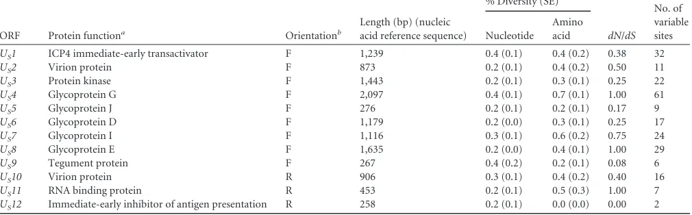
ORF Protein functiona Orientationb
Length (bp) (nucleic acid reference sequence)
% Diversity (SE)
dN/dS No. of variable sites Nucleotide
Amino acid
UL1 Glycoprotein L F 675 0.1 (0.0) 0.2 (0.1) 1.00 10
UL2 Uracil-DNA glycosylase F 1,005 0.2 (0.0) 0.2 (0.1) 0.25 20
UL3 Nuclear phosphoprotein F 702 0.1 (0.0) 0.0 (0.0) 0.00 7
UL4 Nuclear protein R 606 0.3 (0.2) 0.3 (0.2) 0.17 13
UL5 Component of DNA helicase-primase R 2,646 0.2 (0.0) 0.1 (0.1) 0.25 28
UL6 Minor capsid protein F 2,043 0.2 (0.0) 0.3 (0.1) 0.33 33
UL7 Virion egress protein F 891 0.4 (0.1) 0.4 (0.2) 0.20 16
UL8 Component of DNA helicase-primase R 2,259 0.3 (0.1) 0.3 (0.1) 0.40 47
UL9 Ori binding protein R 2,608 0.2 (0.0) 0.1 (0.1) 0.20 42
UL10 Glycoprotein M F 1,404 0.2 (0.1) 0.2 (0.1) 0.14 26
UL11 Myristylated tegument protein R 291 0.1 (0.1) 0.3 (0.2) 1.00 4
UL12 DNase R 1,863 0.2 (0.0) 0.3 (0.1) 0.33 31
UL13 Protein kinase; tegument protein R 1,496 0.2 (0.0) 0.1 (0.0) 0.00 19
UL14 Tegument protein R 660 0.0 (0.0) 0.0 (0.0) 0.00 4
UL15 Role in DNA packaging F 5,862 0.1 (0.0) 0.2 (0.0) 0.50 70
UL16 Proposed initiator CTG codon R 1,119 0.1 (0.1) 0.2 (0.1) 0.50 12
UL17 Tegument protein; DNA packaging R 2,118 0.2 (0.0) 0.3 (0.1) 0.67 29
UL18 Capsid protein R 957 0.1 (0.0) 0.0 (0.0) 0.00 6
UL19 Major capsid protein R 4,125 0.1 (0.0) 0.1 (0.0) 0.50 45
UL20 Virion membrane protein R 669 0.2 (0.1) 0.6 (0.3) 3.00 6
UL21 Tegument protein F 1,599 0.0 (0.0) 0.0 (0.0) 0.00 6
UL22 Glycoprotein H R 2,517 0.3 (0.1) 0.3 (0.1) 0.17 39
UL23 Thymidine kinase [2 possible poly(A) sites] R 1,131 0.2 (0.1) 0.3 (0.1) 1.00 16
UL24 Nuclear protein F 846 0.4 (0.1) 0.4 (0.2) 0.22 21
UL25 Virion protein F 1,758 0.1 (0.0) 0.1 (0.0) 0.00 20
UL26 Capsid maturation protease F 1,923 0.3 (0.1) 0.4 (0.1) 0.29 55
UL26.5 Capsid assembly protein F 990 0.4 (0.1) 0.6 (0.2) 0.38 39
UL27 Glycoprotein B R 2,718 0.2 (0.0) 0.5 (0.1) 0.67 45
UL28 Role in DNA packaging R 2,358 0.2 (0.0) 0.2 (0.1) 0.17 36
UL29 Single-stranded DNA binding protein R 3,609 0.2 (0.0) 0.2 (0.1) 0.14 61
UL30 DNA polymerase catalytic subunit F 3,723 0.1 (0.0) 0.1 (0.0) 0.50 32
UL31 Virion egress protein R 918 0.1 (0.0) 0.0 (0.0) 0.00 11
UL32 Role in DNA packaging R 1,797 0.2 (0.1) 0.3 (0.1) 0.17 29
UL33 Role in DNA packaging F 393 0.2 (0.1) 0.1 (0.1) 0.00 4
UL34 Membrane-associated phosphoprotein F 831 0.1 (0.1) 0.2 (0.2) 0.50 7
UL35 Capsid protein F 339 0.1 (0.0) 0.1 (0.1) 0.00 4
UL36 Very large tegument protein R 9,412 0.2 (0.0) 0.2 (0.0) 0.25 160
UL37 Tegument protein R 3,345 0.1 (0.0) 0.2 (0.1) 0.50 43
UL38 Capsid protein F 1,401 0.4 (0.1) 0.4 (0.1) 0.22 36
UL39 Ribonucleotide reductase large subunit F 3,441 0.4 (0.1) 0.5 (0.1) 0.18 97
UL40 Ribonucleotide reductase small subunit F 1,014 0.1 (0.0) 0.0 (0.0) 0.00 11
UL41 Tegument protein; host shutoff factor R 1,479 0.1 (0.0) 0.1 (0.1) 0.00 14
UL42 DNA polymerase subunit F 1,413 0.2 (0.1) 0.4 (0.1) 1.00 18
UL43 Probable membrane protein F 1,245 0.3 (0.1) 0.4 (0.1) 0.40 22
UL44 Glycoprotein C F 1,443 0.4 (0.1) 0.7 (0.2) 0.80 24
UL45 Tegument/envelope protein F 519 0.1 (0.0) 0.1 (0.1) 1.00 5
UL46 Tegument protein R 2,169 0.3 (0.1) 0.4 (0.1) 0.33 40
UL47 Tegument protein R 2,091 0.1 (0.0) 0.3 (0.1) 1.00 26
UL48 Tegument protein; transactivator of immediate-early genes
R 1,473 0.2 (0.1) 0.1 (0.1) 0.00 17
UL49 Tegument protein R 912 0.5 (0.1) 0.8 (0.3) 0.40 26
UL49A Probable virion membrane protein R 264 0.4 (0.2) 0.2 (0.1) 0.07 7
UL50 Deoxyuridine triphosphatase F 1,110 0.2 (0.1) 0.3 (0.1) 0.25 23
UL51 Tegument protein R 735 0.1 (0.1) 0.0 (0.0) 0.00 5
UL52 Component of DNA helicase-primase F 3,204 0.2 (0.0) 0.3 (0.1) 0.25 49
UL53 Glycoprotein K F 1,017 0.4 (0.1) 0.7 (0.2) 0.50 27
UL54 ICP27 immediate-early regulatory protein F 1,539 0.1 (0.0) 0.2 (0.1) 0.50 22
UL55 Nuclear protein F 561 0.1 (0.0) 0.2 (0.1) 0.50 7
UL56 Membrane protein R 708 0.2 (0.1) 0.4 (0.2) 0.50 13
(Continued on following page)
on November 7, 2019 by guest
http://jvi.asm.org/
SD90e; therefore, the 34 new genomes are more closely related to
SD90e than to HG52 ILMN.
To determine if the levels of diversity and divergence seen in
the ORFs of these 34 HSV-2 genomes were reflected in a larger
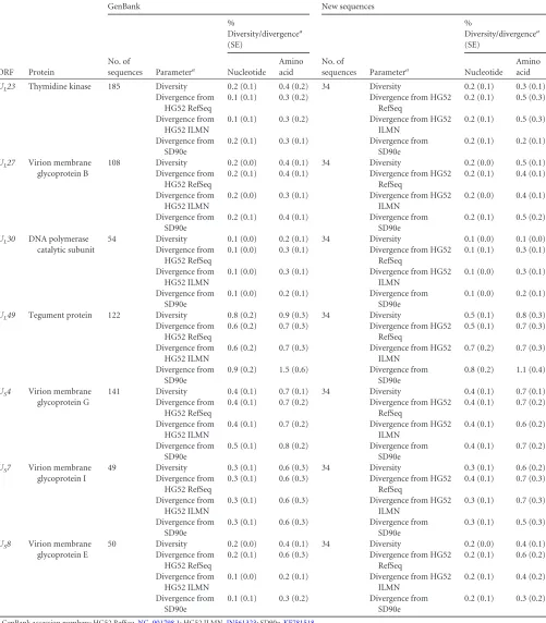
data set, we calculated the levels of diversity in the GenBank
se-quences available for 7 ORFs (
Table 5
), as well as the levels of
divergence from the HG52 RefSeq, HG52 ILMN, and SD90e
strains. We observed that the nucleotide and amino acid
diversi-ties in the larger, independent sequence sets were similar to what
we had observed for the 34 full-length HSV-2 genomes examined.
While larger numbers of sequences for ORFs that were most
divergent from HG52 (
UL11
and
US1
) were not available in
GenBank, the levels of amino acid divergence of the available
ORFs from the two HG52 sequences were similar to those seen for
our 34 new sequences. An additional 122 sequences were available
for one of the ORFs that displayed
⬎
1% amino acid divergence
from SD90e (
U
L49
). Again, we found that divergence of these
UL49
GenBank sequences was greater for SD90e than for HG52
ILMN (
Table 5
).
Analysis of HSV-2 recombination.
To determine if HSV-2
genomes display the extensive recombination reported for HSV-1
sequences (
6
,
30
,
31
), we employed boot-scanning and
phyloge-netic analyses of full-length HSV-2 strain alignments. First, to
confirm that we could detect recombination, we performed our
analysis on HSV-1 and were able to detect recombination
cross-over events cross-over large segments (2,500 to 8,500 bp) at levels
com-parable to those previously seen (references
6
,
30
, and
31
and
results not shown). In contrast, our analysis of recombination in
HSV-2 showed only five major crossover events, with detectable
recombination seen only over small segments of the aligned
se-quences (700 to 1,170 bp) (
Fig. 2
). To confirm the recombination
signals observed in the HSV-2 boot scans, we performed
phyloge-netic analysis of these five small regions between recombination
breakpoints. These analyses showed few highly supported
branches and could confirm potential recombination over 1,170
bp and 700 bp between HG52 and two U.S. strains, BethesdaP5
and 8937_1999_3336, respectively (data not shown). The weak
signal for recombination in HSV-2 suggested either that
recombi-nation does not occur in HSV-2 genomes as frequently as is seen in
HSV-1 or that the high level of sequence similarity among HSV-2
genome sequences makes lateral gene transfer difficult to detect.
Additional analyses using a variety of methods to confirm the lack
of recombination in these HSV-2 strains are described in more
detail in the accompanying paper by
Lamers et al.
(
32
).
Phylogenetic analysis.
Alignment and subsequent
phyloge-netic analysis of the newly sequenced HSV-2 genomes with
exist-ing HSV-2 sequences usexist-ing the ChHV genome sequence as an
outgroup allowed us to determine the relationship between these
geographically distinct HSV-2 strains. As expected, there was
dis-tinct clustering of HSV-2 sequences away from ChHV sequences
in the whole-genome phylogeny (
Fig. 3
). The dendrogram also
showed close relationship among all HSV-2 sequences regardless
of geographic origin. This was in contrast to HSV-1 genome
se-quences, which exhibit robust geographical clustering (
6
). While
clinical HSV-2 strains isolated in Uganda and Japan tended to
cluster together, there was very low bootstrap support for this
clustering, indicating a lack of strong phylogenetic evidence for
grouping of these strains. Similarly, the grouping of HSV-2 strains
isolated from the United States with strains from South Africa also
had low bootstrap support.
To further explore the relationship between geographically
di-verse HSV-2 sequences, we generated a dendrogram of full-length
and nearly full-length HSV-2 sequences (
Fig. 4
). The complete
genome tree recapitulated the clustering of Uganda and Japan
sequences into separate branches and again showed loose
associ-ation of U.S. and South Africa sequences. Similar results were
observed with analyses of U
Lor U
Sregions alone (results not
shown). However, in all phylogenetic analyses, there was strong
support (
⬎
65% bootstrap value in multiple genomic regions) for
the relatedness of U.S. sequence 44_619833 and Uganda sequence
K39924_UG.
DISCUSSION
[image:7.585.41.546.84.241.2]The availability of a large number of nearly complete genome
se-quences from low-passage-number clinical isolates of HSV-2 allowed
us to explore the sequence evolution and diversity of
low-passage-number virus strains isolated from Asia, Africa, and the United States
to infer viral evolution and potential viral determinants of
pathoge-nicity and disease outcome. Here, we report the sequencing and
as-sembly of 34 additional strains, 33 low-passage-number clinical
TABLE 3(Continued)
ORF Protein functiona Orientationb
Length (bp) (nucleic acid reference sequence)
% Diversity (SE)
dN/dS No. of variable sites Nucleotide
Amino acid
US1 ICP4 immediate-early transactivator F 1,239 0.4 (0.1) 0.4 (0.2) 0.38 32
US2 Virion protein F 873 0.2 (0.1) 0.4 (0.2) 0.50 11
US3 Protein kinase F 1,443 0.2 (0.1) 0.3 (0.1) 0.25 22
US4 Glycoprotein G F 2,097 0.4 (0.1) 0.7 (0.1) 1.00 61
US5 Glycoprotein J F 276 0.2 (0.1) 0.2 (0.1) 0.17 9
US6 Glycoprotein D F 1,179 0.2 (0.0) 0.3 (0.1) 0.25 17
US7 Glycoprotein I F 1,116 0.3 (0.1) 0.6 (0.2) 0.75 24
US8 Glycoprotein E F 1,635 0.2 (0.0) 0.4 (0.1) 1.00 29
US9 Tegument protein F 267 0.4 (0.2) 0.2 (0.1) 0.08 6
US10 Virion protein R 906 0.3 (0.1) 0.4 (0.2) 0.40 16
US11 RNA binding protein R 453 0.2 (0.1) 0.5 (0.3) 1.00 7
US12 Immediate-early inhibitor of antigen presentation R 258 0.2 (0.1) 0.0 (0.0) 0.00 2
aReference1. b
F, forward; R, reverse.
on November 7, 2019 by guest
http://jvi.asm.org/
TABLE 4Nucleotide and amino acid divergence of HSV-2 strains in open reading frames
ORF Protein functiona
% Nucleotide divergence from: % Amino acid divergence from:
HG52 RefSeqb
HG52
ILMNc SD90ed
HG52 RefSeqb
HG52
ILMNc SD90ed
UL1 Glycoprotein L 0.1 (0.0) 0.1 (0.0) 0.1 (0.0) 0.1 (0.0) 0.1 (0.0) 0.1 (0.0)
UL2 Uracil-DNA glycosylase 0.5 (0.2) 0.5 (0.2) 0.4 (0.2) 0.5 (0.3) 0.4 (0.3) 0.1 (0.1)
UL3 Nuclear phosphoprotein 0.0 (0.0) 0.0 (0.0) 0.0 (0.0) 0.0 (0.0) 0.0 (0.0) 0.0 (0.0)
UL4 Nuclear protein 0.2 (0.1) 0.2 (0.1) 0.2 (0.1) 0.2 (0.1) 0.2 (0.1) 0.2 (0.1)
UL5 Component of DNA helicase-primase 0.2 (0.1) 0.2 (0.1) 0.1 (0.0) 0.1 (0.0) 0.1 (0.0) 0.1 (0.0)
UL6 Minor capsid protein 0.6 (0.2) 0.2 (0.1) 0.2 (0.1) 0.6 (0.2) 0.4 (0.2) 0.2 (0.1)
UL7 Virion egress protein 0.3 (0.1) 0.3 (0.1) 0.3 (0.1) 0.3 (0.2) 0.3 (0.2) 0.2 (0.1)
UL8 Component of DNA helicase-primase 0.5 (0.1) 0.3 (0.1) 0.3 (0.1) 0.8 (0.2) 0.5 (0.2) 0.3 (0.1)
UL9 Ori binding protein 0.2 (0.1) 0.1 (0.0) 0.2 (0.1) 0.2 (0.1) 0.1 (0.1) 0.1 (0.1)
UL10 Glycoprotein M 0.3 (0.1) 0.2 (0.1) 0.1 (0.0) 0.3 (0.2) 0.3 (0.2) 0.1 (0.0)
UL11 Myristylated tegument protein 1.1 (0.5) 1.0 (0.6) 0.1 (0.0) 2.1 (1.4) 2.1 (1.5) 0.2 (0.1)
UL12 DNase 0.3 (0.1) 0.3 (0.1) 0.1 (0.0) 0.4 (0.3) 0.3 (0.2) 0.2 (0.1)
UL13 Protein kinase; tegument protein 0.3 (0.1) 0.2 (0.1) 0.1 (0.1) 0.3 (0.2) 0.2 (0.2) 0.0 (0.0)
UL14 Tegument protein 0.0 (0.0) 0.0 (0.0) 0.0 (0.0) 0.0 (0.0) 0.0 (0.0) 0.0 (0.0)
UL15 Role in DNA packaging 0.1 (0.0) 0.1 (0.0) 0.1 (0.0) 0.2 (0.1) 0.2 (0.1) 0.2 (0.1)
UL16 Proposed initiator CTG codon 0.1 (0.1) 0.1 (0.0) 0.1 (0.0) 0.1 (0.0) 0.1 (0.0) 0.1 (0.0)
UL17 Tegument protein; DNA packaging 0.3 (0.1) 0.2 (0.1) 0.2 (0.0) 0.3 (0.1) 0.2 (0.1) 0.2 (0.1)
UL18 Capsid protein 0.1 (0.1) 0.1 (0.1) 0.1 (0.1) 0.2 (0.0) 0.0 (0.0) 0.0 (0.0)
UL19 Major capsid protein 0.3 (0.1) 0.1 (0.0) 0.2 (0.0) 0.5 (0.1) 0.2 (0.1) 0.1 (0.0)
UL20 Virion membrane protein 0.3 (0.2) 0.3 (0.2) 0.1 (0.1) 0.4 (0.2) 0.4 (0.2) 0.4 (0.2)
UL21 Tegument protein 0.0 (0.0) 0.0 (0.0) 0.0 (0.0) 0.0 (0.0) 0.0 (0.0) 0.0 (0.0)
UL22 Glycoprotein H 0.3 (0.1) 0.3 (0.1) 0.3 (0.1) 0.3 (0.1) 0.3 (0.1) 0.2 (0.1)
UL23 Thymidine kinase [2 possible poly(A) sites] 0.2 (0.1) 0.2 (0.1) 0.2 (0.1) 0.5 (0.3) 0.5 (0.3) 0.2 (0.1)
UL24 Nuclear protein 0.2 (0.1) 0.2 (0.1) 0.2 (0.1) 0.2 (0.1) 0.2 (0.1) 0.2 (0.1)
UL25 Virion protein 0.2 (0.1) 0.1 (0.1) 0.0 (0.0) 0.3 (0.2) 0.2 (0.2) 0.0 (0.0)
UL26 Capsid maturation protease 0.3 (0.1) 0.3 (0.1) 0.2 (0.1) 0.4 (0.2) 0.4 (0.2) 0.2 (0.1)
UL26.5 Capsid assembly protein 0.4 (0.2) 0.4 (0.2) 0.3 (0.1) 0.8 (0.4) 0.8 (0.4) 0.3 (0.1)
UL27 Glycoprotein B 0.2 (0.1) 0.2 (0.0) 0.2 (0.1) 0.4 (0.1) 0.4 (0.1) 0.5 (0.2)
UL28 Role in DNA packaging 0.2 (0.1) 0.2 (0.1) 0.1 (0.0) 0.2 (0.1) 0.2 (0.1) 0.1 (0.0)
UL29 Single-stranded DNA binding protein 0.3 (0.1) 0.3 (0.1) 0.2 (0.1) 0.2 (0.1) 0.2 (0.1) 0.2 (0.1)
UL30 DNA polymerase catalytic subunit 0.1 (0.1) 0.1 (0.0) 0.1 (0.0) 0.3 (0.1) 0.3 (0.1) 0.2 (0.1)
UL31 Virion egress protein 0.1 (0.1) 0.1 (0.1) 0.1 (0.1) 0.0 (0.0) 0.0 (0.0) 0.0 (0.0)
UL32 Role in DNA packaging 0.3 (0.1) 0.2 (0.1) 0.2 (0.1) 0.4 (0.2) 0.3 (0.1) 0.2 (0.1)
UL33 Role in DNA packaging 0.2 (0.2) 0.2 (0.1) 0.2 (0.2) 0.0 (0.0) 0.0 (0.0) 0.0 (0.0)
UL34 Membrane-associated phosphoprotein 0.1 (0.0) 0.2 (0.1) 0.1 (0.1) 0.1 (0.1) 0.5 (0.4) 0.3 (0.2)
UL35 Capsid protein 0.0 (0.0) 0.0 (0.0) 0.0 (0.0) 0.0 (0.0) 0.0 (0.0) 0.0 (0.0)
UL36 Very large tegument protein 0.2 (0.0) 0.2 (0.0) 0.2 (0.0) 0.3 (0.1) 0.2 (0.1) 0.2 (0.0)
UL37 Tegument protein 0.2 (0.1) 0.1 (0.0) 0.1 (0.0) 0.3 (0.1) 0.2 (0.1) 0.2 (0.1)
UL38 Capsid protein 0.7 (0.1) 0.3 (0.1) 0.4 (0.1) 1.0 (0.3) 0.4 (0.2) 0.6 (0.3)
UL39 Ribonucleotide reductase large subunit 0.4 (0.1) 0.8 (0.1) 0.4 (0.1) 0.7 (0.2) 0.7 (0.2) 0.4 (0.1)
UL40 Ribonucleotide reductase small subunit 0.1 (0.0) 0.1 (0.0) 0.1 (0.0) 0.0 (0.0) 0.0 (0.0) 0.0 (0.0)
UL41 Tegument protein; host shutoff factor 0.1 (0.0) 0.1 (0.0) 0.3 (0.1) 0.0 (0.0) 0.0 (0.0) 0.2 (0.2)
UL42 DNA polymerase subunit 0.3 (0.1) 0.3 (0.1) 0.2 (0.1) 0.5 (0.3) 0.5 (0.2) 0.5 (0.2)
UL43 Probable membrane protein 0.2 (0.1) 0.2 (0.1) 0.2 (0.0) 0.4 (0.2) 0.4 (0.2) 0.2 (0.1)
UL44 Glycoprotein C 0.4 (0.1) 0.3 (0.1) 0.4 (0.1) 0.5 (0.2) 0.5 (0.2) 0.6 (0.2)
UL45 Tegument/envelope protein 0.1 (0.1) 0.0 (0.0) 0.0 (0.0) 0.4 (0.3) 0.0 (0.0) 0.0 (0.0)
UL46 Tegument protein 0.6 (0.1) 0.4 (0.1) 0.5 (0.1) 0.7 (0.2) 0.5 (0.2) 0.5 (0.2)
UL47 Tegument protein 0.1 (0.0) 0.1 (0.0) 0.1 (0.0) 0.2 (0.1) 0.2 (0.1) 0.2 (0.1)
UL48 Tegument protein; transactivator of immediate-early genes
0.1 (0.1) 0.1 (0.0) 0.2 (0.1) 0.0 (0.0) 0.0 (0.0) 0.0 (0.0)
UL49 Tegument protein 0.5 (0.1) 0.7 (0.2) 0.8 (0.2) 0.7 (0.3) 0.7 (0.3) 1.1 (0.4)
UL49A Probable virion membrane protein 0.5 (0.3) 0.4 (0.3) 1.0 (0.6) 0.1 (0.1) 0.1 (0.1) 1.2 (1.1)
UL50 Deoxyuridine triphosphatase 0.2 (0.1) 0.2 (0.1) 0.2 (0.1) 0.3 (0.2) 0.3 (0.2) 0.3 (0.2)
UL51 Tegument protein 0.1 (0.1) 0.1 (0.1) 0.1 (0.1) 0.0 (0.0) 0.0 (0.0) 0.0 (0.0)
UL52 Component of DNA helicase-primase 0.3 (0.1) 0.2 (0.1) 0.2 (0.1) 0.4 (0.2) 0.4 (0.1) 0.3 (0.1)
UL53 Glycoprotein K 0.4 (0.1) 0.4 (0.1) 0.4 (0.2) 0.7 (0.3) 0.5 (0.2) 0.7 (0.3)
UL54 ICP27 immediate-early regulatory protein 0.1 (0.0) 0.1 (0.0) 0.1 (0.1) 0.2 (0.1) 0.1 (0.0) 0.1 (0.0)
UL55 Nuclear protein 0.1 (0.1) 0.1 (0.0) 0.1 (0.0) 0.1 (0.0) 0.1 (0.1) 0.1 (0.1)
UL56 Membrane protein 0.1 (0.1) 0.1 (0.0) 0.5 (0.2) 0.2 (0.1) 0.2 (0.1) 1.0 (0.6)
(Continued on following page)
on November 7, 2019 by guest
http://jvi.asm.org/
strains and 1 laboratory strain, isolated in the United States, Uganda,
South Africa, and Japan. Illumina sequencing of these samples
gen-erated high-quality, nearly complete genome assemblies of the
unique regions of the genome. However, as has been previously
re-ported for both HSV-1 and HSV-2 (
2
,
6
), accurate sequences of both
copies of the terminal and internal large repeat regions (R
Land R
S)
and of intergenic repetitive regions proved difficult. Further
sequenc-ing of the terminal and internal repeat regions with additional
meth-ods, such as single-molecule, long-read sequencing technology, as has
been done for another
Herpesviridae
family member, pseudorabies
virus (PRV), may allow single-base resolution of these difficult
regions of the genome (
33
).
Sequence diversity of HSV-2 isolates.
These newly sequenced
HSV-2 strains showed remarkable sequence conservation,
regard-less of geographic origin. The level of HSV-2 diversity for the 34
full-length genomes was less than reported previously for HSV-1
(
6
), and similar levels of diversity were observed for 7 specific
HSV-2 genes in the larger GenBank database. However, as in
pre-vious studies, problematic areas in or near repeat regions of the
genome were excluded from our analyses due to technical
diffi-culties in sequencing and assembling genome repeats. Because
these regions may represent locations of real biological variability
(
34
), it is likely that future advances in genome sequencing and
assembly technology could accurately fill in these missing regions,
and these improvements could highlight additional diversity
within these genomes.
Although the HSV-2 sequences were generally highly
con-served, certain ORFs showed higher diversity.
U
L49
was more
di-verse than the other ORFs (0.8% at the amino acid level). In
ad-dition, certain ORFs were divergent in specific strains. For
example,
UL49
and
UL49A
showed increased divergence from
SD90e, while
U
L11
and
U
S1
showed increased divergence from
HG52. The origin of the divergence in these ORFs remains to be
defined.
The lower level of diversity in HSV-2 than in HSV-1 has
impli-cations for viral evolution. The decreased diversity seen in HSV-2
is consistent with its diverging more recently than HSV-1 from
ChHV or another herpesvirus progenitor (
15
) but could also be
the result of a greater bottleneck during genital transmission than
in oral transmission or the lower prevalence of HSV-2. While all of
the strains sequenced here were passaged in cell culture prior to
preparation of viral DNA for sequencing, passage numbers were
kept low to minimize the potential accumulation of single
nucle-otide polymorphisms (SNPs) during cell culture. Previous
se-quence comparison of an HSV-2 low-passanumber viral
ge-nome with a derivative that had undergone plaque purification
revealed high levels of sequence conservation and minimal
changes to the viral genome (
2
); therefore, we do not anticipate
high numbers of cell-culture-associated SNPs in these genomes.
An understanding of viral diversity is also important for
vac-cine design. The high level of diversity in human
immunodefi-ciency virus type 1 (HIV-1) is one of the factors that have limited
the development of an effective vaccine. Therefore, the limited
genetic diversity of these 34 HSV-2 strains bodes well for the
po-tential of an HSV-2 vaccine to contain sufficient antigens to
pro-tect against these strains from around the world. Identification of
the key protective antigens will be necessary before this question
can be answered adequately.
Phylogenetic analysis of the full genome sequences of these
HSV-2 strains, as well as of unique regions of the genome, showed
a lack of robust support for geographic clustering. Norberg et al.
previously reported evidence for two genogroups, one from
iso-lates from Tanzania and one from isoiso-lates from Tanzania and
Scandinavia (
9
). This was based on removing isolates that showed
“conflicting phylogenetic signals” from the analysis. The
differ-ences between this study and our finding may be due to the
dif-ference between whole-genome analysis and individual gene
anal-ysis or to the removal of recombination from the genes being
analyzed in the Norberg et al. analysis. Further analysis of the
genes that show the greatest diversity is needed to determine
whether they represent distinct clades in HSV-2.
Recombination.
We found less evidence of recombination in
HSV-2 genomes than in HSV-1, although the low sequence
diver-sity may limit the ability to detect recombination in HSV-2.
Nor-TABLE 4(Continued)
ORF Protein functiona
% Nucleotide divergence from: % Amino acid divergence from:
HG52 RefSeqb
HG52
ILMNc SD90ed
HG52 RefSeqb
HG52
ILMNc SD90ed
US1 ICP4 immediate-early transactivator 0.7 (0.2) 0.5 (0.2) 0.3 (0.1) 1.0 (0.4) 0.7 (0.3) 0.3 (0.1)
US2 Virion protein 0.3 (0.1) 0.3 (0.1) 0.3 (0.1) 0.6 (0.4) 0.6 (0.3) 0.6 (0.4)
US3 Protein kinase 0.2 (0.1) 0.1 (0.0) 0.2 (0.1) 0.4 (0.2) 0.2 (0.1) 0.3 (0.2)
US4 Glycoprotein G 0.4 (0.1) 0.4 (0.1) 0.4 (0.1) 0.7 (0.2) 0.6 (0.2) 0.7 (0.2)
US5 Glycoprotein J 0.1 (0.0) 0.1 (0.1) 0.1 (0.1) 0.1 (0.1) 0.1 (0.1) 0.1 (0.1)
US6 Glycoprotein D 0.2 (0.0) 0.1 (0.0) 0.2 (0.1) 0.6 (0.2) 0.1 (0.0) 0.1 (0.0)
US7 Glycoprotein I 0.4 (0.1) 0.3 (0.1) 0.3 (0.1) 0.7 (0.3) 0.7 (0.3) 0.5 (0.3)
US8 Glycoprotein E 0.2 (0.1) 0.2 (0.1) 0.2 (0.1) 0.6 (0.2) 0.4 (0.2) 0.3 (0.2)
US9 Tegument protein 0.3 (0.2) 0.3 (0.2) 0.3 (0.2) 0.1 (0.1) 0.1 (0.1) 0.1 (0.1)
US10 Virion protein 0.3 (0.1) 0.3 (0.1) 0.2 (0.1) 0.6 (0.1) 0.6 (0.3) 0.5 (0.3)
US11 RNA binding protein 0.1 (0.1) 0.1 (0.1) 0.1 (0.1) 0.3 (0.2) 0.3 (0.2) 0.3 (0.2)
US12 Immediate-early inhibitor of antigen presentation 0.1 (0.1) 0.1 (0.1) 0.4 (0.3) 0.0 (0.0) 0.0 (0.0) 0.0 (0.0) Avg
divergence
0.26 (0.02) 0.22 (0.02) 0.21 (0.02) 0.36 (0.04) 0.30 (0.04) 0.25 (0.03)
aReference1. b
HG52 RefSeq accession no.NC_001798.1.
cHG52 ILMN accession no.JN561323. d
SD90e accession no.KF781518.
on November 7, 2019 by guest
http://jvi.asm.org/
[image:9.585.42.546.77.268.2]berg et al. (
9
) had reported significant recombination in HSV-2
through analysis of three glycoprotein genes. It is conceivable that
recombination is less for HSV-2 than HSV-1, but cell culture
stud-ies show equal frequencstud-ies of recombination between HSV-2
[image:10.585.43.544.79.651.2]mu-tants versus HSV-1 mumu-tants (C. Zhou and D. M. Knipe,
unpub-lished results). Thus, this is not likely to be the explanation for the
apparent low level of recombination evidenced in the HSV-2
strains. More likely, the low level of recombination is due to the
TABLE 5Diversity and divergence of GenBank sequences for select HSV-2 open reading frames
ORF Protein
GenBank New sequences
No. of
sequences Parametera
%
Diversity/divergencea (SE)
No. of
sequences Parametera
% Diversity/divergencea (SE) Nucleotide Amino acid Nucleotide Amino acid
UL23 Thymidine kinase 185 Diversity 0.2 (0.1) 0.4 (0.2) 34 Diversity 0.2 (0.1) 0.3 (0.1)
Divergence from HG52 RefSeq
0.1 (0.1) 0.3 (0.2) Divergence from HG52
RefSeq
0.2 (0.1) 0.5 (0.3)
Divergence from HG52 ILMN
0.1 (0.1) 0.3 (0.2) Divergence from HG52
ILMN
0.2 (0.1) 0.5 (0.3)
Divergence from SD90e
0.2 (0.1) 0.3 (0.1) Divergence from
SD90e
0.2 (0.1) 0.2 (0.1)
UL27 Virion membrane glycoprotein B
108 Diversity 0.2 (0.0) 0.4 (0.1) 34 Diversity 0.2 (0.0) 0.5 (0.1)
Divergence from HG52 RefSeq
0.2 (0.1) 0.4 (0.1) Divergence from HG52
RefSeq
0.2 (0.1) 0.4 (0.1)
Divergence from HG52 ILMN
0.2 (0.0) 0.3 (0.1) Divergence from HG52
ILMN
0.2 (0.0) 0.4 (0.1)
Divergence from SD90e
0.2 (0.1) 0.4 (0.1) Divergence from
SD90e
0.2 (0.1) 0.5 (0.2)
UL30 DNA polymerase catalytic subunit
54 Diversity 0.1 (0.0) 0.2 (0.1) 34 Diversity 0.1 (0.0) 0.1 (0.0)
Divergence from HG52 RefSeq
0.1 (0.0) 0.3 (0.1) Divergence from HG52
RefSeq
0.1 (0.1) 0.3 (0.1)
Divergence from HG52 ILMN
0.1 (0.0) 0.3 (0.1) Divergence from HG52
ILMN
0.1 (0.0) 0.3 (0.1)
Divergence from SD90e
0.1 (0.0) 0.2 (0.1) Divergence from
SD90e
0.1 (0.0) 0.2 (0.1)
UL49 Tegument protein 122 Diversity 0.8 (0.2) 0.9 (0.3) 34 Diversity 0.5 (0.1) 0.8 (0.3)
Divergence from HG52 RefSeq
0.6 (0.2) 0.7 (0.3) Divergence from HG52
RefSeq
0.5 (0.1) 0.7 (0.3)
Divergence from HG52 ILMN
0.6 (0.2) 0.7 (0.3) Divergence from HG52
ILMN
0.7 (0.2) 0.7 (0.3)
Divergence from SD90e
0.9 (0.2) 1.5 (0.6) Divergence from
SD90e
0.8 (0.2) 1.1 (0.4)
US4 Virion membrane glycoprotein G
141 Diversity 0.4 (0.1) 0.7 (0.1) 34 Diversity 0.4 (0.1) 0.7 (0.1)
Divergence from HG52 RefSeq
0.4 (0.1) 0.7 (0.2) Divergence from HG52
RefSeq
0.4 (0.1) 0.7 (0.2)
Divergence from HG52 ILMN
0.4 (0.1) 0.7 (0.2) Divergence from HG52
ILMN
0.4 (0.1) 0.6 (0.2)
Divergence from SD90e
0.5 (0.1) 0.8 (0.2) Divergence from
SD90e
0.4 (0.1) 0.7 (0.2)
US7 Virion membrane glycoprotein I
49 Diversity 0.3 (0.1) 0.6 (0.3) 34 Diversity 0.3 (0.1) 0.6 (0.2)
Divergence from HG52 RefSeq
0.3 (0.1) 0.6 (0.3) Divergence from HG52
RefSeq
0.4 (0.1) 0.7 (0.3)
Divergence from HG52 ILMN
0.3 (0.1) 0.6 (0.3) Divergence from HG52
ILMN
0.3 (0.1) 0.7 (0.3)
Divergence from SD90e
0.3 (0.1) 0.6 (0.3) Divergence from
SD90e
0.3 (0.1) 0.5 (0.3)
US8 Virion membrane glycoprotein E
50 Diversity 0.2 (0.0) 0.4 (0.1) 34 Diversity 0.2 (0.0) 0.4 (0.1)
Divergence from HG52 RefSeq
0.2 (0.1) 0.6 (0.3) Divergence from HG52
RefSeq
0.2 (0.1) 0.6 (0.2)
Divergence from HG52 ILMN
0.1 (0.0) 0.2 (0.1) Divergence from HG52
ILMN
0.2 (0.1) 0.4 (0.2)
Divergence from SD90e
0.1 (0.1) 0.3 (0.2) Divergence from
SD90e
0.2 (0.1) 0.3 (0.2)
aGenBank accession numbers: HG52 RefSeq,NC_001798.1; HG52 ILMN,JN561323; SD90e,KF781518.
on November 7, 2019 by guest
http://jvi.asm.org/
low level of genetic diversity, making recombination less
detect-able in the HSV-2 genomes.
HSV-2 reference genome.
The HSV-2 HG52 genome has
served as the reference genome because, until recently, it was the
only sequence available. However, HG52 is very attenuated in
animals relative to other HSV-2 strains (
10
,
35
). Upon sequencing
of the SD90e low-passage-number clinical isolate (GenBank
ac-cession no.
KF781518
), which shows pathogenicity in mice similar
FIG 2Boot-scanning analysis for evidence of recombination within HSV-2 strains. Shown is a boot-scanning plot of the HSV-2 reference sequence HG52 (accession no.NC_001798.1) versus all other HSV-2 full genome sequences. Thexaxis reflects the position in the aligned set of sequences, and theyaxis shows the percentage of permuted trees in which an individual HSV-2 strain clusters with the query. A recombination cutoff value of 70% is indicated by the dashed line. Positive signals for recombination with HG52 are indicated by a small circle at the peak recombination score and with the name of the strain that most closely resembles the query strain. Directly below the boot-scanning plot, the black lines indicate the recombinant breakpoint regions and their lengths.
FIG 3Phylogenetic relationships of HSV-2 genome sequences to that of chimpanzee herpesvirus. Shown is a maximum-likelihood tree of 34 nearly full-length HSV-2 genome nucleotide sequences generated as part of this study and the publicly available HSV-2 sequences for HG52 (accession no.NC_001798.1and
JN561323) and SD90e (accession no.KF781518), along with ChHV sequence from strain 105640 (accession no.NC_023677). Problematic regions from the multiple-sequence alignment were removed from all sequences. The tree is rooted using the ChHV sequence, and all horizontal branch lengths are drawn to a scale of nucleotide substitutions per site. Bootstrap resampling (1,000 replications) was performed. Bootstrap support values for each node, other than that separating ChHV from HSV-2 sequences, were⬍10% and are not shown on the tree.
on November 7, 2019 by guest
http://jvi.asm.org/
[image:11.585.59.527.68.235.2] [image:11.585.126.456.363.663.2]to that of other HSV-2 strains (
10
), Colgrove et al. proposed that
the SD90e genome should serve as a new HSV-2 reference genome
(
2
). In this study, we found that, on average, SD90e is closer to the
new group of HSV-2 genomes than even the revised HG52
se-quence at both the whole-genome and individual ORF levels.
Therefore, the results from this study further support the proposal
that SD90e serve as the HSV-2 reference genome sequence.
Taken together, the low level of sequence diversity, low rate of
recombination, and relative lack of geographic clustering of
HSV-2 strains are in contrast to what has been reported for
geo-graphically diverse HSV-1 strains. Several studies report that
HSV-1 genomes display high levels of DNA diversity, as well as
extensive recombination (
6
,
30
,
31
). Furthermore, analysis of
ge-netic distances among HSV-1 strains isolated from Asia, Africa,
North America, and Europe shows strong sequence clustering of
strains based on geographic location. Possible explanations for the
differences in genome diversity between HSV-1 and HSV-2 could
be (i) that HSV-2 entered the human population later than HSV-1
and has not borne the cumulative selection pressures that HSV-1
has endured or (ii) that differences between HSV-1 and HSV-2
infection rates and age at the time of infection could lead to fewer
opportunities for divergence and recombination in HSV-2.
Re-cent analysis of the evolutionary origins of HSV-1 and HSV-2
supports the idea that HSV-2 entered the human lineage through
divergence from ChHV only around 1.6 million years ago, while
HSV-1 diverged from ChHV about 6 million years ago (
15
).
How-ever, the increased worldwide prevalence of HSV-1 compared to
HSV-2 (
36
) and subsequent interaction with host selective
pres-sures could also account for the increased sequence diversity seen
in HSV-1. The reduced sequence diversity and genome
recombi-nation that we see in HSV-2 clinical isolates is consistent with
either of these hypotheses, and further work is necessary to
dis-criminate between these and other hypotheses.
The nearly complete genome sequences of geographically
di-FIG 4Phylogenetic relationships of HSV-2 genome sequences. Shown is a maximum-likelihood tree of 34 nearly full-length HSV-2 genome nucleotide sequences generated as part of this study and the publicly available HSV-2 sequences for HG52 (accession no.NC_001798.1andJN561323) and SD90e (accession no.KF781518). Problematic regions from the multiple-sequence alignment were removed from all sequences. The tree is unrooted, and all horizontal branch lengths are drawn to a scale of nucleotide substitutions per site. Bootstrap resampling (1,000 replications) support values are shown at the nodes.
on November 7, 2019 by guest
http://jvi.asm.org/
[image:12.585.80.502.68.470.2]verse HSV-2 low-passage-number isolates reported here permits
assessment of the genetic diversity of HSV-2 strains/isolates in
circulation and will facilitate study of the relationship of this
di-versity to pathogenicity and epidemiology. Metagenomic analysis
with the relevant reference genomic sequences could assist
re-search aimed at diagnosis and the evaluation of clinical
manifes-tations and transmission of HSV-2. An understanding of HSV-2
genetic variation may also contribute to deciphering aspects of
disease transmission and pathogenesis. For example, variation in
T-cell or B-cell epitopes would extend the concept of immune
selection from RNA to DNA viruses. HSV-2 proteins interact with
and are restricted by host proteins at many points. As human
genome data accumulate, viral sequence variation from
geograph-ically distinct specimens will be important. HSV-2 has likely
trav-eled with humans during migrations over the millennia (
15
), and
definition of clades and tag SNPs will allow analysis of how
pop-ulations of a sexually transmitted, persistent latent pathogen
co-vary among and between isolated and cosmopolitan human
pop-ulations. Examination of the biological and clinical implications
of specific SNPs is under way. Additional mining of these genome
sequences could yield insights into the sequence determinants of
HSV-2 pathogenicity and can serve as a tool in the design of future
therapies and vaccines.
ACKNOWLEDGMENTS
This project has been funded in part with federal funds from the National Institute of Allergy and Infectious Diseases, National Institutes of Health, Department of Health and Human Services, under contract no. HHSN272200900018C to the Broad Institute’s Genomic Sequencing Center for Infectious Diseases; grant AI057552 to D. M. Knipe; and grant AI030731 to D. M. Koelle. This work was also supported by the Division of Intramural Research of the National Institute of Allergy and Infectious Diseases.
We thank Tatsuo Suzutani from the Fukushima Medical University School of Medicine and Takashi Kawana from Teikyo University in Japan for supplying the Japanese HSV-2 isolates.
REFERENCES
1.Roizman B, Knipe DM, Whitley RJ.2013. Herpes simplex viruses, p 1823–1897.InKnipe DM, Howley PM (ed), Fields virology, 6th ed. Lip-pincott Williams & Wilkins, Philadelphia, PA.
2.Colgrove R, Diaz F, Newman R, Saif S, Shea T, Young S, Henn M, Knipe DM.2014. Genomic sequences of a low passage herpes simplex virus 2 clinical isolate and its plaque-purified derivative strain. Virology
450-451:140 –145.http://dx.doi.org/10.1016/j.virol.2013.12.014. 3.Roizman B, Jacob RJ, Knipe DM, Morse LS, Ruyechan WT.1979. On
the structure, functional equivalence, and replication of the four arrange-ments of herpes simplex virus DNA. Cold Spring Harbor Symp Quant Biol43:809 – 826.http://dx.doi.org/10.1101/SQB.1979.043.01.088. 4.Hayward GS, Jacob RJ, Wadsworth SC, Roizman B.1975. Anatomy of
herpes simplex virus DNA: evidence for four populations of molecules that differ in the relative orientations of their long and short components. Proc Natl Acad Sci U S A72:4243– 4247.http://dx.doi.org/10.1073/pnas .72.11.4243.
5.McGeoch DJ, Moss HW, McNab D, Frame MC.1987. DNA sequence and genetic content of the HindIII l region in the short unique component of the herpes simplex virus type 2 genome: identification of the gene en-coding glycoprotein G, and evolutionary comparisons. J Gen Virol68:19 – 38.http://dx.doi.org/10.1099/0022-1317-68-1-19.
6.Szpara ML, Gatherer D, Ochoa A, Greenbaum B, Dolan A, Bowden RJ, Enquist LW, Legendre M, Davison AJ.2014. Evolution and diversity in human herpes simplex virus genomes. J Virol88:1209 –1227.http://dx.doi .org/10.1128/JVI.01987-13.
7.Szpara ML, Parsons L, Enquist LW.2010. Sequence variability in clinical and laboratory isolates of herpes simplex virus 1 reveals new mutations. J Virol84:5303–5313.http://dx.doi.org/10.1128/JVI.00312-10.
8.Dolan A, Jamieson FE, Cunnigham C, Barnett BC, McGeogh DJ.1998. The genome sequence of herpes simplex virus type 2. J Virol72:2010 – 2021.
9.Norberg P, Kasubi MJ, Haarr L, Bergstrom T, Liljeqvist JA. 2007. Divergence and recombination of clinical herpes simplex virus type 2 iso-lates. J Virol81:13158 –13167.http://dx.doi.org/10.1128/JVI.01310-07. 10. Dudek TE, Torres-Lopez E, Crumpacker C, Knipe DM.2011. Evidence
for differences in immunologic and pathogenesis properties of herpes simplex virus 2 strains from the United States and South Africa. J Infect Dis203:1434 –1441.http://dx.doi.org/10.1093/infdis/jir047.
11. McGeoch DJ, Cook S.1994. Molecular phylogeny of the Alphaherpes-virinae subfamily and a proposed evolutionary timescale. J Mol Biol238:
9 –22.http://dx.doi.org/10.1006/jmbi.1994.1264.
12. McGeoch DJ, Cook S, Dolan A, Jamieson FE, Telford EA.1995. Mo-lecular phylogeny and evolutionary timescale for the family of mamma-lian herpesviruses. J Mol Biol 247:443– 458.http://dx.doi.org/10.1006 /jmbi.1995.0152.
13. Luebcke E, Dubovi E, Black D, Ohsawa K, Eberle R.2006. Isolation and characterization of a chimpanzee alphaherpesvirus. J Gen Virol87:11–19.
http://dx.doi.org/10.1099/vir.0.81606-0.
14. Severini A, Tyler SD, Peters GA, Black D, Eberle R.2013. Genome sequence of a chimpanzee herpesvirus and its relation to other primate alphaherpesviruses. Arch Virol158:1825–1828.http://dx.doi.org/10.1007 /s00705-013-1666-y.
15. Wertheim JO, Smith MD, Smith DM, Scheffler K, Kosakovsky Pond SL.
2014. Evolutionary origins of human herpes simplex viruses 1 and 2. Mol Biol Evol31:2356 –2364.http://dx.doi.org/10.1093/molbev/msu185. 16. Wang K, Kappel JD, Canders C, Davila WF, Sayre D, Chavez M,
Pesnicak L, Cohen JI.2012. A herpes simplex virus 2 glycoprotein D mutant generated by bacterial artificial chromosome mutagenesis is se-verely impaired for infecting neuronal cells and infects only Vero cells expressing exogenous HVEM. J Virol86:12891–12902.http://dx.doi.org /10.1128/JVI.01055-12.
17. Lai W, Chen CY, Morse SA, Htun Y, Fehler HG, Liu H, Ballard RC.
2003. Increasing relative prevalence of HSV-2 infection among men with genital ulcers from a mining community in South Africa. Sex Transm Infect79:202–207.http://dx.doi.org/10.1136/sti.79.3.202.
18. Chatis PA, Crumpacker CS.1991. Analysis of the thymidine kinase gene from clinically isolated acyclovir-resistant herpes simplex viruses. Virol-ogy180:793–797.http://dx.doi.org/10.1016/0042-6822(91)90093-Q. 19. Taguchi F, Toba M, Tada A.1979. Establishment of a permanent cell line
(HEL-R66) from human embryonic lung cells with high susceptibility to viruses. Brief report. Arch Virol60:347–351.http://dx.doi.org/10.1007 /BF01317506.
20. Koelle DM, Chen HB, Gavin MA, Wald A, Kwok WW, Corey L.2001. CD8 CTL from genital herpes simplex lesions: recognition of viral tegu-ment and immediate early proteins and lysis of infected cutaneous cells. J Immunol166:4049 – 4058.http://dx.doi.org/10.4049/jimmunol .166.6.4049.
21. Denniston KJ, Madden MJ, Enquist LW, Vande Woude G. 1981. Characterization of coliphage lambda hybrids carrying DNA fragments from herpes simplex virus type 1 defective interfering particles. Gene15:
365–378.http://dx.doi.org/10.1016/0378-1119(81)90180-3.
22. Grad YH, Lipsitch M, Feldgarden M, Arachchi HM, Cerqueira GC, Fitzgerald M, Godfrey P, Haas BJ, Murphy CI, Russ C, Sykes S, Walker BJ, Wortman JR, Young S, Zeng Q, Abouelleil A, Bochicchio J, Chauvin S, Desmet T, Gujja S, McCowan C, Montmayeur A, Steelman S, Frimodt-Moller J, Petersen AM, Struve C, Krogfelt KA, Bingen E, Weill FX, Lander ES, Nusbaum C, Birren BW, Hung DT, Hanage WP.2012. Genomic epidemiology of the Escherichia coli O104:H4 outbreaks in Eu-rope, 2011. Proc Natl Acad Sci U S A109:3065–3070.http://dx.doi.org/10 .1073/pnas.1121491109.
23. Bradley RK, Roberts A, Smoot M, Juvekar S, Do J, Dewey C, Holmes I, Pachter L.2009. Fast statistical alignment. PLoS Comput Biol5:e1000392.
http://dx.doi.org/10.1371/journal.pcbi.1000392.
24. Tamura K, Peterson D, Peterson N, Stecher G, Nei M, Kumar S.2011. MEGA5: molecular evolutionary genetics analysis using maximum likeli-hood, evolutionary distance, and maximum parsimony methods. Mol Biol Evol28:2731–2739.http://dx.doi.org/10.1093/molbev/msr121. 25. Kearse M, Moir R, Wilson A, Stones-Havas S, Cheung M, Sturrock S,
Buxton S, Cooper A, Markowitz S, Duran C, Thierer T, Ashton B, Meintjes P, Drummond A. 2012. Geneious Basic: an integrated and
on November 7, 2019 by guest
http://jvi.asm.org/
extendable desktop software platform for the organization and analysis of sequence data. Bioinformatics28:1647–1649.http://dx.doi.org/10.1093 /bioinformatics/bts199.
26. Stamatakis A.