ResearchOnline@JCU
This file is part of the following work:
Sakhaei Manesh, Vahid (2017)
Quantitative assessment of covariants of root canal
treatment efficacy in human teeth.
PhD Thesis, James Cook University.
Access to this file is available from:
https://doi.org/10.25903/5bfc70d7e4155
Copyright © 2017 Vahid Sakhaei Manesh
The author has certified to JCU that they have made a reasonable effort to gain
permission and acknowledge the owners of any third party copyright material
included in this document. If you believe that this is not the case, please email
i Quantitative assessment of covariants of root canal treatment efficacy in human teeth
Thesis submitted by:
Dr Vahid Sakhaei Manesh
Doctor of Dental Surgery (Isfahan University of Medical Sciences, Isfahan, Iran)
in October 2017
for the degree of Doctor of Philosophy
in the College of Medicine and Dentistry
ii
Acknowledgements
I am indebted to many people to have reached this stage in my PhD study. I am deeply grateful
to Prof Richard Stoll and Dr Paul Giacomin for their guidance and patience through every step
of this journey. I wish to thank them for giving me the opportunity to learn not only how to do
research, but also what makes amazing teachers and academics.
I would like to thank everyone in the Discipline of Dentistry family; Head of School, Prof Neil
Meredith and Prof Andrew Sandham for their support of my research. Special thanks to our
laboratory technicians, Sharron Long and Farid Kazemy and our Team Leader Dentistry, Brenda
Been for being there whenever anything in the laboratories needed attention. I am grateful to
Associate Prof Geoffrey Booth who gave me the opportunity to experience teaching at JCU,
which helped me discover my passion for teaching and made my study period immensely
satisfying.
I wish to thank Prof Sarah Larkins and Prof Beverley Glass for their support, particularly with
every effort they made to assure we overcame the obstacles we faced during the early years
of my study and through to the end. I would also like to thank Emma Anderson, Susan Wright
and Diane Chandler from the College of Medicine and Dentistry who made sure everything
was going well in the backstage of my candidature.
I am thankful to Prof Helene Marsh, for her advice and guidance when I needed to make
critical choices. I wish to thank the Graduate Research School and James Cook University for
believing in my research, for the scholarship they honoured me with and for this unique
experience.
iii
Statement of contribution of others
I am deeply grateful for the contributions detailed below.
The research described was undertaken by the author under the supervision of Prof Richard
Stoll and Dr Paul Giacomin. Prof Anahita Jablonski-Momeni (Philipps University of Marburg,
Germany) collaborated with our team for the project described in the fourth chapter of this
thesis. Details regarding the contribution of others in each stage of the projects are
demonstrated in the table below:
Chapter 2 (Biofilm attachment) Chapter 3 (File motion) Chapter 4 (File design) Chapter 5 (File reuse)
Study design VSM, RS, PG VSM, RS, PG VSM, RS, PG, AJM VSM, RS, PG
Experiments VSM, PG VSM VSM VSM
Statistical analysis VSM VSM, RS VSM, RS, AJM VSM, RS
Thesis and paper
preparation
VSM VSM VSM VSM
Thesis and paper review RS, PG RS, PG RS, PG, AJM RS, PG
* RS (Prof Richard Stoll); PG (Dr Paul Giacomin); AJM (Prof Anahita Jablonski-Momeni); VSM (Dr Vahid Sakhaei Manesh).
[image:4.595.96.538.267.475.2]Statistical analyses were conducted by the author and checked by Prof Stoll as described in the
table. The analyses were also later checked by Dr Susan Jacups as part of the Graduate
Research School StatsHelp program.
Funding for the conduction of the experiments was provided by the James Cook University
Graduate Research Scheme Grant (grant numbers QLD-537191, QLD-565281 and
JCU-QLD-602531). I wish to thank James Cook University for the James Cook University
Postgraduate Research Scholarship (JCUPRS) that provided the financial support to make this
iv
Abstract
Clinically relevant cofactors that can demonstrate aspects of root canal treatment quality are
of importance to clinicians, researchers and dental instrument manufacturers. Endodontics
has been one of the most developing fields of dental science in recent years. There have been
new instruments, materials, and methods introduced, which have been very rapidly adopted
since most facilitate the root canal treatment process. Considering the current rate of
technological developments and the long-term follow-ups required for clinical evaluation of
root canal treatment success, clinical trials are not feasible for assessing every variable in
treatment. In search of cofactors that could be used to demonstrate the efficacy and quality of
a root canal treatment, the effect of surface roughness was investigated in the present thesis.
Clinical relevance of surface roughness and its effect on endodontic treatments was assessed
in the second chapter. This aim was achieved by comparing biofilm formation on rough and
smooth dentine surfaces. Enterococcus faecalis was the microorganism tested to form biofilms
on these surfaces because of its role as one of the most important endodontic pathogens in
persistent endodontic infections. A novel methodology utilizing flow cytometry to quantify
bacteria attached to the surfaces was designed for this experiment. The results showed that
rough surfaces harboured a significantly higher number of bacteria compared to smooth
surfaces. This indicated that achieving a final smooth surface in root canal treatment reduces
the chance of bacterial biofilm formation. Considering the wide range of instrument designs
and functions that are used in endodontic treatments, the results demonstrated the necessity
for further investigations into their effect on a treated canal’s final surface quality.
Practical aspects of root canal treatment that may be effective on the canal surface roughness
were the focus of the next experiments of this thesis. The third chapter compares the effect of
two different filing motions, continuous rotary and adaptive reciprocation, on root canal
v filing techniques in root canal instrumentation. In this experiment, a filing system that was
compatible to work in both rotary and adaptive reciprocation modes was used to answer
whether filing motion can affect surface roughness of a root canal. Experiments showed that
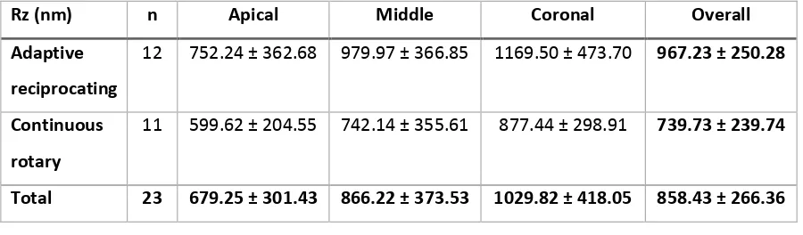
surface roughness was significantly higher overall in the root canals of teeth prepared with
adaptive reciprocation compared to continuous rotary. The results of this chapter showed that
roughness of the root canal is a cofactor that can be modified by the clinician. Treatment
strategies with different techniques can be implemented even while using identical
instruments to achieve smoother treated surfaces. Based on the findings of this study, using a
continuous rotary system to prepare canals or to finish the cleaning and shaping stage of a
root canal treatment can be beneficial to reducing roughness of the canal surface.
Differences between filing systems consists of differences in a mixture of variables including
alloy, surface treatment, cross-section, taper, motion, design, etc. The fourth chapter in this
series was aimed to evaluate the effect of three different filing systems with different
concepts, on the final root canal surface quality. Cleaning and shaping was carried out on
teeth with either a single-file reciprocating (Reciproc), continuous rotary (HyFlex EDM) or
oscillating self-adjusting file (SAF) system. The results from this chapter showed that the three
completely different filing systems resulted in similarly rough root canal surfaces. The high
level of roughness in all groups suggested that the three filing systems tested in this
experiment were relatively aggressive.
File wear results in reduced cutting efficiency and aggressiveness. Since each file undergoes a
life cycle and it is eventually worn out, the fifth chapter of this thesis was designed to assess
how the effect of file wear translates into changes on the treated root canal surface
roughness. In order to evaluate the impact of file wear effectively, Reciproc single-file
reciprocating instruments were used for this study. Reciproc files endure the same stress that
vi amount of wear during three uses, which is within the range of use recommended by the
manufacturer, does not affect the final root canal surface roughness. Without consideration of
safety of these files in terms of file separation risk, these files can be used up to three times
while expecting a similar treatment outcome. However, similar to the previous study, these
files left a relatively rough surface in all cases.
The key findings in the present thesis were that root canal surface roughness is an effective
and modifiable cofactor that can be used to determine the quality of root canal
instrumentation and the performance of the instruments used. The two new methodologies
developed can be used to test other available endodontic instruments and techniques. These
methods can provide a foundation for generating comparable and quantitative data regarding
the roughness values and thresholds associated with biofilm formation and different
endodontic instruments. Standard levels can be set for future instrument designs once enough
research is available regarding the performance of the current instruments and the ideal levels
of surface roughness.
vii
Table of contents
Acknowledgements ... ii
Statement of contribution of others ... iii
Abstract ... iv
Table of contents ... vii
List of figures ... xi
List of tables ... xiii
Glossary ...xiv
Chapter 1 Introduction and literature review ... 1
1.1 Introduction ... 1
1.2 Literature review ... 5
1.2.1 Oral diseases and oral microbiology ... 5
1.2.1.1 Oral biofilm... 6
1.2.1.2 Biofilm formation and bacterial adhesion ... 7
1.2.1.2.1 Surface charge ... 8
1.2.1.2.2 Hydrophobicity and surface energy ... 8
1.2.1.2.3 Surface topography ... 9
1.2.1.2.4 Surface stiffness ... 9
1.2.1.2.5 Surface roughness ... 9
1.2.2 Endodontic microbiology ... 14
1.2.2.1 Bacteriology of endodontic related infections ... 16
1.2.3 Treatment of endodontic-related infections ... 18
1.2.3.1 Microbiological considerations in root canal debridement ... 19
1.2.3.2 Microbiological considerations in the obturation of the root canal ... 20
1.2.3.3 Cofactors that influence root canal treatment ... 21
viii
1.2.3.3.2 Apical debris extrusion ... 23
1.2.3.3.3 Smear layer and root canal surface debris ... 23
1.2.3.3.4 Biofilms and bacterial infection ... 24
1.2.3.3.5 Root canal surface roughness ... 26
1.2.3.3.5.1 Surface roughness characterization and measurement parameters.... 29
1.2.4 Evolution of nickel-titanium endodontic filing systems ... 31
1.2.4.1 First generation ... 31
1.2.4.2 Second generation ... 31
1.2.4.3 Third generation ... 32
1.2.4.4 Fourth generation ... 33
1.2.4.5 Fifth generation ... 35
1.3 Research questions ... 37
1.4 Hypotheses ... 38
Chapter 2 Quantitative comparison of biofilm formation on rough and smooth root canal surfaces using flow cytometry ... 39
2.1 Chapter overview ... 39
2.2 Introduction ... 39
2.3 Materials and methods ... 44
2.3.1 Sample preparation ... 44
2.3.2 Bacterial contamination ... 47
2.4 Results ... 49
2.5 Discussion ... 51
2.6 Conclusion ... 55
Chapter 3 Quantitative evaluation of root canal surface roughness after filing with adaptive reciprocating and continuous rotary instruments ... 56
3.1 Chapter overview ... 56
3.2 Introduction ... 57
ix
3.3.1 Sample preparation and root canal treatment ... 63
3.3.2 Sample scanning and surface roughness evaluation ... 64
3.4 Results ... 66
3.5 Discussion ... 68
3.6 Conclusion ... 70
Chapter 4 Quantitative evaluation of root canal surface roughness after filing with conventional rotary, single-file reciprocating or self-adjusting filing systems ... 71
4.1 Chapter overview ... 71
4.2 Introduction ... 72
4.3 Materials and methods ... 75
4.3.1 Study design and ethics ... 75
4.3.2 Sample preparation and root canal treatment ... 76
4.3.2.1 Group 1 (HFEDM): Continuous rotary filing system (Hyflex EDM, Coltene/Whaledent GmbH + Co. KG, Langenau, Germany) ... 77
4.3.2.2 Group 2 (SAF): Self-adjusting filing system (ReDent-NOVA, Ra’anana, Israel) ... 78
4.3.2.3 Group 3 (RCP): Single-file reciprocating system (Reciproc, VDW GmbH, Munich, Germany)... 79
4.3.3 Sample scanning and surface roughness evaluation ... 80
4.4 Results ... 81
4.5 Discussion ... 86
4.6 Conclusions ... 89
Chapter 5 Quantitative evaluation of root canal surface roughness after repeated use of files with a reciprocating single-file system ... 91
5.1 Chapter overview ... 91
5.2 Introduction ... 92
5.3 Materials and methods ... 94
5.3.1 Study design and ethics ... 94
5.3.2 Sample preparation and root canal treatment ... 95
x
5.4 Results ... 98
5.5 Discussion ... 102
5.6 Conclusions ... 106
Chapter 6 Conclusions and future directions ... 107
xi
List of figures
Figure 2-1 Precision saw used for sectioning teeth and preparing dentine blocks. ... 45
Figure 2-2 Scanning electron microscope surface height map of an (A) rough (Rz=35.10 µm)
and (B) smooth (Rz=16.74 µm) sample obtained using 3D roughness reconstruction. ... 46
Figure 2-3 Vortex shaker that was utilized to dislodge the attached biofilm. ... 48
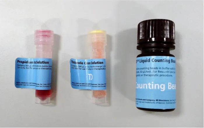
Figure 2-4 Cell viability kit and liquid counting beads used to carry out flow cytometry assay.
Solutions from left to right contain Propidium Iodine (PI), Thiazole Orange (TO) and BD Liquid
Counting Beads. ... 48
Figure 2-5 BD FACSCanto II flow cytometer. ... 49
Figure 2-6 Representative flow cytometric plots of bacterial samples derived from a smooth
(A) and rough surface (B). Number of bacterial cells were assessed by analysing the
frequencies of gated TO-positive bacteria relative to gated counting beads. SSC-A denotes side
scatter area. ... 50
Figure 2-7 Box plot of bacteria count per mL displaying median and distribution of results.
Conventional mean bacteria count per mL ± Standard deviation indicated in writing based on
experimental group. Statistical comparison by post-hoc Tukey tests. Different superscript
letters indicate statistically significant difference between groups (p<0.05). ... 50
Figure 3-1 (a) SPI-MODULE sputter coater (b) Tooth samples loaded into the chamber before
sputter coating. ... 65
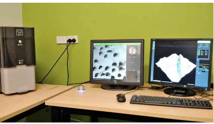
Figure 3-2 Phenom G2 Pro scanning electron microscope. ... 65
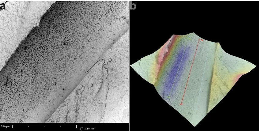
Figure 3-3 a) Scanning electron microscope image of a filed root canal surface at 550x
magnification and b) its surface height map. ... 67
Figure 3-4 Scanning electron microscope images of the Twisted Files. a) The tip and d) middle
third of the SM1 (#20/.04) file. b) The tip and e) middle third of the SM2 (#25/.06) file. c) The
tip and f) middle third of the SM3 (#35/.06) file... 67
Figure 4-1 Root embedded in an acrylic cylinder with a hollow space designed above the
orifice to simulate the pulp chamber. ... 77
Figure 4-2 Continuous rotary filing system consisting of the (a) CanalPro CL motor handpiece
and (b) HyFlex EDM files [Left to right: Orifice Opener (#25/0.12), Glidepath file (#10/.05),
HyFlex OneFile (#25/~) and Finishing file (#40/.04)]. ... 78
Figure 4-3 Self-adjusting filing system consisting of the (a) EndoSTATION motor and the (b) SAF
SYSTEM file set [Left to right: SAF 1.5, SAF-OS (#40/0.10), SAF-1 (#15/0.02) and
xii Figure 4-4 Reciprocating single file system consisting of the (a) VDW.Silver Reciproc motor and
the (b) Reciproc (R40) file. ... 80
Figure 4-5 Box plot illustrating the median and distribution of the Ra (µm) of canal surfaces in
different thirds of the root after cleaning and shaping with each filing system. ... 82
Figure 4-6 Box plot illustrating the median and distribution of the Rz (µm) of canal surfaces in
different thirds of the root after cleaning and shaping with each filing system. ... 83
Figure 4-7 Scanning electron microscope images of canals instrumented with a) HyFlex EDM c)
self-adjusting and e) Reciproc (R40) files. Height maps and roughness parameter calculations
of the scans performed for the b) HyFlex EDM d) self-adjusting file and f) Reciproc (R40) group
surfaces. ... 84
Figure 4-8 Scanning electron microscope images of HyFlex EDM files. a) Apical tip and b)
middle third of the HyFlex Glidepath file (#10/.05). c) Apical tip and d) middle third images of
the HyFlex Finishing file (#40/.04). ... 85
Figure 4-9 Scanning electron microscope images of the self-adjusting files. a) Tip, b) and c)
mesh design connections and the d) abrasive outer surface of the file. ... 85
Figure 4-10 Scanning electron microscope images of the Reciproc (R40) files. a) Tip, b) apical
third and c) middle third surfaces of the file. ... 86
Figure 5-1 Radiography of samples to determine canal curvatures. ... 96
Figure 5-2 Reciprocating single file system consisting of the (a) VDW.Silver Reciproc motor and
the (b) Reciproc (R25) file. ... 97
Figure 5-3 Box plot illustrating the median and distribution of the Ra (µm) of canal surfaces in
different thirds of the instrumented root after use of files for the first, second and third times.
... 99
Figure 5-4 Box plot illustrating the median and distribution of the Rz (µm) of canal surfaces in
different thirds of the instrumented root after use of files for the first, second and third times.
... 100
Figure 5-5 Scanning electron microscope images of canals surfaces instrumented from the a)
first use, c) second use and e) third use groups. The height maps, Ra and Rz calculations of the
scans performed for samples from the b) first use, d) second use and f) third use groups can be
seen in the images on the right. ... 101
Figure 5-6 Scanning electron microscope images of the Reciproc (R25) files with wear after the
xiii
List of tables
Table 3-1 Rz (nm) means ± standard deviation by experimental groups and root thirds. ... 66
Table 5-1 Mean ± Standard deviation of curvature degree, radius and length for roots
xiv
Glossary
Ra Roughness average. Mean height deviation from the mean plane
surface that represents the average distribution of height values.
Rz A ten-point extreme value parameter calculated by measuring the
mean difference between the five highest peaks and the five lowest
valleys of the surface, used to demonstrate average maximum profile
and roughness depths of a surface.
Rq Root mean square deviation roughness. An average between the
mean line and the height deviations. This parameter is mostly used to
demonstrate the skewness and kurtosis properties of roughness.
Sa Arithmetical mean height of an area or in other words Ra (roughness
average) extension onto a surface. It is the difference in height of
every point compared to the surface’s arithmetical mean.
Radial lands Presence of neutral cutting angles in the filing instrument. These
types of instruments tend to burnish the cut debris onto the surface.
Rake angles Angle used to describe the cutting segment of a file. The angle is
subtended between the line from the cutting tip to the centre of the
instrument and the line from the cutting tip that is tangential to the
1
Chapter 1
Introduction and literature review
1.1 Introduction
Root canal treatment failures occur in a significant portion of cases and researchers aim to
reduce risk of failure by identifying the factors that can decrease chances of reinfection.
Success rates of root canal treatment based on strict assessing criteria range from 31 to 96
percent, which reflects the significantly heterogenic distribution of the results. Variable
combination of factors assessed in these studies, different follow-up periods and study designs
make the comparison and interpretation of these studies difficult. Although randomized
controlled trials are considered the gold standard, similar to many other areas of medical
research endodontics is in shortage of such level of evidence.1 Clinical and radiographic
evaluation of endodontic treatments require at least 1 year and in many cases up to 4-5 year
follow-up,1,2 which has made it fall behind with the rate of advancements in technology used
in root canal treatment. The effect of new instruments and materials that are introduced for
clinical use is unclear apart from the advantages that are claimed in their mechanical
properties, efficiency and working times. Therefore, it is of critical importance to evaluate how
these changes may affect the treatment quality and outcome. Success of a root canal
treatment is determined based on long-term clinical and radiographic assessments that
provide evidence of healing. Controversy regarding the factors and thresholds indicating
treatment success has led to different “strict” and “loose” criteria in reports.1 Meta-analysis of
the clinical studies from the last five decades shows the success rates have not improved.
Pooled data even suggests the highest success rates were reported during 1960-80.1 Although
the efficiency of the chemicals and instruments used in treatment have improved over the
years, the unaltered success rates suggests that overall these advances have not affected the
2 technological advances has been comparable to older treatment methods, some of these
innovations may be having detrimental effects while others have been improving the
treatment quality.
Pre-operative clinical factors such as periapical lesions have been widely researched and their
effect on success of root canal treatments has been established. However, intra-operative
factors which are in control of the clinician have been poorly researched. Meta-analyses of
these factors identified fillings within 0-2 mm of the apex and absence of voids in root canal
fillings to be effective in increasing the treatment success rate. However, data regarding
variables of the instrumentation stage such as preparation size and taper is not sufficient for
meta-analysis.3 Individual studies on these factors have conflicting results. Hoskinson et al.
reported a decreasing trend in success rates with larger master apical file sizes, although this
was not statistically significant. They also found no difference between 0.05 and 0.10 tapered
canals.4 In contrast, Smith et al. reported higher taper to be associated with higher success
rates.5
Cofactors of clinical treatment efficiency and success which represent the quality of work can
act as a much needed bridge between clinical and laboratory research. Currently, only few
quantitative cofactors are available that are used to reflect treatment quality. Recent years
has seen some of the previously reliable factors such as root canal seal questioned because of
the errors seen in the methodologies used in their studies.6-8 The identification of relevant
cofactors requires a deeper look into the dynamics of root canal infections.
Root canal treatment failures occur when the persistent microorganisms in the root canal or
invading microorganisms from the outside find a way to grow again.9 Effectiveness of chemical
antimicrobials in eradicating bacteria has been of research interest for decades. The
antimicrobial activities of many disinfectants are weakened in contact with the chemical
3 of the microorganisms involved also complicate their eradication.11,12 Recent concepts of
infection suggest that low levels of microorganisms may be present at sound sites but do not
cause a clinical threat. In this model, disease can occur when environmental changes disturb
the balance of the existing flora towards growth of better adapters to the new conditions.13
Much research is now being carried out to discover means of reducing chances of bacterial
growth.
Root canal infections are biofilm-mediated infections, meaning that the bacteria are not
floating freely in the tissue.14 This explains much about how they develop to mature
communities over time and become resistant to treatment. Biofilms have a different path of
formation, growth and maturity.11 Use of the agar diffusion method for testing antimicrobial
susceptibility has been discouraged since it does not replicate the growth mode and resistance
of bacteria in clinical conditions.6
Biofilm formation is initiated with attachment of microorganisms to the substrate surface,
known as adhesion. Many factors have been identified to alter chances and modes of biofilm
growth. Surface energy, charge, stiffness, chemistry, and roughness of the surface are some of
the factors that are effective in biofilm formation. These factors can have a different effect
magnitude based on the type of microorganisms involved.15 There is currently little
information about the effects of these factors in endodontic treatments and the effect that
they may have on microbial species involved in root canal infections.
Surface roughness has been shown to be dominant factor among the substrate’s properties
that can affect biofilm formation. Surface charge and surface energy have a less significant
effect in rough surfaces.16 In the oral cavity, roughness can increase the amount and maturity
of the biofilm formed by oral microorganisms on dental implants.17 Moreover, roughness is a
surface property that could be modified by both the chemical erosion caused by irrigants and
4 Irrigants and antimicrobials used in the root canal system can cause changes in the physical
properties of dentine.18,19 Chemical erosion and surface changes have been experimented over
the last decades with the available irrigants and also the newer chemicals that have been
introduced. Although most these experiments showed a significant difference in the amount
of roughness that various irrigants left, these differences are in a nanometre scale.19-21
Roughness caused by mechanical instrumentation has not been thoroughly researched. The
few reports available use qualitative or semi-quantitative methods that make them
incomparable to other instruments outside of the study.22-26 Even so, the difference in surface
roughness after using different filing systems seems so obvious that some researchers such as
Barthel et al. compared surfaces without magnification.22
In conclusion, the present research was designed towards achieving two goals. The first goal
was to develop a method to quantitatively investigate the effect of roughness on endodontic
bacteria and determine the clinical relevance of roughness in root canal infections. The second
goal of this series of experiments was to develop a quantitative method to evaluate roughness
of root canals and determine whether the current methods and instruments used for filing
canals can affect the root canal surface roughness.
In the following section of this chapter, the available research on this topic, the knowledge
5
1.2 Literature review
1.2.1 Oral diseases and oral microbiology
The oral cavity can support the growth of one of the most complex and divergent communities
of microorganisms in the human body with over a thousand species.27 These microorganisms
are constantly subjected to a wide range of physical and chemical changes. The oral cavity is
the only part of the body that has externally exposed hard tissue (teeth). Bacteria can adhere
to the teeth and create a biofilm known as dental plaque. Keratinized and non-keratinized soft
tissues of gingiva, tongue, palate, mucosa, and floor of the mouth also provide environments
for various types of microorganisms.28,29
Overall, the oral microbiota is believed to have major health benefits for the human body
under normal conditions28. The commensal microbiota can prevent exogenous infection by
multiplying and covering the binding sites for exogenous pathogens, which is known as
colonization resistance.30 However, commensal microorganisms may also become the cause of
oral disease if normal conditions change. Environmental stresses that alter the haemostatic
mechanisms of the oral biofilms are the main reason that start the pathogenic cycle.28
Dental caries is the most prevalent cause of pulpitis and pulpal infection.31 Caries and
infectious disease in the oral cavity occur when the environmental conditions of the oral
microflora change. These stresses can cause an impediment to the equilibrium between
remineralisation and demineralisation of teeth. If the progress of these events is not stopped
or reversed, it can promote further selective development and multiplication of the acidogenic
and acidophilic bacteria in dental plaque. This process can eventually lead to extensive carious
lesions in enamel and dentine, pulpal inflammation and infection.29 Bacterial by-products can
stimulate the pulpal immune response through dentinal tubules and the bacteria can infect
6 1.2.1.1 Oral biofilm
Oral epithelium sheds around 3 times daily which significantly reduces the amount of bacterial
adhesion and biofilm formation on its surface.15 Exposed hard tissues in the oral cavity have a
very different interaction with the oral environment, especially with saliva and the oral
microbiota. This interaction can start within seconds upon the exposure of the enamel to
saliva. Saliva usually coats all hard and soft tissues in the oral cavity creating a conditioning
film. Salivary proteins are absorbed to the enamel hydroxyl apatite and form what is known as
the acquired enamel pellicle (AEP). This pellicle matures with the absorption of other proteins,
lipids, carbohydrates and adhesion and colonization of microorganisms. 32
The adapting potential of bacteria gives them unlimited mechanisms to overcome the barriers
that prevent them from colonizing inside the oral cavity. The addition of saliva, especially with
its protein content, to this environment, adds further complexity to the system.
The growth mode of oral bacteria is much more complex than the growth of single or multiple
species. Microorganisms in the oral cavity grow in biofilms. The biofilms consist of a
polymer-rich matrix, which have the microorganisms colonizing both inside and on the surface of it.
Dental plaque is a very well-known form of biofilm that is present in the mouth. Aside from
normal oral tissues, microorganisms can also adapt and form biofilms on dental materials used
inside the mouth. Biofilms develop on surfaces of restorative, prosthodontic and endodontic
materials and can cause many problems in treatments. This has led to interest in anti-biofilm
properties in dental materials. 33
Interspecies associations develop in biofilms and help the bacterial community’s nutrition,
adherence and stability. However, these interactions may change with alterations to the oral
environment and become pathogenic. The most noted example would be a change in the diet
that can lead to caries (tooth decay). Intake of a high level of carbohydrates can lead to higher
7 the plaque and subsequently the oral cavity. The altered pH inhibits the growth of some of the
other non-cariogenic bacterial species that are acid-sensitive.28,34
Biofilm bacteria are more resistant to antimicrobials. Antibiotics have not been designed to
eradicate biofilm populations.35 Therefore, treatment of a biofilm-mediated infection is more
difficult.36 Many oral diseases including post-treatment root canal infections are
biofilm-induced infections.37 The maturity level of a biofilm is also effective in its resistance to
antimicrobials. This resistance is believed to be have a major role in the persistence of
infections and recolonization of microorganisms after antimicrobial treatments.38 Biofilms
have a higher chance of being associated with longer standing lesions. Slower metabolism of
microorganisms in a biofilm and the presence of an extracellular matrix, that may act as a
barrier itself, reduces the effectiveness of antimicrobials.39
1.2.1.2 Biofilm formation and bacterial adhesion
Development of a biofilm initiates with attachment and adhesion of the microorganisms to the
substrate surface.40 This stage is also believed to be the most important stage of biofilm
formation. After the initial adhesion to the surface, the bacteria start forming ligand-receptor
binding to the surface which makes the adhesion irreversible.36 Attachment of microorganisms
is followed by development of micro-colonies and microbial growth.41
Initial interaction between bacteria and the surface, which is known by adhesion, is the
foundation of biofilm formation. Adhesion mainly takes place between the bacterial cell wall
and the extracellular components of the substrate or the medium covering it. The dynamic
nature of the bacteria’s response and how it adapts in response to the surface also adds more
complexity to the infinite number of combinations that are possible in this interaction.
8 but they have systems in place to sense their environments and then respond accordingly or
adapt to changes.
Streptococcus mutans has the most prominent role in caries etiology which is rooted in its
high adhesion capability to dental tissues, even though it is less acidogenic and pathogenic
than some other species involved in caries development.42 Susceptibility to bacterial
attachment is considered one of the most important factors in determining a restorative
material’s longevity.42 Therefore, it is logical that a wide range of research has been done on
factors that have the potential to prevent or limit their attachment.
Surface charge, surface energy (hydrophobicity), roughness, topography, stiffness and
chemistry of the surface are some of the most important substrate properties found to affect
adhesion,15 which are reviewed in the following section.
1.2.1.2.1 Surface charge
The negative charge present in most bacterial cell walls adheres better to surfaces with a
positive charge. Negative charge of a surface on its own factor cannot always prevent
adhesion because some bacteria have mechanisms to attach to these surfaces too.15 In
addition, different environmental ions, proteins and mediums such as saliva that coat the
substrate surface, have an important effect on the final role of surface charge.43
1.2.1.2.2 Hydrophobicity and surface energy
Superhydrophobic and superhydrophilic materials are both used to create non-fouling
surfaces because of their non-adhesive properties. This demonstrates the different role
surface energy has on bacterial adhesion. An average range of hydrophilicity or hydrophobicity
9 environment (e.g. saliva film covering the surface).15 In subgingival areas of the mouth where
saliva flow is less significant, surface energy has less effect on biofilm formation. However, on
supragingival areas of the mouth, shear stresses caused by saliva flow seems to detach
biofilms easier from hydrophobic surfaces compared to the hydrophilic ones.44,45
1.2.1.2.3 Surface topography
Topographic patterns of certain shape and size can inhibit biofilms. These patterns that are
mostly in nanometre or micrometre scales can be used to create non-fouling surfaces or even
surfaces that can kill bacteria upon contact. This may be one of the only instances where a
surface with roughness is less suitable for biofilm formation compared to a flat and smooth
surface.15 Although some of these properties may be someday used in prefabricated
treatments, it is highly unlikely to apply directly inside the oral cavity.
1.2.1.2.4 Surface stiffness
Softer materials allow better adhesion and more rapid growth of biofilms. Stiffness is one of
the most recent and least known surface properties that affects bacterial response and
physiology. This effect has only been investigated on a limited number of bacterial types and
requires much more research. However, there is evidence that saliva forms different viscosity
films on intraoral surfaces.15,46 These differences may influence the surface stiffness properties
too.15
1.2.1.2.5 Surface roughness
Roughness has a very distinct yet variable effect on biofilm formation. This effect has been the
10 multiply the amount of surface area available for adhesion by a factor of 2-3.47 These areas can
also trap bacteria or provide shelter and shield for them against shear forces that can detach
the biofilm. The magnitude and threshold of this effect relies on the bacteria type and species.
Roughness in nanometre scale can significantly increase the biofilm formation of certain
bacteria while others might be much less affected by the same scale roughness.15
Teeth and various dental materials’ roughness attract formation of different types and
amounts of biofilm. The first bacterial attachments and biofilm formations on dental tissues
and restorative materials occur in irregular and rough surfaces such as cracks and grooves.42
Intraoral plaque formation on polymer surfaces increases significantly with a 2 µm increase in
surface roughness. 47 Experimentation of subgingival microbiological changes based on
roughness differences have been technically challenging. These studies require surgical
interventions and alteration of subgingival hard surfaces48 that were not common before
introduction of dental implants.
Dental implants’ optimal roughness properties have been extensively experimented to achieve
lower levels of biofilm formation. Early studies showed plaque accumulation can be as much
as 25 times on rough intraoral surfaces compared to smooth ones.48 Implant research has
focused on surface roughness extensively since the implant surface characteristics is important
to both osseointegration of the implant and adhesion of the microorganisms. Peri-implantitis,
the inflammation of the tissues surrounding the implant, caused by plaque and
microorganisms can result in implant failure. Rough implant surfaces harbor and colonize
more bacteria, therefore, increase the risk of peri-implantitis.49,50 On the other hand,
moderate roughness of the implant surface has been reported to promote bone response and
osseointegration.51 An ideal surface would have a balance in which it is rough enough to
provide osseointegration but not too rough to significantly boost plaque accumulation and
11 Quirynen et al. showed in 1993 that there were 25 times more microorganisms on subgingival
rough surfaces compared to smooth ones. In addition, more motile bacteria and a larger
proportion of spirochetes were seen on rough surfaces. This effect was especially seen in
supragingival plaque after 3 months which suggests the effect of roughness on plaque
maturity as well as bacterial quantity. This effect was seen by only increasing the surface
roughness average (Ra; for more information regarding roughness parameters please refer to
section “1.2.3.3.5.1 Surface roughness characterization and measurement parameters”) of the
implant abutments from Ra=0.35 µm to Ra=0.81 µm.17
Roughness threshold of 200 nm was suggested for implants. Further reduction of roughness
beyond this point is expected to cause no change in biofilm formation. Clinical assessment of
patients that had implant abutments that were highly polished (Ra=0.05 µm) with standard
(Ra=0.21 µm) abutments showed no significant difference in the biofilms formed on them
after 3 months. This meant that roughness values lower than 200 nm have less clinical
significance and do not impact the biofilm composition. Furthermore, the standard abutments
showed less probing depths, which suggests better attachment gain for the gingival cells in
this group.48
Long-term effects of roughness below the 200 nm threshold on biofilm formation and
composition was experimented in a split-mouth study by Bollen et al. in 1996. Implant
abutments made of machined titanium (Ra=200 nm) were compared to polished ceramic
abutments (Ra=60 nm). Clinical examinations, differential phase-contrast microscopy and
bacterial cultures were carried out for the implants in 3 and 12 months after abutment
placement. The results in general show that the two types of surfaces did not differ
significantly in quantity or quality of their biofilms.52
Xing et al. found a strong correlation between the amount of biofilm accumulation on TiZr
12 being tested in this study since TiZr discs were placed in a removable splint inside the mouth
of the ten participants for 11 hours. The roughness of each disc was assessed with 50×
magnification on four areas of 255 µm × 191 µm using a blue light laser profilometer. Biofilms
were stained using safranin and released from the discs using acetic acid. The amount of
bacteria from each sample was tested with spectrophotometry.53
Atomic force microscopy (AFM), scanning electron microscopy (SEM), fluorescence in situ
hybridization (FISH), and confocal laser scanning microscopy (CLSM) were carried out on 6
different implant surface disks and bovine enamel slabs in an in vivo study by Al-Ahmad et al.
in 2010. The average surface roughness (Ra) of the surfaces were calculated with AFM on a
surface area of 50 × 50 µm. Twelve volunteers wore the splints containing the disks for 3 and 5
days. After 3 days, the biofilm thickness in 6 groups out of 7 was found to be correlated to the
surface roughness. The correlation between surface roughness and the biofilm thickness
decreased significantly after 5 days. The biofilm composition assays with FISH and CLSM
showed no difference between enamel slabs and the implant material. The materials did not
have a significant effect on the bacterial composition. This was assumed to be due to the fact
that the acquired pellicle has a more dictating role in the biofilm composition than the
materials.54
Quantitative assessments of the effect of surface roughness (Ra) and surface free energy on
the amount of biofilm formation were done by Burgers et al.55 They used fluorescent
microscopy and an automated multi-detection fluorescence reader to examine the amount of
biofilm formation which was more sensitive than the methods previously introduced. Surface
free energy and surface roughness (Ra) of the two types of titanium materials were calculated
with a goiniometer and perthometer, respectively. Splints that carried the rough (Ra=0.15 µm)
and smooth (Ra=0.95 µm) titanium specimens were worn by volunteers for 12 hours.
13 on the titanium specimens showed that both were significantly higher for the rough
specimens.55
Lin et al.56 showed that 0.3-1.4 µm range of roughness (Sa) of titanium disks does not have an
impact on the quantity of biofilms developed by Streptococcus mutans or Porphyromonas
gingivalis species after 1 and 3 days. The two levels of roughness that were experimented in
this study with low (Sa=0.3 µm) and moderately (Sa=1.4 µm) roughened titanium disks were
both above the 200 nm threshold mentioned earlier that was described earlier for titanium
implants and the range of microorganisms involved on their trial.48 Roughness did however
have an effect on how effective chlorhexidine was on the biofilms. The colony forming unit
(CFU) counts of both 1 day and 3 day biofilms reduced significantly less after treatment on the
rougher surfaces,56 which suggests a more resistant biofilm on rougher surfaces.
Saliva can also affect the topography and roughness of oral and dental surfaces by its uneven
distribution in a nanometre scales.32 Once the pellicle is formed inside the oral cavity, the
proteins and enzymes in the extracellular matrix can affect the pellicle’s surface properties.
The polysaccharides that are produced by the exoenzymes that come in contact to sugar can
change the surface topography and create high affinity binding sites for bacteria on the
pellicle.57
Pellicle formation and its effect on the substrate surface properties has been controversial.
Although some research point out the role that saliva has in masking some of the qualities of
the substrate’s surface, there is some evidence that the substrate surface properties are also
effective in presence of saliva.42 Research shows surface roughness can enhance S. mutans
binding to parotid saliva on composite resins and glass ionomers.58 Therefore, it seems that
the presence of saliva is another variable partially affecting bacterial attachment in the oral
cavity. This effect can be minimized in endodontics if the root canal environment is sufficiently
14 Interaction between the biofilm and substrate that alters surface properties is dependent on
the type of substrate material and microorganisms.42 S. mutans biofilms can increase surface
roughness on resin composites in vitro. This effect can in return accelerate bacterial
attachment and biofilm formation and therefore, start a cycle that eventually compromises
the restoration.59
The overall trend of research seems to show that roughness affects the amount and
characteristics of biofilm formed on implants.60 This effect that roughness shows is different
depending on the types of microorganisms and substrates tested. The magnitude of effect
varies in studies based on the methods utilized and the types of roughness parameters used to
describe surfaces; e.g. Ra, Rz and Sa.
1.2.2 Endodontic microbiology
Endodontology represents the study of the diseases of the pulp-dentine complex and
periapical tissues. The dental pulp is a sterile and protected tissue surrounded by dentine and
enamel. The embryonic origin of dentinal and pulpal tissues is similar. These tissues form a
functional organ that is responsible for producing dentine and tooth sensitivity. Dentine is in
contact with the enamel (dentinoenamel junction or the DEJ) or cementum (dentinocemental
junction or DCJ) on its outer surface. During the development of dentine, the odontoblasts
form a porous structure with the dentinal tubules running from the DEJ and DCJ to the inner
surface.61 When the integrity of the tooth is somehow compromised (caries, trauma,
periodontal disease, etc.), an inflammatory response will occur. Traditionally, endodontic
disease is a sequel to caries, and non-infectious pulpal inflammation is much less common
compared to infectious conditions. The infectious diseases affecting the pulp also exhibit a
progressive nature.62 Reversible pulpitis can transform into an irreversible state where pulp
15 against infection because it is surrounded by mineral tissue and lacks collateral circulation.63
The temperature, humidity, available nutrition, anaerobic conditions in the root canal system,
which are largely inaccessible to the host defence, are ideal for many microorganisms to
colonize.64 This leads to a rapid loss of vitality in the tissue which is believed to be “higher than
any other tissue in the body”.63
The landmark research of Kakehashi et al.65 in 1965 revealed the pathogenic nature of pulpal
inflammation. Their research demonstrated this for the first time by examining pulpal
exposure in germfree and conventionally-reared rats. The report showed that exposed pulpal
tissue in germ-free rats could initiate repair by creating dentine bridges.61,65 The vital role of
microorganisms in this process provides an understanding of why endodontic treatment
largely focuses on eliminating infection and preventing reinfection.66
Prognosis of root canal treatment in cases that are associated with preoperative infection are
lower than of teeth with vital pulps. Ng et al. reported a success rate of above 80% for primary
and secondary root canal treatments in a prospective study on 2484 roots. However, presence
of a preoperative periapical lesion decreased the odds of success by 49% compared to roots
without a lesion (OR=0.51, 95% Confidence Interval 0.32-0.80).67 Another large-scale
prospective cohort study on 1369 roots by Riccuci et al. reported a success rate of 93.1% for
vital roots. The success rates for roots with necrotic pulps and roots with a combination of
pulp necrosis plus apical periodontitis were 92.3% and 84.1%, respectively.68
Invasion of dentinal tubules with microorganisms or exogenous substances can initiate from
exposed dentine in the oral cavity. This process can be initiated by bacteria that are common
in dental plaque but obligate anaerobic bacteria are dominant in the infected root canals.
Although the pulp-dentine complex has some defensive mechanisms, if the source of infection
is not eliminated, it may result in pulpitis, pulp necrosis, and pulp infection that may
16 root canal infections. These findings are consistent with the research that show some species
like Enterococcus faecalis can resist periods of starvation, sodium hypochlorite, heat, hydrogen
peroxide, and highly alkaline conditions (that could be caused by calcium hydroxide dressings).
Survival through these stages can provide a bacterial source that could cause failure in root
canal treatment.61
1.2.2.1 Bacteriology of endodontic related infections
Root canal infections are usually endogenous, where oral bacteria contaminate the root canal
and cause the infection.69 Given the highly complex and diverse nature of the microbial
communities in the oral cavity,28 a polymicrobial community is seen in most oral infections.30
The organisms that often invade the root canal are opportunistic pathogens. These pathogens
are not the most virulent or invasive species, e.g. E. faecalis. However, these organisms are the
more resistant to antimicrobial agents and pH fluctuations.69-71 Development of mature
biofilms and also the ability of E. faecalis to invade dentinal tubules, where it is protected from
antibacterial irrigants, can make its eradication even more difficult.38
Studies investigating the species of bacteria associated with certain infections in the oral cavity
have been hindered by the fact that approximately half of the oral bacteria cannot grow on a
conventional culture media.30,72 Recent research using molecular techniques, PCR, and
anaerobic culture methods for defining the bacterial composition of endodontic infections
have revealed contradictory results.66,71,72
Dentine tubule invasion with microorganisms is important for understanding the mechanism
of root canal infection and treatment. From the hundreds of bacterial species in the oral
microflora, only a small number can invade dentine and cause infection in the root canal.
Microflora that are involved in caries development are from the streptococci, lactobacilli and
17 to bind to collagen type I and invade dentinal tubules. The superficial layers of caries are
mostly populated with Gram-positive rods. The deeper layers of caries in dentine harbour
more anaerobic species of the Gram-positive rods.31,74 Streptococci which are more dependent
on the nutrients in saliva have less chance of thriving in deeper depths of the lesion.31
Coronal dentinal tubules are wider in deeper levels that are close to pulp (approximately 4.3
µm diameter) and narrower in superficial depths of dentine close to enamel (approximately
2.4 µm diameter).75,76 The size of the tubules both in the surface and depths close to the canal
decreases towards the apical region of the tooth.77 However, it is important to note that these
sizes are still large enough to harbour the bacteria that invade tubules.
Sampling errors from root canals infections are inevitable amid the different techniques used
by researchers. The samples are categorized based on their recovering site, which is usually
either the pulp chamber (non-vital or containing some vital tissue) or apical tissues.
Maintaining the integrity of the samples acquired and preventing oral and saliva
contamination of the sample remains a challenging task.14,66 Sample collection is usually done
with use of paper points which is believed to be biased towards collecting free-floating
bacteria rather than the biofilms attached to canal walls. In addition, this method cannot
specify which part of the canal the microorganisms are acquired from.14,63
The microbial communities that are recovered from the primary and secondary infections of
the root canal are different. Bacteria which are recovered from a primary root canal infection
are usually polymicrobial communities of 2-8 species. Obligate anaerobes are dominant in
these communities. The flora of secondary infections of the root canal (failed cases) usually
consists of 1-2 species per canal. In these cases, mainly gram positive facultative anaerobes
are recovered. Enterococcus faecalis is the predominant species in failed root canal
treatments,61,70,71 whereas this species has shown to be less commonly recovered from the
18
E. faecalis has a number of potential virulence factors that give it the opportunity to thrive in
root canal reinfections. This microorganism has low sensitivity to antimicrobial agents and
chemicals used in the root canal. In addition, it has the ability to invade dentinal tubules where
it can be sheltered from the medications and chemicals used in treatment until the conditions
are suitable for it to reinfect the root canal system.78
1.2.3 Treatment of endodontic-related infections
Eliminating microorganisms and their by-products from the root canal system and preventing
reinfection are the primary objectives of root canal therapy.79,80 After endodontic access
preparation, debridement of the root canal system is carried out. During and after
debridement, the root canals are shaped in a way that can be filled. Reinfection may occur due
to coronal penetration of oral bacteria or the remaining bacteria in the root canal system after
cleaning and shaping. The root canals are therefore filled to prevent reinfection by sealing the
remaining space (against bacterial penetration from the oral cavity) and also entombing the
remaining bacteria.80,81
A range of endodontic instruments, techniques and materials have been experimented to
optimize the results that are achieved. Different filing systems, sonics, ultrasonics, irrigation
solutions, smear layer removal methods, and intracanal medicaments are some of the
different options that a clinician may consider at this stage. The ideal result of this treatment
stage would be a root canal and pulpal chamber that are free of microorganisms and would be
ready to be filled. These conditions would allow periapical healing and osseous regeneration.
However, these conditions cannot always and practically be met due to the complexities of the
19 1.2.3.1 Microbiological considerations in root canal debridement
Debridement of the root canal is defined as elimination of the substances (organic and
inorganic) and microorganisms from the root canal.82 This stage of treatment is referred to as
the foundation of a successful endodontic treatment and the importance of it has been
emphasized since 1931.83
Debridement is achieved by cleaning and shaping the root canal system. Cleaning, which is
done before and during shaping, is often carried out with a combination of chemical and
mechanical approaches.83 Studies show that large areas of the canal remain intact during sole
instrumentation and emphasize the importance of adequate irrigation. Even though
mechanical instrumentation does reduce the number of microorganisms infecting the canals,
combining instrumentation with irrigation has shown to result in 100-1000 times more
reduction in the number microorganisms compared to instrumentation alone.83 Irrigation also
allows chemical disinfection and elimination of bacteria from the canal, which are key to root
canal treatment success.84,85
Time, physical restrictions and the complex morphology of the root canal system do not allow
complete disinfection and removal of the smear layer and debris.86-90 In practice, the aim is
towards minimizing the number of microorganisms. The root canal debridement is usually
limited to the main canal, which also has some remote areas that might be unprepared at the
end of treatment.91
Residual microorganisms that are infecting dentinal tubules are one other source of bacteria
that may jeopardise the final clinical outcome.86 Since these microorganisms grow in biofilms,
root canal biofilm resistance to various irrigation solutions and medicaments has also been a
focus of research in this field. The efficacy of these treatments has been tested on biofilms
20 New instruments have been introduced to reduce the procedural errors of instrumentation.
Much effort has been made also to reduce the chances of instrument fractures.95 There have
been changes in the type of materials used with the introduction of more flexible Ni-Ti
instruments instead of stainless-steel. Instrument designs and cutting efficiency have also seen
many improvements.95
1.2.3.2 Microbiological considerations in the obturation of the root canal
None of the current techniques employed in endodontics can entirely eliminate root canal
bacteria.96 The aim of a root filling is to achieve a seal against bacteria and their by-products.
The result of an ideally debrided root canal would be a disinfected hollow space in the tooth.
However, even in such conditions, this space is in proximity of the bacteria of the oral cavity.
This space is not accessible by the host’s immune system if contaminated, and would
therefore be a potential site for reinfection. Incomplete filling of the root canal is suggested to
be associated with up to 60 percent of endodontic treatment failures.97,98
Orstavik et al.99 carried out a multivariate analysis on the factors influencing the final outcome
of endodontic treatment. They reported that root filling density and other technical qualities
of the filling such as the apex-to-filling distance have a significant effect on the clinical
outcome.99
Current filling materials cannot entirely seal the root canal space and they do allow leakage.
Gutta-percha does not bond to the canal walls and is often used with a sealer to help fill this
gap. If the sealer has bonding qualities, it may also help prevent dislodgment of the root
filling.100 More recently, heated and pre-heated gutta-percha methods have been introduced
21 1.2.3.3 Cofactors that influence root canal treatment
The possible combinations that can be made from the wide range of instruments, chemicals
and techniques to carry out a root canal treatment may seem almost infinite. However, many
of these combinations may not have been thoroughly tested. Rapid technological
developments and innovations in the available materials and instruments also need to be
experimented against conventional treatments. Although clinical studies provide the highest
level of evidence, they require long-term follow-ups that are difficult to achieve with the rate
of advancements. The quality of a root canal treatment is assessed by evaluating various
factors that directly or indirectly affect the treatment outcome. The following section will
review the factors that have been utilized to test instruments and methods used in the
cleaning and shaping stage of root canal treatments.
1.2.3.3.1 Dentinal integrity, defects, cracks and craze lines
Vertical root fractures are one of the relatively common reasons (8.8-13.4%) for extraction of
teeth after root canal treatments.102,103 This has led to a large amount of research regarding
the factors that could create craze lines, cracks and ultimately fractures in dentine.104
Root canal preparation and obturation were both shown effective in creating dentinal defects
in a study by Shemesh et al.105 Horizontal teeth sections were observed under a
stereomicroscope with x12 magnification after different treatments. The number of teeth with
defects were significantly higher after preparation with Gates Glidden drills and rotary files
compared to unprepared teeth. Obturation of the canals with lateral condensation technique
also created more dentinal defects compared to the prepared teeth without any filling.105 In
contrast to this study, which found significantly more dentinal defects in teeth using a lateral
condensation technique compared to no compaction of gutta-percha, Onnink et al. reported
22 Exposure of dentine to sodium hypochlorite, which is frequently used in root canal irrigation,
decreases its flexural strength. This effect is more significant when 5.25% NaOCl is used
compared to the 0.5% concentration solution. Endodontically treated teeth were previously
claimed to be more susceptible to fracture because of loss of dentinal tissue and changes in
proprioception and nociception. The findings regarding the changes in dentine’s physical
properties further supported this idea.107
Calcium hydroxide is another chemical that decreases dentine fracture resistance especially
when exposed to it long-term. This is of importance since calcium hydroxide is used as a root
canal dressing and should therefore be applied with caution considering this effect.18,108
Post space preparation and post placement generally weakens the endodontically treated
tooth structure. 109 Recent developments in adhesive luting of posts and availability of
materials other than the rigid metal posts have decreased the chances risk of root fracture
failures,109,110 however, they should still only be placed when essential.109
Micro-computed tomography methods that were developed to research dentinal cracks
allowed comparison of defects before and after procedures. Some studies utilizing the new
methods question the clinical relevance of previous research and the effect of micro-cracks as
a cofactor in root canal treatments. The recent studies using micro-computed tomography
indicate there is no causal relationship between canal instrumentation and formation of
microcracks.111-113 It is also important to interpret the results from in-vitro experiments with
caution since they usually require the tooth to be in dehydrating conditions and without the
support of the periodontal tissues.114
23
1.2.3.3.2 Apical debris extrusion
Apical debris extrusion may complicate a root canal treatment outcome by causing pain,
swelling and flare-up. The type of filing technique and instrument seem to affect the amount
of extruded debris.115 However, it is difficult to compare data between studies since they have
conflicting results while using the same instruments. An example for this is the different
results obtained for HyFlex CM and ProTaper Next files in reports from Capar et al.116 and
Kocak et al.115 Filing motion has also been linked to the amount of debris extrusion. Rotary
filing leads to less debris extrusion compared to certain modes of reciprocation.117
Clinical relevance of in-vitro studies of apical extrusion is questionable since it is not clear what
amount of debris would actually cause clinical complications. The threshold may vary in each
case depending on the infectious potency of the debris. Debris extrusion in a clinical scenario
would be greatly influenced by the amount of resistance from periradicular tissues and the
dimension of apical preparation too, which is difficult to simulate in laboratory settings.118
1.2.3.3.3 Smear layer and root canal surface debris
Smear layer is a 1-5 µm thick surface film of debris that is formed on the root canal surface
after instrumentation with endodontic files.119 The smear layer may harbour bacteria and their
byproducts and it can also be packed as far as 40 µm deep into dentinal tubules.120 It has been
a focus of research since it can prevent irrigants from reaching deeper parts of dentine and
reduce the bond of adhesives and sealers to the root canal wall.119 Various chemical,121
mechanical, ultrasonic122 and laser methods120 have been tested to remove the smear layer.
EDTA is the most common solution used for removal of the smear layer in endodontic
treatment.123 It is also referred to as the gold standard for removal of the smear layer in
research.121 Multiple studies regarding the effect of EDTA on the final seal of root canal
24 However, the meta-analysis by Shahravan et al. in 2007 concluded an improvement in the seal
of the root canal system when the smear layer is removed.124 The use of ultrasonic activation
of irrigants in combination with EDTA to achieve cleaner root canal surfaces has been claimed
to be beneficial in some studies.122,125 In contrast, a randomized clinical trial by Beus et al.
showed no difference in achieving bacteria free canals when comparing use of only 1% NaOCl
with use of a passive ultrasonic multi-irrigant (1% NaOCl, 17% EDTA and 2% chlorhexidine)
protocol to clean the canals.126 Evidence from another randomized clinical trial by Liang et al.
indicates that periapical healing of endodontically treated teeth also seems to be similar with
or without ultrasonic activation of 5% NaOCl.127 The methods used in these studies widely vary
and the effect of smear layer as a cofactor has not been completely clear. There has also lately
been more debate regarding whether some experiments may be biased due to not
differentiating sclerotic dentine from the smear layer.121
1.2.3.3.4 Biofilms and bacterial infection
Microbial infection is an integral part of pulpal and periapical pathosis.65 This role has led to
the development of various research regarding the microbial status in root canals128 and study
models to test the effect of treatments on their reduction and elimination. Disc diffusion
methods and testing antimicrobial effects of chemicals on bacteria grown on agar plates that
were common earlier are now questioned for their clinical relevance and the level of evidence
that they provide.6 Experiments on the antimicrobial efficacy of irrigants on planktonic
cultures is also of limited value since bacteria express different phenotypes and have much
higher resistance to antimicrobials in biofilms compared to when they are in a suspension.129
Plastic, glass and stainless steel, which are often used as the substrate in biofilm experimental
25 attributed to the ability of the microorganism to adhere to the substrate as the first stage in
biofilm formation.130
Bacteria generally do not adhere well to mineral tissue.131 A biofilm model with a substrate
that resembles dentine with a combination of organic and inorganic components was
introduced by Shen et al.129 Disks made of collagen-coated hydroxyapatite (C-HA) were used as
the substrate in this study. The results showed that multi-species biofilm in this experiment
was killed faster using CHX-Plus (Vista Dental Products, Racine, WI), which has surface
modifiers in addition to chlorhexidine, compared to 2% chlorhexidine gluconate.129
Mono-species biofilm models have been more popular in endodontic research. Although the
number of species in the root canal infections are much less than the oral microbial flora,
poly-microbial biofilm models and their characteristics better resemble root canal infections.132
Quantification of bacteria in relation to treatments and disinfectants has remained a major
challenge in endodontics. Starved biofilms, similar to the populations in the root canal, may be
via