0022-538X/85/120757-10$02.00/0
Copyright © 1985,American Society for Microbiology
Effect of
Herpes
Simplex
Virus
Types
1and 2
on
Surface
Expression of
Class I Major
Histocompatibility
Complex
Antigens
on
Infected
Cells
STEPHEN R. JENNINGS, PATRICIA L. RICE, EDMOND D. KLOSZEWSKI, ROBERT W. ANDERSON, DAVID L. THOMPSON, ANDSATVIR S. TEVETHIA*
Department of Microbiology andCancer Research Center, The Pennsylvania State University College of Medicine, Hershey, Pennsylvania 17033
Received 31 May 1985/Accepted 30July 1985
Cytotoxic T lymphocytes (CTL) generated in C57BL/6 (H-2b) mice in response to infection with the serologically distinct herpes simplex virus type1 (HSV-t)ortype2(HSV-2)werecross-reactiveagainst target cells infected with eitherserotype. However, HSV-2-infected cellswere showntobe muchlesssusceptible to CTL-mediated lysis, and analysis through the useof HSV-1 x HSV-2 intertypicrecombinants mapped the reduced susceptibility toa region contained within 0.82to 1.00 mapunits of the HSV-2 genome. Thestudy reported here was undertaken to determine the possible reasons for the reduced susceptibility of HSV-2-infected cellstolysis by CTL.Competition for the specific lysis of labeled HSV-1-infectedcellsby either HSV-1-or HSV-2-infected, unlabeled inhibitor cells and frequency analysis of the CTL precursorable to recognize HSV-1-and HSV-2-infected cells suggested that the reduced susceptibility of HSV-2-infectedcells tolysiscould be explained, at least in part, by reduced levels of target cell recognition. A determination of the surface expression of the critical elements involvedintarget cell recognition byCTLfollowinginfection with HSV-1or HSV-2 revealed that all the major HSV-specific glycoprotein specieswereexpressed. InfectionwithbothHSV-1 and HSV-2causedareductionintheexpression of theclass I H-2antigens.However,this reductionwasmuch greaterfollowing infection with HSV-2. This suggestedthatoneimportant factor contributingtoreducedlysis ofHSV-2-infected cellsmaybe the alteredorreduced expressionof the class I H-2 self-antigens.
The recognition of viral antigens, inserted into the host cell membraneas aconsequenceofinfection, by cytotoxicT
lymphocytes (CTL) is restrictedby theassociation of these
antigenswith the class Iself-antigensof themajor
histocom-patibility complex. In the mouse, these so-called restricting elements are the gene products encoded within theK-, D-, and L-loci of the H-2 complex (9, 40). Several studies have confirmed that these basic principles are true in the CTL responsetoherpes simplexvirus(HSV)in mice(5, 6, 14, 18, 22, 26, 33, 34). Wefocused ontheprimaryCTLresponsein
C57BL/6 (H-2b) mice following the local injection of infec-tious virus inthe hindfootpads,and itwasdemonstrated that one or more ofthe HSV-encoded envelope glycoproteins, designated gB, gC, gD,andgE (23), expressedonthesurface ofHSV-infected H-2b (B6/WT-3) cells serve as the target antigens forHSV-specific CTL(6, 18); that CTLgenerated
inresponse toHSVtype 1 (HSV-1)orHSVtype 2(HSV-2)
exhibit cross-reactive recognition of target cells infected with either serotype (5); and that the effector CTL
popula-tion recognizes HSV-encoded glycoprotein(s) primarily in association with the H-2Kb self-antigen (14, 18).
Despitetheobservation thatHSV-specificCTLare cross-reactive, it hasbeen shown that HSV-2-infectedtargetcells areconsistently less susceptibletolysis than HSV-1-infected
cells(5).The reducedsusceptibilityisnotduetotheinability
of HSV-2toreplicate withinB6/WT-3 cells, norisit dueto
an inability of the infected cells to express serologically recognizableHSV-encoded glycoproteinsonthe cell surface (5). The HSV-2-encoded function(s) responsible for the
reducedsusceptibilitymapsbetween0.82and1.00mapunits
of the HSV-2 genome (4),a region which encompasses the
* Correspondingauthor.
shortunique and terminal repeatsequences (21). However,
neither the viralproduct(s) responsiblefor this phenomenon northemechanism by which it is facilitated areknown.
The complex, multistaged process by which CTL effect thelysisoftargetcells has been describedpreviouslyingreat detail (1). The critical initial stage is the formation of
CTL-target cell conjugates. The efficiency of this step is
dependent on the levels of expression of the appropriate antigenic structuresonthetargetcell surface. Inthis study, we examined theeffect of HSVinfectionon theexpression of the class I H-2 antigens on the infected cell surface to examine the possibility that the reduced susceptibility of HSV-2-infected cells to lysis by cross-reactive,
HSV-specific CTL may be the consequence ofa reduced CTL recognition of the target cell. The results show that both HSV-1 and HSV-2cause areduction inserologically detect-able H-2 but that the effect of HSV-2 is morepronounced.
Furthermore, theabilityof HSV-2toreduce H-2expression maps to the same region of the HSV-2 genome associated
withreducedsusceptibilitytolysis. Finally, the reducedH-2
expression correlates with a reduced susceptibility to lysis by CTL specific fora third-party antigen expressed on the
surface ofB6/WT-3cells, namelythe simianvirus 40(SV40) tunmor-specific transplantation antigen. The significance of
thesefindings inthecontext ofrecognition of HSV-infected
cellsby CTL is discussed.
MATERIALS AND METHODS
Mice. Male C57BL/6 (B6, H-2b)mice were obtained from JacksonLaboratories,BarHarbor,Maine,at4to5 weeks of age and were used routinely between the ages of 6 and 12 weeks.
Cells and cell culture. The clonally derived cell line
757
on November 10, 2019 by guest
http://jvi.asm.org/
B6/WT-3, an SV40-transformed mouse embryo fibroblast line of B6 origin, has been described previously (28). The cells were maintained in monolayer culture at 37°C in Dulbecco modified Eagle medium supplemented with 5%
(vol/vol) heat-inactivated fetal calf serum-0.075% NaHCO3-20 mM HEPES (N-2-hydroxyethylpiperazine-N'-2-ethanesulfonic acid) buffer-100 U ofpenicillinper ml-100 ,ug of streptomycin sulfate per ml-20
pg
ofgentamicin perml-0.03% glutamine.
Viruses. The original stocks of HSV-1 strain KOS and HSV-2 strain 186 were kindly provided by P. A. Schaffer, theDana-Farber Cancer Institute, Boston, Mass. The orig-inalstocks of the HSV-1 x HSV-2 intertypic recombinants
R5OBG10, RH1G44, and RS1G25 were kindly provided by B. Roizman, the University of Chicago, Chicago, Ill. The
origin and genetic characteristics of these intertypic recom-binants has beensummarizedpreviously (4). Inaddition, the HSV-1 x HSV-2 intertypic recombinant R50BG13 lAl (designated lA1) and a derivative (designated 3-3) were
kindly provided byP. G. Spear, the University ofChicago.
lAl (11) is a TK+, arabinosylthymidine-sensitive
modifica-tion of the original stock isolated by Tognan et al. (38).
Derivative 3-3 is an insertion mutant oflAl which has the HSV-1 gD gene (Sacl fragment, 0.906 to 0.924 map units)
inserted into the tk locus. This mutant expresses gD of both HSV-1 and HSV-2 (11).
Generation of HSV and SV40-specific CTL. Effector cells
capableoflysing HSV-infected or SV40-transformed target cellsweregenerated asdescribed previously(6,26). Briefly,
donormicewereimmunizedby injectionof105PFUof HSV
or 107 PFU of SV40 virus into each hind footpad. Five or
nine days later, respectively, the draining popliteal lymph
nodes were excised and collected in RPMI 1640 medium (GIBCO Laboratories, Grand Island, N.Y.) containing 10% fetal calf serum, 2 x 10⁻⁵ M 2-mercaptoethanol, 20 mM
HEPESbuffer,0.225%NaHCO3, 100 Uofpenicillinperml,
100
jig
ofstreptomycin sulfateperml,and 0.03%glutamine.Single-cell
suspensions were incubated in tissue culturedishes (60 by 15 mm; Costar, Cambridge, Mass.) at a final
concentration of2 x 107cellsin 6 mlofmediumat37°Cand in5%CO2for 3 days. Control culturesof lymphocyteswere
isolated from the inguinal,superficial and deepaxillary, and
cervicallymphnodesof nonimmunizedmice and established underidentical conditions.
Generation ofSV40-specificCTLclones.The
SV40-specific
CTL clones used in thisstudywere1OH5(H-2Kb-restricted), 20C7, and B12
(H-2D&-restricted).
They were generated asdescribed by Campbellet al. (3). Briefly, mice were
immu-nized twice by the intraperitoneal route with the SV40-transformed celllineB6/WT-19. A single-cell suspension
of
9-day-old immune spleen cells was cultured in vitro at a
concentration of 1.5 x 10⁷ with 3 x 10⁵ gamma-irradiated B6/WT-19 stimulatorcells per well in 12-well tissue culture plates (Costar). On day 4ofculture, the lymphocytes were
harvested, fractionated on lymphocyte-separating media
(Litton
Bionetics,
Kensington, Md.), and cultured at aconcentrationof6 x 104cellsper well inthepresenceof 3 x
i05
stimulator cells and interleukin-2 (CollaborativeRe-search, Inc.,Lexington, Mass.) in 12-well plates. The clones
were isolated from mass cultures by limiting dilution in the presence of irradiated, syngeneic spleen cells as fillers,
stimulatorcells,
and
interleukin-2intoflat-bottomed,96-well microtiterplates and were expanded.Assay for lymphocyte-mediated cytolysis. The ability of
lymphocytes obtained from mice immunized with HSV to
lyseHSV-infected target cells wasdetermined asdescribed
previously (6). Briefly, confluent monolayers of target cells
were infected by adsorption for 1 h with HSV diluted in Tris-buffered
physiological
saline at amultiplicity
of infec-tion of 2.5. At this time, the inoculum was removed andreplaced with Dulbecco modified Eagle medium containing
10% fetal calf serum and labeled with 250 ,uCi of
51Cr
(Amersham Corp.,
Arlington
Heights,
111.) for 12 to 14 h. Infected cells were removedby treatment withtrypsin (0.5mg/ml,
1 min, 37°C), washed threetimes,
andsuspended
in RPMI 1640mediumat2 x 105cells per ml.Cytolytic
activitywas determined in a standard 5-h assay by adding 2 x 10⁴
labeled target cells in 100
jl
toglass culturetubes(10by
75mm)containing100
[lI
of effector cellsat aneffectortotarget cell ratio of 50:1. Spontaneousreleasefromtargetcellsover the 5-h incubation period ranged from 15 to 25%. Maximal release was determined by the addition of 5%(wt/vol)
sodiumdodecyl sulfate.
Cold target inhibition assay. The
ability
ofunlabeled cellpopulations
to competeagainst
labeled target cells for thelyticactivityofHSV-specific CTLwasdetermined inacold target inhibitionassay. Briefly, lymphocytes were added to
glass tubes (10 by 75 mm) at a concentration to give the
required
effector
to labeled target cell ratio.Unlabeled
inhibitor cells were added at various concentrations, fol-lowed immediately
by
the addition ofafixed concentrationof
5'Cr-labeled
targetcells. Theability
of the unlabeled cellsto compete was determined by
measuring
the reduction ofspecific 51Cr releasefollowing a5-h incubationat 37°C.
Antibody-dependent complement-mediated cytolysis of
HSV-infectedcells.Theexpression of HSV-encoded antigens
onthe cell surface wasdetermined as
described
previously
(5).
HSV-specific antibody,
used at adilution of 1:10, wasprepared by immunizing rabbits three times with HSV-infected rabbit
kidney
cells. Rabbit complement (C'; Low ToxM;Cedarlane, London,
Ontario, Canada)
wasusedat a final dilution of1:20.Determination of CTL precursor
frequency
by limiting dilution analysis. Thefrequency
ofHSV-specific
CTLpre-cursors
(CTLp)
thatgive
rise tolytic
progeny wasdeter-mined as described
previously (14,
30,31).
Briefly, graded
numbers
(usually
2.5 x 10² to 3.2 x10⁴)
oflymphocytes
from mice immunized for 5
days
were cultured in vitro inflat-bottomed,
96-well microtiterplates
(Costar) containing
3 x 10⁵irradiated (1200 R)syngeneic spleen cells as fillers, 2.0half-maximal units of interleukin-2
(AMGen
Biologicals,
Thousand Oaks,
Calif.),
withHSV-infected,
mitomycin-C-treated
peritoneal washing
cells as stimulators. Control cultures were established as describedabove,
except noresponder
cellswereincluded. Cultureswereincubatedfor 5 to 7days
at37°C
and in 5%CO2.
Cultures weresplit
three-orfourfold,
and thecytolytic activity
wasdeterminedagainst
2 x 10³labeled target cells. Individual cultures were
consid-ered
positive
ifthe level of51Cr
release exceeded the meanvalue of the control cultures
by
at least 3 standard devia-tions. Precursorfrequencies
wereestimatedby
theminimumchi-square
method(36).
Theestimateswereconsideredvalidonly
ifthesingle-hit hypothesis
was fulfilled with aproba-bilityofP >0.05.
Monoclonal antibodies.Themonoclonalantibodies
specific
for theH-2
antigens
wereEH-144(H-2Kb [27])
and H-141-51(H-2Db
[19]),
bothkindly
provided by
T. V.Rajan,
the Albert EinsteinCollege
ofMedicine,
NewYork,
N.Y.Monoclonal antibodies
specific
for the HSVglycoprotein
species were1-144(gB-1 [15]) and 11-436 (gD-1
[15]),
which were kindlyprovided by P. G. Spear; HC-1(gC-1
[25])
andH600(gE-1
[24])
wereprovided by
L.Pereira,
theCalifornia
on November 10, 2019 by guest
http://jvi.asm.org/
Department of Health, Berkeley, Calif.; 20 D4 (gB-2,1), 17 A2(gC-2), 17 A3 (gD-2), 17 C2 (gE-2), and 13 C5 (gG-2) were provided by W. Rawls and N. Balachandrin, McMaster University, Hamilton, Ontario, Canada (16).
Preparation of target cells for analysis by flow cytometry. HSV- andmock-infected cells were removed from theflasks by treatment with trypsin (0.5 mg/ml, 1min, 37°C), washed once in phosphate-buffered saline (pH 7.4) containing 2% fetal calf serum and 0.1% NaN3, and 106 cells were dis-pensed inglass culture tubes (10 by 75 mm). The cellswere
pelleted by centrifugation (400 x g, 5 min), thoroughly drained, and suspended in 100 ,ul of theappropriate
mono-clonalantibody, and incubated at4°C for 1 h. Controlswere
suspended in phosphate-buffered saline. The cells were washed once, suspended in 100 p.1 of a 1:25 dilution of fluorescein isothiocyanate-conjugated goat anti-mouse immunoglobulin G (heavy and light chain specific; Becton
Dickinsonand Co., Mountain View,Calif.), and incubatedat
4°Cfor1 h.
Samnples
(400 ,u1) wereanalyzedimmediatelyorfixed by the addition of1 ml of2% (wt/vol) paraformalde-hydeinphosphate-buffered saline(pH7.4) and storedat4°C fornolongerthan 48 h.
Flow cytometry analysis. Samples were analyzed with an
Epics V Flow cytometer/sorter (Coulter Electronics, Inc., Hialeah, Fla.) with excitation of fluorescence of 500 mWat
488 nm. Instrument calibration was performed with 10-p.m fluorospheres (grade II; Coulter Electronics) and the
coefficientsof variation were < 2.5. Ninety-degree light
scatter was collected with a 488-nm dichroic mirror and a
neutral densityfilter.Greenfluorescencewascollected with
a515-nm interferencefilter,a560-nmshort-passfilter,anda
515-nm long-pass glass filter. Foreach
sample,
10,000 cells were analyzed at 500/s by collecting the logarithm of inte-grated green fluorescence gated on forward angle lightscatterand900 lightscatter. Deadcells, usuallyless than5%,
were effectively gated out with forward angle light scatter gates or by the exclusion of red fluorescing cells after the addition ofpropidium iodide. Histograms wereanalyzed by simple integration and the immuno program provided by
Coulter Electronics. Resultswerepresentedasfluorescence
profile histogramswith the number of cellsonthe yaxis,and
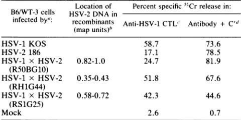
TABLE 1. Mappingof the HSV-2-encodedfunction(s) causing reducedsusceptibility tolysisby HSV-specificCTL
Locationof Percentspecific 51Crreleasein: B6/WT-3 cells HSV-2 DNA in
infectedby": recombinants Anti-HSV-1CTL' Antibody + C" (mapunits)'
HSV-1 KOS 58.7 73.6
HSV-2 186 17.1 78.5
HSV-1 x HSV-2 0.82-1.0 24.7 81.9
(RS0BG10)
HSV-1 x HSV-2 0.35-0.43 51.8 67.6
(RH1G44)
HSV-1 x HSV-2 0.58-0.72 42.3 44.6
(RS1G25)
Mock 2.6 0.7
"'Targetcells wereinfected with thedesignatedvirus strain at amultiplicity of infection of 2.5 or mock infected with Tris-buffered saline. The cells were labeledwith 250,uCiof51Crandharvested 14hpostinfection.
b Approximatelocation.
cLymphocyte cytotoxicitywasdetermined in a 5-h51Crrelease assay at
37°C.CTLwereused at aneffector-to-targetcell ratio of 50:1.
d Targetcells were treated with hyperimmune rabbit anti-HSV sera (1:10) at 4°Cfor 1 h followedbyrabbit lowtoxicityC' (1:20) for1 hat37°C.
NUMBER OF RESPONDING CELLS
(103
PER WELL)2 4 8 16
V,
-j
w
C,
z
2
L.)
FIG. 1. Frequency of HSV-specific CTLp able to recognize
KOS-, 186-, and RSOBG10-infected B6/WT-3 cells.CTLpfrom mice immunized 5days previously with HSV-1 KOS (A) orHSV-2 186 (B) were expanded under limiting dilutionconditions(seethetext). Thelytic activity of individualcultureswasdeterminedby splitting each culture fourfold and assaying against KOS- (@), 186- (0),
RSOBG10- (O), or mock-infected (data not shown) target cells.
HSV-specific CTLp frequencyestimatesagainstinfected target cells were as follows: panel A, KOS, f= 1/6030, P = 0.55; 186,f = 1/24290,P=0.10; R5OBG10,f= 1/9003,P=0.15;panelB, KOS, f= 1/1914, P = 0.82; 186, f= 1/10219, P = 0.21;R50BG10, f=
1/3932, P=0.90. Thefrequency ofpositive cultures against mock-infected cells was too low to determine.
thefluorescenceintensityispresentedasarbitraryunitson a
three-decade logarithmicscaleon thexaxis. RESULTS
Differential susceptibility of HSV-1- and HSV-2-infected B6/WT-3 cellstolysis by CTL. In Table 1is summarizedthe
relative susceptibility ofB6/WT-3 cells infected with HSV-1 strain KOS or HSV-2 strain 186 to lysis by HSV-specific CTL. First, HSV-2-infected cells exhibited a reduced
sus-ceptibilitytolysis by CTL compared with cells infectedwith
HSV-1. Second, by using HSV-1 x HSV-2intertypic
recom-binants with a precisely characterized genome structure, it
waspossible to map the HSV-2-encodedfunction(s) associ-ated with the reducedsusceptibilityto0.82to1.00 mapunits ofthe HSV-2 genome. Cells infected with the recombinant
RSOBG10, the genome of which contains only these
se-quences of HSV-2 DNA, exhibited a levelofsusceptibility comparable to that of HSV-2-infected cells. It should be noted that therewas not an absolute reductionto the same levels asthoseof HSV-2. R50BG10-infected cellsexhibited
a susceptibility to lysis intermediate to those of KOS- and
on November 10, 2019 by guest
http://jvi.asm.org/
[image:3.612.48.288.531.651.2]LLJ
(I)
LLJ
cr
nU
40
186 R5OBGIO
[image:4.612.85.297.62.291.2]B6/WT3CELLS INFECTEDBY:
FIG. 2. Lytic activity of individual oligoclonal cultures against KOS-, 186-, and R5OBG10-infected B6/WT-3 cells. Each point
represents the percentspecific51Cr releaseobtained from individual
culturesat the 32,000 respondercell inputlevel. The broken lines
represent the 95% tolerance limits forthedetermination ofpositive lytic activity againsteach labeled target cell.
186-infectedcells.Recombinantgenomesexpressing regions of HSV-2 DNA other than the 0.82- to 1.00-map-unit se-quencesexhibitedasusceptibilitytolysis comparabletothat ofHSV-1-infected cells. These resultsareinagreementwith ourpreviously published findings (4, 5).
Despitethe differentialsusceptibilitytolysis by CTL,cells infected with the wild-type and the recombinant strains of HSV were equally susceptible to lysis mediated by
poly-clonal anti-HSVantibodyinthepresenceof C'(Table1 [4]).
This indicates that the infected cells are abletoprocessand
express viral antigens importanttothe humoralresponse to HSVinfection and that the reduced susceptibility of HSV-2-infected cells to lysis by CTL is probably not due to an innate resistance of the cells tothe mechanisms of immune
cytolysis.
Theability of unlabeled HSV-1- and HSV-2-infected cells toinhibit thespecific lysisof labeled HSV-1-infected targets was investigated in acold target inhibition assay. Although the overall levels of inhibition obtained were low, it ap-peared that HSV-2-infected cells were less effective. This suggests that HSV-2-infected cells may be less efficiently recognized by the CTLpopulation (data notshown).
Frequency of CTL precursors with specificity for HSV-1-andHSV-2-infected cells. There were potentially two
possi-ble explanations for the reduced susceptibility of HSV-2-infected cellstolysis by HSV-specificCTL. Onepossibility, as mentionedpreviously, may be that HSV-2-infected cells are recognized less efficiently by the cross-reactive,
HSV-specific CTLpopulation. An alternativeexplanationmaybe that fewer CTLp with the potential ofrecognizing HSV-2-infected cells are generated in vivo during the primary
responseto HSV. Thesepossibilities weretested by assess-ingthefrequencyandlytic potentialof theprogenyof limited numbers ofHSV-specific CTLp. Lymphnode cells obtained frommiceimmunized 5days previouslywerecultured under
limiting dilution conditions under whichpotentially all virus-specific CTLp may expand (30, 31). We determined that the effector cells that lyse HSV-infected cells in this assay express the Thyl+, Lyt2+ phenotype, with a lower but significant level ofLytl expression(data not shown). Figure
1 shows the relative frequencies of CTLp generated in response toimmunization with HSV-1 KOS. The frequency of cultures with lytic activity against HSV-1 KOS-infected cells
(f
= 1/6030; P = 0.55) was higher than for either R50BG10-infected (f= 1/9004; P = 0.15) or HSV-2 186-infected(f
= 1/24920;P = 0.10)cells. Moreover, this finding was not due to the generation of an HSV-1 KOS-specific response, assimilar results (Fig. 1) were observed for CTLp generated in response to HSV-2 186 (KOS-infected, f =1/1914, P = 0.82; R50BG10-infected, f= 1/3932, P = 0.90;
186-infected,
f
= 1/10219, P = 0.26), which indicates that cross-reactive HSV-specific CTL were generated. There-fore, the consistent finding of these experiments is that the reducedsusceptibilityof HSV-2 186-infected cells to lysis by CTL reflectsalowerfrequencyof CTLp with the potential to recognize the HSV-2-infected cells.The results shown in Fig. 2 illustrate an important point obtained from this assay system. At aresponder cell input number at which all lethal dose cultures were positive
against KOS-, 186-, and R5OBG10-infected cells, the levels of the percentage of specific 5tCr release obtained from 186-infected targets were demonstrably lower than those
obtained from the KOS-infected cells. The lysis of the
R5OBG10-infected cells was intermediate. No individual culture exhibitedequivalentlevels oflytic activity against all three targetcells,whichsuggests afundamental difference in theabilityof the different targetcells to be recognized by the
lytic progenyofthe HSV-specific CTLp generated in vivo.
Levels of expressionofHSV-encoded glycoproteins on the surfaceofHSV-infectedB6/WT-3cells.If the reducedlysis of HSV-2-infected cells were due to a decreased level of
recognition by CTL, this impliesareduction or alteration in the essentialantigenic structures expressed on the infected celi surface. Virus-specific CTL recognize antigenic deter-minants resulting from an association between the viral
antigen and the class I H-2 antigens on the surface of infected cells (9, 40). Therefore, reduced recognition may
indicate aninadequate expression of either or both of these
antigens. Theexperimentsshown in Table1indicatethat all
wild-type, mutant, and intertypic strains of HSV so far
tested are susceptible to lysis by anti-HSV antisera in an
antibody-dependent, complement-mediated cytolysis assay. While providingevidencefor the presence of the
glycopro-tein specieson the surface of infectedcells, noinformation
on the relative expression of the individual glycoproteins
couldbedeterminedbythis method. Therefore, the expres-sionofHSV-specific glycoproteinswasassessedby
fluores-cent flow cytometric analysis with monoclonal antibodies
specific for individual glycoprotein species of HSV-1 and HSV-2. Themonoclonal antibodieswereextensivelytitrated and used at thedilution correspondingto maximal
binding.
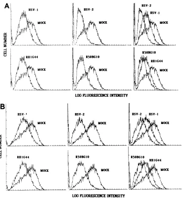
Thefluorescentprofilesof cells infected with HSV-1KOS,
HSV-2 186, or the intertypic recombinants RH1G44 or
R5OBG1O for12 hand stained forthe surfaceexpressionof the individual glycoprotein species are shown in
Fig.
3.Negligible bindingto the mock-infected cellswasobserved. It is evident from the fluorescence profiles obtained for KOS- and 186-infected cells that each of the
glycoprotein
speciesisexpressed onthecell surface. Bythisanalysis, it
was found that strain 186-infected cells express
approxi-mately atwofold lower level of all theglycoproteinstested
on November 10, 2019 by guest
http://jvi.asm.org/
HSV-2 (sB-2)
Il
LOGFLUORENCE INTENSITY
E
, HSV-2 (gC-2)
1.~~~~~~A
! ~~~R50BGIO (sC-I)
,.,.,,,,,
LOGFLUORESCENCE INTENSITY
I
LOG FLUORESCENCE INTENSITY
LOGFLUORESCENCEINTENSITY
[image:5.612.66.450.54.602.2]HSV-2 (gD-2)
FIG. 3. Expression ofHSV-specific glycoprotein speciesonthe
surface of infected B6/WT-3 cells. B6/WT-3 cells infected for 12 h
with HSV-1 KOS, HSV-2 186, orthe HSV-1 x HSV-2 intertypic recombinantsRH1G44orR5OBG10wereassessed for the levels of
surfaceexpression ofgB (A), gC (B), gD (C), gE(D),orgG (E) by
fluorescenceflow cytometry(seetextfor the monoclonal antibodies
used). Each infected cell population was compared with
mock-infectedcells stained under identical conditions.
LOGFLUORESCENCE INTENSITY
compared with KOS-infected cells. Consistently
quantita-tive differences in the expression of certain glycoproteins, especially gB,wereobserved,but results ofrepeated exper-iments suggest that this is most likely attributable to
varia-tions in the affinity or avidity of the individual monoclonal
antibodies. With this qualification, the results suggest that
thereducedsusceptibilityof strain186-infected cellstolysis by HSV-specific CTL may be attributable in part to an inappropriateor inadequate expression of certain
glycopro-teinspecies, but thisalone couldnotaccountfor the
obser-vation.
The HSV-2-encoded function(s) responsible for the re-ducedsusceptibilitytolysis byCTLmapto0.82to1.00map units of the HSV-2 genome. It is possible that the reduced
lysis may be directly attributable to the expression of a glycoprotein speciesencoded in thisregionof HSV-2 DNA. This could be because a glycoprotein in this region is the
major target antigenfor CTL, but the HSV-2 glycoprotein
A
sSV-I (gB-1)...
I
... ^.IHIG44 (gB-2)
t<I
B
2 C-)
HSV-I (gC-l)
I
4,
pJ
....<.... *^X~
I.
HSV-I (3D-I)
011
i
C
2
C-)
on November 10, 2019 by guest
http://jvi.asm.org/
¾l~, BSV-2
II MOCK
,li
BSV-2 sv- OC
hK
MOCLOGFLUORESCENCEINTENSITY
: z
[image:6.612.133.505.58.459.2]LOGFLUORESCENCEINTENSITY
FIG. 4. Expression of the class I H-2 antigenson thesurfce ofHSV-infected B6/WT-3 cells. B6/WT-3 cellswereinfected for 12 h with
HSV-1KOS, HSV-2 186,orthe HSV-1 x HSV-2intertypic recombinants RH1G44orR5OBG10, and assessed forthe expression of H-2Kb
(A)orH-2Db(B) antigensonthe cell surfaceby fluorescence flowcytometry(seetextfor themonoclonal antibodies used). Each infected cell populationwascompared with mock-infected cells stained under identical conditions.
may not serve as well as its HSV-1 counterpart in this regard. As pointed out above, primary HSV-specific CTL arecross-reactive, and it has been shown that ofthe glyco-proteins encoded within this region, gD-2 and gE-2express mainly cross-reactive determinants (23), whilegG is
appar-ently type specific (20). We were interested to determine whether the reduced susceptibility to lysis could be over-comeifbothgD-1 andgD-2were expressed ontheinfected cell surface. B6/WT-3 cellswereinfected with the intertypic recombinant 3-3, an insertion mutant which coexpresses gD-1 and gD-2 (11). Using a monoclonal antibody that
recognizesdeterminants exclusivetogD-1andasecond that recognizes cross-reactive determinants, we were able to confirmthat bothgD speciesareexpressedonthesurface of infectedB6/WT-3 cells (datanotshown). As showninTable
2, 3-3-infected cells were lysed to the same level as cells infected withtheparentalrecombinantR5OBG13 and similar to the levelobserved for 186-infected cells. Therefore, the
coexpression ofgD-1 and gD-2 did not overcome the
re-ducedsusceptibility tolysis, whichindicates either that the HSV-2-encoded function(s) responsible was indepenent of
gD expression or that the HSV-2 encoded function was dominant.
Expression of H-2antigens onthesurface of HSV-infected B6/WT-3 cells. If the reduced susceptibility of HSV-2-infected cellsto lysis byCTL is the result of reducedtarget cellrecognition,butnottotallyattributabletotheexpression
of HSV-2 glycoprotein species on the surface of infected
cells, an alternative explanation may be that HSV-1 and HSV-2 cause a differential reduction or alteration in the
expression of the class I H-2 self-antigens. This possibility wasalsoinvestigated byfluorescentflowcytometric analysis of infected cells stained with monoclonal antibodies specific
for H-2Kb or H-2Db. Mock-infected B6/WT-3 cells ex-pressed high levels of H-2Kb and H-2Db (Fig. 4). Although not formally tested by two-color fluorescence, it was ex-pected that the antigens are coexpressed, because greater
than90% of cells weregradedas positivefor eachantigen.
HSV-I
MOCK
A
: z C-)
on November 10, 2019 by guest
http://jvi.asm.org/
TABLE 2. Coexpression ofgDofHSV-1 and HSV-2 does not overcomeHSV-2-induced lowered susceptibility tolysisby
HSV-specific CTL
Percentspecific91Crreleasein: B6/WT-3 cells anti-HSV-i CTLh Antibody +C"'
infectedby":
Expt1 Expt2 Expt1 Expt2
HSV-1 KOS 57.1 66.3 76.5 80.9
HSV-2186 8.1 26.4 85.0 84.6
HSV-1 x HSV-2 10.7 18.7 30.8 31.2
(R50BG13-1Aj)d
3-3d 15.3 24.6 64.1 61.9
Mock 3.1 6.1 1.7 1.6
aTarget cellswereinfected withthedesignatedvirus strain at amultiplicity ofinfection of2.5 ormockinfected with Tris-buffered saline.The cells were labeled with250pLCiof51Crandharvested14hpostinfection.
bLymphocytecytotoxicitywasdetermined ina5-h51Crreleaseassay at 37°C. CTLwereusedat aneffectortotargetcell ratio of50:1.
cTarget cellsweretreated withhyperimmunerabbit anti-HSVsera(1:10)at 4°C for1 hfollowed by rabbit lowtoxicity C' (1:20) for1h at37°C.
dR50BG13-1A1 is the HSV-1 xHSV-2recombinant virus R50BG13 made TK+ and araTs.3-3isaninsertionmutantcontainingthe DNAfor HSV-1gD inthetkgeneofR50BG13-lA1.
However, it was consistently found that H-2Kbexpression was lower than
H-2Db
expression, as indicated by a lower mean peakfluorescence.Infectionwith both HSV-1 KOSorHSV-2 186 resulted in a decrease in the expression of serologically detectable H-2Kb and H-2Db (Fig.4) butwas morepronounced follow-ing HSV-2 infection. This was evidentfrom theshift to the
leftandlowermeanpeakfluorescence oftheHSV-2-infected cells, which indicates that a greater number of cells are
registering in the lower fluorescence channels. This effect could be attributedtofunction(s) associatedwith the 0.82- to
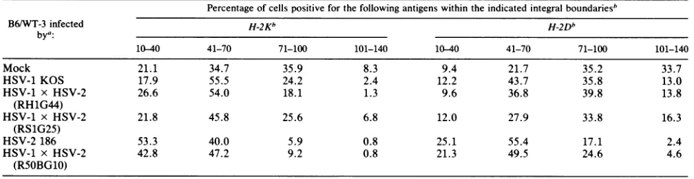
1.00-map-unit region oftheHSV-2genome, asinfectionwith theintertypic recombinant R5OBG10had asimilar effect on H-2 expression as that with HSV-2 186. This was empha-sized by the results presented inTable 3, which represents an integration analysis of the fluorescence profiles of
in-fected and uninfected cells, showing the percentage ofthe total cells that registered within certain arbitrary
fluores-cence intensity limits. It can be seen that HSV-1 KOS and the recombinants RH1G44 and RS1G25 caused a similar reduction inH-2expression, asshownby theincrease inthe percentage ofcells thatregister inthelowerintensity
chan-TABLE 4. Lysis of HSV-infected B6/WT-3 cells by SV40-specific CTL
Percentspecific5"Crreleaseinthefollowing CTL and theindicated expt no.":
B6/WT-3 cells
infected by: Anti-SV40 Anti-HSV
1 2 3 1 2 3
Mock 58.9 49.6 79.0 0.5 2.9 25.6
HSV-1KOS 27.7 21.7 71.4 33.3 42.1 75.3
HSV-2 186 7.8 19.7 37.8 4.7 15.0 45.5
HSV-1 x HSV-2 18.4 20.1 54.6 12.3 22.2 64.0 (R50BG10)
aSV40-specificandHSV-specific CTLweregeneratedinC57BL/6miceand used at aneffectortotarget ratio of 50:1 in a 5-h assay.
nels. HSV-2 186 and the recombinant R50BG10 had a more pronounced effect.Therefore, the ability of a particular virus toreduce H-2expression closely paralleled the susceptibility of cells infected with that virus to lysis by HSV-specific CTL.Furthermore, the increased ability of HSV-2 to reduce H-2appearedto map to the 0.82- to1.00-map-unit regionof the HSV-2 genome.
Recognition ofHSV-infectedB6/WT-3cellsbySV40-specific CTL. The results presented above suggest that the reduced susceptibility of HSV-2-infected cells to lysis by HSV-specific CTL is a direct consequence of the reduction in H-2 expression on the surface of the infected cell. However, it
cannot be assumed thata reduction in serologically detect-able H-2 necessarily reflects a decreased ability of these molecules to act as restriction elements for self-restricted CTL.Therefore, weinvestigated the effect of infection with HSV-1, HSV-2, and the intertypic recombinants on the recognition and lysis of B6IWT-3 cells by
H-2b-restricted
CTL clonesspecific foranunrelatedthird-party antigen,the
SV40-specific large T antigen present on the surface of
B6/WT-3 cells. Large T antigen has been shown to be the mostlikely candidate for the SV40 tumor-specific transplan-tation antigen (37). The results obtained (Tables 4 and 5) demonstrate three distinct groups withrespect to
suscepti-bility tolysis by SV40-specific CTL. These groups directly
reflect theability of the virus strain to reduce the expression of class I H-2 antigen expression. Furthermore, infection withHSV did not cause adecrease in thesurface expression
ofSV40largeTantigenonthecellsurface (datanotshown).
Therefore, it appears that the reduction in expression of
TABLE 3. Integration analysis ofH-2expression fluorescence profiles of HSV-infectedanduninfected B6/WT-3 cells
Percentage ofcellspositive forthefollowing antigenswithin the indicatedintegralboundariesb
B6/WT-3 infected H-2Kb H-2Db
bya:
10-40 41-70 71-100 101-140 10-40 41-70 71-100 101-140
Mock 21.1 34.7 35.9 8.3 9.4 21.7 35.2 33.7
HSV-1 KOS 17.9 55.5 24.2 2.4 12.2 43.7 35.8 13.0
HSV-1 x HSV-2 26.6 54.0 18.1 1.3 9.6 36.8 39.8 13.8
(RH1G44)
HSV-1 x HSV-2 21.8 45.8 25.6 6.8 12.0 27.9 33.8 16.3
(RS1G25)
HSV-2 186 53.3 40.0 5.9 0.8 25.1 55.4 17.1 2.4
HSV-1 x HSV-2 42.8 47.2 9.2 0.8 21.3 49.5 24.6 4.6
(RSOBG10)
aTargetcells wereinfectedwiththedesignatedvirusstrain atmultiplicityof infection of 2.5for14h or mockinfectedwithTBS. The cells were reacted with monoclonalantibodiesspecific forH-2KborH-2Dbandlabeledwith afluoresceinisothiocyanate-conjugated second-step antibody.
bBoundariesaredefinedbyfluorescence units.
on November 10, 2019 by guest
http://jvi.asm.org/
[image:7.612.49.289.95.206.2] [image:7.612.48.552.562.692.2]TABLE 5. Lysis ofHSV-infected B6/WT-3 cellsby SV40-specificCTL clones
Percentspecific5'Crreleaseby thefollowing B6/WT-3 cells SV40-specific CTLclonesa:
infected by: Expt 1 Expt2
10H5 20C7 1OH5 B12
Mock 54.8 92.3 83.3 87.6
HSV-1KOS 39.0 78.5 78.8 85.5
HSV-1 x HSV-2 39.1 75.5 77.5 84.0
(RH1G44)
HSV-2186 14.5 52.8 59.5 72.2
HSV-1 x HSV-2 14.0 60.6 62.5 78.2
(R5OBG10)
a51Cr-labeledtarget cells were reacted withSV40-specific CTL clonesat an
effector to target ratio of 10:1 in a 5-h assay.
serologically detectable H-2 does indeed reflect a decreased
ability ofthesemolecules to function as restriction elements. DISCUSSION
Murine virus-specific CTL recognize infected cells viaa receptor specific for virus-encoded antigens present in the
infected cell membrane only in association with the class I
antigens ofthe H-2 complex (9, 40). Reducedrecognition by
CTLmaybe duetoeither loworinappropriate expression of
a particular viral antigen that serves as the major target
antigen or to a reduced or altered expression of H-2K or H-2D self-antigens or both. However, the first explanation
appears to be unlikely because both HSV-1- and
HSV-2-infectedcells were equally susceptible tolysis byanti-HSV antibody and C' (Table 1 [4, 5]), and analysis of the
expres-sion of individual HSV-1- andHSV-2-specific glycoprotein species by fluorescent flow cytometry confirmed that cells
infected with either virus strainexpressedafullcomplement
of viralglycoproteins ontheir surface. Itwasobserved that the detectable levels ofgB-2 expression were consistently lowerthan those of gB-1. This either may be due to a real
inability ofgB-2 tobe expressed at high levels on infected cells or may reflect a lower affinity of the gB-2-specific monoclonal antibody for this glycoprotein species.
Never-theless, it is unlikely that the low expression of gB-2 is
responsible for the reducedsusceptibility of HSV-2-infected cellstolysis,astheintertypic recombinant RH1G44, which confers the HSV-1 level ofsusceptibility to infected cells, alsoexpresses relatively lowlevels ofgB-2.
Itwasfoundthat the expression of both H-2K and H-2D
antigens werereduced after infection with both HSV-1 and
HSV-2. However, HSV-2 causedagreater reductionin H-2
expression than HSV-1, and the HSV-2-encoded function
responsible for the greater reduction in expression of H-2
mappedto0.82 to 1.00 mapunits of the HSV-2 genome. This
providesstrongcircumstantialevidencethat thereductionin the class I H-2antigens may bedirectly responsible for the HSV-2-associated reduction in susceptibility to lysis by HSV-specificCTL. Thepossibilitythat thereduction in H-2
expression causes the reduced susceptibility to lysis of HSV-2-infected cells was further reinforced in an assay in which an unrelated third-party antigen present on the surface ofB6/WT-3cellswasused. It wasfound thatB6/WT-3cells infected with HSVwerelesssusceptibletolysis byboth bulk
culture CTL and CTL clones specific for the SV40
tumor-specific transplantation antigen and that the ability of a
particularvirus straintoreduce target celllysiswasdirectly
related to its ability to alter H-2 expression. The surface
expressionofSV40largeTantigen, thelikely candidatefor SV40tumor-specifictransplantationantigen (37), was
unaf-fected by HSV infection, further emphasizing the
impor-tanceofH-2 reductionin reducedsusceptibilityto lysis.
Thereduction inH-2 expressiononhost cellsas a
conse-quence ofviral infection has beenreported in a numberof
investigations(2, 10, 12, 29).Therearemany ways in which the interaction between an infecting virus and a host cell
couldleadtothequalitative changeorquantitative reduction
in theexpression ofH-2antigens. One possibilityis that the virus may inhibit host cell protein synthesis, leading to
decreased denovosynthesisof H-2. It is known that HSV-2 shutsoffhostcellprotein synthesismorerapidlythan HSV-1 (8), and it islikelythatthispropertyplayssome roleinH-2
reduction in infected B6/WT-3 cells. There is reason to suggestthat this is not thesole explanation. The HSV-1 x HSV-2 intertypic recombinant R50BG10, with a genome
containing
HSV-2 DNAfrom0.82to1.00 mapunits,causes a decrease in H-2 expression equivalent to that of HSV-2186. However, the HSV-2-encoded functions associated
with the rapid shutoff ofhost cell protein synthesis mapto
0.52to0.59mapunits(7, 13), andR5OBG10 shuts downthe
protein synthesis of B6/WT-3 cells much like HSV-1 KOS (S. R. Jennings,P.L. Rice,and S. S.Tevethia,unpublished data).
A second possibility is that the interaction between the
viral antigenand H-2 in thecellmembrane that leads to the
formation of determinants recognizable by H-2-restricted CTL causes aqualitative alterationin the native H-2
mole-cule (10). Such an altered H-2 molecule may no
longer
express the epitopes
recognized
by theH-2-specificmono-clonalantibodies used in this study. We have shown
previ-ouslythat
H-2b-restricted,
HSV-specific CTLrecognizeviralantigen primarily in association with theH-2Kb gene
prod-uct, suggesting a preferential association with this
self-antigen (14), and it was observed in this study that HSV
infectionapparentlycauses agreaterreductionin the
expres-sion ofH-2Kb than
H-2Db.
The difference in the ability ofHSV-1 and HSV-2toreduce H-2expressionin alllikelihood
represents one aspect of a series ofbiological differences
between the two serotypes. Other
possible
explanations
includeavirus-induceddestabilization ofthe H-2molecules within the cell membrane, leading to increased shedding, destabilization of the
protein itself,
leading
to morerapid
turnover, areduction in thetranscription ofthe class I H-2
mRNA, or a destabilization of the class I H-2 mRNA, leadingto ahigherrateofdegradation ordecreased transla-tion. These
possibilities
are currentlyunderinvestigation.A question that arises from this study is the
potential
biological
significance
of a reduction in the class I H-2antigensfollowinginfection with HSV. It has been shown in the
experimental
adenovirus model that theability
of ade-novirus 12to affect theexpression
of the class I moleculeswasdirectly related toits
oncogenic ability
invivo (32,35).
The oncogenic potential presumably reflects the
ability
of the transformed cells to evade the immune system(35).
Whether such aneffectof HSV is
occurring
invivo has yetto be established, but the differential effects ofHSV-1 and HSV-2 on class I H-2
expression
mayexplain
in part thehigher
pathogenicity
of HSV-2 in mice (17). It should be noted that Yasukawa andZarling
(39),studying
humanHSV-specific CTL clones, didnot observe a reduced
sus-ceptibility
of HSV-2-infected target cellscompared
withHSV-1-infected cells. However, the CTL in their
experi-ments were restricted to the
recognition
of HSVglycopro-teins in association with the class II
major
histocompatibility
on November 10, 2019 by guest
http://jvi.asm.org/
[image:8.612.71.310.84.200.2]complex antigens.
AdifferentialeffectofHSV-1 and HSV-2 onclassI-restricted humanHSV-specific
CTL has not been determined.An
important
furtherconsiderationis thecriticallevels of surface H-2expression
that willprovide
anadequate
target for CTLrecognition. Although
wedemonstrated that there is a reduction in theexpression
ofclass I H-2 antigens and a concomitantreduction inlysis by CTL,
thereduction in H-2expression
wasrelative,
not absolute. The results suggest that ahigh
level of H-2 must beexpressed
for adequaterecognition by HSV-specific
CTLand thatexpression
below this critical level leads to reducedrecognition.
This issupported by
thefinding
that cell lines that are able toprocessandexpressHSV
glycoproteins
but that express lowlevels of
H-2, compared
with those ofB6/WT-3,
are poor targets forHSV-specific
CTL(S.
R.Jennings,
unpublisheddata).
Another considerationalluded toearlier is thepossi-bility
ofaninnate resistanceofHSV-2-infectedcellstolysis
by
CTL.Thus, although
the cell isrecognized
andthe lethal hit isdelivered,
lysis
does not occur.Although
theequal
susceptibility
ofall HSV-infected cells tolysis by antibody
and C' makes this
possibility unlikely,
it has not beendefinitively
proven. Ourcurrentresearch isconcerned witha moredetailed
analysis
oftheeventsfollowing
theinterac-tion of CTL withtheHSV-infectedtarget cell.
ACKNOWLEDGMENTS
This workwassupported byPublic Health Serviceresearch grants CA27503 and CA 25000 from the National Cancer Institute andin part by Institutional grant IN-109H from the American Cancer
Society.
We greatly appreciate the excellent assistance of Patricia
Thompson in thepreparationof thismanusript.
LITERATURE CITED
1. Berke, G. 1980. Interaction ofcytotoxic T lymphocytes and target cells. Prog. Allergy27:69-133.
2. Bubbers, J. E., S. Chen, and F. Lilly. 1978. Non-random inclusion of H-2K and H-2Dantigensin Friend virusparticles
from mice of various strains. J. Exp. Med. 147:340-351. 3. Campbell,A.C.,F. L.Foley,and S. S. Tevethia. 1983.
Demon-strationofmultiple antigenic sites of the SV40transplantation
rejection antigen by using cytotoxic T lymphocyte clones. J. Immunol. 130:490-492.
4. Carter, V. C., S. R. Jennings, P. L. Rice, andS. S. Tevethia. 1984. Mappingofa herpes simplexvirus type2-encoded func-tion that affects the susceptibility of herpes simplex
virus-infected target cells to
lysis
by herpes simplex virus-specificcytotoxicT lymphocytes.J. Virol. 49:766-771.
5. Carter, V. C., P.L. Rice, and S. S. Tevethia. 1982. Intratypic
and
intertypic
specificityoflymphocytesinvolvedinthe recog-nition ofherpes simplex virus glycoproteins. Infect. Immun. 37:116-126.6. Carter, V. C., P. A. Schaffer, and S. S. Tevethia. 1981. The involvement of herpes simplex virus type 1 glycoproteins in cell-mediatedimmunity. J.Immunol. 126:1655-1660.
7. Fenwick,M.L.,L.S.Morse,and B. Roizman.1979.Anatomyof
herpes simplexvirusDNA. XI.Apparentclusteringoffunctions
effecting rapidinhibition of host DNA andprotein synthesis. J. Virol. 29:825-827.
8. Fenwick, M. L., and M. J. Walker. 1978. Suppressionof the
synthesisof cellular macromoleculesby herpes simplexvirus.J. Gen. Virol. 41:37-51.
9. Finberg, R., and B. Benacerraf. 1981. Induction, control and consequencesof virusspecificcytotoxicTcells.Immunol. Rev. 58:157-180.
10. Gardner, I. D., N. A. Bowern, and R. V. Blanden. 1975. Cell-mediated cytotoxicity against ectromelia virus-infected target
cells. Eur. J. Immunol. 5:122-127.
11. Gibson, M. G., and P. G. Spear. 1983. Insertion mutants of herpes simplex virus have a duplication oftheglycoprotein D geneand express two different forms ofglycoproteinD.J.Virol. 48:396-404.
12. Hecht, T. T., and D. F. Summers. 1976. Interactions ofvesicular stomatitis virus with murine cell surface antigens. J. Virol. 19:833-845.
13. Hill, T. M., R. R. Sinden, and J. R. Sadler. 1983. Herpes simplex virus types 1 and 2 induce shutoff of host protein synthesis by different mechanisms in Friend erythroleukemia cells. J. Virol.45:241-250.
14. Jennings, S. R., P. L. Rice, S. Pan, B. B. Knowles, and S. S. Tevethia. 1984.Recognition of herpes simplex virus antigenson
the surface of mouse cells of the H-2b haplotype by virus-specific cytotoxic Tlymphocytes. J. Imnmunol. 132:475-481. 15. Johnson, D. C., and P. G. Spear. 1982. Monensin inhibits the
processing of herpes simplex virus glycoproteins, their
trans-porttothe cellsurface, andthe egress of virions from infected cells. J. Virol. 43:1102-1112.
16. Killington,R.A., L. Newhook, N. Balachandrin, W. E. Rawls, and S. Bacchetti. 1981. Production of hybrid cells secreting antibodies to herpes simplex virus type 2. J. Virol. Methods 2:223-236.
17. Kirchner, H. 1982. Immunobiology of infection with herpes simplexvirus. InJ. L.Melnick(ed.), Monographs in Virology, vol13. S. Karger,Basel.
18. Lawman, M. J. P., R. J. Courtney, R. Eberle, P. A. Schaffer, M. K. O'Hara,and B. T. Rouse. 1980. Cell-mediated immuniity
toherpes simplexvirus: specificityofcytotoxic T cells. Infect. Immun. 30:451-461.
19. Lemke, H.,G.J. Hammerling,andU.Hammerling. 1979. Fine specificity analysis with monoclonal antibodies to antigens controlled bythemajor histocompatibilitycomplex and by the Qa/TL regionin mice. Immunol. Rev.47:175-206.
20. Marsden,H.S.,A.Buckmaster, J. W. Palfreyman, R. G. Hope, and A. C. Minson. 1984. Characterization of the92,000-dalton glycoprotein induced by herpes simplex virus type 2. J. Virol. 50:547-554.
21. Marsden,H.S.,N. D.Stow,V.G. Preston, M. C. Timbury, and N. M. Wilkie. 1978. Physical mappingofherpes simplex virus-inducedpolypeptides. J. Virol. 28:624-642.
22. Nash, A. A., R. Quartey-Papafio, and P. Wildy. 1980. Cell-mediatedimmunityinherpes simplexvirus-infected mice: func-tionalanalysisoflymphnodecellsduring periodsof acute and latentinfection, with referencetocytotoxicand memory cells. J. Gen. Virol.49:309-317.
23. Norrild, B. 1980. Immunochemistry of herpes simplex virus glycoproteinsCurr. Top. Microbiol. Immunol. 290:67-109. 24. Pereira, L. 1982. Useof monoclonalantibodies toHSV-1 and
HSV-2forserological analysis oftheviralglycoproteins. Dev. Biol. Standard. 52:115-131.
25. Pereira, L.,T.Klassen,and R.J.Baringer.1980.Type-common andtype-specificmonoclonalantibodiestoherpes simplex virus type 1. J. Virol. 29:724-732.
26. Pfizenmaier, K.,H.Jung, A. Starzinski-Powitz, M.Rollinghoff, and H.Wagner.1977. The role of Tcells inanti-herpes simplex virus immunity. I. Induction of antigen-specific cytotoxic T lymphocytes. J.Immunol. 119:939-944.
27. Potter,T.A.,C.Boyer,A.Schmitt-Verhulst,P. Goldstein,and T. V. Rajan. 1984. Expression ofH-2Dbonthe cellsurface in the absence ofdetectable
P-2
microglobulin. J. Exp. Med. 160:317-322.28. Pretell,J.,R.S.Greenfield,andS. S.Tevethia. 1979.Biology of simian virus 40(SV40) transplantation rejection antigen (TrAg). V. In vitro demonstration of SV40 TrAg in SV40 infected nonpermissive mouse cells by the lymphocyte mediated cytotoxicityassay.Virology97:32-41.
29. Rager-Zisman, B.,G.Ju, T. V.Rajan,and B. R. Bloom. 1981. Decreasedexpressionof H-2antigens followingacute measles virusinfection.Cell. Immunol. 59:319-329.
30. Reddehase,M.J.,G.M.Kiel,andU. H. Koszinowski. 1984. The cytolyticTlymphocyteresponsetomurinecytomegalovirus. I.
on November 10, 2019 by guest
http://jvi.asm.org/
Distinct maturationstagesofcytolytic T lymphocytes constitute the cellular immune response during acute infection of mice
with murine cytomegalovirus. J. Immunol. 132:482-489. 31. Rouse, B. T., and H. Wagner. 1984. Frequency of herpes
simplex virus specific cytotoxic T lymphocyte precursors in
lymph node cells of infected mice. Immunology 51:57-64. 32. Schrier,;P.I.,R. Bernards, R. T. M. J. Vaessen, A.Houweling,
and A. J. van der Eb. 1983. Expression of class I major
histocompatibility antigens switched off by highly oncogenic adenovirus 12 in transformed rat cells. Nature (London) 305:771-775.
33. Sethi,K. K., and H. Brandis. 1977. Specifically immunemouse
Tcellscandestroy H-2 compatible murinetargetcells infected with herpes simplex virustype 1 and 2. Z Immunitaetsforsch. Immunobiol. 153:162-173.
34. Sonado, S., Y. Hitsumoto, S. Utsumi, S. Takami, M.Oseto, and
Y. Minimishima. 1981. Preparation of stable target cells for anti-herpes simplex virus cytotoxic T lymphocytes. Microbiol. Immunol. 25:999-1010.
35. Tanaka, K., K. J. Isselbacher, G. Khoury, and G. Jay. 1985.
Reversal ofoncogenesis by the expression ofamajor
histocom-patibility complex classIgene. Science 228:26-30.
36. Taswell, C.1981.Limiting dilutionassaysfor thedetermination of immunocompetent cell frequencies. I. Data analysis. J.
Immunol. 128:1614-1619.
37. Tevethia,S. S.1980.Immunology of simian virus 40,p.581-601.
InG. Klein(ed.),Viral oncology.RavenPress, NewYork.
38. Tognan, M., D. Furlong, A. J. Conley, and B. Roizman. 1981.
Moleculargenetics of herpes simplex virus. V. Characterization ofamutantdefective in ability toform plaquesatlow temper-ature and in a viral function which prevents accumulation of coreless capsids at nuclear pores late in infection. J. Virol.
40:870-880.
39. Yasukawa, M., and J. M. Zarling. 1984. Humancytotoxic T cell clones directed against herpes simplex virus-infected cell. 1. Lysis restricted by HLA class II MB and DR antigens. J.
Immunol. 133:422-427.
40. Zinkernagel, R. M.,and P. C. Doherty. 1979. MHC-restricted cytotoxic^T cells: studiesonthe biological roleofpolymorphic major transplantation antigens determining T-cell restriction specificity, function and responsiveness. Adv. Immunol. 27:51-177.