0022-538X/92/126953-07$02.00/0
Copyright X 1992, AmericanSociety for Microbiology
Sulfation of the
Human
Immunodeficiency
Virus
Envelope
Glycoprotein
HELENEB.BERNSTEIN ANDRICHARD W. COMPANSt*
DepartmentofMicrobiology, UniversityofAlabamaatBirningham, Birmingham, Alabama 35294 Received 8 June 1992/Accepted 4 September 1992
Sulfation isaposttranslational modificationofproteins which occurs oneither the tyrosine residuesorthe
carbohydratemoietiesofsomeglycoproteins. In thecaseofsecretoryproteins,sulfation has been hypothesized to actas a signal for export from the cell. We have shown that the human immunodeficiency virustype 1 (HIV-1) envelope glycoprotein precursor (gpl60) aswell as the surface (gpl20) and transmembrane (gp4l)
subunitscanbespecificallylabelledwith35SO42-.SulfatedHIV-1envelope glycoproteinswereidentified inH9
cells infectedwith the IIIB isolate of HIV-1 and in the cell lysates and culture media of cells infected with vacciniavirus recombinants expressing a full-length ortruncated, secreted form of the HIV-1 gpl60 gene.
N-glycosidaseFdigestion of 355042--labelled envelope proteins removedvirtually all radiolabel fromgpl60, gpl20, and gp4l, indicating that sulfatewaslinkedtothecarbohydratechainsoftheglycoprotein.The
3S042-labelwasatleastpartiallyresistanttoendoglycosidaseHdigestion,indicating thatsome sulfatewaslinkedto complex carbohydrates. Brefeldin A,acompound thatinhibits the endoplasmic reticulumtoGolgitransport of glycoproteins, was found to inhibit the sulfation of the envelope glycoproteins. Envelope glycoproteins synthesizedincellstreated withchloratefailedtoincorporate
35s042-.
However,HIVglycoproteinswerestillsecreted fromcells inthepresenceofchlorate, indicating thatsulfation isnot arequirement for secretion of
envelopeglycoproteins.Sulfationof HIV-2 and simian immunodeficiency virus envelopeglycoproteinshas also been demonstrated by using vaccinia virus-based expression systems. Sulfation is a major determinant of
negative charge and could playarole inbiological functionsandantigenicproperties of HIVglycoproteins.
Human immunodeficiency virus type 1 (HIV-1) is the
etiologicagentof AIDSand related disorders(5, 13, 27,38). The HIV-1 envelope glycoprotein mediates attachment of the virusto cellsvia the cellular receptor, CD4(24, 29, 30,
40). Previous studies on the transport of HIV envelope
proteinsindicate that theyarefirst synthesized asa polypro-tein precursor(gp160),themajorityof which is retained inan intracellular compartment and undergoes subsequent degra-dation (1, 43, 50, 52). Afraction ofgpl60is proteolytically
cleaved in the Golgi complex toyield the transmembrane
(gp4l)andsurface (gpl20)subunits of thematureprotein(9,
11, 15, 36,47). gpl20andgp4lareexpressedonthe surfaces of infected cells and are incorporated into buddingvirions
(48). Following proteolytic cleavage,
gp120
andgp4lremain associated via noncovalent interactions which appear to besufficiently weak to allow the shedding large amounts of
gpl20 from thesurfaces ofcells into theextracellular envi-ronment (42, 44). The envelope glycoproteins are highly
glycosylated, with carbohydrate moieties estimated to ac-count for one-half of the molecularweightofgpl20 (1, 43). Sulfation is a
posttranslational
modification ofproteins
thatoccursinvirtuallyall animal cellsandhas been
hypoth-esized toplay a role in the transportof secretory proteins
from the cell (20). Other possible functions of sulfation include contributions to cell differentiation and cell-cell
interactions (17). Sulfation is also a
major
determinant ofnegative charge. Inorganic sulfate is transferred from a carriermolecule,3'-phosphoadenosine
5'-phosphosulfate,
to proteins by sulfotransferases located in the transGolgi
network (TGN) (18, 20). Two types of protein sulfation
* Correspondingauthor.
tPresentaddress:Departmentof
Microbiology
andImmunology,
EmoryUniversity,Atlanta,GA 30322.
modifications have beenreported: the addition of sulfate to tyrosine, and the addition of sulfate to the carbohydrate moieties ofglycoproteins. The Vl capsid protein of polyo-mavirus is modified by tyrosine sulfation (28).
Carbohy-drates have been found to be modified by the addition of sulfate to N-acetylglucosamine or terminal mannose resi-dues (31, 39, 53). Sulfation of thecarbohydratemoieties of viral envelope glycoproteinshas also beenreported(19, 31, 37, 39).
Wehaveinvestigatedtheincorporation of sulfate into the envelopeglycoproteins of HIV-1 and other lentiviruses. The association of the sulfate with the HIV-1envelope
glycopro-tein has been examinedby using enzymaticdeglycosylation.
We have also determined the effects of brefeldin A(BFA)
and chlorateon envelopeglycoprotein synthesis, sulfation,
and transport, using recombinant vaccinia viruses which expressfull-lengthortruncated, secreted forms of the enve-lopeglycoproteins.
MATERIALSANDMETHODS
Reagents.Brefeldin AwasobtainedfromEpicenter Tech-nologies (Madison, Wis.). Sodium chlorate was purchased
from AldrichChemicalCo.(Milwaukee, Wis.). 5'-Bromo-2'-deoxyuridine, N-glycosidase F (PNGase F), and
endogly-cosidaseH(endo
H)
wereobtained fromBoehringer
Mann-heim(Indianapolis, Ind.).Cells and viruses. TK- 143 cellswere obtained from the AmericanType CultureCollectionand grown in Dulbecco's modification of Eagle's medium containing 10% newborn calfserumand 25 ,ug of
5'-bromo-2'-deoxyuridine
perml. H9 cells were also obtained from the American Type Culturedescribed previously (14). Vaccinia virus recombinants VV-K1 (22), VV-SC11 and VV-env-1 (35), VV-env-4 (16), VV-ST and VV-ROD (32), VV-ST2 (33), and
VV-SIV1A11
(41)were grown as described previously (23).
Protein expression. Confluent TK- 143 cells in 35-mm disheswereinfectedwith recombinant vaccinia viruses ata multiplicity of infection of 5. For radiolabelling, the cells werestarvedfor 30 min in deficientEagle'smedium and then
metabolicallylabelled with either50,Ciof
[35S]methionine-cysteine (Met-Cys) (New England Nuclear) per ml or 100
,Ci
ofNa235SO4(ICN)perml inMet-Cys-deficientorso42-deficient Eagle's medium, respectively, for various time
periods. Cellswere lysed inabuffer containing50 mM Tris
(pH 7.4), 150 mMNaCl, and 1%Nonidet P-40.Lysates were
preclearedwithStaphylococcus aureuspreloaded with nor-mal human antisera, and immunoprecipitations were then carriedout by using pooled HIV-positive antisera and pro-teinA-agarose beads (Pierce) as described previously (35).
Immunoprecipitated proteins were resolved by sodium
do-decyl sulfate (SDS)-polyacrylamide gel electrophoresis
(PAGE)
and autoradiography.PNGase F and endo H digestions. Immunoprecipitated
sampleswereremoved from the agarose beads by boiling the
samplesfor5 min in 10
RI
ofsample loading buffer containing1% SDS, 10% glycerol, 5% mercaptoethanol, and 62 mM Tris (pH 6.8). Aliquots of eluted protein samples were
deglycosylated with 0.4 U of PNGase F for 17 h at 37°C in 100
RI
of buffercontaining 1%N-octylglucoside, 50 mM Tris(pH 7.4), and 10 mM EDTA. Other aliquots were treated with 5 mU of endo H in 100 ,ul of buffer containing 1%
N-octylglucoside, 10 mMTris (pH 7.4), 100 mM NaCl, and
1% SDSfor 17 hat37°C.Thesampleswerethenprecipitated
with 10 volumes of acetone for 2 h at -20°C, and pellets wereredissolved insample loadingbuffer(35)and subjected to SDS-PAGE.
RESULTS
HIV-1envelope
glycoproteins
aresulfated.Atimecourseof sulfate incorporation was performed to establish that the HIV-1envelope glycoproteinsaresulfated andtodetermine the time ofpeak sulfateincorporation. TK- 143 cellswere infectedwithVV-env-1, arecombinantvaccinia virus which expressesthecompleteHIV-1envelopegene,orVV-SC11, a recombinant vaccinia virus which expresses ,-galactosi-dase.Figure
1A shows theincorporationofsulfate into bothgpl60andgpl20atvarious intervalspostinfection. The peak level of sulfateincorporationwasfound to occur between 8 and 12 hpostinfection. Bothgpl60andgpl20werefound in the sulfate-labelled samples; however, in the amino acid-labelled sample, the predominant envelope species found wasgpl60 (Fig. 1B).The observation that onlygpl60is seen in the amino acidpulse-labelledsample, whereas bothgpl60
and gpl20 are seen in the sulfate pulse-labelled sample, indicates that sulfate incorporation is a posttranslational event. However, the finding that a fraction of gpl60 is sulfatedindicatesthat the sulfotransferase enzyme is present at alocationproximaltothe protease which causes cleavage of theenvelopeglycoprotein. To verify that HIV-1 envelope
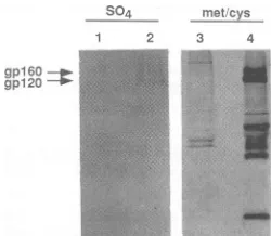
glycoproteins incorporate sulfate during viral infection,
per-sistentlyHIV-IIIB-infected H9 cells were metabolically la-belled with
35S042-,
andviral proteins were examined bySDS-PAGE
(10%
polyacrylamide gel). HIV-1-specificpro-teins with apparent molecular masses of 160 and 120 kDa wereimmunoprecipitatedfrom sulfate-labelled, infected cell
lysates
by pooledHIV-1-positive
sera(Fig. 2, lane 2).gpl60,
A
hourspostinfection 4 6 8 10 12 14 16t
,-U)"zmP"O
B
.4-gpl60 4- gpl20
hourspost infection 4 6 8 10 12 14 16
gp160
FIG. 1. Timecourseof sulfationof the envelope glycoprotein. TK- 143 cells infected withVV-env-1werelabelled with 100 ,Ci of 3SO42-perml(A)or50,Ci of [35S]Met-Cysperml(B) for 2 h,and cellswerelysedatthe indicated timespostinfection. VV-SC11 cells
werelysedat 16 hpostinfection as acontrol. Immunoprecipitated celllysateswereanalyzed by SDS-PAGE (10% polyacrylamide gel) andfluorography.
gpl20, andvariousformsof the capsidproteinswerefound in
immunoprecipitates
from the amino acid-labelled infected celllysates (Fig.
2,lane4).Lanes 1 and 3areradiolabelled,immunoprecipitated
cell lysates from uninfected H9 cells. These results demonstrate that the HIV-1 envelopeglyco-proteins incorporate
sulfate invirus-infectedcells as well as in recombinantexpressionsystems.Sulfation of other lentivirus
glycoproteins.
TK- 143 cells were infected with several recombinant vaccinia virusesexpressing
HIV-1, HIV-2,orsimianimmunodeficiencyvirus(SIV) glycoproteins
todetermine whether the glycoproteins of otherlentiviruses arealso sulfated. Viruses used includeVV-ST, VV-ST2,
and VV-ROD (vaccinia virus recombi-nants which express HIV-2 envelope glycoproteins),VV-SIV1All
(a
vaccinia virusrecombinantwhich expresses the SIVenvelope glycoprotein), VV-env-1,
and VV-SC11. In-fected cellswerelabelled with 100 ,uCiof35SO42-perml or 50,Ci
of[35S]Met-Cys
perml at 10 hpostinfectionfor 4 hprior
tolysing
ofthecells. Celllysateswereimmunoprecip-itatedwith
HIV-2-positive
antisera(obtainedfrom the AIDSrepository)
with theexceptionof theVV-env-1-infectedcelllysate,
for whichpooledHIV-1-positive antisera were used. As shown inFig.
3A, the amino acid-labelledimmunopre-S04 met cys
1 2 3 4
gpl60
[image:2.612.345.531.75.221.2]--pw-gpl20
FIG. 2. Incorporation of sulfate into the HIV-IIIB
envelope
glycoprotein.H9(lanes1and3)andpersistentlyHIV-IIIB-infected H9(lanes2and4)cellsweremetabolicallylabelledwith 50 pCiof
[image:2.612.372.497.559.668.2]4 dayexposure 1dayexposure
A S04 met cys met cys
1 2 3 4 5 6 1 2 3 4 5 6 1 2 3 4 5 6
SU -_- a """"OmI.v
ule
nN "S04
1 2 3 4 5 6
metcys
1 2 3 4 5 6
.~
.4- o
1 2 3 4 5 6 7 8
gp16O
_--o
-4-ogp160tr
gpl120 -A
SO4 meti S04 met/
cys cys
lysates media
FIG. 4. Transport of sulfated glycoprotein. TK- 143 cells in-fected withVV-env-1 (lanes 1, 3, 5, and 7) and VV-env-4 (lanes 2, 4, 6, and8) were labelled with 100p.Ciof S042-per ml or 50pCiof [35S]Met-Cys per ml for4 h at 10 hpostinfection. Media and cell lysatesfrominfected cells wereimmunoprecipitated and subject to SDS-PAGE(10% polyacrylamidegel) and fluorography.
FIG. 3. Modification of other lentiviral envelope glycoproteins by the addition ofsulfate. Vaccinia virus-infected TK- 143 cells
weremetabolically labelled with100,uCi of 35SO42 permlor50pCi
of[35S]Met-Cyspermlfor 4 hat10hpostinfection. Immunoprecip-itated cell lysates were resolved by SDS-PAGE (10%
polyacryl-amidegel). (A)Precursor(Pre)and surface(SU) units of envelope glycoproteins immunoprecipitated from cells infected with W-SC11(lane 1), W-env-1 (lane 2),W-ST(lane 3), W-ST2 (lane 4), W-SIV1All (lane 5), and W-ROD (lane 6); (B) transmembrane (TM)units ofenvelopeglycoproteinsimmunoprecipitatedfrom cells infected withW-SC11(lane 1),W-env-1(lane2), W-ST(lane 3), W-ST2 (lane 4), W-ROD (lane 5), and
W-SIVlAll
(lane 6) (4-week exposure).cipitates yielded precursor and surface subunit proteins of the expected molecular weights, which vary among the
different lentiviruses. All of the envelope glycoproteins examined were found to be labelled with sulfate, as evi-dencedbythefluorograph. The levelof[35S]Met-Cys incor-poration into envelope glycoproteins was higher than the level of 35S042 incorporation, as demonstrated by the
increased intensity ofthe bands in the amino acid-labelled lanescomparedwiththesulfate-labelled lanes(Fig. 3A).On prolongedexposureof thegel, thetransmembrane proteins
were alsofoundtobe labelled with sulfate (Fig. 3B).These results indicatethatsulfationis amotifshared by envelope
glycoproteins ofseveral lentiviruses andthat both the
sur-facesubunit and transmembrane unitcleavageproducts are
sulfated.
Transport offull-lengthand truncatedHIV-1 glycoproteins. Sulfation ofproteinshas beenproposedtoactas asignalfor theexportof secretoryproteinsfromthe cell(20). VV-env-4 is avaccinia virus recombinant whichexpresses a soluble,
truncated form of theenvelopeglycoprotein(gpl60tr)which issecreted into the media of infected cells(16).Wewishedto determine whether sulfated forms of gpl60tr are exported
fromthe cell andtoinvestigatethepossibleroleofsulfation in the transport of secreted envelope glycoproteins. Media andlysateswerecollectedfrom35SO42_- and
[35S]Met-Cys-labelled TK- 143 cells infected with VV-env-1orVV-env-4,and immunoprecipitated proteins were analyzed by SDS-PAGE (10% polyacrylamide gel) (Fig. 4). Sulfate-labelled gp160andgp120wereimmunoprecipitatedfromlysates,and gp120waspresent inthemedium of VV-env-1-infectedcells. Sulfatedgp160tranditscleavageproduct,gp120,wereboth immunoprecipitated from themediumofcells infected with W-env-4, but only a low level of sulfate-labelled gpl60tr
was identified in cell lysates. This result indicates that the
gpl60tr molecules which are secreted into the medium are more highly sulfated than thecell-associated forms.
Tofurtherexamine the role ofsulfation in thetransportof viralglycoproteins, cells expressing viral glycoproteinswere
treated with chlorate, an inhibitor of ATP-sulfurylase, the
firstenzymein 3'-phosphoadenosine 5'-phosphosulfate
bio-synthesis. Chlorate treatmenthas been reportedtoproduce 99% inhibition ofsulfation when used with reduced amino acid concentrations (3, 20). Sodium chloratewas added to themediaatafinalconcentration of 0, 1, or10 mM at 10 h postinfection, 30min priortolabelling of cells,and through-out the
labelling
period. In this experiment, the cellswerelabelled in S04 --deficient and 99% Met-Cys-deficient Ea-gle's media as described by Baeuerle and Huttner (3). As showninFig.5,chlorate hadnoeffectonthelevel ofamino acid-radiolabelled envelope glycoproteins foundin the
me-dium of cells infected with VV-env-1 or VV-env-4. There
was, however, marked inhibition of sulfate incorporation, whichis apparent ata concentration of1 mMchlorate and
appears to bevirtually complete at 10 mM chlorate. Even
under conditions of complete inhibition of sulfation, at 10 mMchlorate, gpl60trwasstill foundtobe secreted intothe medium(Fig.5B) and gpl20wasalsopresentin themedium (Fig. SA). These results indicate that sulfation is not a
requirement for the secretion of HIV envelope glycopro-teins. Chlorate was also examined for its abilityto inhibit
A
A B C A B C
g p120 -10 SWam t
B
A B C A B C
gp160tr_ "
met.' met! S04
[image:3.612.87.287.74.241.2]cys S4cys
FIG. 5. Chlorate inhibition of sulfation. TK- 143 cells were
infected withW-env-1(A)orW-env-4(B).At10 hpostinfection, the cellswerestarved inS042--deficientand 99%Met-Cys-deficient Eagle'smediacontaining0(lanes A),1(lanes B),or10(lanes C)mM
chlorate for 0.5h and thenmetabolically labelled with 100 pCiof
35S042-permlor50p,Ciof[35S]Met-Cysperml for 10 h in media of
thesamecomposition. Celllysatesand mediawere
immunoprecip-itated and resolved by SDS-PAGE(10% polyacrylamide gel) and
fluorography.
[image:3.612.366.525.75.148.2] [image:3.612.329.566.506.639.2]A
VV-env-1 VV-SCl1 VV-env-4
pIg!mlBFA 0 1 5 10 0 1 5 10 0 1 5 10
gp160 t _eso_ _ 4-gpl60tr
; > >,- > >
) a) C ) w C
B
gpl60-_o gp120
-_-)C)CC C) C)cl)n nc:
>>>»>V~
IC>>n
. 1-gp160tr gpl20-_
-BFA +BFA
gow"
-BFA +BFA -BFA +BFA
SO4 metcys
FIG. 6. BFAtreatmentof TK- 143 cellsexpressingtheenvelope glycoprotein.TK- 143 cellswereinfected withVV-env-1, VV-env-4,or
VV-SC11andmetabolicallylabelled with 100ACiof3 S042-permlor50p,Ciof[35S]Met-Cysperml for 4 hat 10 hpostinfection.BFAwas
initially added tocells 30min priortolabellingandwaspresent throughoutthelabelling period. (A) Immunoprecipitatedcelllysatesfrom [35S]Met-Cys-labelledcells with various concentrations ofBFA; (B) immunoprecipitated lysatesfrom35SO42--labelledcellstreated with10 ,ugof BFAperml; (C)immunoprecipitatedmediafrom35S042-_ and[35S]Met-Cys-labelled cells treated with 10p,gof BFA per ml.
syncytium formation in VV-env-1-infected CD4+ HeLa cells. At concentrations ofupto25 mMchlorate,no reduc-tion in HIV-induced cell fusion was observed (data not shown), indicating that sulfation of the HIV-1 envelope glycoproteinisnotadeterminant of thefusionactivityof the HIV-1envelope glycoprotein.
BFA treatmentinhibits sulfation and transport of theHIV-1 envelope glycoprotein. We examined the effect ofBFA, a
fungalmetabolite whichreversiblyinhibitsproteintransport fromthe endoplasmic reticulumto the Golgi complex (12),
onsulfation of the HIV-1 envelope glycoproteins. Vaccinia
virus recombinant-infected cells weretreated with BFA 30
min prior to and throughout the labelling period. We first examined the effect of BFAtreatmenton thesynthesis and
processingofgp160. Figure6A shows that the synthesis of gpl60andgpl60trwasunaffected in VV-env-1- and
VV-env-4-infected cells treated with0, 1, 5,or 10,ugof BFAperml. There was, aspreviously reported (11, 36),a lack ofgpl60 cleavage, which islikelytobe due to the inaccessibility of the envelope glycoprotein to the protease under the condi-tions of BFA treatment. BFA also affected carbohydrate processingofgpl60andgpl60tr, asevidenced bythe lower
apparent molecular weight of the envelope glycoproteins synthesized in the presence of BFA. BFA was found to
inhibit the incorporation of sulfate into the HIV envelope proteins, as demonstrated by the absence of radiolabelled
gpl60, gpl20, and gpl60tr in lysates of sulfate-labelled vacciniavirusrecombinant-infected cellstreated with 10 ,ug of BFA per ml (Fig. 6B). No radiolabelled glycoproteins
were detected in the medium ofcellstreated with 10 ,ug of BFA per ml, whereas radiolabelled glycoproteins were
readilydetected in the medium ofuntreated cells (Fig. 6C). These results indicate that sulfation of the HIV envelope glycoprotein occurs after transport from the endoplasmic reticulum.
Sulfate is linkedtotheenvelope glycoproteinvia
carbohy-drate moieties. To determine thenatureof thesulfatelinkage totheenvelopeglycoproteins,theeffect ofenzymatic degly-cosylationof the HIV-1 envelope glycoproteinswas
exam-ined. TK- 143 cells were infected with VV-env-1 or
VV-SC11 and metabolically labelled for 10 h at 10 h postinfection. Aliquotsofimmunoprecipitated proteinswere
treated with thedeglycosylating enzymePNGaseForendo
H. After PNGase F or endo H treatment, amino acid-labelled glycoproteins (Fig. 7) yielded deglycosylated
pro-teins of theexpectedmolecularweights (15). In the caseof
thesulfate-labelledglycoproteins, PNGase F digestion elim-inated all of the detectable35s42- from all three species of the sulfate-labelled envelope glycoproteins, indicating that thesulfatewaslinkedtothe carbohydrate moieties ofgpl60, gpl20, andgp4l.Inthe endoH-digested samples, however, therewas not complete removal of the35S042, indicating that sulfate was linked to endo H-resistant carbohydrate moieties. Similarresultswerealsoobtainedaftertworounds of enzymatic digestion to ensure that deglycosylation was
complete (data notshown).
DISCUSSION
We havedemonstrated that theenvelopeglycoproteins of theIIIBisolate of HIV-1 and therecombinant vaccinia virus VV-env-1 are radiolabelled with inorganic sulfate and that
[image:4.612.136.475.78.350.2]met cys S04
N P H N P H N P H N P H
I
geglcdgpl60
gipl20_-Mo
deglycosylated
gpl60-'P
deglycosylated gp120_-*
gp4l -_--,
vv-sc11 VV-env-1 VV-SC11
FIG. 7. Deglycosylation of sulfate-labelled envelope
glycopro-tein. Immunoprecipitated cell lysates fromW-env-1-infected TK-143 cells labelledwith lO0 LCi of35so42- per ml or 50 pCi of
[35S]Met-Cysperml for10hat10h postinfectionweretreated with
PNGaseF(lanes P)orendoH(lanesH)or wereuntreated(lanes N),
and samples were analyzed by SDS-PAGE (10% polyacrylamide
gel).
this sulfate is linked to the carbohydrate moieties of the glycoprotein. Sulfated forms ofthe HIV-1 envelope glyco-protein include theprecursor,gpl60,and both thegp120and
gp4l cleavageproducts. Incontrast,the HIV-1 Gag proteins ofpersistentlyHIV-IIIB-infectedH9cellswerenotfoundto
incorporate sulfate. Sulfate-labelled immunoprecipitated proteins fromTK- 143 cells infected withVV-K1, a
recom-binant vaccinia virus which synthesizes Gag proteins, also containedno
sulfated_proteins
(datanotshown). Radiolabel incorporation inthe350S42--labelled
samples canbeattrib-uted only to the incorporation of inorganic sulfate into proteins,asmammaliancells lack the biochemical pathways
necessarytoconvertinorganic sulfate into amino acids (6). HIV-2 and SIV envelopeglycoproteinswerealso foundtobe labelled with sulfate. This posttranslational modification of envelope glycoproteins has also been reported for several other enveloped viruses, including the retrovirus Rauscher leukemia virus (19, 31, 37, 39). Chlorate appeared to
com-pletely inhibit the sulfationof HIV-1 envelope proteins, with
no effecton envelope protein synthesis, proteolytic
cleav-age, or secretion intothe culture media.
Sulfate ispresentonboth theprecursorandcleaved forms
of the HIV envelope glycoprotein. Incontrast,theprecursor
formof the herpesvirusgDprotein isnot sulfated (19). The
presence of both cleaved (gpl20 and gp4l) and uncleaved
(gpl60) forms of sulfated glycoprotein indicates that the sulfotransferases, which are located in the Golgi complex
(20), reside at a location proximal to the cellular protease
whichcausescleavageof theprecursorintothesurfaceand
transmembrane units andthatsomegpl60maybe cleavedat
averylatestepinthetransportprocess.Glycosaminoglycan sulfotransferases and tyrosyl sulfotransferases have been specificallylocalizedtotheTGN(20,46). Since BFA inhibits thesulfation of the envelope glycoprotein,our results
indi-catethat theglycoprotein sulfotransferases arealso located
intheTGN. This result is inagreementwith those of Spiro etal.(46), whoreportedtheinhibitionofchondriotin sulfate
glycosaminoglycanelongationandsulfation of the melanoma proteoglycan core protein with BFA treatment. Endo H
digestion of the sulfate-labelled glycoproteins resulted in incomplete removal of the
35s042-
label, indicating that sulfate isadded, atleast inpart, to complexcarbohydrates which are resistant to endo H digestion. This view is consistent with the idea that sulfation is a lateposttransla-tional protein modification (20), mediated by sulfotrans-ferases in the TGN.
Our results show that the HIV-1 envelope glycoproteins are sulfated and that the sulfate is linked to the carbohydrate moieties of the glycoproteins. Since the carbohydrate com-ponents linked to viral glycoproteins are specified by host cell enzymes, host cell-dependent differences may exist in oligosaccharide structures. The oligosaccharide structures of the viral glycoproteins clearly resemble those present on host cell glycoproteins, and evidence has been obtained that they may represent host cell-specific antigens that are incor-porated into virus particles (21, 25). In the case of the influenza virus HA glycoproteins, the host antigen activity has beencorrelatedwith the presence of a sulfated oligosac-charide linked to the N-terminal region of the HAl subunit (10, 51). The association of a host cell-specific antigen with lentiviruses has recently been demonstrated in the case of SIV, in studies in which protection of rhesus monkeys against virus challenge involved the induction of an immune response to a host cell-specific antigen (8, 26, 34, 45, 49). It will therefore be of interest to further identify the precise structure of the sulfated viral oligosaccharides and their possible role in induction of an immune response. In regard to the role of sulfation in the transport of secretory proteins from the cell, our results indicate that sulfation is not required for the secretion of the envelope glycoproteins. It is possible that sulfate protects glycoproteins from exoglyco-sidicdegradation oraffectsthe proteolytic processing of the envelope glycoprotein. Sulfate contributes negative charge to the surface of the envelope glycoprotein, and this may affect the interaction of virus with cells or extracellular matrix components. It is also of interest that sulfated com-ponents, including the monosaccharide glucosamine-6-sul-fate, have been reported to inhibit HIV-induced cell fusion (4) and appear to affect the early stages of virus-cell inter-action (2).
ACKNOWLEDGMENTS
WethankSimonTucker for helpful discussion and Mark Mulligan forpooled HIV-positive antisera. VV-K1 and HIV-2-positive anti-serawereobtained from the AIDS Research and Reference Reagent Program, Division ofAIDS, NIAID. We also thank Cherie Oliver fortechnicalassistance, Eugene Arms for photographic assistance, andBettyJeffreyfor assistance in preparing the manuscript.
This work was supported by research grants AI 28147 and Al 27290fromtheNationalInstitute of Allergy and Infectious Diseases. HeleneB. Bernsteinwassupported by Institutional Research Ser-vice AwardHLO 7553fromtheNational Heart, Lung, and Blood Institute.
REFERENCES
1. Allan, J. S., J. E. Coligan, F. Barin, M. F. McLane, J. G. Sodroski, C. A. Rosen,W.A.Haseltine, T. H. Lee, and M. Essex. 1985. Major glycoprotein antigens that induce antibodies in AIDSpatients are encoded by HTLV-III. Science 228:1091-1094.
2. Baba, M.,R.Pauwels, J. Balzarini, J. Arnout, J. Desmyter, and E.DeClercq. 1988. Mechanism of inhibitory effect of dextran sulfate andheparin onreplication of human immunodeficiency virusinvitro. Proc. Natl.Acad. Sci. USA 85:6132-6136. 3. Baeuerle, P. A., and W.B. Huttner. 1986. Chlorate-a potent
inhibitorofproteinsulfation inintact cells. Biochem. Biophys. Res. Commun. 141:870-873.
4. Bagasra,O.,P.Whittle, B.Heins, and R. J. Pomerantz. 1991. Anti-humanimmunodeficiencyvirustype 1 activity of sulfated monosaccharides: comparison with sulfated polysaccharides andotherpolyions.J. Infect. Dis. 164:1082-1090.
[image:5.612.60.298.78.186.2]IsolationofaT-lymphotropic retrovirusfromapatientatrisk of acquired immune deficiency syndrome (AIDS). Science 220: 868-870.
6. Cantarow,A., and B.Schepartz. 1962. Metabolismofproteins, p. 529-593.In A. Cantarowand B. Schepartz (ed.), Biochem-istry. W. B.SaundersCo.,Philadelphia.
7. Chege,N.W.,and S. R.Pfeffer. 1990.Compartmentation of the Golgi complex: brefeldin-Adistinguishes trans-Golgi cisternae fromthetrans-Golgi network. J. Cell Biol. 111:893-899. 8. Cranage, M. P., L. A. E. Ashworth, P. J. Greenaway, M.
Murphey-Corb,and R.Derosiers.1992. AIDS vaccine
develop-ments.Nature355:685-686.(Letter.)
9. Dewar, R. L., M. B.Vasudevachari, V. Natarajan, and N. P. Salzman. 1989.Biosynthesisandprocessing of human immuno-deficiencyvirustype 1 envelopeglycoproteins: effects of mon-ensinonglycosylationand transport. J. Virol. 63:2452-2456. 10. Downie, J. C. 1978. Host antigen as the sulphated moiety of
influenzavirushemagglutinin. J.Gen. Virol. 41:283-293. 11. Earl,P.L., B.Moss,andR. W. Doms.1991.Folding, interaction
withgrp78-BiP, assembly, andtransportof the human immuno-deficiencyvirustype 1envelope protein. J.Virol. 65:2047-2055. 12. Fujiwara,T.,K.Oda, S.Yokota, A.Takatsuki, and Y. Ikehara. 1988.Brefeldin A causesdisassembly of the Golgi complex and accumulation of secretory proteins in the endoplasmic reticu-lum. J. Biol. Chem. 263:18545-18552.
13. Gallo,R. C., S.Z.Salahuddin, M.Popovic, G. M. Shearer, M. Kaplan,B. D. Haynes, T.J.Palker, R.Redfield,J.Oleske, B. Safai, G.White, P. Foster, and P. D. Markham. 1984. Frequent detection and isolation ofcytopathic retroviruses (HTLV-III) frompatients withAIDSandatrisk for AIDS. Science 224:500-503.
14. Gartner, S.,and M.Popovic. 1990. Virusisolationand purifica-tion, p. 53-69. In A. Aldovini and B. D. Walker (ed.), Tech-niquesinHIVresearch. Stockton Press, NewYork.
15. Geyer, H., C. Holschbach, G. Hunsmann, and J. Schneider. 1988.Carbohydrates of humanimmunodeficiencyvirus. J. Biol. Chem.263:11760-11767.
16. Hallenberger, S., S. P. Tucker, R.J. Owens, H. B. Bernstein, andR. W.Compans. Secretion ofatruncatedform of the human immunodeficiencytype1 envelope glycoprotein. Submitted for publication.
17. Heifetz, A., and M. D.Prager. 1981. The effect ofbutyrateon sulfated glycoprotein biosynthesis by human kidney tumor cells. J.Biol. Chem. 256:6529-6532.
18. Heifetz, A.,C.Watson, A. R. Johnson, and M. K. Roberts. 1982. Sulfatedglycoproteins secreted by human vascularendothelial cells. J. Biol. Chem.257:13581-13586.
19. Hope, B. R. G., J. Palfreyman, M. Suh, and H. S. Marsden. 1982.Sulphated glycoproteins induced by herpes simplexvirus. J.Gen. Virol. 58:399-415.
20. Huttner, W. B. 1988. Tyrosine sulfation and the secretory pathway.Annu. Rev.Physiol. 50:363-376.
21. Jackson,D.C.,T.A. Dopheide, R. J. Russell, D.0.White, and C. W. Ward. 1979.Antigenic determinants of influenza virus hemagglutinin.Virology 93:458-465.
22. Karacostas, V.,K.Nagashima, M. A. Gonda, and B. Moss. 1989. Human immunodeficiency virus-like particles produced by a vaccinia virusexpressionvector.J.Virol. 64:2653-2659. 23. Kilpatrick, D. R., R. V. Srinivas, E. G. Stephens, and R. W.
Compans. 1987. Effects of deletionof the cytoplasmic domain upon surface expression and membrane stability of a viral envelope glycoprotein.J. Biol. Chem. 262:116-121.
24. Lasky,L. A., J. E. Groopman, C. W. Fennie, P. M. Benz, D. J. Capon,D.J.Dowbenko, G. R. Nakamura, W. M. Nunes, M. E. Renz, and P. W. Berman. 1986. Neutralization of the AIDS retrovirus by antibodies to a recombinant envelope glycopro-tein. Science 233:209-212.
25. Laver, W. G., and R. G. Webster. 1966. The structure of influenza viruses. IV. Chemical studies of the host antigen. Virology30:104-115.
26. LeGrand,R., B.Vaslin, G. Vogt, P. Roques, M. Humbert, D. Dormont, and A. M. Aubertin. 1992. AIDS vaccine develop-ments.Nature(London)355:684. (Letter.)
27. Levy,J. A.,A. D. Hoffman, S. M. Kramer, J.A.Landis, J. M. Shimabukuro, and L.S.Oshiro. 1984. Isolation of lymphocyto-pathic retroviruses from San Francisco patients with AIDS. Science 225:840-842.
28. Ludlow, J. W., and R. A. Consigli. 1987. Polyomavirus major capsid proteinVP1ismodified by tyrosine sulfuration. J.Virol. 61:1708-1711.
29. Matthews, T. M., K. Winhold, H. Lyerly, A. Langlois, H. Wigzell,and D.Bolognesi. 1987.Interaction betweenthe human T-cell lymphotropic virus type IIIB envelope glycoprotein gpl20 and the surface antigen CD4: role of carbohydrate in binding and cell fusion. Proc. Natl. Acad. Sci. USA 84:5424-5428.
30. McDougal, J., J.Nicholson,G.Cross,S.Cort,M.Kennedy,and A.Mawle. 1986.Binding of the human retrovirusHTLV/LAV/ ARV/HIV tothe CD4(T4) molecule: conformationdependence, epitopemapping, antibodyinhibitionand potential foridiotypic mimicry.J. Immunol. 137:2937-2944.
31. Merkle, R.K.,A. D.Elbein, andA.Heifetz. 1985. The effect of Swainsonine and Castanospermineon the sulfation of the oli-gosaccharide chainsofN-linked glycoproteins. J. Biol. Chem. 260:1083-1089.
32. Mulligan,M. J., P. Kumar, H. Hui, R. J. Owens, G. D. Ritter, Jr., B. H.Hahn, andR. W. Compans. 1990. The env protein of an infectious noncytopathic HIV-2 is deficient in syncytium formation. AIDS Res. Hum.Retroviruses 6:707-720.
33. Mulligan, M. J., G. D. Ritter, Jr., M. A. Chaikin, G. V. Yamshchikov,P.Kumar, B. H. Hahn,R.W. Sweet, and R.W. Compans. 1992. Human immunodeficiencyvirus type 2 enve-lopeglycoprotein: differentialCD4 interactions of solublegpl20 versus theassembledenvelopecomplex. Virology 187:233-241. 34. Osterhaus,A., P.DeVries,andJ. Heeney. 1992. AIDS vaccine
developments. Nature(London) 355:685.(Letter.)
35. Owens, R. J., and R. W. Compans. 1989. Expression of the human immunodeficiency virus envelope glycoprotein is re-stricted to the basolateral surfaces of polarizedepithelial cells. J.Virol. 179:827-833.
36. Pal, R., S. Mumbauer, G. M. Hoke, A.Takatsuki, and M. G. Sarngadharan. 1991. Brefeldin A inhibits the processing and secretion of envelope glycoproteins of human immunodefi-ciency virus type 1. AIDS Res.HumanRetroviruses 7:707-712. 37. Pinter, A., and R. W. Compans. 1975. Sulfatedcomponents of
envelopedviruses. J. Virol. 16:859-866.
38. Popovic, M., M. G. Sarngadharan,E. Read, and R. C. Gallo. 1984. Detection, isolation, and continuous production of cyto-pathic retroviruses (HTLV-III) from patients with AIDS and pre-AIDS. Science 224:497-500.
39. Prehm, P., A. Scheid, andP. W. Choppin. 1979. The carbohy-dratestructureof the glycoproteins of the paramyxovirusSV5 growninbovine kidney cells. J. Biol. Chem. 254:9669-9677. 40. Putney, S. D., T. J. Matthews, W. G. Robey, D.L.Lynn,M.
Robert-Guroff, W. T. Mueller, A. J.Langlois, J.Ghrayeb, S. R. Petteway,Jr., K. J. Weinhold, P. J. Fischinger, F.Wong-Staal, R. C. Gallo, and D. P. Bolognesi. 1986. HTLV-III/LAV-neutral-izing antibodies to an E. coli-produced fragment of thevirus envelope.Science 234:1392-1395.
41. Ritter, G. D., M. J. Mulligan, and R. W. Compans. Cellfusion activity of thesimian immunodeficiency virus envelopeprotein is modulated by the intracytoplasmic domain. Submitted for publication.
42. Robey, W. G., L.0.Arthur, T. J.Matthews, A.Langlois,T. D. Copeland, N. W. Lerche, S. Oroszlan, D. P. Bolognesi, R. V. Gilden, and P. J. Fischinger. 1986. Prospect for preventionof human immunodeficiency virus infection: purified 120kD enve-lope glycoprotein induces neutralizing antibody. Proc. Natl. Acad.Sci. USA 83:7023-7027.
43. Robey, W. G., B. Safai, S. Oroszlan, L.0. Arthur, M. A. Gonda, R. C. Gallo, and P. J. Fischinger. 1985. Characterization of envelopeand core structural gene products of HTLV-III with serafrom AIDS patients. Science 228:593-595.
immunodefi-ciencyvirus. J. Gen. Virol. 67:2533-2538.
45. Schwartz, D. H. 1992. AIDS response. Nature (London)354: 439.(Letter.)
46. Spiro, R. C., H. H. Freeze, D. Sampath,and J. A.Garcia.1991. Uncouplingof chondroitinsulfateglycosaminoglycansynthesis byBrefeldin A.J. Cell Biol. 115:1463-1473.
47. Stein,B.S.,and E.G. Engleman.1990. Intracellularprocessing
of the gpl60HIV-1 envelope precursor. J. Biol. Chem. 265: 2640-2649.
48. Stein,B.S.,S. D.Gowda,J. D.Lifson,R.C. Penhallow, K.G. Benach, and E. G. Engleman. 1987. pH-independent HIV-1 entryintoCD4-positive T cellsvia virusenvelope fusiontothe plasma membrane. Cell 49:659-668.
49. Stott, E. J. 1992. Anti-cell antibody in macaques. Nature (London) 353:393.
50. Veronese, F. D., A. L. DeVico,T. D. Copeland, S. Oroszlan,
R C.Gallo,andM.G. Sarngadharan.1985. Characterizationof gp4l as the transmembrane protein coded by the HTLV-III/
LAV envelopegene.Science229:1402-1405.
51. Ward, C. W., J. C. Downie, L. E. Brown, and D. C. Jackson. 1980.Identification ofthe sulphated oligosaccharideof A/Mem-phis/102/72 influenza virus hemagglutinin, p. 233-240. In G.
Laver and G. Air(ed.), Structure and variation in influenza virus.Elsevier,NewYork.
52. Willey, R. L., J. S. Bonifacino, B. J. Potts, M. A. Martin, and R. D. Klausner. 1988. Biosynthesis,cleavage, anddegradation of the humanimmunodeficiency virus 1envelope glycoprotein gpl60.Proc. Natl.Acad. Sci.USA85:9580-9584.
53. Yamashita, K., I. Ueda, and A. Kobata. 1983. Sulfated
![FIG. 3.glycoproteinswerebyW-SIV1AllW-ST2SC11ofitatedamide(TM)infected(4-week [35S]Met-Cys Modification of other lentiviral envelope glycoproteins the addition of sulfate](https://thumb-us.123doks.com/thumbv2/123dok_us/1304258.83609/3.612.366.525.75.148/glycoproteinswerebyw-ofitatedamide-infected-modification-lentiviral-envelope-glycoproteins-addition.webp)
![FIG. 6.VV-SC11initially[35S]Met-Cys-labelled,ug BFA treatment of TK- 143 cells expressing the envelope glycoprotein](https://thumb-us.123doks.com/thumbv2/123dok_us/1304258.83609/4.612.136.475.78.350/fig-initially-labelled-treatment-cells-expressing-envelope-glycoprotein.webp)
![FIG. 7.PNGasetein.and143[35S]Met-Cys Deglycosylation of sulfate-labelled envelope glycopro- Immunoprecipitated cell lysates from W-env-1-infected TK- cells labelled with lO0LCi of 35so42-per ml or 50 pCi of per ml for 10 h at 10 h postinfection were treated with F (lanes P) or endo H (lanes H) or were untreated (lanes N), samples were analyzed by SDS-PAGE (10% polyacrylamidegel).](https://thumb-us.123doks.com/thumbv2/123dok_us/1304258.83609/5.612.60.298.78.186/pngasetein-deglycosylation-labelled-immunoprecipitated-infected-postinfection-untreated-polyacrylamidegel.webp)
