JOURNAL OFVIROLOGY, Sept. 1990, p. 4364-4374 Vol.64, No. 9 0022-538X/90/094364-11$02.00/0
Copyright C) 1990,American Society forMicrobiology
In
Vitro
Processing
of
Dengue
Virus
Type
2 Nonstructural
Proteins
NS2A, NS2B,
and
NS3
FRANK PREUGSCHAT, CHEN-WENYAO,t AND JAMES H. STRAUSS* Divisionof Biology, 156-29, CaliforniaInstitute ofTechnology, Pasadena, California 91125
Received19March1990/Accepted4June1990
We have tested the hypothesis that the flavivirus nonstructural protein NS3 is a viral proteinase that generates thetermini ofseveral nonstructural proteins by usingan efficient in vitro expression system and monospecific antisera directedagainst the nonstructuralproteinsNS2BandNS3. A series of cDNA constructs was transcribed by using T7 RNA polymerase, and the RNA was translated in reticulocyte lysates. The resulting protein patterns indicated thatproteolytic processingoccurred in vitrotogenerateNS2B andNS3. The aminoterminiof NS2BandNS3producedin vitro werefoundtobe the sameasthe termini of NS2B and NS3 isolated from infectedcells.Deletionanalysisof cDNA constructs localized theproteasedomain withinNS3 tothefirst 184aminoacids but did noteliminatethepossibilitythatsequenceswithinNS2Bwerealsorequired forproper cleavage. Kinetic analysisofprocessingeventsin vitro andexperimentstoexamine thesensitivity ofprocessing to dilution suggested that an intramolecular cleavage between NS2A and NS2B preceded an intramolecular cleavagebetweenNS2BandNS3.The datafromtheseexpressionexperimentsconfirmthatNS3 is the viral proteinaseresponsible for cleavage events generatingthe amino termini of NS2B and NS3 and presumably for cleavages generatingthe termini ofNS4A andNS5aswell.
ThedenguevirusesbelongtotheFlaviviridae,a
family
ofapproximately 70 viruses, most of which are arthropod borne,which can begrouped intoeight antigenic complexes
(6,7). There arefour serotypes ofdengue
virus,
and allare involved in worldwide epidemics of increasing proportion(18,24, 45).Ourlaboratory has beenworking withthePR159
Si(candidate vaccine) strain of dengue virus type2(1,2, 17, 27, 46). The PR159 genome is a positive-stranded RNA molecule 10,712 nucleotides inlengththatiscappedatthe 5' terminus with a type 1 cap and has at the 3' terminus a uridineresidue (11, 27, 53). Translationbeginsatnucleotide 97 andcontinuesfor10,173basestoproducea 3,391-amino-acid polyprotein that is both co- and posttranslationally
processed intoatleast 10differentpolypeptides (47).
Sequence analysis of flavivirus proteins and in vitro expression experiments have implicated host cell signalases in the amino-terminal processing of prM, E, and NS1 and
possibly NS2A and NS4B (4, 9, 12, 19, 23, 35, 37, 44, 54). These host cell signalases cleave on the carboxy-terminal side ofhydrophobic leader sequences and function in the lumen of the endoplasmic reticulum (51). On the other hand, thecleavages that generate the amino termini of NS2B, NS3,
NS4A, and NS5, which follow two basic amino acids and occur in the cytosol, are believed to be due to a viral
proteinase (39, 41).
The short half-lives of polyprotein precursors in vivo combined with the poor fidelity of in vitro expression sys-temshave made the study of flaviviral polyprotein
process-ingdifficult. Early experiments that translated genomic RNA invitro did not produce detectable amounts of nonstructural
proteins, and correct processing of structural proteins oc-curred only in the presence of exogenously supplied mi-crosomal membranes (50, 52). In vivo pulse-chase experi-mentsthat utilized amino acid analogs or starved cells prior
*Corresponding author.
tPresentaddress:Department of Molecular Biology, Instituteof Preventive Medicine, National Defense Medical Center, Taipei, Taiwan.
to amino acid labeling enabled the detection of dengue virus-specific higher-molecular-weight proteins, but the lack of specific immune reagents and the inability to chase these putative precursors limited the experimental analysis (10, 38). Recent studies utilizing improved expression systems have made progress in analyzing signalase-mediated cleav-age events but have not addressed the cleavcleav-ages thatoccur afterdibasic amino acids (19, 35,37, 44).
Recently, two laboratories haveproposed models predict-ing that the amino terminus of NS3 is atrypsin-like protease (3, 20), on the basis of limited sequencesimilaritytocellular serine proteases and more extensive similarityto the prote-ase domain of the capsidprotein of the alphavirus Sindbis virus (25, 26). These molecularmodeling studies predictthat His-51, Asp-75, and Ser-135 of NS3 form a classic serine protease catalytictriad and that the entire protease domain lies within the first 180 amino acids of this protein. The
proteolytic activity of this NS3 enzyme is thought to be
responsible for cleavages generating the amino termini of
NS2B, NS3, NS4A, and NS5 and possibly the carboxy terminus of the capsidprotein. To test this model, we have
developed anefficient in vitro expression system for NS2B and NS3 and generated specific immune reagents reactive with these proteins. In this system, the processing events that generate the amino termini ofNS2B and NS3 occur
faithfully in vitro, mediated by the nonstructural protein NS3. By deletion analysis, the protease domain has been mapped to within the first 184 amino acids of NS3, and from the kinetics ofcleavage we propose that an intramolecular cleavage between NS2A and NS2B precedes an intramolec-ularcleavage between NS2B and NS3.
MATERIALS AND METHODS
Construction of trpE fusions. cDNA clones of the PR159 strain of dengue virus type 2 were used for all plasmid constructions (27). All plasmids were constructed by using standardrecombinant DNA techniques and were purified on CsCldensity gradients (34). To create a gene fusion between trpE and dengue virus NS2B, the vector pATH3 (49) was 4364
on November 10, 2019 by guest
http://jvi.asm.org/
FLAVIVIRUS NONSTRUCTURAL PROTEIN PROCESSING 4365 digested withBamHI and the 3' recessed end was filled in by
usingthe Escherichia coli DNA polymerase I Klenow frag-ment and then digested with ClaI. A fragment fromStuI to
HpaII (from cDNA clone 2, referred to here as pDN2)
containing nucleotides 4077 to 4463 of the dengue virus genome was inserted toyield the construct pTNS2.
To create a genefusion with NS3, the vector pATH3 was
digested with EcoRI, treated with Klenow fragment, and
digested with Sall. A fragment from Asp718 (Klenow treated) to Sall (from cDNA clone pDN2) containing
nucle-otides 4497 to 4944ofthedenguevirus genomewasinserted to produce pTNS3. Recombinant clones were screened by
analysis of protein expression patterns, restriction endonu-clease digestion, and DNA sequencing.
Expression of fusion proteins and immunizations. trpE
fusion proteins were prepared from large-scale induced cultures essentially as described previously (28), with the
following modifications. All trpEplasmids werepropagated
in bacterial strain XL1 (Stratagene). After induction,
pel-leted cells were frozen and thawed (two cycles) in 50 mM Tris (pH 7.5)-0.5 mM EDTA-300 mM NaCl (TEN) buffer. To promote efficient lysis, cellswere treatedfor 15 min ina sonicating water bath (Bransonic 12; Branson Sonic Power
Co., Danbury, Conn.) at 4°C. Insoluble inclusion bodies
were purified by two successive pelletings through a 25%
sucrose-1 mM EDTA cushion andwere solubilized
directly
into sodium dodecyl sulfate (SDS) loading buffer (33) for
electrophoresisinpreparative 10%
polyacrylamide-SDS gels
(SDS-PAGE). Proteins were visualized
by
staining
with 250 mM KCI at 4°C. Gels werehomogenized
in sterilephos-phate-buffered saline (pH 7.5)andmixedwith MPL
adjuvant
(RIBIImmunochem) priorto
injection.
New Zealand Whiterabbitswereinjected
intramuscularly
andsubcutaneously
on a3-to 4-week schedulewith 100to 150 ,ugoffusionprotein
perinjection sessionandwere bled 10to14
days
afterbeing
givena booster dose. Serum
samples
were stored at-20°C
until furtheruse.Cells and virus stocks. Stocks ofthe PR159
dengue
virus type 2Si
isolate (1, 2,46)wereprepared
onAedesalbopic-tusC6/36 cellsessentiallyasdescribed
previously (13).
C6/36cells were
propagated
at30°C
in Dulbecco modifiedEagle
medium
containing
10% fetal calfserum andsupplemented
with nonessential amino acids. BHK-21 clone 15
cells,
obtainedfromJoel
Dalrymple,
U.S.Army
MedicalResearchInstitute for Infectious
Diseases,
Bethesda,
Md.,
wereused toplaquepurifythe virus andtoprepareinfectedcelllysates
for
immunoprecipitation.
BHK cells werepropagated
at 37°C inminimum essential mediumsupplemented
with non-essential amino acids andcontaining
5% fetal calf serum. Plaque assays wereperformed
at34°C
on BHK cellsby
usinga1%
low-melting-temperature
agarose(SeaKem;
FMCCorp., Marine Colloids
Div.,
Rockland,
Maine)
overlay
containing
minimum essential medium and 5% fetal calf serum.Plaques
werereadily
visible after 4days
by
directvisualization or after
staining
themonolayer
with neutralred.
Labelingof infected cells.
Equivalent
conditionswereused to prepare labeled viralproteins
forimmunoprecipitation
assays and amino-terminal
sequencing.
BHK cells wereinfectedwith
dengue
virusat amultiplicity
of5inminimum essential medium. At30 hpostinfection,
cellswerelabeled inminimumessentialmedium
containing
2%dialyzed
fetalcalfserum, 1/40thenormalconcentration of
methionine,
and 75,uCi of
[35S]methionine (Amersham
Corp., Arlington
Heights,
Ill.)
per ml for 12 h. Forpreparation
of leucine-labeledproteins, cellswereincubatedinminimum essential medium
containing
2%dialyzed
serum, 1/100the normal amountofleucine,
and 75,uCi
of[3H]leucine
(New England
NuclearCorp.,
Boston,
Mass.)
per ml for 12 h. Celllysates
wereprepared by solubilizing
themonolayers
inadenaturing lysis
buffer
containing
0.5%SDS,
50 mMTris(pH
7.5),
150 mMNaCl,
1 mMEDTA,
20 ,ugofphenylmethylsulfonyl
fluoride(Sigma
ChemicalCo.,
St.Louis, Mo.)
perml,
2 jig ofaprotinin (Boehringer
MannheimBiochemicals,
Indianapo-lis, Ind.)
perml,
and 2 ,ug ofleupeptin (Sigma)
perml.Translationconstructs. Vector
pS'L213
wasconstructedto facilitatehigh-level expression
ofdengue
virus sequences inan in vitro translation system.
Briefly,
afragment
from EcoRItoPstIencoding
nucleotides 16to213 ofthedengue
virus type 2 genome
(from pDNC3)
wasclonedinto EcoRI-andPstI-digested pGEM1
(Promega Biotec, Madison,
Wis.)
to
yield
pGem16/213.
Adouble-stranded DNA oligonucleo-tide that contained a T7 RNApolymerase
promoter
imme-diately adjacent
tothefirst 55nucleotides ofthedengue
virus genome(the
sequence of the first 15 nucleotides wasas-sumedtobe thesameasforthe
dengue
virustype 2Jamaicastrain
[15])
wassynthesized
and was inserted intoSacl-PvuII-digested
pGeml6/213, yielding
vectorpS'213.
pS'213
andpGEM4
weredigested
with PstI andNheI,
and poly-linker sequences ofpGem4
(a
fragment
fromPstI toNheI)
were inserted toyield
vectorpS'L213.
VectorpS'L213
contains several
unique
restriction sites thatallowfusionofheterologous
sequences with thedengue
viruscapsid
genereading
frame.Vector
pS'L213
wasdigested
withPstI,
and the 3'over-hang
wasmadebluntby
treatmentwith T4 DNApolymerase
anddigested
with EcoRI. An AseI(Klenow
treated)-
toXhoI-digested
fragment
containing
nucleotides 3778to5427ofthe
dengue
virusgenome,fromcDNA clonepDN2,
andafragment
fromXhoItoEcoRIcontaining
nucleotides5428to 6351ofthedengue
virusgenome,from cDNA clonepDN5,
wereinserted to createpT10.
Thisconstructcontainsthe 5' untranslatedregion
of thedengue
virus genome and thenucleotides
encoding
thefirst37 aminoacids ofthedengue
viruscapsid
protein
fused inframe to codon 101 ofNS2A,
followed
by
thesequencesencoding
theremainderof NS2A(amino
acids 101 to218),
thecomplete coding
sequencesof NS2B(amino
acids1to129),
and the first 610amino acids ofNS3
(the
carboxy-terminal
8 amino acids ofNS3,
aminoacids 611 to
618,
are notpresent).
Proteins
produced
in vitro with thesametermini asthosein vivo have been
designated
by
theprefix
NS,
whereasproteins
with an altered structure have beendesignated
by
the
prefix
P and aprime superscript.
Forexample,
pTlO-programmed
translationsyield
the precursorP2A2B3,
theprocessing
intermediateP2B3,
and theproducts
P2A, NS2B,
and P3.
Precursors,
intermediates,
andproducts
(described
below)
generated
by
theproteolytically
active deletion con-structspTll
andpT12
aredesignated
P2A2B3', P2A2B3",
P2B3',
P2B3", P2A, NS2B,
P3',
andP3",
respectively.
Plasmid
pTlO
wasusedtoproduce
deletionclonestomap the boundaries of theprotease
domain,
by using
XhoI,
AsuII,
Sall,
andKpnI
(Asp718)
restriction sites within NS2B and NS3. In each case,pTlO
wasdigested
withEcoRI
andthesecondenzyme
(XhoI,
AsuII,
etc.),
the3'-recessedendswere filled in with
Klenow,
and theplasmid
wasrecircular-ized
by
using
T4DNAligase
tocreatethedeletedconstructsdesignated pTll, pT12, pT13,
andpT14.
Two additional deletion clones were constructed that
together
removed almost all the NS2A- andNS2B-coding
sequences
represented
inpT10.
pTll
wasdigested
withKpnI,
blunt ended with T4 DNApolymerase,
and thenVOL. 64,1990
on November 10, 2019 by guest
http://jvi.asm.org/
4366 PREUGSCHAT ET AL.
digested with SnaBI and cyclized to createpT15.
pT12
was digested with PvuII and SnaBI and then cyclized to create pT16.In vitrotranscriptionand translation. Allplasmids used for in vitro transcription and translation were purified by CsCl density gradient centrifugation. After linearization with
ap-propriaterestriction enzymes, templates were digested with
proteinase K and phenol extracted. Ethanol-precipitated
templates were suspended in diethylpyrocarbonate-treated RNase-freewater at a concentration of 0.5
,ug/ml.
Transcrip-tion reaction mixtures containing 0.5 mM ribonucleoside triphosphates (Pharmacia, Inc., Piscataway, N.J.), 0.5 mM cap analog m7G(5')ppp(5')G (New England BioLabs, Inc., Beverly, Mass.), 50 ng of template perRI,
and 2 U of T7 RNA polymerase (Pharmacia) perRI
in lx transcriptionbuffer (42) were incubated at37°Cfor 45min. Thequality of
synthesized RNA was routinely checked by gel electropho-resis prior to in vitro translation, and the mass of RNA
synthesized
wasquantitated by inclusion of trace amountsof [3H]rGTPin thetranscriptionmix. RNA yields were approx-imately S to 10 p.g of RNA per,ug
of input template. Micrococcal nuclease-treated rabbit reticulocyte lysates containing 0.025 eq of microsomal membranes (Promega) perpul
wereprogrammedwith in vitro-transcribed RNA at a concentration of 5 to 10,ug/ml. All translations were carried out at30°C for 90min unless otherwise noted.Immunoprecipitation. The amount of antiserum necessary to quantitatively immunoprecipitate radiolabeled viral
pro-teins from cell lysates and in vitro translation mixes was
empirically determined. Labeled cell lysates were diluted
fivefold with radioimmune precipitation (RIPA) buffer (1%
TritonX-100, 1% sodium deoxycholate, 0.1% SDS, 0.15 M
NaCl, 50 mM Trishydrochloride, pH 7.5) prior to
immuno-precipitation, and protein A agarose beads (Boehringer Mannheim) were used at a fivefold binding excess over immune sera (31). After a 1-h incubation at room tempera-ture,the immune complexes were washed twice with RIPA
buffer containing 1 mg bovine serum albumin per ml and oncewith RIPA buffer lacking albumin. For
immunoprecip-itation of in vitro translations, reticulocyte lysates were
diluted25-fold in denaturing lysis buffer and heated to
70°C
for 5 min prior to 5-fold dilution with RIPA buffer.
Immu-noprecipitated proteins were eluted by boiling in
SDS-loadingbuffer. Samples were analyzed by 15% SDS-PAGE, andgels werefluorographed at -80°C (8).
Sequencing of viral proteins. Radiolabeled NS2B was
prepared from infected BHK cell lysates by preparative
immunoprecipitation. Cells (2 x 10⁷) labeled with [35S]
methionine or cells (6 x 10⁷) labeled with [3H]leucine were
lysed
in 4 ml ofdenaturing lysis buffer and then diluted in RIPA buffer forimmunoprecipitation.
Immunoprecipitates werewashed three times with RIPA and twice with 10 mM ammoniumbicarbonate buffer (pH 7.0) prior to being eluted into 250pAl
of0.1%trifluoroaceticacid and blotted onto glassfiber discs (30). In vitro-processed NS2B was isolated by
preparative
immunoprecipitation
of denatured reticulocytelysates. Fifty microliters of a
pTll-programmed
[35S]methi-onine- or 200
pul
of a[3H]leucine-labeledtranslation mix wasdenatured and diluted in RIPA buffer prior to
immunopre-cipitation. For sequencing of P2B3' precursors, 50
pI
of apTll
[35S]methionine-labeled
translation mix was fraction-atedby SDS-PAGE and then electroblotted onto Immobilon PVDF (Millipore Corp., Bedford, Mass.) membranes byusingacarbonate-based buffersystem (16, 36).
Forsequencing of P3' produced in vitro, 200
jil
of apTll-programmed
[3H]leucine-labeled
translation mix or 300RI
ofa
[3H]valine-labeled
translation mix was fractionatedby
SDS-PAGE and then electroblotted onto Immobilon
mem-branes. Immobilon membranes were
autoradiographed
to locate[35S]methionine-labeled
P3'markers,
andadjacent
lanes containing
[3H]leucine-
or[3H]valine-labeled
P3' were excised. Samples labeled invivoorin vitroweresequenced
on a gas-phase sequenator(model
477A;Applied
Biosys-tems) without
phenylthiohydantoin
derivatizationby
using
acustomized ATZ-1 program. Radioactivity in the eluate of
each cycle was determined in a
liquid
scintillation counter, and3H-counting
efficiency was determined to be 40%by
using calibration standards. RESULTS
Productionof fusionproteinsandspecificity of antisera. We haveproduced monospecific immune reagentsreactive with NS2B and NS3 by constructing plasmids
(pTNS2
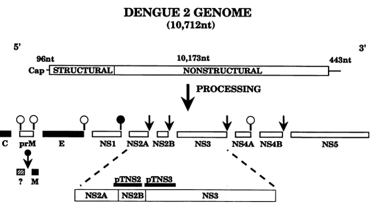
and pTNS3) which expressthesemoieties asfusionproteinswith trpE. Thelocations in the denguevirus genome of the inserts used are shown in Fig. 1,which alsoillustratesschematicallytheposttranslationalprocessing ofthedengue virus
polypro-tein togive thematurestructuralandnonstructuralproteins.
When bacterial cultures harboring either pTNS3 or pTNS2
were induced and insoluble inclusion bodies were purified and analyzed by SDS-PAGE, fusion proteins of51 and 54 kilodaltons (kDa), respectively, were found (Fig. 2A),
indi-cating that the fusion proteins were stable when overex-pressed. Fusion proteins werepurified bypreparative
SDS-PAGE and used to inject two rabbits each. High-titer
antiserum was usually obtained with three injections.
Denatured lysates of dengue virus-infected cells were analyzed by using antisera directed against pTNS2 and pTNS3 fusion proteins. Serum from a rabbit injected with the pTNS2 fusion protein immunoprecipitated a protein of approximately 14 kDa, identified as NS2B from its molecular size, immunoreactivity, and N-terminal
sequence
(see be-low), from infected but not from mock-infected cell lysates (Fig. 2B), and the serum was therefore designatedaNS2B
serum. NS2B was immunoprecipitated from denatured but not from nondenatured lysates, implying that its immunore-active epitopes are linear in nature or are masked in nonde-natured lysates (data not shown). Since pTNS2 contains the last 19 amino acids of NS2A as well as the first 111 amino acids of NS2B (Table 1), antiserum raised against the en-coded fusion protein could theoretically have reacted with NS2A sequences. However, no NS2A protein (predicted size, 24 kDa) was detected when either denatured (Fig. 2B) or nondenatured (data not shown) lysates were immunopre-cipitated with otNS2B.Serum from a rabbit injected with the pTNS3 fusion protein precipitated a polypeptide of 69 kDa, identified as
NS3, from infected cell lysates (Fig. 2B). In addition, three larger proteins (85, 93, and 130 kDa) were present in signif-icant amounts in the immunoprecipitates. All four species were also immunoprecipitated by a polyvalent anti-dengue virus mouse hyperimmune ascitic fluid (Fig. 2B, lane HIS). The 93-kDa protein appears to be
NS5,
and the reason it should precipitate with the atNS3 antiserum is obscure. The size and reactivity of the 85-kDa protein are consistent with it being polyproteinNS34A.
The identity of the 130-kDa species is unknown; it may be a polyprotein containingNS3.
Since pTNS3 encodes the last 8 amino acids of NS2B as well as the first 141 amino acids of
NS3
(Table 1), antiserum raised against the encoded fusion protein could, in theory, also precipitate proteins containing NS2B sequences. aNS3 J. VIROL.on November 10, 2019 by guest
http://jvi.asm.org/
FLAVIVIRUS NONSTRUCTURAL PROTEIN PROCESSING 4367
DENGUE
2
GENOME
(10,712nt)
5, 3'
96nt
Cap STRUCTURAL
10,173nt 443nt
I
PROCESSINGC prM E NS1 NS2A NS2B NS3 NS4A NS4B NS5
/
's~~~~~~~~~~~'
NS2A |NS2B| NS3
FIG. 1. Genome organization and expression ofdengue virus type 2. A schematic diagram aligning the trpE cDNA fusions to their corresponding regions ofthedenguevirus genome is shown. The upper third of the figure details the genomeorganizationof thePR159 strain. Themiddlethird of the figure depicts the processing events generating the structural andnonstructuralproteins. Virion structural proteins aredesignated by shaded boxes, and nonstructuralproteins are designated by open boxes. Cleavages after dibasic residues are designated by arrows. Hostcell signalase cleavages aredesignatedby openball-and-stickicons, and apotential viral-encoded signalase-like cleavage is designated bya crosshatchedball-and-stickicon.A late, cellmembrane-associatedcleavagegeneratingthemembrane structural protein is designated byaballand arrow. Theboundariesof the dengue virus inserts in the pTNS2 and pTNS3 cDNA fusions are mapped on the expanded viewofNS2A, NS2B, and NS3 shown inthelowerthird ofthefigure. Fulldetails ofconstructs pTNS2 and pTNS3 aregivenin Table1. nt,Nucleotides.
serum immunoprecipitated small amounts of NS2B from nondenatured lysates (data not shown) but notfrom dena-tured lysates (Fig. 2B). Thus, with denatured lysates, the aNS2BandoaNS3 seradescribed hereareessentially
mono-specific,anddenaturedlysateswereusedfor all
immunopre-cipitations shown inthefigures.
Invitro translation and processing kinetics. Processing of flavivirusnonstructuralproteinsoccursveryrapidlyinvivo, and viral polyproteins can be detected only in pulse-chase experiments under special labeling conditions (10).
How-ever,itseemedlikely that these processingeventscould be
monitored upon translation in vitro. Our initial expression experiments used RNA transcripts ofdengue virus type 2 cDNA clones that were notengineered to have specific 5' leader sequences or initiation codons in an appropriate
context. Onlylow levels ofprotein synthesis wereobtained in vitro, and variable amounts of specific (i.e., at the 5' methionine in theRNA)versus nonspecific(internal) initia-tionof translationwereobserved. Levels ofexpressionwere
increased approximately fivefold by inserting the dengue virus 5' nontranslated region upstream of the region to be translated. The efficiency of translation was increased an
additional two- to threefold when the reading frame of interestwasfusedtothat of thedengueviruscapsidprotein, such that the mRNA translated had the authentic dengue
virus type 2 leader and the initiation codon in the normal dengue virus context (data not shown). The yellow fever virus 5' nontranslated region has also been shown to
en-hance expression ofheterologous coding sequences by
re-ticulocyte lysates (43).
The firstconstructtested forproteaseactivitywaspT10.
RNAtranscribed fromthisconstructwith T7 RNA
polymer-ase was translated into a polyprotein (P2A2B3) containing
the first 37 amino acidsof thecapsidprotein, theC-terminal 118 amino acids of NS2A, all ofNS2B, and most of NS3 (Fig. 3; Table 2). Protein patterns resulting from different times of translation of this RNA in reticulocyte lysatesare
shown in Fig. 4A. A protein with the molecular mass
predictedfor the slightly truncated NS3(P3) wasproduced
(Fig. 4A), showingthatproteolyticprocessinghad occurred in this in vitro system. Proteins with molecular masses
predictedfor thefull-length (unprocessed)translated precur-sorP2A2B3 and for theprocessingintermediate P2B3 were
also observed(Fig. 4A).
Since the pTlO construct contains two cleavage sites, different processing intermediates would be predicted, de-pending upon the temporal order in which the cleavages
occur.IfthecleavagebetweenP2Aand NS2Bprecededthe
[image:4.612.125.500.74.280.2]cleavage between NS2B and P3, intermediate P2B3 would begenerated.Conversely,ifcleavagebetween NS2B andP3
TABLE 1. trpE/dengueviruscDNAfusions
Construct Vector" Gene Insertb Aminoacidsb Proteinc
pTNS3 pATH3(Eco(K)lSal) NS2B3 4497-4944 (Asp(K)/Sal) 122-129/1-144 NS3
pTNS2 pATH3
(Bam(K)/Cla)
NS2A2B 4077-4463(StulHpaII) 200-218/1-111 NS2BaVector andinsertDNAswerepreparedasdescribedinMaterialsand Methods. (K),5'overhangsthatwerebluntedbyKlenowtreatment.
bNucleotidesarenumberedrelativetothedenguevirusgenomicsequence,and amino acidnumbersof individualgeneproductsarelisted.
cDenguevirus-specificproteinrecognizedbyeach antiserum underdenaturingconditions.
IkTA-tlkyclrwv" v TA-Irww rrb AV
VOL. 64, 1990
I.
on November 10, 2019 by guest
http://jvi.asm.org/
4368 PREUGSCHAT ET AL.
A.
.:r :rJ
- ?:Z I_ _ _
x1 A _;
1.16-
9741-43
-13.
~
XI-a z
I U UT 1-1 1 1 I
NS5- - -1:3:
NS.4A- O.
NS3-4 do -69
NS- - -1
4
NS2;B - --11
FIG. 2. trpE/dengue virus fusionproteinsandcharacterization of antisera. (A) Coomassie blue-stained SDS-PAGEgelwithsamples of purified inclusion bodies from induced cultures ofXL1aloneand XL1culturescontaining plasmidspTNS3 and pTNS2.Thepositions ofprotein standards (Bio-Rad Laboratories, Richmond, Calif.)are
showntothe left with themolecular masses(kilodaltons) indicated. (B) Immunoprecipitates of [35S]methionine-labeled infected and uninfected BHK cells analyzed by SDS-PAGE. A total of 105 infected(IandPI) ormock-infected cells(U) werelabeled for12h, beginning at 30 hpostinfection. Lysates wereimmunoprecipitated with a polyvalent mouse hyperimmune asciticfluid(HIS)or mono-specific aNS2B and aNS3 immune (I) and preimmune (PI) sera. Locations of bands containing the viral proteins E, NS1, NS2B, NS3,andNS5and theputativeprecursorNS34Aareindicated.The molecularmasses(kilodaltons)of14C-labeledproteinmarkers (Am-ersham)areshown.
preceded cleavage between P2A and NS2B, P2A2B rather than P2B3 would be produced. The potential precursor P2A2B was not observed by SDS-PAGE (Fig. 4A) or by immunoprecipitation with xNS2B serum (see below), but P2B3 was readily detected as described above, suggesting that cleavage at the 2A/2B junction occurred first. The amountsand processing kinetics of P2B3 were also consis-tent with the view that it is the major intermediate in the
processingpathway (see below).
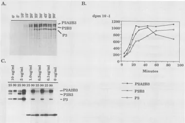
Bands containing P2A2B3, P2B3, and P3 (Fig. 4A) were excised from the dried gel, solubilized, and assayed for
[image:5.612.80.279.78.294.2]radioactivity(Fig. 4B). The translations were conducted by
TABLE 2. Deletion constructs
Construct Gene" Amino aciddeletionb
pTlO NS3 611-618
pTll NS3 303-618
pT12 NS3 184-618
pT13 NS3 141-618
pT14 NS2B/NS3 122-129/1-618
pT15 NS2B/NS3 43-121/303-618
pT16 NS2A/NS2B/NS3 146-218/1-42/184-618
aGenesaffectedby deletion.
bThe amino acids deleted in individual gene products arelisted. When
more than one gene product containsadeletion, aminoacid numbers are
separatedbyashill.
usingan RNAconcentration of10 ,ug/ml, conditions under which initiation oftranslation occurs nearly synchronously
and the reticulocytes lose theirability toinitiate new poly-peptidechains afterapproximately30 min of translation(14). Thus, the kinetics ofaccumulation andprocessingexhibited twophases. All threespecies accumulatedwithsimilar half timesduringthefirst 30 min(synthesis phase). After 30 min
of translation (postsynthesis phase), there was no further
incorporation of label into newly synthesized protein. The label in the full-length P2A2B3 band remained constant between 30 and 90min,indicatingthat these moleculeswere stable andwere notprocessed, whereasP2B3decayedwith a half-life of42 min and P3 accumulated at the same rate,
suggesting a precursor-product relationship between these twospecies.Thefactthat P2A2B3appearedtobeprocessed rapidlyinthe first 30 min butappearedtobestablethereafter suggeststhatfolding ofthisproteindoesnotoccuroptimally during translation in reticulocyte lysates and that those molecules that fold correctly during the initial synthesis
phaseareprocessed rapidly, while misfoldedmolecules are processed slowlyor not atall. The fraction of P2A2B3 that wasprocessed varied fromexperimenttoexperiment,being
sensitive to the lotofreticulocyte lysate used and to other
unknownvariables.
Cleavage of the dengue virus polyprotein in cis. Intermo-lecular (trans) cleavages exhibit second-order kinetics and are concentration dependent, whereas intramolecular (cis)
cleavagesarefirst order and concentrationindependent. To determine whether thecleavagesatthedenguevirus 2A/2B and2B/3siteswereconcentrationdependent, we examined
the protein products obtained upon translation of different amountsofinput RNA, which effectively changes the
con-centration ofthe proteinase (Fig. 4C). At the highest con-centrations ofRNA tested, the components of the transla-tion system were limiting (29) and processing appeared to
proceed more rapidly. At lower concentrations of RNA,
pTlO
(1-37) (101-218) (1-129)
T7
1 P2A NS2BSSSS
1
1-213+<(1-610) amino acids
..
_/- nucleotides
FIG. 3. Schematicdiagram of dengue virus type 2 protein and nucleic acid sequences expressed in the translation vectorpT10.T7 RNA polymerasetranscribes a chimeric RNA which contains nucleotides 1 to 213 fused to nucleotides 3778 to 6351 of the dengue virus genome. Aninitiatormethionine codon at position 96 is designated by a closed diamond. Upon translation, the encoded polyprotein contains amino acids 1 to 37ofthedengue virus capsid protein, amino acids 101 to 218 of NS2A, all ofNS2B,and the first 610 amino acids ofNS3( ). Thepredicted proteasedomain of NS3 is indicated (M).
J. VIROL.
on November 10, 2019 by guest
http://jvi.asm.org/
[image:5.612.315.556.82.173.2] [image:5.612.85.527.613.678.2]FLAVIVIRUS NONSTRUCTURAL PROTEIN PROCESSING
A. B.
- - Ci 0 L:5 K C
O at--_ > NM vr =
""Ideme"I
,P9421B3
P3
dpin 10-1
IZVUJ
800-600 400200
-fBi
(3
2590259025390
_o@*
2590 2590" _-P2A2B3 -P2B3
_ - -_
-_
P320
''''I
40 60 80 100
Minlutes
a--- P2A2B3 P2B3
--- P3
FIG. 4. Processing of proteins during in vitro translationofconstructpT10.(A)SamplesofapTlO-programmed translationwereremoved
atthedesignated times(min) after initiation of translationand analyzed by SDS-PAGE. Precursors P2A2B3 and P2B3aswellasmatureP3
areindicated. (B) Bands containing P2A2B3, P2B3, andP3wereexcised from gels like thatshown in panel A and quantitatedasdescribed
inthetext.(C)Samples of RNA transcribed frompTlOwereseriallydilutedand translated in vitro.Samplesof the translation mixtureswere
analyzed bySDS-PAGE. Inordertoequalize for[35S]methionineincorporation,variousamountsof lysatewereloaded in each lane. Lanes
1to4contained 1 p.l of lysate; lanes 5,6,8, and 10contained 2 p.l of lysate; and lanes7, 9, and 11contained 4 pulof lysate. Massof RNA
permilliliter and the length of the translation incubation (min)areindicated.Locations of bands containingnonstructuralproteinprecursors
P2A2B3 and P2B3 as wellasmature P3areindicated.
total incorporation appearedto increase between25 and 90 min of translation and proportionately more label was
presentina46-kDareticulocyte-specific band. However, the
ratio ofP2A2B3 to P3 after 90 min of translation at 30°C appeared to be independent of the concentration of input RNA (Fig. 4C), suggesting that cleavage occurs in cis,
although extremely efficient trans cleavage by the dengue virus proteinasecannot beruled out.
We have usedasecondapproach toexamine whether the dengue virus polyprotein can be cleaved in trans. Polypro-teinsproduced bypT13-andpT14-programmedtranslations contain aninactive protease, but thepotentialcleavagesites
are intact (see below). These polyproteins were used as
substrates for the active proteinase produced by a
pT10-programmedtranslation,andnodetectable cleavageofthese polyproteins occurred (datanotshown).The lackof detect-able trans cleavage in these experiments is consistent with the results of the kinetic and dilutionsensitivity experiments which suggested that cleavage occurred at the 2A/2B and 2B/3junctions only in cis.
Deletionmappingof thedenguevirusprotease.On the basis of molecularmodeling studies,both Bazan and Fletterick(3) andGorbalenyaetal. (21)havepredictedtheprecise
bound-aries of the flavivirus protease domain. This predicted
do-main is illustrated schematically in Fig. 5 in relation to the
dengueviruspolyprotein. The entire domainishypothesized
tospanthe first 180amino acids of NS3(solid boxes)and to
contain four subregions or boxes ofhomology with serine proteases,the first three of whichcontain the threeelements
of thecatalytic triad and the fourth ofwhich is involved in
substratebinding.Totestthesepredictions,we constructed
a series ofdeletion constructsthat together spanthe entire
lengthofNS3 and thatdelete large regions ofP2AandNS2B (Fig. 5; Table 2); translation mixes wereprogrammed with
RNA transcribed from these constructs, and the resulting protein productswereimmunoprecipitated withaNS2B and
cxNS3 seraand analyzed by SDS-PAGE (Fig.6).
ConstructspT10, pTll, and pT12 contain the entire
pro-posed protease domain(Fig. 5), and from these constructs polypeptideswiththemolecularmassesand
immunoreactiv-ities ofmatureNS2B andofthe truncatedformsof NS3(P3, P3', and P3") were detected (Fig. 6A). In contrast, pT13-programmed translations did not produce any (truncated)
NS3 or NS2B (Fig. 6B), indicating that this deletion abol-ishedactivity, presumably by invadingtheproteasedomain (Fig. 5). Thus, the protease domain encompasses a maxi-mumof 184 amino acidsatthe Nterminus of NS3. Polypep-tides consistent in molecular mass and immune reactivity with aP2B3 processingintermediate could also be detected
inpTlO-, pTll-, andpT12-programmedtranslations but not inpT13-programmed translations (Fig. 4 and6).
Translation of RNA transcribed from pT14 yielded a
polypeptide which was consistent in molecular mass and immunoreactivitywiththat predictedfor P2A2B'. Process-ingdidnotoccuratthe2A/2B site. The location of the band and theamount expressedfrom pT14were suchthat if this
precursor had been presentupon translation ofRNA from
constructs such as pT10, it would have been readily
de-tected.Thissupportsthe earlier conclusionsthat P2A2Bwas
nevergenerated duringprocessingin vitro.
To examine the N-terminal boundary of the protease domain, that is, whether sequences in P2A or NS2B are C.
VOL.64, 1990 4369
on November 10, 2019 by guest
http://jvi.asm.org/
[image:6.612.124.504.78.332.2]4370 PREUGSCHAT ET AL.
BOXI BOX2
His51 Asp75
BOX3 BOX4
Serl35 SBP
/
~~~~~~~~~~~~~~
4 NS2A | NS2B _
P2T. NS2B ...2
L P2A.4 Ns2BP
A.
3 2 ]3 2 3 2
li'l- Ir
__
4:j -I
a P2A W82BNS2 _
B.
3 2 3 2 2
2) C3 2 3 2:1 2:1
-PE>3
- `2AA1"3 -1'321133
PW3~
12A'2113"'-so
*_ -461l'S.12A2B13
--31
pT13 K P2A NS2B
pTl4 VA - - -
-pT14
E3I;- ---________-pT15 a:iii ---1 _ ~
-pTl6 -m-h-_*_.- - - -
-I I I'
A P Sn K D S As X E
FIG. 5. Schematicmapofdeletionconstructsusedtomap
pro-tease activity. The relative positions of the fourhomology boxes
proposed by Bazan and Fletterick (3) and Gorbalenyaetal.(21)are
shown. The proposed catalytic histidine, aspartate, and serine residuesarelisted within boxes1, 2, and 3 and indicated byvertical linesontheschematicmap. SBPreferstotheproposed substrate-binding pocket. Gene products P2A and NS2B are designated by
openand stippled boxes, respectively. Capsid proteinis indicated by hatched lines. The putative protease and helicase domains of NS3aredesignated by solid and crosshatched boxes, respectively.
Thestructuresof deletion clonesaremapped below.The
abbrevia-tions forrestriction sites used togeneratedeletion constructs are
indicated as follows: A, Asel; P, PvuII; Sn, SnaBI; K, KpnI (Asp718); D, DraIll; S, Sall; As, AsulI; X, XhoI; and E, EcoRI. Cleavagesatthe2A/2Bor2B/3junctionsareindicatedbyarrows.A detaileddescription of eachconstructisgiveninTable 2.
required for proteolytic activity, we analyzed the in vitro
translationpatternsproduced fromtwodeletionconstructs, pT15andpT16, which togetherdelete almost all the P2A and NS2B sequences. When pT15-programmed translations were fractionated by using aoNS2B, only low levels of
immunoreactive species were observed upon overexposure
ofautoradiographs (Fig. 6B).Themolecularmassesofthese
species were not consistent with correctprocessing at the 2A/2B or 2B/3 cleavage site. The low level of
immunoreac-tivity suggests that the major antigenic epitopes recognized by aNS2B have been deleted in this construct. Upon
pre-cipitation with aNS3, a small amount ofaberrantly
proc-essed P3' was detected. When pT16-programmed
transla-tions were immunoprecipitated and analyzed by
SDS-PAGE, both oaNS2B- and aNS3-immunoreactive species of aberrantmolecularmasscouldbe detected. Since the 2A/2B
cleavage site had been removed by this deletion, only cleavageatthe2B/3 boundary could be examined.Thus, it is unclearwhethersequencesinP2AorNS2B arerequired for
proteolytic activityorwhether the deletionsinduce
misfold-ing of the molecule such that the correct cleavage sites are
notrecognized.
Sequencing ofviral proteins. In orderto confirm that the cleavageeventsobserved in vitrooccuratthesamesitesas
those utilized in vivo and to confirm the identities of the proteinsproduced in vitro,wecompared the amino-terminal
amino acid sequences ofNS2B and NS3 produced in vitro
withthose of proteins isolatedfrom infected celllysatesand obtained the N-terminal sequence of P2B3'. NS2B from
IH- _ A -14
_NS2B
FIG. 6. Immunoprecipitations of in vitro translations pro-grammed with RNAtranscribed fromNS3deletion constructs. (A) Samples of reticulocytelysatesweredenatured and immunoprecip-itated with aNS2B (marked by 2)oraNS3 (marked by 3)immune
sera, and immune complexes were dissolved in SDS-containing loading buffer and analyzed by SDS-PAGE. The construct from which RNAwas transcribedisindicated above each lane. Protein species which were identified asP2A2B3", P2B3", P3, P3', P3",
and NS2B by immunoreactivity andmolecularmassare indicated. The molecularmasses(kilodaltons) of"'C-labeledproteinstandards (Amersham) are indicated. (B) Immunoprecipitations of in vitro translations from proteolytically inactive templates. Unprocessed
precursorsP2A2B3"and P2A2B'aredesignatedas arethepositions of molecularmassmarkers.
dengue virus type 2-infected cells labeled with [35S]methio-nine or [3H]leucine was isolated by preparative immunoaf-finity chromatography. Approximately 85% of the label in thepurified NS2Bused forsequencing consisted ofmature NS2B,asdeterminedbyelectrophoresis. The resultsclearly
showed that serine 1345 of the dengue virus polyproteinis the amino-terminal residue ofNS2Bproduced invivo(Fig. 7A). This assignment is in agreement with data for other sequencedNS2Bspecies (9, 48, 54),onthe basis ofsequence
homology.
In order to sequence NS2B produced in vitro, pTll-programmed translations were immunoprecipitated with
aNS2B. Approximately 85% of the NS2B immunoprecipi-tatedwasfully cleaved, andapproximately10% of the label
was in P2B3', which is coterminal with mature NS2B and contributes to the observed NS2B signal (see below). The amino terminus of in vitro-produced NS2B was indistin-guishable from thatfound invivo(Fig. 7B).
Sequencing of P3' labeled in vitro in pTll-programmed translations identified the amino-terminal residue of P3' as
Ala-1476 of the dengue virus type 2 polyprotein (Fig. 7C). Peaks of[3H]leucine atcycles4and 18 and of [3H]valine at
cycles 3, 7,and13 makethisassignment unambiguous. This result agrees perfectly with the sequence of NS3 isolated
from denguevirus type 2-infected cells (4). The P3' results
arealso of interestbecause thecleavage which produces the
amino terminusofdengue virustype2NS3 is unique in that
it appears to occur after a single basic amino acid; the
cleavagestoproduceNS3 in allother sequencedflaviviruses
occurafter dibasicresidues. Thefact that the amino termi-nus of P3' produced in vitro is the same as that of NS3
isolated from infected cells suggests that the dengue virus 2B/3 cleavage site is infact different and that the observed terminusof NS3 doesnotarisefrom cleavageatanupstream
pT1O
pTll
pT12
-69
J. VIROL.
40
on November 10, 2019 by guest
http://jvi.asm.org/
[image:7.612.60.297.63.275.2] [image:7.612.318.555.76.237.2]FLAVIVIRUS NONSTRUCTURAL PROTEIN PROCESSING 4371
CYCLE
[35S] Met
[3H1
Leu[35S]
Met
---131H
Leu[3H]
Leu[3H]
Val
[35S]
MetED
[image:8.612.75.561.79.369.2]CYCLE
FIG. 7. Amino-terminal sequencingofviral proteins.Datain each panelareplottedasradioactivityperEdmandegradation cycleversus
cycle number. (A) Invivo-labeled NS2B. (B) Invitro-labeled NS2B. (C) In vitro-labeled P3'. (D) PrecursorP2B3'. The amino-terminal
sequencesofNS2B, NS3, and NS2B3 deduced from the nucleotidesequenceofthe RNAgenome areindicated below eachsequencepanel,
with theleucine,valine, and methionine residues identified by Edmandegradation highlighted in boldfacecapital letters. The burstof35S disintegrationsperminute observed in the first sequencing cycleofimmunoaffinity-purifiedNS2B (both in vivo and invitro) isnotconsistent with thededucedsequenceofNS2B and is presumably contributedbycontaminantswhichcoprecipitate with NS2B.Thesingle-letteramino acidcodeused isasfollows: A, alanine; C, cysteine; D,aspartate;E,glutamate;F,phenylalanine;G,glycine;H,histidine;I,isoleucine;K,
lysine; L, leucine; M,methionine;N,asparagine;P,proline; Q,glutamine;R,arginine;S,serine; T,threonine;V,valine; W,tryptophan;and Y, tyrosine.
site characterized by dibasic residues followed by amino-terminal nibbling(4).
Inordertodefine further thespecificity of cleavageevents anddefinitively identify processing intermediates, the puta-tiveP2B3' wasisolatedfrompTll-programmed translations
by preparative SDS-PAGE and sequenced. Peaks of 35S
were observed at positions 9 and 13, aligning this species perfectly with theamino terminus of NS2B andconfirming theprevious identification basedongel mobilityand immuno-reactivity (Fig. 7D).
DISCUSSION
Cleavage of the dengue virus polyprotein in vitro. The kinetic experiments indicated that in vitro cleavage at the 2A/2B junction precedes that at the 2B/3 junction. These junctions were inactive as substrates in a trans cleavage
assayand cleavagewasinsensitivetodilution,implyingthat
cleavageatthesetwojunctionsoccursbyan intramolecular mechanism. It is formally possible that NS3 is extremely active in trans such thatsensitivity to dilution couldnotbe demonstrated and that the negative results of the
trans-processing experimentsresulted from theperturbedtertiary structureofsubstrates,althoughwehave usedsimilar meth-ods to demonstrate trans cleavage of the Sindbis virus
nonstructural polyprotein (29). Differences in the rate of
translation and in the association of membranes with
proc-essing intermediates could also influence the observed rate ofcleavage in vivo.
Cleavageatthe2A/2Band2B/3 junctionsin vitroproceeds withthe samespecificity asthatfound in vivo. Translation products are cleaved at the correct sites of cleavage, and nonspecificcleavage israrelyobserved. While the kineticsof cleavage in vitro are much slower than those observed in vivo, it is highly probable that the fidelity and order of in vitrocleavageare atruereflection ofinvivoprocessing.The
use ofimproved expressionsystems and in vitro mutagene-sisof cleavage sites will be useful inestablishingwhether the order of cleavageobservedisobligatoryorwhether itsimply represents thekinetically favored order.
Testing the flavivirus protease model. Positive identifica-tion of NS3as aflavivirus proteinasewillstimulateresearch
on the nature of the catalytic and substrate recognition residues involved inproteolysis. Thespecific predictionsas
to which histidine, aspartate, and serine residues form the catalytictriadaretestablethroughsite-specific mutagenesis, and mutants which are catalytically active in vitro can be
tested for biological activity in vivo through the use of
infectious cDNA clones (40). NS3 shares the greatest amountofsequence similaritywith theSindbisviruscapsid protein protease domain, and site-directed mutagenesis of
IwpLneaiMavgMvsiLassLLkn
VOL.64, 1990
on November 10, 2019 by guest
http://jvi.asm.org/
4372 PREUGSCHAT ET AL.
the NS3 protease domain will be greatly assisted by molec-ular modeling studies once the coordinates of the Sindbis capsid protein become available (5).
Four specific predictions of the protease model were addressed by experiments presented here (3, 21). Our exper-imental results are consistent with the predictions that the protease domain consists of approximately 180 amino acids at the N terminus of NS3 and could retain function when severed from the remainder of NS3, which forms a putative helicase domain. However, our results are not consistent with the prediction that an internal conserved cleavage site in NS3 is utilized to separate the helicase and protease domains or the prediction that cleavage of the
flavivirus
polyprotein by NS3 is an ATP-dependent process. The antiserum used in our experiments was directed specifically
against the protease domain, and therefore any internal cleavage of NS3 in vitro or in vivo should have been detected. Proteolysis occurred even in those cases in which thehelicase domain had been deleted, making itunlikely that hydrolysis of ATP is absolutely required for proteolysis, although the possibility that it has an effect in vivo has not been eliminated.
The presence of protease and helicase domains within a single viral protein is a common structural motif found in alphavirus, pestivirus, potyvirus, and coronavirus proteins as well as flaviviruses (22). The primary function of a viral proteinase is to posttranslationally regulate the production of individual gene products from a polyprotein precursor. The viral proteinase can also produce processing intermediates which maythemselvesbefunctional components of the viral life cycle. The choice between emphasizing proteinase func-tion andemphasizinghelicase function would depend on the specific needs of the virus at that point in the life cycle. It appears likely that the main function of mature NS3 is as a helicase, since the initial cleavages releasing NS2B and NS3 most likely occur in cis and mature NS3 does not appear to workefficiently in trans.
Implications for dengue virus polyprotein processing. A number of viralproteinases function both in cis and in trans tocleavepolyproteins in vivo (32). Like other proteinases of
positive-stranded RNA viruses, NS3 may also function both in cis and in trans. The amino terminus of NS4B is believed tobegenerated by a signalase cleavage event which presum-ably occurs cotranslationally upon insertion of the hydro-phobic tail of NS4A into the lumen of the endoplasmic reticulum (9, 48). Once this event occurs, the dengue virus
polyprotein backbone is severed, and by definition, any
processing events that occur downstream of this scission mustoccur in trans, and if NS3 is the proteinase responsible for cleavage at the 4B/5 boundary then it must cleave in
trans.
Processing events consistent with scission at the 3/4A
junction areobservable in vitro (F. Preugschat, unpublished data), and putative NS34A intermediates can be detected in vivo (Fig. 2B). Thisimplies that the kinetics of 3/4A cleavage areslower than the kinetics of 2A/2B and 2B/3 cleavage. It is possible that NS34A is a precursor that is restricted to the plane of the endoplasmic reticulum by a carboxy-terminal
membrane-spanning segment and that this is the form of proteinase that isresponsible fortrans cleavage at the 4B/5
junction. By restricting the proteinase to the plane of the endoplasmicreticulum,theconcentration dependence of the trans cleavage step would be reduced and result in rapid kinetics of cleavage in infected cells.
The flavivirus capsid protein undergoes a complex two-step maturation process in infected cells. As the carboxy
terminus of the capsid protein is translated, it acts as a membraneinsertion sequenceforacotranslational
signalase-mediated cleavage that generates the aminoterminus ofthe prM protein. Nascent intracellularcapsid
protein
possesses a C-terminal membrane-spanning segment that is cleavedprior to assembly of the virion (37). This cleavage event occurs on thecarboxy-terminalside of severalbasicresidues and could be potentially mediated in trans by NS3. The initialsignalase-mediated cleavage can beobservedby
using
aninvitro translation system, but the secondary maturation cleavagethatremovesthemembrane insertion sequence has not been observed in vitro (37, 44). By using an in vitro expression system designed to analyze cleavages at the
3/4A,
4B/5,
and C/prM junctions, it should be possible to reconstruct theflaviviral
protein-processing pathway
by
using the experimental
approaches
described inthis paper.ACKNOWLEDGMENTS
We thank Richard Kuhn and Ellen Strauss for stimulating discus-sions and editorial prowess; David Teplow and Tammy Bauer for expertise and assistance with proteinsequencing; and Joel Dalrym-ple for furnishing cells, virus, and hyperimmune antiserum. We also thank A. E. Gorbalenya for communicating results prior to publica-tion.
Frank Preugschat gratefully acknowledges the financial support of the Canadian Natural Sciences and Engineering Research Council (NSERC) and Gordon Ross. This work was supported by Public Health Service grantA120612 from the National Institutes of Health and by a grant from the World Health Organization.
LITERATURE CITED
1. Bancroft, W. H., R. M.
Scott,
K. H. Eckels, C. H. Hoke, T. E. Simms, K. D. T. Jesrani, P. L. Summers, D. R. Dubois, D. Tsoulos, and P. K. Russell. 1984. Dengue virus type 2 vaccine: reactogenicity and immunogenicity in soldiers. J. Infect. Dis. 149:1005-1010.2. Bancroft, W. H., F. H. Top, Jr., K. H. Eckels, J. H. Anderson, Jr., J. M. McCown, and P. K. Russell. 1981. Dengue-2vaccine: virological, immunological, and clinical responses of six yellow fever-immunerecipients. Infect. Immun. 31:698-703.
3. Bazan, J. F., and R. J.
Fletterick.
1989. Detection of a trypsin-like serine protease domain inflaviviruses
and pestiviruses. Virology 171:637-639.4. Biedrzycka, A., M. R. Cauchi, A. Bartholomeusz, J. J. Gorman, and P. J. Wright. 1987. Characterization of protease cleavage sites involved in the formation of envelope glycoprotein and three nonstructural proteins of dengue virus type 2, New Guinea C strain. J. Gen. Virol. 68:1317-1326.
5. Boege, U., M. Cygler, G. Wengler, P. Dumas, and J. Tsao. 1989. Sindbis virus core protein crystals. J. Mol. Biol. 208:79-82. 6. Calisher, C. H., N. Karabatsos, J. M. Dalrymple, R. E. Shope,
J. S. Porterfield, E. G. Westaway, and W. E. Brandt. 1989. Antigenic relationships between
flaviviruses
as determined by cross neutralization tests with polyclonal antisera. J. Gen. Virol. 70:37-43.7. Calisher, C. H., R. E. Shope, W. Brandt, J. Casals, N. Karabat-sos, F. A. Murphy, R. B. Tesh, and M. E. Wiebe. 1980. Proposed antigenic classification of registered arboviruses.
I.
Togaviridae, Alphavirus. Intervirology 14:229-232.8. Chamberlin, J. P. 1979. Fluorographic detection of radioactivity in polyacrylamide gels with the water-soluble fluor, sodium salicylate. Anal. Biochem. 98:132-135.
9. Chambers, T. J., D. W. McCourt, and C. M. Rice. 1989. Yellow fever virus proteins NS2a, NS2b and NS4b: identification and partial N-terminal amino acid sequence analysis. Virology 169: 100-109.
10. Cleaves, G. R. 1985. Identification of dengue type 2 virus-specific high molecular weight proteins in virus-infected BHK J. VIROL.
on November 10, 2019 by guest
http://jvi.asm.org/
FLAVIVIRUS NONSTRUCTURAL PROTEIN PROCESSING 4373 cells. J. Gen. Virol. 66:2767-2771.
11. Cleaves, G. R., and D. T. Dubin. 1979. Methylation status of intracellular dengue type 2 40S RNA. Virology96:158-165. 12. Coia, G., M. D. Parker, G. Speight, M. E. Byrne, and E. G.
Westaway. 1988. Nucleotide and complete amino acid se-quences of Kunjin virus: definitive gene order and characteris-tics of the virus-specified proteins. J. Gen. Virol. 69:1-21. 13. Condreay, L. D., R. H. Adams, J. Edwards, and D. T. Brown.
1988. Effect of actinomycin D and cycloheximide on replication of Sindbis virus in Aedes albopictus (mosquito) cells. J. Virol. 62:2629-2635.
14. Dasso, M. C., and R. J. Jackson. 1989. On the fidelity of mRNA translation in the nuclease treated rabbit reticulocyte lysate system. Nucleic Acids Res. 17:3129-3144.
15. Deubel, V., R. M. Kinney, and D. W. Trent. 1988. Nucleotide sequence and deduced amino acid sequence of the nonstructural proteins of dengue type 2 virus, Jamaica genotype: comparative analysis of the full-length genome. Virology 165:234-244. 16. Dunn, S. D. 1986. Effects of the modification of transfer buffer
composition and the renaturation of proteins in gels on the recognition of proteins onwesternblots by monoclonal antibod-ies. Anal. Biochem. 157:144-153.
17. Eckels, K. H., V. R. Harrison, P. L. Summers, and P. K. Russell. 1980. Dengue-2 vaccine: preparation from a small-plaque virus clone. Infect. Immun. 27:175-180.
18. Ehrenkranz, N., A. Ventura, R. Cuadrado, W. Pond, and J. Porter. 1971. Pandemic dengue in Caribbean countries and the southern United States-past, present, and potential problems. NewEngl.J. Med. 285:1460-1469.
19. Falgout,B., R. Chanock, and C.-J. Lai. 1989. Proper processing of dengue virus nonstructural glycoprotein NS1 requires the N-terminal hydrophobic signal sequence and the downstream nonstructural protein NS2a. J. Virol. 63:1852-1860.
20. Gorbalenya, A. E., A. P. Donchenko, V. M. Blinov, and E. V. Koonin. 1989. Cysteine proteases of positive strand RNA vi-ruses and chymotrypsin-like serine proteases: a distinct protein superfamily with a common structural fold. FEBS Lett. 243: 103-114.
21. Gorbalenya, A. E., A. P. Donchenko, E. V. Koonin, and V. M. Blinov. 1989. N-terminal domains of putative helicases of Flavi-and Pestiviruses may be serine proteases. Nucleic Acids Res. 17:3889-3897.
22. Gorbalenya, A. E., E. V. Koonin, A. P. Donchenko, and V. M. Blinov. 1988. A conserved NTP-binding motif in putative heli-cases. Nature (London) 333:22.
23. Gruenberg, A., W. S. Woo, A. Biedrzycka, and P. J. Wright. 1988. Partial nucleotide sequence and deduced amino acid sequence of the structural proteins of dengue virus type 2, New Guinea C and PUO-218 strains. J. Gen. Virol. 69:1391-1398. 24. Gubler, D. J. 1989. Aedes aegypti and Aedes aegypti-borne
disease control in the 1990s: top down or bottom up. Am. J. Trop. Med. Hyg. 40:571-578.
25. Hahn, C. S., E. G. Strauss, and J. H. Strauss. 1985. Sequence analysis of three Sindbis virus mutants temperature-sensitive in the capsid autoprotease. Proc. Natl. Acad. Sci. USA 82:4648-4652.
26. Hahn, C. S., and J. H. Strauss. 1990. Site-directedmutagenesis of the proposed catalytic amino acids of the Sindbis virus capsid protein autoprotease. J. Virol.64:3069-3073.
27. Hahn, Y. S., R. Galler, T. Hunkapiller, J. Dalrymple, J. H. Strauss, and E. G. Strauss. 1988. Nucleotide sequence of dengue 2 RNA and comparison of the encoded proteins with thoseof other flaviviruses. Virology 162:167-180.
28. Hardy, W. R., and J. H. Strauss. 1988. Processing the nonstruc-tural polyproteins of Sindbis virus: study of the kinetics invivo by using monospecific antibodies. J. Virol. 62:998-1007. 29. Hardy, W. R., and J. H. Strauss. 1989. Processing the
nonstruc-tural polyproteins of Sindbis virus: nonstrucnonstruc-tural proteinase is in the C-terminal half of nsP2 and functions both in cisandin trans. J. Virol. 63:4653-4664.
30. Hewick, R. M., M. W. Hunkapiller, L. E. Hood, and W. J. Dreyer. 1981. A gas-liquid-solid phase peptide and protein sequenator. J. Biol. Chem. 256:7990-7997.
31. Kelley,P. M., and M. J. Schlesinger. 1982. Antibodiesto two major chicken heat shock proteins cross-react with similar proteinsinwidelydivergent species.Mol.Cell. Biol. 2:267-274. 32. Kraeusslich, H. G., and E. Wimmer. 1988. Viral proteinases.
Annu.Rev. Biochem. 57:701-754.
33. Laemmli, U. K. 1970.Cleavage ofstructuralproteinsduringthe assembly ofthe head ofbacteriophage T4. Nature (London) 227:680-685.
34. Maniatis, T., E. F. Fritsch, and J. Sambrook. 1982. Molecular cloning:a laboratorymanual. ColdSpringHarborLaboratory, ColdSpringHarbor, N.Y.
35. Markoff,L.1989.Invitroprocessing ofdengue virus structural proteins: cleavage of the pre-membrane protein. J. Virol. 63: 3345-3352.
36. Matsudaira, P. 1987. Sequence from picomole quantities of proteins electroblotted onto polyvinylidene difluoride
mem-branes.J. Biol. Chem. 262:10035-10038.
37. Nowak, T., P. M. Farber, G. Wengler, and G. Wengler. 1989. AnalysesoftheterminalsequencesofWestNile virusstructural proteinsand of the in vitrotranslation of theseproteins allowthe proposal of a complete scheme ofthe proteolytic cleavages involved in theirsynthesis.Virology 169:365-376.
38. Ozden,S., and B.Poirier. 1985. Denguevirus induced polypep-tide synthesis. Arch. Virol.85:129-137.
39. Rice, C. M., R. Aebersold, D. B. Teplow, J. Pata, J. R. Bell, A. V.Vorndam,D. W.Trent,M. W.Brandriss, J. J.Schlesinger, and J. H. Strauss. 1986. Partial N-terminal amino acid
se-quences of three nonstructural proteins of two flaviviruses. Virology 151:1-9.
40. Rice, C. M., A.Grakoui, R. Galier, and T.J.Chambers. 1989. Transcription ofinfectious yellow fever RNA fromfull-length cDNA templates produced by in vitro ligation. New Biol. 1:285-296.
41. Rice, C. M., E. M.Lenches,S. R.Eddy, S. J.Shin,R.L. Sheets, and J. H. Strauss. 1985. Nucleotide sequence ofyellow fever virus:implications for flavivirusgeneexpression andevolution. Science 229:726-733.
42. Rice, C. M., R. Levis, J. H. Strauss, and H. V. Huang. 1987. Production of infectious RNA transcripts from Sindbis virus cDNA clones: mapping of lethalmutations, rescueofa temper-ature-sensitive marker, and in vitro mutagenesis to generate defined mutants. J. Virol. 61:3809-3819.
43. Ruiz-Linares, A., M.Bouloy,M. Girard, and A.Cahour. 1989. Modulations of the in vitro translational efficiencies of yellow fevervirusmRNAs:interactions betweencodingand
noncoding
regions. Nucleic AcidsRes. 17:2463-2474.
44. Ruiz-Linares, A., A. Cahour, P. Despres, M. Girard, and M. Bouloy. 1989. Processing ofyellow fever viruspolyprotein: role ofcellular proteases inmaturation ofthestructuralproteins.J. Virol. 63:4199-4209.
45. Schatzmayer,H.G.,R.M. R.Nogueira,and A. P. A. Travassos da Rosa. 1986. An outbreak ofdengue virus at Rio deJaneiro (1986). Mem. Inst. Oswaldo Cruz RioJ. 81:245-246.
46. Scott, R. M., K. H. Eckels, W. H. Bancroft, P. L. Summers, J.M.McCown,J. H.Anderson,and P. K. Russell.1983.Dengue 2 vaccine: dose response in volunteers in relation to
yellow
fever status. J.Infect. Dis. 148:1055-1060.47. Smith, G. W., and P. W.Wright. 1985.Synthesisofproteinsand glycoproteins in denguetype 2 virus-infectedVero and Aedes albopictus cells. J.Gen. Virol. 66:559-571.
48. Speight, G., G.Coia,M. D.Parker,and E.G.Westaway.1988. Genemapping and positive identification ofthe non-structural proteins NS2A, NS2B, NS3, NS4B and NS5 of the flavivirus Kunjinandtheircleavagesites. J. Gen. Virol. 69:23-34. 49. Spindler, K. R., D. S. E.Rosser,and A. J.Berk. 1984.Analysis
ofadenovirustransforming proteins from
early
regions
1Aand 1B with antisera to inducible fusion antigens produced in Escherichiacoli. J. Virol. 49:132-141.50. Svitkin, Y. V., V. N. Lyapustin, V. A. Lashkevich, and V. I. Agol. 1984. Differences between translation
products
of tick-borneencephalitisvirus RNA incell-freesystemsfrom Krebs-2 cells andrabbitreticulocytes: involvementof membranes inthe processing of nascent precursors of flavivirus structuralpro-VOL.64, 1990
on November 10, 2019 by guest
http://jvi.asm.org/
4374 PREUGSCHAT ET AL. teins. Virology 135:536-541.
51. von Heijne, G. 1986. A new method for predicting signal sequencecleavage sites. Nucleic Acids Res. 14:4683-4690. 52. Wengler, G., M. Beato, and G. Wengler. 1979.In vitro
transla-tion of 42S virus-specific RNA from cells infected with the flavivirus West Nile virus. Virology 96:516-529.
53. Wengler, G., G. Wengler, and J. Gross. 1978. Studiesonvirus
specific nucleic acids synthesized in vertebrate and mosquito cells infected with flaviviruses. Virology89:423-437.
54. Wright, P. S., M. R. Cauchi, and M. L. Ng. 1989. Definition of the carboxy termini of the three glycoproteins specified by denguevirustype2.Virology171:61-67.
J. VIROL.