0022-538X/85/110607-06$02.00/0
Copyright ©D 1985,American Society for Microbiology
NOTES
Posttranslational Processing
of p21
ras
Proteins Involves
Palmitylation of
the
C-Terminal
Tetrapeptide
Containing
Cysteine-186
ZHANG-QUN CHEN,1 LINDA S.
ULSH,'
GARRETTDuBOIS,2 ANDTHOMAS Y. SHIH1*Laboratory of MolecularOncology, Division ofCancerEtiology, NationalCancer
Institute,'
andProgramResources,Inc.,
NCI-Frederick CancerResearchFacility,2
Frederick,Maryland
21701Received29April 1985/Accepted 12 July 1985
The p21 proteins ofras oncogenes are synthesized as precursors in thecytosol. After processing, which involves acylation, the products are associated with the plasma membrane in eucaryotic cells. The p21 overproduced inEscherichia coli, however, isnotprocessed byacylation. Asynthetic tetrapeptideofthep21 C terminus is usedtoidentify the acylation siteineucaryotic p21ascysteine-186.The samepeptideofbacterial p21 is not acylated. Although p21 of Harvey murinesarcomavirus-transformedNRKcellscanbemetabolically labeled with either[3H]palmitateor[3H]myristate,thelipid moiety ofthehydrophobic peptideisidentifiedas palmitic acid. We suggest that the enzymatic mechanism for p21 palmitylation may be different from N-terminal myristylation of manyother membraneproteins.
The viral transforminggenes ofHarvey murine sarcoma virus (Ha-MuSV) and Kirsten murine sarcoma virus
(Ki-MuSV) encode the 21,000-daltonp21rasproteins(5,7, 8, 29,
31, 32, 35). A family of structurally highly conserved p21
proteins is encoded by cellular homologs of the viral ras
genes (v-ras). These cellular homologs activated by point
mutations have been detected in a variety of human
neo-plasms. Posttranslational processing ofp21 appears tobe a common pathway for the synthesis ofall p21 ras proteins
(30, 34). Sefton et al. (26) demonstratedacylation of p21 in
the processed products. The importance ofp21acylation to
its transforming functionwas mostclearly demonstrated by
Willumsen et al. (36, 37) in a series of genetic mutations
located near the p21 C terminus. Studies of these mutants
indicate that cysteine-186 is essential, although not
suffi-cient, for p21 processing. The exactrole ofcysteine-186 in
p21 processing is uncertain from the mutant studies;
how-ever,wedemonstrate here directchemical evidence
indicat-ing that cysteine-186 is the palmitylation site. We suggest
thatthe sequence surrounding this sitemay be required for
recognition by cellular palmitylationenzymes.
Figure 1 shows a comparison oftryptic peptide maps of
pro-p21 and p21 labeled with
[35S]cysteine.
It is clear thatthere is a peptide (indicated byan arrow) in pro-p21 thatis absent in the processedp21because of eithermodification or
cleavage during posttranslational processing. To determine
thesiteofacylation moredirectly,wetook advantageof the
abundantsourceofp21overproduced in Escherichia coli as
an equivalent ofpro-p21 (14). The p21 overproduced in E.
coliwasnotlabeled by [3H]palmitate (Fig. 2A), as isp21 in
Ha-MuSV-transformedNRK (Ha-NRK) cells. p21in E.coli
isnot processed asit is inmammalian cells, as revealed by
slight mobility changes in short pulse-labeling experiments
(30). There was no change in the mobility of E. coli p21
during
labeling periods of1.5, 5, 10, 30, and 120 min (data* Correspondingauthor.
not shown). Therefore, E. coli p21 serves as a convenient equivalent of pro-p21 forcomparison ofits peptide map. E. coli p21 is a fusion protein (24,000 daltons), with 4 N-terminal residues ofv-ras p21 replacedby 14residues from the expression vector, but there is no cysteine residue in these sequences to complicatethe labelingexperiments.
An Ha-NRK cell line wasgrown in 60-mm tissue culture dishes (31). Afterreaching confluence, cellswere labeledat 37°C for 18 h with 5 ml ofamino acid-free Dulbecco-Vogt
mediumsupplementedwith2% dialyzedfetal calfserumand
containing 2 mCi of
L-[35S]cysteine
(1,000 Ci/mmol;Amersham Corp., Arlington Heights, Ill.) and 5 mCi of
9,10(n)-[3H]palmitate (50 Ci/mmol; Amersham). The latter
isotope in toluene was dried undera streamofN2 and was
dissolved in 50 ,ul ofdimethyl sulfoxide before mixingwith the medium.p21 wasisolated from celllysatesby
immuno-precipitationwithamonoclonalantibody (YA6-172), andthe
precipitated proteins were resolved by sodiumdodecyl
sul-fate-polyacrylamidegel electrophoresis(SDS-PAGE)as
pre-viously described (28, 31, 32). After autoradiography, the
wet gel pieces containing the p21 band were excised and
elutedasdescribedpreviously(28) with 100 ,ugofunlabeled
purified p21 overproduced in E. coli as a carrier (11). To
obtain
[35S]cysteine-labeled
E. colip21, bacterial cellscar-rying theplasmid pJLcIIrasI (constructed by Lautenberger
et al. [14])were grownat32°Candwereinducedby shifting
the temperature to 410C aspreviously described (14). After
SDS-PAGE, p21 was eluted asabove. Free thiol groups of
p21 were carboxymethylated with iodoacetate after
reduc-tion withdithiothreitol bytheprocedure describedby Allen (1), but with proteins in 1% SDS instead of guanidine
hydrochloride (Fig. 3). In most otherexperiments, p21 was
notchemically treated to preserve the attached lipid. After
dialysisandlyophilization,thelabeledproteins (120,000 cpm
ofE. colip21; 80,000cpmofHa-MuSVp21),with 100 ,ugof
carrier p21, were digested with 18 ,ug of tolylsulfonyl phenylalanyl chloromethyl ketone-trypsin (Sigma Chemical
Co., St. Louis, Mo.) in 100 ,ul of 0.2 M sodium phosphate
607
on November 10, 2019 by guest
http://jvi.asm.org/
A. Pio-p21 B. p21
.e'P
SF'
9:
0 CL
E
a
U
Electrophoresis
FIG. 1. Comparisonof trypticpeptide maps ofpro-p21 and p21. African green monkey kidney cells infected with a simian virus 40-Harvey-ras recombinant virus (10) were labeled with [35S]cysteine for30 minforpro-p21 or20h forp21.After immuno-precipitation and SDS-PAGE, the p21 and pro-p21 bands were eluted. The two-dimensional tryptic peptide map wasprepared as previously described(8, 31). The arrowindicatestheunique peptide inpro-p21. (A)pro-p21; (B)p21.
buffer(pH 8.0)for 11 h at 370C. The reaction mixture was
dilutedto 500
[lI
with 10 mM phosphoric acid beforeinjec-tionintoareversed-phase high-pressure liquid
chromatogra-phy(HPLC) column (,uBondapak Phenyl, 3.9 mm by 30 cm; Waters Associates, Inc., Milford, Mass.). Peptides were eluted with linear gradients of acetonitrile (HPLC grade;
FisherScientific Co., Pittsburgh, Pa.) in 10 mM phosphoric
acid first from0 to48%in 90 min and then from 48 to80%in
20 min at aflowrateof1ml/min.Theeffluent was monitored at 210 nm for the carrierp21, andfractions of each major
peak and the interpeak regions (ca. 1 ml) were collected.
Differential counting of 3H and 35S was performed in a
Beckman liquid scintillation system at window settings of narrow3Hand3Hplus14Cfor35Swith samples in Aquassure
(New EnglandNuclearCorp.,Boston, Mass.).Thecounting
ratio for the 3H standard was 0.94; for the 35S standard, it
was 0.16. All the major peaks in the effluent had a ratioof
0.17, similartothatofthe35S standard. The exceptionwas
the peptide b peak, indicated inthe double-hatched areain
Fig. 3B,
which
had a ratioof 0.8, indicatingdouble labelingby3H and
35S.
HPLC patterns of the unlabeledE. colip21peptides were very similar (Fig. 3). We noticed a small
reproducible
[35S]cysteine-labeled
peptide of E. coli p21,indicatedaspeptideainFig.3A, whichwasabsentinp21of
Ha-NRK cells. (The peptide peak is split in this
carboxy-methylated sample perhaps because ofincomplete reaction
with iodoacetate in anSDS-protein micelle. In most tryptic
digests without
modification,
it is seen as asingle peak.)
Instead, the Ha-MuSV p21 displaysa hydrophobic peptide
(peptide b in Fig. 3B) that eluted at high concentrations of
acetonitrile and was double labeled with
3H-fatty
acid inaddition to
[35S]cysteine.
To identify thesepeptides,
wesynthesizedaCys-Val-Leu-Sertetrapeptide (CVLS peptide)
ofthe sequenceNH2-Cys-Val-Leu-Ser-COOH, byan auto-mated solid-phase method (18) according to the expected C-terminal sequence of v-rasH p21 following the trypsin cleavage site at lysine-185 (7). This CVLS peptidewaseluted from HPLC at the position of the uniquepeptide a of E. coli p21 as indicated in Fig. 3A. (The CVLS peptide was also carboxymethylated in this experiment.) To further confirm theidentity of these peptides, the [35S]cysteine-labeled pep-tide a obtained by HPLC was mixed with synthetic CVLS peptide and was cochromatographed as shown in Fig. 4A. Two sets of experiments were performed by modifying the thiol group of CVLS peptide either with oxidation or carboxymethylation. The oxidized form is shown in Fig.4A. The 35S-labeled peptide a was obtained by HPLC from a trypsin digest of E. coli p21 without any prior chemical treatment.After lyophilization, it was mixed with 5
[Lg
ofthe synthetic CVLS peptide, and the mixture was treated with performic acid according to the method of Beemon and Hunter(2) to oxidize the thiol group. After lyophilization, the peptides were analyzed by the ,uBondapak Phenyl col-umneluted with a linear acetonitrile gradient from 0 to 23.3%in0.05%trifluoroacetic acid (Fisher) in 35 min. The flow rate
was 1 ml/min, and 1-ml fractions were collected. The
35S-labeled peptide a coeluted precisely with the synthetic CVLSas monitored by A210 (Fig. 4A). Similar results were obtained with the carboxymethylated form of peptide a,
A
B
1
2
3 4
7,..,
..i
1 9 3 4
p21_
-26K---18K-
I
1 '1 -1
4K-FIG. 2. [3H]palmitatelabeling ofp21 inHa-NRKand Ki-MuSV-transformedNRK(Ki-NRK) cellsandp21expressed inE.coli.(A) Ha-NRKcellswerelabeled with[3H]palmitate(1mCi/ml)for18h, andE. colioverproducing p21was labeled fora periodof 30 min, beginning 10 min after induction of p21 synthesis. p21 was im-munoprecipitated from cell lysates containing 107 trichloroacetic acid-precipitable counts per minute, with a monoclonal antibody againstp21 (YA6-172).Theprecipitatedproteins wereresolved by SDS-PAGE and were visualized by fluorography. Lanes: 1, Ha-NRKlysatewithnormalimmunoglobulinG;2,Ha-NRKlysatewith antibody; 3,E.colilysatewithnormalimmunoglobulin G; 4,E.coli lysatewithantibody. (B)Ha-NRKand Ki-NRK cellswerelabeled with [3H]palmitate and immunoprecipitated as above. Lanes: 1, Ha-NRKlysatewithnormalimmunoglobulin G; 2,Ha-NRKlysate withantibody;3, Ki-NRKlysatewith normalimmunoglobulin G; 4, Ki-NRK lysate with antibody. K, Protein size standards (103 daltons).
A.
0
MP
I.,*
'wo,so
,i-A-Ai6l.
"S.',4
-43K-. .::
.. .110..'-w .0
on November 10, 2019 by guest
http://jvi.asm.org/
[image:2.612.58.298.72.307.2] [image:2.612.337.538.353.565.2](A)
E. colip21
0
0
IrE
0. U
0A
*_
cnCu
Lf
Elution Time
(min)
(B)
Ha-MuSVp21
E
c
CD
O
CM
.0-0
0
.0
Elution Time (min)
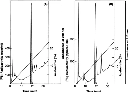
FIG. 3. ComparativeHPLC oftryptic peptides ofHa-MuSV p21 andp21 overproduced in E. coli. p21 labeledwith [35S]cysteine and [3H]palmitatewasisolated from Ha-NRKcells orfrom bacterialcellsoverproducingp21byimmunoprecipitation withananti-p21 antibody andSDS-PAGE. Theradioactively labeledp21wasmixed with100,ugofpurified unlabeledE.colip21as acarrier.Tryptic peptides of the carboxymethylated p21wereresolvedby HPLC. (A) E.colip21; (B) Ha-MuSVp21. Arrowsindicateunique peptides: peptideainE.coli cochromatographs with the synthetic tetrapeptide (CVLS), and peptide b inHa-MuSV p21 contains both 35Sand 3H radioactive labels. Tracings ofA210andblocks ofradioactivityoffractionsare as shown. Theelutionpositionof the synthetic CVLSpeptide is indicated by CVLSarrows.
which eluted 3 min later on HPLC. Therefore, we conclude that the unique E. coli peptide a is the C-terminal tryptic
peptide ofp21. A radioactive sequencing of
[35S]cysteine-labeled peptide a also confirmed the expected sequence of
Cys-Val-Leu-Serwithmostof theradioactivityfound in the
firstcycle of Edman degradation.
The identity of the hydrophobic peptide b of Ha-MuSV
p21 in the transformed NRK cells is also established by
cochromatography with the synthetic CVLS peptide after hydroxylamine cleavage of the fatty acid (Fig. 4B). Peptide b was obtained by HPLCfrom a tryptic digest of Ha-MuSV p21 double labeled with ["H]palmitate and [35S]cysteine. Afterlyophilization, the material was dissolved in 200 ,ul of 1 MNH2OH-HCl, thepHwas adjustedto 8.0 with NaOH, and the sample was incubatedat25°C for 8 h. Thereaction mixture was extracted with 400 ,ul ofchloroform, and the
on November 10, 2019 by guest
http://jvi.asm.org/
[image:3.612.111.505.78.563.2]E
E
400cL
0.
'> 300
200
0
0
la
< 100
CD
E
.0
E
0.
-U ._
-W
0 10 20 30
E c
0
N
0
0
U C
o 0 0
.0
Time (min)
Time
(min)
FIG. 4. Cochromatography of the synthetic tetrapeptide CVLS with peptide a and the hydroxylamine-treated peptide b. (A) The unique peptide a of E. colip21 tryptic digest labeled with[35Slcysteine(notcarboxymethylated) was mixed with the synthetic peptide (CVLS) and wasanalyzed by HPLC after performic acid oxidation. (B) The unique hydrophobic peptide b of Ha-MuSV double labeled with[3H]palmitate and[35S]cysteine washydrolyzed with hydroxylamine. After chloroform extraction of fatty acids, the material in the aqueous phase was mixed with the synthetic CVLS peptide and was analyzed by HPLC after performic acid treatment. The hatched areas indicate the radioactivity peaks.
aqueous phase was repeatedly lyophilized. After the
addi-tion of10
jig
of synthetic CVLS peptide, the mixture wastreated with performic acid as above. The material was
lyophilized and dissolved in500 ,ulofwaterandwasapplied
tothe HPLCcolumn. The columnwaselutedwith0 to60%
acetonitrile gradient in
0.05%
trifluoroacetic acid, and anarea (2 ml) around the CVLS peptide position was pooled.
After lyophilization, the mixture was again analyzed by
HPLC under the same conditions as in Fig. 4A. This
recovered radioactive peptide coeluted with the synthetic
CVLS (Fig. 4B). Although the
hydrophobic
peptide bpos-sesseda 3H-to-35S
counting
ratio of 0.8, the peptiderecov-ered inthe aqueous phase hadaratioof 0.18, characteristic
of the 35Sstandard. The ratiofor differential
counting
inthechloroform extract was similar to that ofthe 3H standard,
indicating
the presence of3H-lipid.
Thus, weconcludethatpeptide b is an acylated peptide of the p21 product and
peptide ais the precursor form found inE. colip21.
As
p21
intransformed cellscanbereadily labelednotonlyby
[3H]palmitate, a C16 saturated fatty acid, but alsoby
[3H]myristate,
which is twocarbon atoms shorter than theformer (datanot shown), we are interested in the natureof
the fatty acid attached to peptide b. Ha-NRK cells were
double labeled with
[35S]cysteine
(1,000Ci/mmol;
Amersham) and either
9,10(n)-[3H]palmitate
(50Ci/mmol;
Amersham) or
9,10(n)-[3H]myristate
(50Ci/mmol;
Amersham) under conditions described above. The
hybrophobic peptide b was isolated by HPLC after trypsin
digestion. The fatty acid was releasedfrom the peptide by
hydroxylamine cleavage. Thereaction mixtures in200 ,ulof
1 MNH20H-HCIwereextracted with400
RI1
ofchloroform.Thechloroformphasewasfurtherextracted threetimes with
200 ,ul of a chloroform-water mixture (1:10). The organic
phase from all extractions was pooled and dried under a
streamofN2.Thematerialsweredissolvedin 400pu1of 50% methanol and wereappliedtothe,uBondapak PhenylHPLC
column. Elution was with 70% methanol in 0.05%
trifluoroacetic acid at 1 ml/min. Fractions of 1.4 ml were
collected, and the
radioactivity
was counted. Palmitic acidand myristic acid standards were well separated. The
fatty
acid of
peptide
b waspalmitic
acidirrespective
of in vivolabeling either with
[3H]palmitate
or[3H]myristate.
Sincesomeofthefattyacids released from
proteins
may be in theform ofhydroxamates,theelution
positions
onHPLCof therespective fatty acid hydroxamates were also determined.
Hydroxamateswereobtainedby
hydroxylamine
cleavageofthe model thioester
compounds
[14C]palmityl
and[14C]myristyl
coenzyme A(Amersham). Hydroxamates
ofthese fatty acids eluted threefractions earlier than did the
corresponding free fatty acids under present HPLC
condi-tions (data not shown).
Thus,
radioactivity
released frompeptide b appears
mostly
as freepalmitic acid,
not as itshydroxamate. This is furtherconfirmed by complete
hydro-lysis ofthe3H-labeled
products
frompeptide
bwith 6 NHCIon November 10, 2019 by guest
http://jvi.asm.org/
[image:4.612.94.526.73.384.2]300
E
N.
0
,._w
0-0
0
100
co)
0
10
20
30
15
E
L0
co
0
x
E
10
0af
U
l.
co
co
54<
-W0
-W
-W
IP.
C,)
Fraction Number (1.4 ml)
FIG. 5. Identification of the fatty acid associated withthehydrophobic peptide b. Thefattyacid of Ha-MuSVp21 double labeled either with[35S]cysteineand[35H]palmitatemixture orwith[3S]cysteineand[3H]myristatewasanalyzedwithreversed-phaseHPLC. Thefattyacids wereobtained from the chloroform extracts ofHPLC-purified peptide b after hydroxylaminetreatment. The solid blocks depictstandard
[3H]palmitic acid and [3H]myristic acid. 1, HPLC profiles of3H radioactivity fromp21 labeledwith [3H]palmitate (fractions 29 and30)
performed under thesameconditions; 1,profilesofp21labeled with3H-myristate (fractions27to29). at 110HC for 16 h (4). The latter treatment did not further
change the elution position of the 3H products (Fig. 5).
Therefore, it is concluded that the fatty acid incorporated
into peptidebis palmiticacid,notmyristic acid. Apparently,
thesetwo3H-fatty acidsaremetabolically interconvertiblein
NRK cells.
We conclude that posttranslational processing of p21
involves palmitylation ofthe C-terminaltetrapeptide ofthe
sequence, Cys-Val-Leu-Ser. Although the chemical evi-dence does not allowus to ruleoutpalmitylation atthe last
serine residue, we consider this possibility very unlikely
sincepalmitylation alsooccurs inp21 of
rasK
(Fig.2B) andperhaps
rasN
genes, in which the last amino acids are both methionine rather than serine. It is most likely that thepalmitic acid moiety is attached to cysteine-186 of p21
molecules throughathioesterlinkage. Cysteine-186 is
con-served in all ras-related genes, and the specificity of
palmitylationappears tobe thecysteine residue4amino acid
from the C termini. The present studies, however, do not
rule outthepossibility thatp21 C-termini may be heteroge-neous withproductsfurtherprocessed by proteolytic cleav-ageafterpalmitylation atcys-186, since recovery of peptide
b isnotstoichiometric.Preliminaryattempts to removelipid
fromp21bytreatmentwith either hydroxylamine or alkaline
methanol do not restore
p21
mobility on SDS-PAGE to thatofpro-p21.
It is interesting to notethat although lipidation ofp21 is
required for membrane association and transformation, the
pro-p21 and the p21 overproducedinE. coli have all known
p21 biochemical activitiesof GTP/GDPbinding (11, 25, 27),
autokinase (11, 25, 27),and GTPase (9, 11, 17, 33). Perhaps
thep21 moleculehas an enzymedomain and another
mem-brane-binding domainat thepalmitylation site.
Some oncogene proteins, such as p60 src, and several
cellular
proteins
have recently been shown to bemyristy-latedattheir Nterminithrough amidelinkages (4, 6, 12, 16,
23, 24). Palmitylationofp21 throughathioesterlinkagenear
thep21 C terminusmay representadifferentmechanism for
posttranslational lipidation of proteins (13, 15, 19). Many
reports have described palmitylation of membrane-associated proteins probably occurring through thioester
linkages with internal cysteine residues (3, 20, 21, 22). The
exact palmitylation sites of these proteins have not been determined. The present identification ofthe p21 palmityla-tion site with a defined sequence may allow direct
experi-mentation to identify and to characterize the enzyme
sys-tems for protein palmitylation by using the p21 precursor
overproduced inE.coliasthe enzymesubstrate. Studieson these enzyme systems will be veryimportantfor
understand-ing posttranslational processing of membrane proteins with
criticalbiological consequences.
WethankSeisukeHattori and David J. Clantonfor many invalu-able discussions andforapreparationofpurifiedE. colip21(from S.H.),Darrell T. Liu and the late John J. Pisanofor valuable advice
on November 10, 2019 by guest
http://jvi.asm.org/
[image:5.612.128.489.75.393.2]onpeptide chemistry, and Milton J.Schlesinger for suggestionson fatty acid identification.
LITERATURE CITED
1. Allen, G. 1981. Sequencing of proteins and peptides. Elsevier BiomedicalPress, Amsterdam.
2. Beemon, K., and T. Hunter. 1978. Characterization of Rous sarcomavirus srcgeneproducts synthesized in vitro.J.Virol. 28:551-566.
3. Bolanowski, M. A., B. J. Earles, and W.J. Lennarz. 1984. Fatty acylation of proteins during development of sea urchin em-bryos. J. Biol. Chem. 259:4934-4940.
4. Buss, J. ,L.,and B. M. Sefton. 1985. Myristic acid, ararefatty acid, is ihe lipid attachedto the transforming protein ofRous sarcoma virus and its cellularhomolog.J. Virol. 53:7-12. 5. Chang,E.H.,J. M.Maryak,C.-M.Wei,T. Y.Shih,R.Shober,
H. L. Cheung, R. W. Ellis, G. L. Hager, E. M. Scolnick, and D. R. Lowy. 1980. Functional organization of the Harvey murine sarcomavirus genome.J. Virol. 35:76-92.
6. Cross, F. R., E. A. Garber, D.Pellman,and H. Hanafusa.1984. Ashortsequence in the p60src Nterminus is required forp60 src myristylationand membrane associationand for cell trans-formation. Mol.Cell. Biol. 4:1834-1842.
7. Dhar, R., R. W. Ellis, T. Y. Shih, S. Oroszlan, B. Shapiro, J. Maizel, D. Lowy, and E. M. Scolnick. 1982. Nucleotide se-quence of the p21 transforming protein of Harvey murine sarcomavirus. Science217:934-937.
8. Ellis,R.W., D.DeFeo,T. Y.Shih, M. A.Gonda,H. A.Young, N. Tsuchida, D. R. Lowy, and E.M. Scolnick. 1981. The p21 genes of Harvey and Kirsten sarcoma viruses originate from divergent members of a family of normal vertebrate genes. Nature(London) 292:506-511.
9. Gibbs, J. B., I. S. Sigal, M. Poe, and E. M. Scolnick. 1984. Intrinsic GTPase activity distinguishes normal and oncogenic ras p21 molecules. Proc. Natl. Acad. Sci. USA 81:5704-5708. 10. Gruss, P., R. W.Ellis, T.Y. Shih,M.Koenig, E. M.Scolnick,
andG. Khoury. 1981. SV40recombinant moleculesexpress the gene encoding p21 transforming protein of Harvey murine sarcomavirus. Nature(London)293:486-488.
11. Hattori, S., L. S. Ulsh, K. Halliday, and T. Y. Shih. 1985. Biochemical propertiesofa highly purified v-rasH p21protein overproduced inEscherichia coli and inhibition of its activities by amonoclonal antibody. Mol. Cell. Biol. 5:1449-1455. 12. Henderson, L. E., H. C. Krutzsch, and S. Oroszlan. 1983.
Myristyl amino-terminal acylation of murine retrovirus pro-teins: an unusual post-translational proteinmodification. Proc. Natl. Acad. Sci. USA 80:339-343.
13. Kaufman, J. F., M. S. Krangel, and J. L. Strominger. 1984. Cysteines in thetransmembrane region ofmajor
histocompati-bility complexantigensarefatty acylatedvia thioester bonds. J. Biol. Chem. 259:7230-7238.
14. Lautenberger,J. A.,L.Ulsh,T.Y.Shih,and T.S.Papas. 1983. High level expression in Escherichia coli of enzymatically active Harvey murine sarcoma virus p21 ras protein. Science 221:858-860.
15. Magee, A. I., A. H. Koyama, C. Malfer, D. Wen, and M. J. Schlesinger. 1984. Release offatty acids from virus
glycopro-teins byhydroxylamine. Biochim. Biophys. Acta798:156-166. 16. Marchildon, G. A., J. E. Casnellie, K. A. Walsh, and E.G. Krebs. 1984. Covalentlybound myristate inalymphoma tyro-sineproteinkinase. Proc. Natl. Acad. Sci. USA81:7679-7682. 17. McGrath, J. P., D. J. Capon, D. V. Goeddel, and A. D. Levinson. 1984. Comparativebiochemicalproperties of normal and activated human ras p21 proteins. Nature (London) 310:644-649.18. Merrifield, R. B. 1963. Solid phase peptide synthesis. I. The synthesisofatetrapeptide.J. Am. Chem. Soc. 85:2149-2156. 19. Rose, J. K., G. A. Adams, and C. J. Gallione. 1984. The
presence ofcysteinein thecytoplasmicdomain ofthevesicular
stomatitis virusglycoproteinisrequiredforpalmitateaddition. Proc. Natl. Acad. Sci. USA81:2050-2054.
20. Schlesinger, M. J., A. I. Magee,and M. F. G. Schmidt. 1980. Fattyacidacylationofproteinsin cultured cells.J. Biol. Chem. 255:10021-10024.
21. Schmidt, M. F. G., M. Bracha, and M. J. Schlesinger. 1979. Evidence for covalent attachmentoffattyacidstoSindbis virus
glycoproteins. Proc. Natl. Acad. Sci. USA 76:1687-1691.
22. Schmidt, M. F. G., and M. J. Schlesinger. 1979. Fatty acid
bindingtovesicular stomatitisvirusglycoprotein:a newtype of
posttranslatinal modification of the viral glycoprotein. Cell
17:813-819.
23. Schultz,A.M.,L. E.Henderson,S.Oroszlan,E. A.Garber,and H.Hanafusa. 1985. Amino terminalmyristylationof the
protein
kinase p60 src, a retroviral transformingprotein.
Science 227:427-429.24. Schultz,A.M.,andS. Oroszlan. 1984.Myristylationofgag-onc fusionproteins inmammalian transformingretroviruses. Virol-ogy 133:431-437.
25. Scolnick, E. M., A. G. Papageorge, and T. Y. Shih. 1979. Guanine
nucleotide-binding
activity
as an assay for the srcprotein of rat-derived murine sarcoma viruses. Proc. Natl.
Acad. Sci.USA76:5355-5359.
26. Sefton, B., I. S.Trowbridge,J.A.Cooper,andE. M.Scolnick. 1982.Thetransforming
proteins
ofRoussarcomavirus,Harvey
sarcoma virus and Abelson virus contain
tightly
boundlipid.
Cell31:465-474.27. Shih, T. Y., A. Papageorge, P. E. Stokes, M.0. Weeks, and E. M. Scolnick. 1980. Guanine
nucleotide-binding
andautophosphorylation
activities associated withp21
srcprotein
ofHarveymurinesarcomavirus.Nature(London)287:686-691.
28. Shih, T. Y., P. E. Stokes, G. W. Smythers, R. Dhar, and S. Oroszlan. 1982. Characterization of the
phosphorylation
sites and thesurroundingamino acidsequencesof thep21transform-ing proteins coded for by the Harvey and Kirsten strains of
murinesarcomaviruses. J. Biol. Chem.257:11767-11773. 29. Shih,T.Y.,and M.0.Weeks.1984.Oncogenesandcancer:
p21
rasgenes. CancerInvest.2:109-123.
30. Shih,T.Y.,M.0.Weeks,P.Gruss,R.Dhar,S.Oroszlan,and
E. M.Scolnick.1982.Identification ofaprecursorin the
biosyn-thesis of the p21 transformingprotein
ofHarvey murinesar-comavirus. J. Virol. 42:253-261.
31. Shih, T. Y., M. 0. Weeks, H. A. Young,and E. M.Scolnick. 1979.Identification ofa sarcomavirus-encoded
phosphoprotein
innonproducercellstransformed
by
HarveyorKirsten murinesarcoma virus.
Virology
96:64-79.32. Shih,T. Y., M. 0. Weeks, H. A. Young, and E. M. Scolnick.
1979.p21 of Kirsten murinesarcomavirus is thermolabile ina
viralmutanttemperature sensitive for the maintenance of
trans-formation. J. Virol.31:546-556.
33. Sweet, R. W., S. Yokoyama, T. Kamata, J. R. Feramisco, M.
Rosenberg,andM. Gross. 1984. The
product
ofrasisaGTPaseand T24
oncogenic
mutant is deficient in thisactivity.
Nature(London)311:273-275.
34. Ulsh,L.S.,and T. Y.Shih.1984. Metabolicturnoverofhuman
c-rasH p21
protein
of EJ bladder carcinoma and its normal cellular and viralhomologs.
Mol. Cell. Biol. 4:1647-1652.35. Wei, C. M., D. Lowy, and E. M. Scolnick. 1980.
Mapping
oftransforming region of the Harvey murine sarcoma virus
genome by
using
insertion-deletion mutants constructed in vitro. Proc. Natl. Acad. Sci. USA77:4674-4678.36. Willumsen, B. M., A. Christensen, N. L. Hubbert, A. G. Papageorge, andD. R. Lowy. 1984. The
p21
rasC-terminus isrequiredfortransformationand membraneassociation. Nature
(London)310:583-586.
37. Willumsen,B.M.,K.Norris,A.G.Papageorge,N. L.Hubbert, andD. R. Lowy. 1984. Harvey murine sarcomavirus
p21
rasprotein:
biological
andbiochemicalsignificance
of thecysteine
nearestthe carboxyterminus. EMBO J. 3:2581-2585.
![FIG.1.40-Harvey-rasAfricanprecipitationin[35S]cysteineeluted.previously pro-p21. Comparison of tryptic peptide maps of pro-p21 and p21](https://thumb-us.123doks.com/thumbv2/123dok_us/1395500.92648/2.612.337.538.353.565/fig-harvey-rasafricanprecipitationin-cysteineeluted-previously-comparison-tryptic-peptide.webp)
![FIG. 3.TracingscochromatographsCVLScarboxymethylatedand[3H]palmitate Comparative HPLC of tryptic peptides of Ha-MuSV p21 and p21 overproduced in E](https://thumb-us.123doks.com/thumbv2/123dok_us/1395500.92648/3.612.111.505.78.563/tracingscochromatographscvlscarboxymethylatedand-palmitate-comparative-hplc-tryptic-peptides-musv-overproduced.webp)
![FIG. of [35S]cysteine obtained 5. Identification with under acid from the and and the same [35H]palmitate [3H]myristic chloroform 3H-myristate to (fractions 27 p21 conditions; labeled either acid](https://thumb-us.123doks.com/thumbv2/123dok_us/1395500.92648/5.612.128.489.75.393/cysteine-identification-palmitate-myristic-chloroform-myristate-fractions-conditions.webp)