City, University of London Institutional Repository
Citation:
Martinelli, C., Rigoli, F. & Shergill, S.S. (2017). Aberrant Force Processing in Schizophrenia. Schizophrenia Bulletin, 43(2), pp. 417-424. doi: 10.1093/schbul/sbw092This is the accepted version of the paper.
This version of the publication may differ from the final published
version.
Permanent repository link:
http://openaccess.city.ac.uk/19139/Link to published version:
http://dx.doi.org/10.1093/schbul/sbw092Copyright and reuse: City Research Online aims to make research
outputs of City, University of London available to a wider audience.
Copyright and Moral Rights remain with the author(s) and/or copyright
holders. URLs from City Research Online may be freely distributed and
linked to.
Aberrant force processing in schizophrenia
Cristina Martinellia*, Francesco Rigolib, Sukhwinder S. Shergilla
aDepartment of Psychosis Studies, Institute of Psychiatry, Psychology & Neuroscience,
King’s College London, London, United Kingdom.
bWellcome Trust Centre for Neuroimaging, University College London, London, United
Kingdom.
2
Abstract
Initially considered as mere side effects of antipsychotic medication, there is now evidence
that motor and somatosensory disturbances precede the onset of the illness and can be found
in drug-naïve patients. However, research on the topic is scarce. Here, we were interested in
assessing the accuracy of the neural signal in detecting parametric variations of force linked
to a voluntary motor act and a received tactile-sensation, either self- or externally-generated.
Patients with a diagnosis of schizophrenia and healthy controls underwent functional
magnetic resonance imaging while asked to press, or abstain from pressing, a lever in order to
match a visual target force. Forces, exerted and received, varied on 10 levels from 0.5N to 5N
in 0.5N increments. Healthy participants revealed a positive correlation between force and
activity in contralateral primary somatosensory area (S1) when performing a movement as
well as when receiving a tactile sensation, but only when this was externally, and not self-,
generated. Patients showed evidence of altered force signalling in both motor and tactile
conditions, as well as increased correlation with force when tactile sensation was
self-generated. Findings are interpreted in line with accounts of predictive and sensory integration
mechanisms and point towards alterations in the encoding of parametric forces in the motor
and somatosensory domain in patients affected by schizophrenia.
Introduction
Motor and somatosensory disturbances have been consistently reported in the literature on
schizophrenia. With regards to motor deficits, patients have shown to be slow when
generating motor acts1, to exhibit abnormal involuntary movements2, altered coordination3 and finger sequencing4. Other motor impairments include catatonia5 and neurological soft signs (NSS6). With regards to somatosensory disturbances, evidence shows reduced sensory
gating in the somatosensory domain7 and pain sensitivity8. Patients are also thought to show
lack of somatosensory attenuation, whereby self-, but not externally-, generated sensations
are attenuated, putatively as a result of them being predicted9-12. In line with this,
somatosensory brain response has shown to be increased when sensation is externally
generated, as opposed to self-generated, in controls and not in patients10. In schizophrenia,
lack of somatosensory attenuation has been associated with aberrant sense of agency and may
lead to a misattribution of self-generated actions to external sources, thus contributing to the
development of psychotic symptoms, such as auditory hallucinations and delusions of
3
There is now ample evidence showing that motor and somatosensory impairments are present
in antipsychotic naïve schizophrenia patients14-15, in individuals at risk of developing the
illness14, 16-17 and in patients’ biological relatives18-20, suggesting that these are not merely
consequent on antipsychotic treatment. To this extent, recent models support the view that
such symptoms constitute an important clinical dimension and may provide important insight
into the biological and neurodevelopmental mechanisms underlying the disorder21. However,
despite their clinical importance, research has yet to characterise the determinants of motor
and somatosensory dysfunction in schizophrenia. In particular, very little is known regarding
the level of accuracy of such systems in patients.
Accuracy of force processing, for example, is an important component in motor and
somatosensory functional domains and it is likely that inappropriate tuning of motor and
somatosensory brain areas to varying levels of force may account for some of the
aforementioned deficits in schizophrenia. In particular, failure to appropriately encode
generated motor force might give rise to problems associated with the correct execution and
monitoring of movements22. Moreover, as the motor and somatosensory systems are largely
intertwined, appropriate signalling of somatosensory feedback associated to motor acts is also
likely to contribute to the same issues23. Lastly, altered somatosensory feedback of force,
associated with received tactile sensations, might account for some of the sensory
impairments detected in experimental settings, including reduced somatosensory attenuation,
and at such may play an important role in psychosis. Nevertheless, accuracy of force
processing in schizophrenia has received little investigation.
There is general agreement that motor and somatosensory areas increase activity with
increasing force24-26. Here, we employed functional magnetic resonance imaging (fMRI) to
research the neural mechanisms underlying force production and sensation with the
hypothesis that schizophrenia patients would demonstrate aberrant signalling of force
intensity in the motor and somatosensory network. In light of previous neuroimaging findings
on healthy individuals27-29, 25, analysis was focused on response in contralateral (to the hand
engaged in task) primary motor cortex (M1), primary somatosensory cortex (S1) and
supplemental motor area (SMA). Whereas force-related activation in M1 and S1 has been
4
and somatosensory stimuli, force-related activation in SMA has been predominantly
associated with its specific role in motor planning and volitional control29.
The task employed here has been previously used in our lab for the investigation of basic
sensory attenuation in patients9-10. We now exploit its features (described in Materials and
Methods) to independently analyse the accuracy of the motor and somatosensory system in
encoding parametric variations of force. Moreover, by comparing brain activation to tactile
forces when these are self- as opposed to externally- generated, we could also investigate the
effects of sensory attenuation on parametric force signal. Specifically, we predicted that a)
healthy individuals would show a positive correlation with forces in contralateral M1 and
SMA when performing a movement; b) commensurately in contralateral S1 when receiving a
tactile sensation; c) but also in contralateral S1 when performing a movement as the result of
the somatosensory feedback linked to the motor act; d) patients would show altered motor
and sensory force signalling in all of the above; finally e) we predicted that healthy controls
would show absent or reduced correlation between applied force and brain activation within
contralateral S1 when the force could be predicted (i.e., was self-generated), as opposed to
when the force could not be predicted (i.e., was externally generated), and that patients would
fail to demonstrate this difference.
Materials and Methods
Participants
Volunteers were 21 patients with a diagnosis of schizophrenia (based on assessment using
criteria from DSM-IV-TR30) being treated with stable antipsychotic medication, and 26
healthy individuals with no reported history of psychiatric illness assessed with the MINI
International Neuropsychiatric Interview31 (Table 1). Ethical approval was provided by South
London and Maudsley Research and Ethics Committee. All participants provided informed
written consent and were given a monetary inconvenience allowance for study participation.
Participants met the following inclusion criteria: 1) capacity to consent; 2) age between 18-60
years; 3) sufficient command of the English language to understand task instructions and 4)
right-handedness, as measured by the Edinburgh Handedness Inventory32. Participants were excluded if they had: 1) current drug or alcohol dependence; 2) brain disease or damage or if
5
confirmed by an experienced clinician and severity of symptoms was assessed with the
Positive and Negative Symptoms Scale (PANSS33).
Data acquisition
Blood oxygenation level-dependent (BOLD) functional images were acquired on a GE 3
Tesla system (Signa Excite; General Electric) with an 8-channel head coil using an echo
planar imaging sequence with the following parameters: repetition time, 2600 milliseconds;
echo time, 30 milliseconds; and flip angle, 90°. In each of the three runs, 330 volumes that
comprised 40 descending, sequentially ordered 2-mm axial slices (with 1-mm gap between
slices) and an in-plane resolution of 3 × 3 mm were acquired.
Task paradigm
Participants performed a sensorimotor task with the use of the experimental apparatus
depicted in Figure 19-10 which enabled forces to be measured through the use of two pressure
sensors mounted one above the other. The upper sensor was fixed in space, and the lower
sensor was mounted on the end of a lever attached to a small torque motor. This apparatus
permitted a press (by the right index finger) on the upper sensor to either be transmitted to the
left index finger or not. Moreover, the tactile stimulus on the left finger could also be
presented in the absence of any corresponding right finger press. Thus, four experimental
conditions were presented: movement with self-produced tactile sensation (M1S1), externally
produced tactile sensation with no movement (M0S1), self-produced movement without
tactile sensation (M1S0) and a no movement and no tactile sensation rest (M0S0). Forces
exerted and received varied on 10 levels from 0.5N to 5N in 0.5N increments. Thus, this set
up allowed investigating brain activation associated to parametric force when both
performing a motor act and receiving a tactile sensation, the latter being either self- or
externally-generated. Note that in M1S1, the intensity of the self-generated force was
transferred to the left finger and was thus predictable, whereas in M0S1, the force was
generated by the apparatus and thus its intensity could not be predicted by subjects.
The experimental session comprised three 13-minute runs, containing a total of 468 trials
split in alternating blocks of 13 trials each. Each block included 10 experimental trials from
one sole condition and 3 null trials, yielding a total of 90 experimental trials and 27 null trials
per condition. Within each run, 3 blocks of each condition were presented in random order.
6
would receive a non-self generated tactile sensation on the left finger in M0S1, they were not
able to predict its force. At the start of each block, participants were shown an instruction
screen ‘press’ or ‘don’t press’ (depending on condition) for 3s - no instruction was given
regarding tactile sensation. Each trial was 4s long (1s target + 3s response) and inter-trial
interval was 1s. Same timings applied to no movement trials in that the lever pressed
subjects’ left finger after 1s target display. In line with previous studies9-10, the average force
associated with movement and tactile sensation during the last two seconds of the trial was
calculated for each subject and used to run behavioural and neural analyses. We chose to
exclude the first second of response to ensure a more accurate measure of peak forces
generated and received.
Before scanning, all participants received full instructions regarding task features and
underwent a training phase using the same experimental apparatus described above,
comprehensive of all four experimental conditions. In line with previous studies from our
lab9-10,participants learnt a correspondence between target on the screen and force applied. However, they didn’t have to hold this information in memory, but could rather visually
follow the cursor moving towards the target while pressing. To avoid pressing on the lower
sensor, subjects’ left finger was taped to the apparatus. To facilitate the required sustained
attention, sessions were split by a short relaxation period during which the participants
remained in the scanner.
Functional data analysis
The fMRI data were processed using SPM8 (Statistical Parametric Mapping, Wellcome
Department of Imaging Neuroscience, University of London). Data were realigned to the first
image, normalized to a standard template of the Montreal Neurological Institute brain, and
smoothed using an 8-mm full-width at half-maximum Gaussian kernel. BOLD response was
modelled with a canonical hemodynamic response function and a general linear model
(GLM) including four stick function regressors associated with trial onset, one for each
condition, and four stick function regressors associated with the last two seconds of the trial,
one for each condition. To investigate the relationship between force and BOLD activity on a
single-trial basis, condition-specific average forces during the last two seconds of the trials
following right-index finger movement and left-index finger sensation were calculated for
each individual and included as first-order parametric modulators of BOLD activity for
7
parameter vectors as regressors of no interest. To further investigate group differences in
head motion, we performed group comparisons on the computed mean of the absolute values
of the motion correction parameters for translation (X, Y, Z) and rotation (pitch Z, roll Y,
yaw Z) across all runs34 (see Supplemental Materials).
First-level contrast images associated to parametric modulators were entered into a
second-level random-effects model. Across subjects, the parameters associated to the parametric
modulators were used for one-sample (within groups) and independent-samples (between
groups) t-tests. To account for the putative influence of gender and task accuracy on our
results, we included two covariates in our second-level model, reflecting gender and subjects’
accuracy on the motor task. Accuracy of generated forces was estimated by conducting a
linear regression between expected and actual forces, separately for each condition and
subject. Estimated betas (indicating subjects’ accuracy in generating forces according to
target) were then used as covariates in all our second level analyses. To investigate whether
patients’ medication could account for the observed group differences, we correlated chlorpromazine equivalent medication dosage with patients’ fMRI BOLD activation in the
peak significant for the between group contrasts (see Supplemental Materials). Further
analyses were run to investigate the association between severity of symptoms and alterations
of force signalling in the illness. In line with this, contrast images associated with parametric
modulators were further included in a regression model and correlated with PANSS scores
for the positive and negative scales as a covariate of interest for force related conditions.
A region of interest (ROI) approach was adopted to investigate task effects in M1, S1 and
SMA. A priori ROIs in M1, S1 and SMA were created at the group level, with a 5mm-radius
sphere centred on the foci of standard peak activations for right and left finger movement29,
35. The Human Motor Area Template (HMAT36) was used as masking to ensure proper
location of observed activations in M1, S1 or SMA. For hypothesis testing, the peak voxel
statistics were small volume corrected for our a-priori ROIs, with a p < 0.05 Family Wise
Error (FWE) significance criterion. Note that different contrasts may highlight different peak
activation voxels, even if the same ROI is used for both these contrasts. For completeness, we
also ran an exploratory whole brain analysis where statistics were corrected for the whole
8
Results
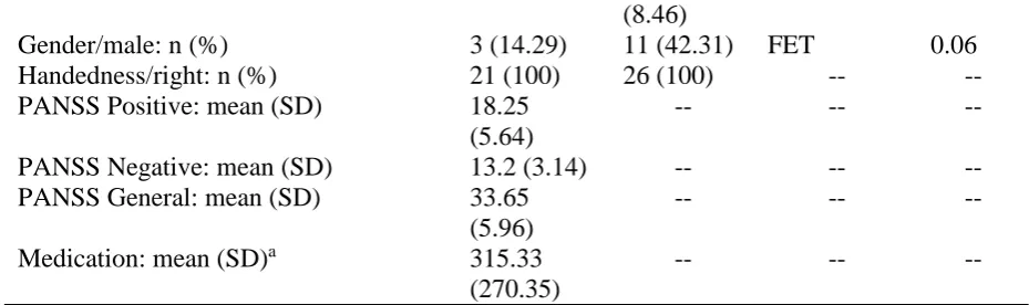
In the healthy controls, we observed a positive correlation between exerted force and brain
activity in left S1 in both conditions with movement, namely M1S1 (-34, -26, 54; Z = 3.08; p
= 0.018 SVC) and M1S0 (-32, -24, 54; Z = 3.49; p = 0.008 SVC). Note that this effect
emerged in left S1, contralateral to the finger used to produce the movement and hence
putatively reflects the somatosensory feedback to the same finger. No difference was found
for the contrast M1S1 vs. M1S0 in left S1. Contrary to expectations, no positive correlation
was detected between exerted force and brain activity in other ROIs for these conditions.
Schizophrenia patients also exhibited a positive correlation between exerted force during
movement and brain activity in left S1 in both M1S1 (-38, -22, 56; Z = 3.55; p = 0.004 SVC)
and M1S0 (-40, -20, 54; Z = 3.09; p = 0.015 SVC). No difference was found for the contrast
M1S1 vs. M1S0 in left S1. Between groups contrasts revealed a trend towards increased
correlation between exerted forces and left S1 activity in patients compared to healthy
controls in M1S1 (Fig 2A: 38, 22, 56; Z = 2.57; p = 0.052, SVC) and M1S0 (Fig 2B: 34,
-20, 48; Z = 2.59; p = 0.067, SVC). No group showed force related activation in the side
ipsilateral to the hand performing the movement in M1S0 in any of our ROIs. Taken together,
these findings suggest that in both groups S1 encodes parametric variation of force associated
with the somatosensory feedback from the finger performing the motor act, and that in
patients such encoding is increased.
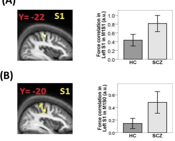
In the right S1 (i.e., associated to the left finger receiving the tactile sensation), healthy
controls exhibited a positive correlation between the intensity of tactile sensation and
activation during M0S1 (32, -24, 50; Z = 3.75; p = 0.003 SVC). On the contrary, patients
failed to show such effect. Healthy controls exhibited no correlation between force on the left
finger and activity in right S1 when this was obtained by a self-generated movement (M1S1).
Patients exhibited some activation for M1S1, but this did not survive SVC (38, -28, 56; Z =
1.95; p = 0.15, SVC). In line with accounts of sensory attenuation, controls showed increased
correlation when the sensation was externally, as opposed to self-, produced (Fig 3A: 34, -26,
50; Z = 3.19; p = 0.014 SVC). On the contrary, patients failed to exhibit such pattern of
results, resulting in reduced difference for the contrast M0S1 > M1S1 compared to healthy
controls (36, -24, 54; Z = 3.48; p = 0.005). Moreover, we found that patients showed an
9
latter was self-generated (i.e., M1S1 > M0S1; Fig 3B: 38, -24, 56; Z = 2.61; p = 0.048). This
seemed to be driven by reduced correlation with force in M0S1 compared to healthy controls
(Fig 3C: 36, -24, 52; Z = 3.51; p = 0.005 SVC). Neither group showed significant correlations
between received force intensity and brain activity in other ROIs. No group showed force
related activation in the side ipsilateral to the hand receiving the tactile sensation in M0S1 in
any of our ROIs. Taken together, these findings suggest that in healthy individuals, S1 tunes
to parametric forces associated to tactile sensation only when these are externally generated
and thus unpredicted. On the contrary, patients fail to show such somatosensory attenuation
and instead exhibit more accurate encoding of parametric tactile forces when these are
predictable.
We found no difference in motion parameters across groups (see Supplemental Materials),
suggesting that residual differences in motion did not contribute to results above. Also, we
found no group differences in baseline condition M0S0, for any of our a-priori ROIs.
Furthermore, patients exhibited no correlation between chlorpromazine equivalent dosage
and peak activation of between groups contrast estimates (see Supplemental Materials),
suggesting that antipsychotic medication could not account for observed group differences.
We found no correlation between PANSS scores on either the negative or positive subscales
and brain activity when investigating the whole brain (FWE correction). However, we did
find positive correlations with negative symptoms when investigating activation within our
ROIs – these results are reported as exploratory findings in the Supplemental Materials.
Whole-brain analysis revealed significant correlation between exerted force and brain activity
in left S1 for the M1S1 condition in healthy controls (-38, -30, 66; Z = 5.08; p = 0.023,
FWE). No significant whole-brain activations were found for other brain regions, conditions
or symptoms in schizophrenia patients after correcting for multiple comparisons. We
therefore report results from a whole-brain analysis with p < 0.001 uncorrected in the
Supplementary Materials.
Discussion
Motor and somatosensory disturbances are well established in schizophrenia. Initially
10
that such disturbances precede the onset of the illness and can be found in drug-naïve patients
or first-degree relatives. In line with this, a few studies attempted to investigate the neural
mechanisms underlying such disturbances. However, research of this kind is relatively scarce,
specifically there is little data regarding the accuracy of the motor and somatosensory system
in patients. Here, we were interested in researching the accuracy of the neural signal in
detecting parametric variations of force intensity linked to a voluntary motor act or a received
tactile-sensation. Moreover, given the hypothesised importance of motor action prediction for
sense of agency disturbance in the illness, we were also interested in investigating how the
accuracy of the parametric force signal would change as a function of its predictability (i.e.,
forces being either self- or externally-generated). Note that whereas the influence of
predictive mechanisms on somatosensory attenuation has been investigated in relation to
general sensory processing9-10, this has never been investigated in relation to specific features
of sensory processing such as parametric force encoding.
Force processing is known to employ key structures of the motor and somatosensory system.
In particular, research on healthy individuals has shown a positive correlation between
exerted force during a movement and brain activity in contralateral sensorimotor areas24-25, 37.
Likewise, positive correlations between force intensity and contralateral somatosensory areas
have also been found in relation to received tactile-sensations38-40. However, how such force
processing differs when the tactile-sensation is self, as opposed to externally, generated is
unknown. Here, we aimed to investigate force processing in schizophrenia in regards to all
the above mentioned aspects. Based on the literature on healthy individuals, we confined our
analysis to M1, S1 and SMA.
In healthy controls, we found a positive correlation between exerted force during movement
conditions and activation in S1 contralateral to the finger used for movement production. This
fits with what has previously been observed in the literature consistent with a somatosensory
feedback linked to performing a movement24-25, 37. Surprisingly, controls failed to exhibit
positive correlation between force and brain activity in motor areas such as M1 and SMA.
Lack of activation in SMA may be related to the fact that movements in motor conditions
(i.e., M1S1 and M1S0) were here triggered by an external cue (i.e., the appearance of the
target on the screen) and were not the product of self-paced voluntary intentions. The role of
SMA in differentiating between self-paced and externally-triggered movements has recently
received increasing empirical support, confirming the crucial role of SMA in transforming
11
activation in M1 is harder, as the latter has shown to correlate with force on several
occasions24-25, 27. It is possible that such discrepancy is attributable to the nature of the task
employed here, which required participants to both press and hold down a lever to a certain
intensity level. In support of this interpretation, recent studies comparing static and dynamic
force movements found that activation with parametric forces was higher when the latter
were dynamic compared to static42. It is thus likely that press and hold movements more
heavily rely on somatosensory feedback than those used to investigate force processing in
other studies (e.g., precision grip force, opposition force, etc.), hence resulting in lack of
force-related M1 activation in our study.
Patients exhibited increased correlation between forces and brain activity in S1 (contralateral
to the movement) when actively pressing the lever (i.e., M1S1 and M1S0). One potential
interpretation for this finding links to sensory attenuation disturbances. As the sensation is
associated to a self-generated motor act, accurate force processing can be attenuated in
healthy individuals. Increased correlation between somatosensory activation and force in
patients compared to healthy individuals might reflect lack of such attenuating mechanism.
However, such interpretation remains speculative, as the current paradigm does not allow
testing sensory attenuation linked to the finger performing the movement.
With regards to processing of applied tactile force, healthy controls exhibited a positive
correlation between the intensity of the sensation on the left finger and activation in
contralateral S1 when the sensation was externally generated, but not when this was
self-produced. This finding resonates with observations that tactile-sensations are attenuated when
self-generated, and thus predicted, as opposed to when they are externally generated and thus
unpredicted9-10. Likewise, one might expect that accurate encoding of forces is only really
necessary when the sensation is not predicted. As a result of this, somatosensory areas would
not tune to parametric variations of forces when these are self-generated. Note that this would
not be the case, in healthy controls, for somatosensory activation associated to body parts
involved in generating movements, as discussed above, as this would still be relevant for the
accurate performance of the task.
Contrary to controls, schizophrenia patients showed no correlation between force and
activation in S1 when receiving an externally generated tactile-sensation (M0S1). This
resulted in patients failing to show increased S1 activation when receiving an externally
12
patients the sensory signal encoding parametric tactile force was higher when the force could
be predicted (M1S1) as opposed to when it was unpredicted (M0S1). A possible explanation
of this result relies on sensory integration accounts, which emphasise the interplay between
predictive and postdictive mechanisms in the experience of agency43. In particular,
individuals with schizophrenia have been shown to rely more on visual postdictive feedback
associated to motor actions44. In our study, an increased weight attributed to visual
postdictive feedback may lead patients to be less accurate in conditions when such visual
feedback is unrelated with the level of tactile stimulation. This lack of relationship
characterizes M0S1 alone (and not M1S1, M1S0, M0S1) where, by design, tactile forces
generated by the apparatus did not correspond to visual information on the screen. The hypothesis that patients’ somatosensory encoding relies largely on visual feedback might
entail a decreased influence of motor predictions and in turn a decreased attenuation43. This
connects to data reported above showing an increased accuracy for both M1S1 and M1S0 in
left S1 (associated to the pressing finger) in patients compared to controls. Future studies
should aim to test whether an increased weight on postdictive visual feedback is directly
associated with a decreased weight on tactile predictions (derived from motor commands)
which in turn may be responsible for decreased attenuation of tactile inputs in schizophrenia.
More insight on the matter may be gained by investigating the anticipatory mechanisms
involved in motor execution and receipt of tactile sensation. It is indeed possible that groups
differ in the way they prepare for the task, also influencing the weight attributed to the
various predictive and postidictive components involved.
This study presents a number of limitations. First, all patients were on stable antipsychotic
treatment at the time of the experiment, possibly affecting our results. The effects of
antipsychotic medication on the motor system are unclear, with studies reporting
improvement, deterioration or no change on motor disturbance as a result of treatment45.
However, we here found no association between medication dosage and brain activity in
patients suggesting that our results were not due to antipsychotic medication. Second, we did
not collect information on constancy of response across time, which may have had an impact
on task performance. Third, this study failed to stratify patients on the basis of their specific
motor profile. Indeed, recent studies suggest that specific motor disturbances may be
associated with specific patterns of brain dysfunction21. Particularly relevant for the current
task are models of motor slowing, which propose that the latter may be associated with
13
compensatory mechanisms from the premotor cortex46. It is hard to reconcile these models
with our findings, as we failed to find activity in premotor areas and did not collect
information regarding motor slowing. In order to investigate whether motor slowing affects
encoding of parametric forces, future studies should stratify patients on the basis of their
motor profile while also using a force task that better taps on SMA and pre-SMA functioning.
Lastly, we failed to show a correlation between positive symptoms and predictive
dysfunctions in patients. One possible reason links to our sample, which did not provide
sufficient sensitivity on the delusional scale, most often associated to dysfunctions in
prediction and sense of agency10, 43.
In sum, our results point towards alterations in the encoding of parametric force in the motor
and somatosensory domain in patients affected by schizophrenia. Particularly affected seems
to be the processing of the somatosensory feedback associated to parametric force, putatively
linked to defective prediction and sensory integration mechanisms. This study also confirms
the utility of using functional imaging to probe cortical response to elementary sensorimotor
stimuli, in addition to the more conventional investigation of higher order cognitive
processing. We suggest that future research on the topic would benefit from the investigation
of the specific functional features involved in motor and somatosensory processing. In
particular, mapping alterations in these components with the different motor profile of
patients may increase our understanding of these often under-recognised symptoms46.
References
1. Morrens M, Hulstijn W, Sabbe B (2007). Psychomotor slowing in schizophrenia.
Schizophrenia bulletin, 33(4), 1038-1053.
2. Compton MT, Fantes F, Wan CR, Johnson S, Walker EF (2015). Abnormal movements in
first-episode, nonaffective psychosis: Dyskinesias, stereotypies, and catatonic-like signs.
Psychiatry research, 226(1), 192-197.
3. Varlet M, Marin L, Raffard S, Schmidt RC, Capdevielle D, Boulenger JP, Bardy BG
(2012). Impairments of social motor coordination in schizophrenia. PLoS One, 7(1), e29772.
4. Delevoye-Turrell Y, Giersch A, Wing AM, Danion JM (2007). Motor fluency deficits in
14 5. Peralta V, Campos MS, Jalón D, García E, Cuesta MJ (2010). Motor behavior
abnormalities in drug‐naïve patients with schizophrenia spectrum disorders. Movement
Disorders, 25(8), 1068-1076.
6. Bombin I, Arango C, Buchanan RW (2005). Significance and meaning of neurological
signs in schizophrenia: two decades later. Schizophrenia Bulletin, 31(4), 962-977.
7. Thoma RJ, Hanlon FM, Huang M, Miller GA, Moses SN, Weisend MP, Cañive JM
(2007). Impaired secondary somatosensory gating in patients with schizophrenia. Psychiatry
research, 151(3), 189-199.
8. Boettger MK, Grossmann D, Bär KJ (2013). Increased cold and heat pain thresholds
influence the thermal grill illusion in schizophrenia. European Journal of Pain, 17(2),
200-209.
9. Shergill SS, White TP, Joyce DW, Bays PM, Wolpert DM, Frith CD (2013). Modulation
of somatosensory processing by action. Neuroimage, 70, 356-362.
10. Shergill SS, White TP, Joyce DW, Bays PM, Wolpert DM, Frith CD (2014). Functional
magnetic resonance imaging of impaired sensory prediction in schizophrenia. JAMA
psychiatry, 71(1), 28-35.
11. Shergill SS, Bays PM, Frith CD, Wolpert DM (2003). Two eyes for an eye: the
neuroscience of force escalation. Science, 301(5630), 187-187.
12. Shergill SS, Samson G, Bays PM, Frith CD, Wolpert DM (2005). Evidence for sensory
prediction deficits in schizophrenia. American Journal of Psychiatry, 162(12), 2384-2386.
13. Frith CD, Done DJ (1988). Towards a neuropsychology of schizophrenia. The British
Journal of Psychiatry, 153(4), 437-443.
14. Wolff AL, O'Driscoll, GA (1999). Motor deficits and schizophrenia: the evidence from
neuroleptic-naive patients and populations at risk. Journal of Psychiatry and Neuroscience,
24(4), 304.
15. Arnfred SM, Chen AC (2004). Exploration of somatosensory P50 gating in schizophrenia
spectrum patients: reduced P50 amplitude correlates to social anhedonia. Psychiatry
research, 125(2), 147-160.
16. Daly MP (2014). Characterization of Somatosensory Processing in Relation to
Schizotypal Traits in a Sample of Nonclinical Young Adults. CITY UNIVERSITY OF NEW
YORK.
17. Hagenmuller F, Heekeren K, Theodoridou A, Walitza S, Haker H, Rössler W, Kawohl W
(2014). Early somatosensory processing in individuals at risk for developing psychoses.
15 18. Flyckt L, Sydow O, Bjerkenstedt L, Edman G, Rydin E, Wiesel FA (1999). Neurological
signs and psychomotor performance in patients with schizophrenia, their relatives and healthy
controls. Psychiatry research, 86(2), 113-129.
19. Chang BP, Lenzenweger MF (2001). Somatosensory processing in the biological relatives
of schizophrenia patients: a signal detection analysis of two-point discrimination. Journal of
abnormal psychology, 110(3), 433.
20. Chang BP, Lenzenweger MF (2005). Somatosensory processing and schizophrenia
liability: proprioception, exteroceptive sensitivity, and graphesthesia performance in the
biological relatives of schizophrenia patients. Journal of abnormal psychology, 114(1), 85.
21. Walther S, Strik W (2012). Motor symptoms and schizophrenia. Neuropsychobiology,
66(2), 77-92.
22. Teale P, Pasko B, Collins D, Rojas D, Reite M (2013). Somatosensory timing deficits in
schizophrenia. Psychiatry Research: Neuroimaging, 212(1), 73-78.
23. Baker SN (2007). Oscillatory interactions between sensorimotor cortex and the periphery.
Current Opinion in Neurobiology, 17(6), 649-655.
24. Cramer SC, Weisskoff RM, Schaechter JD, Nelles G, Foley M, Finklestein SP, Rosen BR
(2002). Motor cortex activation is related to force of squeezing. Human brain mapping,
16(4), 197-205.
25. Kuhtz-Buschbeck JP, Gilster R, Wolff S, Ulmer S, Siebner H, Jansen O (2008). Brain
activity is similar during precision and power gripping with light force: an fMRI study.
Neuroimage, 40(4), 1469-1481.
26. Manganotti P, Formaggio E, Storti SF, Avesani M, Acler M, Sala F, Beltramello A
(2009). Steady-state activation in somatosensory cortex after changes in stimulus rate during
median nerve stimulation. Magnetic resonance imaging, 27(9), 1175-1186.
27. Dettmers C, Fink GR, Lemon RN, Stephan KM, Passingham RE, Silbersweig D, ...
Frackowiak RS (1995). Relation between cerebral activity and force in the motor areas of the
human brain. Journal of Neurophysiology, 74(2), 802-815.
28. Wexler BE, Fulbright RK, Lacadie CM, Skudlarski P, Kelz MB, Constable RT, Gore JC
(1997). An fMRI study of the human cortical motor system response to increasing functional
demands. Magnetic resonance imaging, 15(4), 385-396.
29. Haller S, Chapuis D, Gassert R, Burdet E, Klarhöfer M (2009). Supplementary motor area
and anterior intraparietal area integrate fine‐graded timing and force control during precision
16 30. American Psychiatric Association (2000). Diagnostic criteria from DSM-IV-TR.
American Psychiatric Pub.
31. Sheehan DV, Lecrubier Y, Sheehan KH, Amorim P, Janavs J, Weiller E, ... Dunbar GC
(1998). The Mini-International Neuropsychiatric Interview (MINI): the development and
validation of a structured diagnostic psychiatric interview for DSM-IV and ICD-10. Journal
of clinical psychiatry.
32. Oldfield RC (1971). The assessment and analysis of handedness: the Edinburgh
inventory. Neuropsychologia, 9(1), 97-113.
33. Kay SR, Flszbein A, Opfer LA (1987). The positive and negative syndrome scale
(PANSS) for schizophrenia. Schizophrenia bulletin, 13(2), 261.
34. Haller S, Monsch AU, Richiardi J, Barkhof F, Kressig RW, Radue EW (2014). Head
motion parameters in fMRI differ between patients with mild cognitive impairment and
Alzheimer disease versus elderly control subjects. Brain topography, 27(6), 801-807.
35. Kuhtz‐Buschbeck JP, Mahnkopf C, Holzknecht C, Siebner H, Ulmer S, Jansen O (2003).
Effector‐independent representations of simple and complex imagined finger movements: a
combined fMRI and TMS study. European Journal of Neuroscience, 18(12), 3375-3387.
36. Mayka MA, Corcos DM, Leurgans SE, Vaillancourt DE (2006). Three-dimensional
locations and boundaries of motor and premotor cortices as defined by functional brain
imaging: a meta-analysis. Neuroimage, 31(4), 1453-1474.
37. Sulzer JS, Chib VS, Hepp-Reymond MC, Kollias S, Gassert R (2011, August). BOLD
correlations to force in precision grip: An event-related study. In Engineering in Medicine
and Biology Society, EMBC, 2011 Annual International Conference of the IEEE (pp.
2342-2346). IEEE.
38. Torquati K, Pizzella V, Della Penna S, Franciotti R, Babiloni C, Rossini PM, Romani GL
(2002). Comparison between SI and SII responses as a function of stimulus intensity.
Neuroreport, 13(6), 813-819.
39. Bornhovd K, Quante M, Glauche V, Bromm B, Weiller C, Buchel C (2002). Painful
stimuli evoke different stimulus-response functions in the amygdala, prefrontal, insula and
somatosensory cortex: a single-trial fMRI study. Brain 125, 1326–1336.
40. Timmermann L, Ploner M, Haucke K, Schmitz F, Baltissen R, Schnitzler A (2001).
Differential coding of pain intensity in the human primary and secondary somatosensory
cortex. J. Neurophysiol. 86, 1499–1503.
41. Nachev P, Kennard C, Husain M (2008). Functional role of the supplementary and
17 42. Keisker B, Hepp‐Reymond MC, Blickenstorfer A, Kollias SS (2010). Differential
representation of dynamic and static power grip force in the sensorimotor network. European
Journal of Neuroscience, 31(8), 1483-1491.
43. Synofzik M, Vosgerau G, Voss M (2013). The experience of agency: an interplay
between prediction and postdiction. Frontiers in Psychology, March.
44. Synofzik M, Thier P, Leube DT, Schlotterbeck P, Lindner A (2010). Misattributions of
agency in schizophrenia are based on imprecise predictions about the sensory consequences
of one’s actions. Brain 133, 262–271.
44. Docx L, Morrens M, Bervoets C, Hulstijn W, Fransen E, De Hert M, Sabbe B (2012).
Parsing the components of the psychomotor syndrome in schizophrenia. Acta Psychiatrica
Scandinavica, 126(4), 256-265.
45. Peralta V, Cuesta MJ (2010). The effect of antipsychotic medication on neuromotor
abnormalities in neuroleptic-naïve nonaffective psychotic patients: a naturalistic study with
haloperidol, risperidone, or olanzapine. Prim Care Companion J Clin Psychiatry, 12(2),
e1-11.
46. Walther S (2015). Psychomotor symptoms of schizophrenia map on the cerebral motor
circuit. Psychiatry Research: Neuroimaging, 233; 293-298.
Acknowledgment
This paper presents independent research funded by the National Institute for Health
Research (NIHR) Biomedical Research Centre for Mental Health at South London and
Maudsley NHS Foundation Trust and Institute of Psychiatry, Psychology & Neuroscience
King’s College London via a research studentship awarded to Cristina Martinelli. SSS is
funded by a European Research Council Consolidator Award. The views expressed are those
of the authors and not necessarily those of the NHS, the NIHR or the Department of Health.
Table 1
Demographic and clinical characteristics
SCZ (n = 21)
HC (n = 26)
Test statistic
P value
18
(8.46)
Gender/male: n (%) 3 (14.29) 11 (42.31) FET 0.06 Handedness/right: n (%) 21 (100) 26 (100) -- -- PANSS Positive: mean (SD) 18.25
(5.64)
-- -- --
PANSS Negative: mean (SD) 13.2 (3.14) -- -- -- PANSS General: mean (SD) 33.65
(5.96)
-- -- --
Medication: mean (SD)a 315.33 (270.35)
-- -- --
SCZ: volunteers diagnosed with schizophrenia, HC: healthy controls, SD: standard deviation, PANSS: Positive and Negative Syndrome Scale, FET: Fisher’s Exact Test.
[image:19.595.64.529.73.211.2]a Chlorpromazine equivalent (mg per day) – all volunteers were on stable atypical medication
Figure 1
The apparatus permitted to transmit a force generated by the right index finger to the left index finger (M1S1) or not (M1S0), due to the ability of the torque motor to move so as to reflect (on the left finger) the movement performed by the right finger or to remain still. Likewise, a tactile sensation caused by the lever pressing against the left index finger could be caused by a self-initiated movement with the right finger (M1S1) or by the movement of the torque motor without any involvement of the right finger (M0S1). In movement
19
Figure 2
20
Figure 3