Relating the structure of insect silk
proteins to function
Andrew A. Walker
April 2013
Declaration
This thesis represents my own original research work, and has not been submitted previously for a degree at any university. To the best of my knowledge and belief this thesis contains no material previously published or written by another person, except where due reference is made.
One of the realities of doing research in a modern laboratory is the necessity of working closely with other members of a research team and external collaborators. For this reason some of the experimental results presented in this document were obtained by people other than myself. They are presented here to maintain a coherent narrative. A comprehensive list of these instances follows: liquid chromatography-mass spectrometry results in chapters 3, 4 and 6, and some of those in chapter 5, were obtained by Sarah Weisman; all Raman scattering spectra were obtained by Jeffrey S. Church; in chapters 4 and 6, I make use of a cDNA library constructed by Holly Trueman; all nuclear magnetic resonance spectra were obtained by Tsunenori Kameda; all amino acid analyses are results obtained by a commercial service at the Australian Proteome Analysis Facility.
Andrew Walker April, 2013
Acknowledgements
I would first like to thank my principal supervisor Tara Sutherland, who is the sort of person who can look at a lawn and see all the four-leaf clovers. She has taught me much about protein science but more about good management, good writing, happy workplaces, and how to publish. My supervisors at ANU, comprising one John (Trueman) and two Pauls (Cooper and Carr), have been gentle and constructive in their criticism, creative and bold in their thinking, and I thank them.
I would like to thank each member of the silks group at CSIRO Ecosystem Sciences whom I have been privileged to work with in the course of this project: Sarah Weisman, Holly Trueman, Sri Sriskantha, Peter Campbell, Michelle Williams, and Ros Mourant. I would like to thank the other individuals with whom we have worked over the course of this project: David Merritt, Jeff Church, Tsunenori Kameda, Andrea Woodhead, and John Ramshaw and his team. I would like to thank everyone who has provided me with feedback regarding passages of this thesis including Roger Jones, Holly Trueman, Dek Woolfson, Shoko Okada, Mary Walker, Cameron Ewens, and Ashley Walsh. Thanks go to Ros Mourant, David Rentz, You Ning Su, Alison Rowell and John Trueman for advice and assistance in collecting raspy crickets; to the long list of people who helped collect silverfish and praying mantises; to Paul Cooper and Eric Hines for help with dissection and photography; and to Sri Sriskantha, Michelle Williams, Peter Campbell and Xiaoyi Wang for assistance with laboratory procedures.
Abstract
Silks are extracorporeal fibrous protein materials. Classically, silkworm (Bombyx mori) and orb-spiders (Arachnida: Araneidae) have served as model organisms in which to investigate silk protein structure-function relationships. However, silk production has evolved multiple times in insects. The silk proteins of many insects do not fold into theβ-sheet structures found in silkworm and spider silks but into coiled-coils, collagen helices or polyglycine helices. Therefore, the structure-function relationships elucidated for silkworm and spider silk proteins may be too narrow to apply to insect silk proteins generally.
To increase the available data, I examined silk production by raspy crickets (Orthoptera: Gryllacrididae), silverfish (order Thysanura), praying mantises (order Mantodea), glow-worms (Diptera: Keroplatidae), and sawflies (Hymenoptera: Tenthredinidae). Silk protein primary structures were investigated using transcriptomics, mass spectrometry, and amino acid analy-sis; secondary and tertiary structures were investigated by infrared and Raman spectroscopy, nuclear magnetic resonance, circular dichroism spectroscopy, and bioinformatics.
proteins were found to be more variable in molecular weight and have lower repeat regularity. Based on these data, I propose three major mechanisms of silk fabrication by insects: a) mesogenic ordering of short rod-like proteins, a process for which the coiled-coil and collagen structures are well-suited; b) molecular extension of long flexible protein chains to promote intermolecular bonding, which is suitable for the formation of β-sheet-rich silks; and c) entan-glement of protein chains, which is suited to silks with a high degree of disorder.
Contents
Acknowledgements vii
Abstract ix
1 Introduction 1
1.1 What is a silk? . . . 2
1.2 Silk-producing insects . . . 3
1.3 Silk glands . . . 5
1.4 Silk molecular structure . . . 12
1.5 Silks with extended-β-sheet crystallites . . . 15
1.6 Silks with cross-β-sheet crystallites . . . 17
1.7 Silks with coiled-coil crystallites . . . 18
1.8 Silks with collagen and polyglycine II crystallites . . . 19
1.9 Silk fabrication . . . 19
1.10 Silk mechanical behaviour . . . 23
1.11 A comparative approach to understanding silk . . . 26
2 Silk from crickets: A new twist on spinning 27 2.1 Abstract . . . 29
CONTENTS
2.3 Results . . . 32
2.3.1 Raspy cricket build shelters by using silk fibres and films to join other materials . . . 32
2.3.2 Fibres and films have a similar molecular structure . . . 34
2.3.3 Silk is produced from acinar labial glands . . . 36
2.3.4 Raspy cricket silk proteins . . . 39
2.4 Discussion . . . 43
2.5 Materials and Methods . . . 48
2.5.1 Insects . . . 48
2.5.2 Microscopy . . . 48
2.5.3 Raman Spectroscopy . . . 49
2.5.4 X-ray scattering . . . 49
2.5.5 cDNA library construction . . . 50
2.5.6 Mass spectrometry . . . 50
3 Silverfish silk is formed by entanglement of randomly coiled protein chains 53 3.1 Abstract . . . 54
3.2 Introduction . . . 55
3.3 Results . . . 58
3.3.1 Silverfish produce very fine non-birefringent silk threads . . . 58
3.3.2 Silverfish silk has low chemical stability and is made from high molecular weight proteins . . . 58
3.3.3 Silverfish silk consists of randomly coiled proteins . . . 60
3.3.4 Amino acid composition of silverfish silk . . . 70
3.4 Discussion . . . 73
3.5 Materials and methods . . . 78
CONTENTS
3.5.2 Microscopy . . . 78
3.5.3 Solubilisation and electrophoresis . . . 78
3.5.4 Raman and FTIR spectroscopy . . . 79
3.5.5 Amino acid composition . . . 80
4 Natural templates for coiled-coil biomaterials from praying mantis egg-cases 81 4.1 Abstract . . . 83
4.2 Introduction . . . 84
4.3 Materials and methods . . . 88
4.3.1 Insects and dissections . . . 88
4.3.2 Fourier transform infrared spectroscopy . . . 88
4.3.3 Solid-state nuclear magnetic resonance . . . 88
4.3.4 Construction of cDNA libraries . . . 89
4.3.5 Mass spectrometry . . . 89
4.3.6 Sequence analysis . . . 90
4.3.7 Recombinant protein expression and purification . . . 90
4.3.8 Circular dichroism . . . 91
4.3.9 Fabrication of solid protein materials . . . 92
4.4 Results . . . 93
4.4.1 Oothecae are composed of low molecular weight proteins folded into coiled-coils . . . 93
4.4.2 Two main structural proteins identified by LC-MS . . . 95
4.4.3 Mantis fibroins form coiled-coils with an unusual alanine/aromatic core . 99 4.4.4 Recombinant fibroins form coiled-coils in solution and solids . . . 102
4.5 Discussion . . . 106
CONTENTS
5 Molecular mechanisms enabling prey capture by glow-worm silk fibres 111
5.1 Abstract . . . 112
5.2 Introduction . . . 113
5.3 Results . . . 117
5.4 Glow-worm snares are a composite of silk and mucus . . . 117
5.4.1 Crystallites in glow-worm silk have the cross-β-sheet structure . . . 117
5.4.2 Glow-worm silk contains long hydrophilic proteins protease inhibitors . . . 122
5.5 Discussion . . . 128
5.6 Materials and methods . . . 133
5.6.1 Insects and silk collection . . . 133
5.6.2 Microscopy . . . 133
5.6.3 FTIR spectroscopy . . . 134
5.6.4 Amino acid analysis . . . 134
5.6.5 Solubilisation and electrophoresis . . . 134
5.6.6 Gene discovery . . . 135
5.6.7 Liquid chromatography-mass spectrometry . . . 135
5.6.8 Wide angle X-ray scattering . . . 135
6 A new class of collagens masquerading as an insect silk 137 6.1 Abstract . . . 138
6.2 Introduction . . . 139
6.3 Results and Discussion . . . 141
6.4 Materials and Methods . . . 153
6.4.1 Collection of insects and silk . . . 153
6.4.2 X-ray scattering . . . 153
CONTENTS
6.4.4 Protein solubilisation and gel electrophoresis . . . 153
6.4.5 Gene discovery . . . 154
6.4.6 Mass spectrometry . . . 154
6.4.7 Mechanical testing . . . 155
7 Comparative analysis of silk proteins 157 7.1 Architecture . . . 158
7.2 Repetitive elements . . . 159
7.3 Charge properties . . . 162
7.4 Amino acid composition . . . 166
7.5 Protein folding . . . 171
7.6 Multiple fibroins . . . 172
7.7 Conclusions . . . 174
8 Silk protein structure and function 177 8.1 Fabrication of silk materials . . . 178
8.1.1 Fabrication of silk from rod-shaped proteins by mesogenic ordering . . . 178
8.1.2 Fabrication of silk by extension of flexible protein chains . . . 184
8.1.3 Fabrication of silk by molecular entanglement . . . 184
8.1.4 Are crystallite structures associated with particular fabrication mechanisms? . . . 185
8.2 Silk protein structure in the solid state . . . 186
8.2.1 The five crystallite types as adaptations for particular mechanical tasks . . . 187
CONTENTS
8.2.3 Can features promoting homogenous molecular structure and
orientation be adaptive but crystallite type unimportant? . . . 190 8.3 Contingency in silk protein evolution . . . 191 8.4 Conclusions of this thesis . . . 196
Appendices 201
A Supplementary material for Chapter 2 201
B Supplementary material for Chapter 3 207
C Supplementary material for Chapter 4 215
D Amino acid composition of insect silk proteins and silk 223
E Prediction of secondary structure from silk protein sequences 227
F Molecular orientation in mantis oothecae 229
F.1 Praying mantis oothecae are birefringent . . . 229 F.2 Coiled coils are oriented parallel to striations on the surface of oothecae . . . 229 F.3 Methods . . . 234
G Silk protein sequences 235
List of Figures
1.1 Examples of insect silks . . . 4
1.2 Lineages of silk-producing insects . . . 6
1.2 Lineages of silk-producing insects (continued). . . 7
1.3 Insect silk glands . . . 11
1.4 Semi-crystalline structure of silk . . . 14
1.5 Molecular structure of crystallites in insect silks . . . 16
1.6 Two paradigmatic views of silk fabrication by silkworms . . . 22
1.7 Silk stress-strain plots . . . 24
2.1 Raspy cricket silk-webs . . . 33
2.2 Raman spectra of raspy cricket fibres and films . . . 35
2.3 Anatomy of silk production by raspy crickets . . . 38
2.4 Identification of raspy cricket silk proteins by LC-MS . . . 41
2.5 Method of fibre and film fabrication by raspy crickets . . . 45
3.1 The grey silverfish,Ctenolepisma longicaudata . . . 57
3.2 Micrographs of silverfish silk . . . 59
3.3 SDS-PAGE of silverfish silk proteins . . . 61
3.4 FTIR spectrum of silverfish silk . . . 62
LIST OF FIGURES
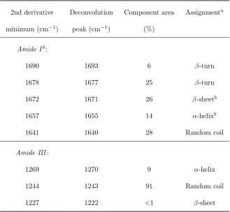
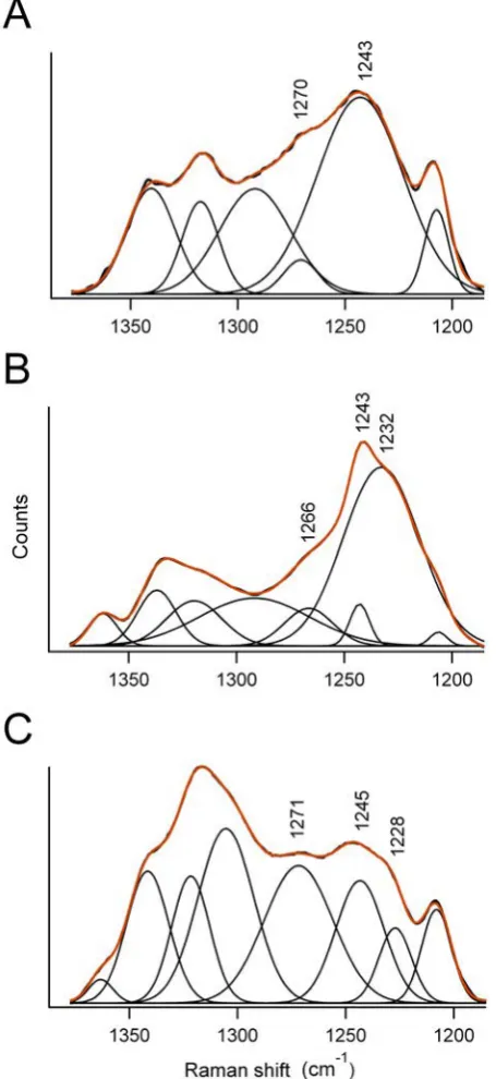
3.6 Deconvolution of the amide III region of Raman spectra obtained from three
kinds of silk . . . 67
3.7 Polarisation dependency of amide I region of Raman spectra obtained from sil-verfish silk . . . 69
3.8 Entanglement as the major form of cohesion in silverfish silk . . . 75
4.1 Praying mantis ootheca . . . 87
4.2 Oothecae proteins separated on an SDS-PAGE gel . . . 94
4.3 Solid state 13C NMR spectra from oothecae of three species of praying mantis . 96 4.4 Primary sequence alignment of mantis fibroins annotated with predicted primary, secondary and supersecondary structural features . . . 101
4.5 Amino acid character differs according to heptad position in mantis fibroin coiled-coil domains . . . 103
4.6 Recombinant expression, refolding and material fabrication of mantis fibroins . . 104
5.1 The New Zealand glow-worm,Arachnocampa luminosa . . . 114
5.2 Glow-worm silk . . . 118
5.3 FTIR spectra of glow-worm silk and mucus . . . 120
5.4 X-ray scattering pattern from a pupal rope . . . 121
5.5 SDS-PAGE of glow-worm silk and mucus preparations . . . 123
5.6 Sequences of proteins detected in glow-worm silk by LC-MS . . . 127
6.1 Cocoon made by the willow sawfly,Nematus oligospilus . . . 140
6.2 X-ray scattering pattern obtained from willow sawfly cocoon . . . 142
6.3 Mass spectrometry of SfColl proteins which have been resolved by denaturing gel electrophoresis . . . 146
6.4 Primary amino acid sequence alignment of sawfly silk proteins . . . 147
LIST OF FIGURES
6.6 Comparison of sawfly collagen proteins with previously described collagens . . . 152
7.1 Architecture of insect silk proteins . . . 160
7.2 Charge properties of insect silk proteins . . . 165
7.3 Amino acid composition of insect silk proteins . . . 167
7.4 Composition of the hydrophobic core of coiled-coil-forming silk proteins . . . 170
8.1 Liquid crystalline order in silk protein solutions . . . 180
8.2 Hypothetical example of a fitness landscape for a silk protein . . . 193
A.1 Wide-angle X-ray scattering pattern from A. illawarra silk fibre bundle . . . 201
A.2 Raspy cricket silk protein sequences . . . 202
A.3 Photographs of a raspy cricket producing silk . . . 203
B.1 Second derivative plot of the amide I region of silverfish silk FTIR spectrum . . . 207
B.2 Second derivative plot of the amide III region of silverfish silk FTIR spectrum . . 208
B.3 Deconvolution of the amide I region of silverfish silk FTIR spectrum . . . 209
B.4 Deconvolution of the amide III region of silverfish silk FTIR spectrum. . . 210
B.5 Second derivative plot of the amide I region of silverfish silk Raman spectrum . . 211
B.6 Deconvolution of the amide I region of silverfish silk Raman spectrum . . . 212
B.7 Second derivative plot of the amide III region of silverfish silk Raman spectrum . 213 C.1 FTIR spectra of mantis oothecae . . . 216
C.2 Hydrophobicity and side-chain length of amino acids at each heptad position of mantis fibroin coiled-coil domains . . . 217
C.3 Comparison of the FTIR spectra of materials made from recombinant fibroins . . 218
F.1 Mantis ootheca viewed using polarising light microscopy . . . 230
List of Tables
1.1 Silk-producing insects . . . 8
1.1 Silk-producing insects (continued) . . . 9
1.1 Silk-producing insects (continued) . . . 10
1.2 Mechanical properties of insect silks and some other materials . . . 25
2.1 Assignment of peaks in scattering pattern obtained from raspy cricket silk . . . . 37
2.2 Comparison ofβ-sheet-rich silks . . . 47
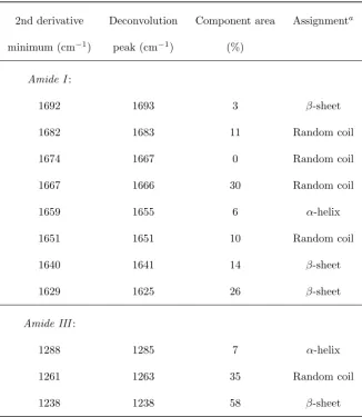
3.1 Amide I and III band components in silverfish silk FTIR spectrum . . . 63
3.2 Amide I and III band components in silverfish silk Raman spectra . . . 66
3.3 Amino acid composition of silverfish silk . . . 71
4.1 Assignment of peaks in NMR spectra from praying mantis oothecae . . . 97
4.2 Identification of praying mantis ootheca proteins by LC-MS . . . 98
4.4 Comparison of the features of Mantis Fibroin 1 and Mantis Fibroin 2 . . . 100
5.1 Amino acid composition of glow-worm silk fibres and mucus . . . 124
5.2 Proteins identified in glow-worm silk using LC-MS . . . 126
5.3 Comparison of glow-worm silk with some similar silks . . . 129
LIST OF TABLES
6.2 Amino acid analysis of cocoon silk from twoNematus species . . . 144 6.3 Tripeptides in collagen forming silk proteins . . . 148
7.1 Periodicities in amino acid sequences of insect silk proteins . . . 163
8.1 Comparison of proteins which exhibit mesogenic ordering . . . 181
A.1 Proteins identified in raspy cricket reservoirs by LC-MS . . . 204 A.2 Homology between raspy cricket cDNA sequences and known proteins . . . 205
C.1 List of cDNA sequences recorded fromP. albofimbriata library . . . 219 C.2 List of cDNA sequences recorded fromT. australasiae library . . . 220 C.3 List of cDNA sequences recorded fromA. monstrosa library . . . 221 C.4 Observed allelic variation in mantis fibroins . . . 222
D.1 Amino acid composition of insect silk proteins and silk . . . 224 D.2 Amino acid composition of insect silk proteins and silk (continued) . . . 225
E.1 Prediction of secondary structure in insect silk proteins . . . 228
List of Abbreviations
AilSP Apotrechus illawarra Silk Protein
AmelF Apis mellifera Fibroin
ANU Australian National University
ATR attenuated total reflectance
BCE before common era
BSA bovine serum albumin
CD circular dichroism
cDNA coding deoxyribonucleic acid
CE common era
CP cross-polarisation
CSIRO Commonwealth Scientific and Industrial Research Organisation
EDTA ethylenediamine tetraacetic acid
FACIT fibril associated collagen with interrupted triple helices
FTIR Fourier transform infrared
LIST OF TABLES
HMM hidden Markov model
Hyl hydroxyproline
Hyp hydroxyproline
LC-MS liquid chromatography - mass spectrometry
MAS magic-angle spinning
mRNA messenger ribonucleic acid
MWCO molecular weight cut-off
NA numerical aperture
NGS normal goat serum
NMR nuclear magnetic resonance
PAGE polyacrylamide gel electrophoresis
PBLG poly-γ-benzyl-L-glutamate
PBS phosphate buffered saline
PBT phosphate buffered saline/Triton X reagent
PCR polymerase chain reaction
pI isoelectric point
SAXS small-angle X-ray scattering
SDS sodium dodecyl sulfate
SfColl sawfly collagen
TMV tobacco mosaic virus
Chapter 1
Introduction
Because of their unique properties, silk materials made by arthropods are used by humans for a diverse range of applications. In pre-industrial societies, silk collected from wild species found use as fishing lures (Waldman, 2005), and as bandages and sutures (Heimer, 1988). Silk use by humans greatly increased after the domestication of the silkworm in China in neolithic times. According to Chinese folklore, a cocoon falling into the teacup of the Empress Leizu as she sat under a mulberry tree in the 27th century BCE led to the invention of sericulture and the silk loom (Lizhu, 2003). While exporting large amounts of silk fabric, the process of its production was a closely guarded secret in China for centuries, and silk fabric was symbolic of wealth, elegance and power. Sericulture eventually spread to the rest of the world after programs of industrial espionage such as that initiated by Justinian the Great around 550 CE. At Justinian’s request, silkworm eggs were smuggled to Byzantium inside the bamboo walking sticks of two monks (Cave & Coulson, 1936). More recently, silks have proved to be ideal materials from which to construct parachutes, diffraction gratings, and the cross-hairs in optical devices.
CHAPTER 1. INTRODUCTION
secondary structures were elucidated (e.g. Marsh et al., 1955a,b; Warwicker, 1956). In the decades that followed, researchers such as K. M. Rudall (Lucas & Rudall, 1968a; Rudall, 1962) amassed X-ray scattering and amino acid composition data from a wide range of arthropod species, demonstrating a diverse range of molecular structure in silks. The advent of modern molecular biology and genome sequencing led to a more detailed understanding of silk protein primary sequences (Craig & Riekel, 2002; Fu et al., 2009). Finally, detailed study of two model systems, the silkworm Bombyx mori and orb-weaving spiders (Arachnida: Araneidae), has provided us with understanding of the mechanisms by which silk is fabricated (for example Asakura et al., 2006; Hu et al., 2006; Jin & Kaplan, 2003).
Despite these advances a detailed understanding of the structure and function of silk pro-teins, and how one relates to the other, remains mysterious. More comprehensive studies on arthropod silks will make possible a comparative approach to understanding how silk protein structure is related to function.
1.1
What is a silk?
Early authors such as Rudall & Kenchington (1971) favoured broad definitions of silks as fibrous materials that ‘once produced . . . do not have contact with humors of the animal body’ (p73). More recent definitions tend to emphasise the mechanism by which fibres are produced, such as this one from Porter & Vollrath (2009):
1.2. SILK-PRODUCING INSECTS
properties of the final thread, fibre, filament, ribbon, or film. (p487)
In this thesis, a silk (figure 1.1) is defined as a solid material used outside the body which is fabricated from a concentrated protein solution (silk dope). This definition excludes non-proteinaceous fibres made by arthropods, such as ‘chitinous silks’ and ‘cuticulin silks’ (Rudall, 1962), as well as fibres such as hair that are fabricated slowly using low concentrations of proteins. Otherwise, it is deliberately broad to accommodate some unusual silks such as mantis ootheca (see chapter 6).
1.2
Silk-producing insects
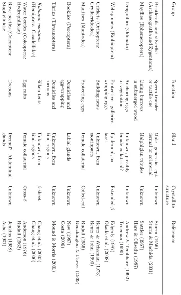
The capacity to produce silk is present in many arthropod groups, including 17 out of the 30 orders of insects (figure 1.2). Some characterisation has been performed on silk materials made by web-spinners (order Embioptera); mantises (order Mantodea); the leafhopper
Ka-haono montana (Hemiptera: Cicadellidae); lacewings (order Neuroptera); some water beetles
1.3. SILK GLANDS
transport by ballooning (table 1.1).
The ability to produce silk is a complex trait requiring a suite of characteristics—silk pro-teins, silk glands, and silk-spinning behaviour. Regardless, the ability to make silk has evolved many times among the arthropods. Sutherland et al. (2010) reviewed the multiple independent origins of silk production, and grouped silk-producing species into ‘lineages’ depending on phy-logenetic distribution, the glandular origin of the silk, and the silk’s molecular structure. Each of the 23 lineages thus designated is likely to constitute an instance where silk proteins, silk glands and silk-spinning behaviour are homologous between all members of the lineage.
1.3
Silk glands
Silk is produced and stored in dedicated glands. The insect silk glands described to date are derived from one of three types of secretory glands that fulfil other functions in related species: labial glands, Malpighian tubules, or dermal glands (figure 1.3; Sutherland et al., 2010). In species that do not produce silk, these glands secrete saliva, venom, pheremones, defensive compounds, or other substances (Britton et al., 1970). Three components are present in all silk glands: protein-producing secretory cells, an internal reservoir in which silk dope can be stored prior to spinning, and an opening through which silk fibres may be drawn into the external environment.
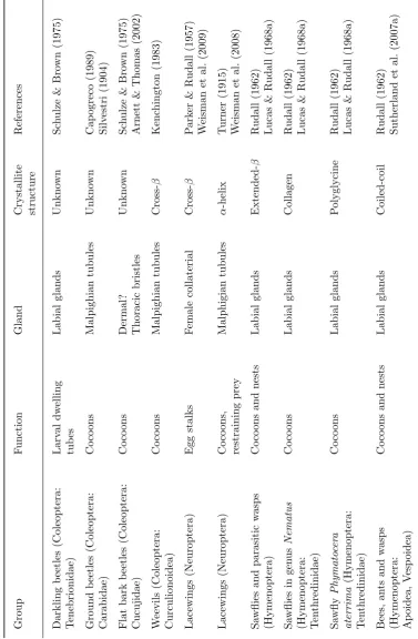
T able 1.1: Silk-pro ducing insects (con tin ued). Group F u nction Gland Crystallite structure References Darkling b eetles (Cole op te ra: T enebrionidae) Larv al dw elling tub es Labial glands Unkno wn Sc h ulze & B ro wn (1975) Ground b eetles (Cole op te ra: Carabidae) Co co ons Malp ighian tubules Unkn o wn Cap ogreco (1 989) Silv estri (1904) Flat bark b eetles (Coleoptera: Cucujidae) Co co ons Dermal? Thoracic bristles Unkno wn Sc h ulze & Bro wn (1975) Arnett & Thomas (2002) W eevils (Coleoptera: Curculionoidea) Co co ons Malp ighian tubules Cross-β Kenc hington (1983) Lacewings (Ne u roptera) Egg stalks F emale collaterial Cross-β P ark er & Rudall (1957) W eisman et al. (2009) Lacewings (Ne u roptera) Co co ons, restraining prey Malphigian tubules α -helix T urner (1915) W eisman et al. (2008) Sa wflies and parasitic w asps (Hymenoptera) Co co ons and nests Labial glands Extended-β Rudall (1962) Lucas & Rudall (1968a) Sa wflies in gen us Nematus
(Hymenoptera: Ten
[image:33.595.147.525.149.726.2]CHAPTER 1. INTRODUCTION
may simply terminate at an aperture (Ganguly, 1960).
Dermal silk glands can be further divided into epidermal glands and internalised glands. All epidermal silk glands are type III epidermal glands, meaning protein is secreted by a glandular cell into a reservoir, and passes to the exterior of the insect by a ductule (Noirot & Quennedey, 1974). Typically, each glandular unit comprises a large secretory cell, a canal cell, and several supporting cells. Often, large numbers of small glandular units are present, and the fibres produced are very fine (Okada et al., 2008).
Internalised dermal silk glands are much larger structures. Most often they are sex ac-cessory (collaterial) glands, so that silk is fabricated at the opening of the reproductive canal (e.g. Kenchington & Flower, 1969). Others have openings located elsewhere, such as those of staphilinid beetles, which have an external opening on the insect’s ninth tergite (Ashe, 1981; Jenkins, 1958).
Malpighian tubules are excretory and osmoregulatory organs that open near the junction of the mid- and hind-guts (Britton et al., 1970). Insects that make silk in Malpighian tubules may have separate regions of their Malpighian tubules devoted to silk secretion and excretory functions (Aoyagi, 1994a,b) or may shut down the excretory activity of their tubules while they produce silk (Maloeuf, 1938). Concentrated solutions of silk proteins collect in the alimentary canal, and the anus acts as a spinneret.
1.4
Silk molecular structure
1.4. SILK MOLECULAR STRUCTURE
mass spectrometry and amino acid analysis. At the level of secondary and supersecondary structure, silk proteins can be investigated using techniques such as infrared, Raman and solid-state NMR spectroscopy, differential scanning calorimetry and X-ray scattering. Molecular orientation can be investigated using polarising light microscopy, X-ray scattering and polarising Raman spectroscopy.
Each of these techniques investigates order of a particular type and on a particular scale. For example, Raman spectroscopy gives information about the amino acid residues present and the secondary structure of the proteins in which they are contained, and can provide orientational infomation in some circumstances; X-ray scattering provides information only about proteins in the crystalline regions, by which we mean any region in which proteins are ordered over a sufficient scale to act as diffraction gratings. The variety of levels on which silk can be described structurally gives rise to the concept ofhierarchical structure (Zhou & Zhang, 2005), in which the macroscopic features of silk materials can be reduced by successive levels through proteins present and their interactions, the secondary structures of the proteins, down to the primary sequence level.
In silkworm and orb-spider silk, two mechanisms are responsible for silk proteins cohering together in a solid form: cross-linking of protein chains through intermolecular bonding, and en-tanglement. Crystallites are the main site of cross-linking, while entanglement occurs primarily in the amorphous regions. Although hydrogen bonding is a weak interaction, hydrogen bonds are the principle form of cross-linking between protein chains, and many weak interactions sum to produce strong cross-linking between protein chains (Fu et al., 2009).
1.5. SILKS WITH EXTENDED-β-SHEET CRYSTALLITES
orientation—for example, extended- and cross-β-sheet silks. Variations such as these produce different overall patterns of bonding to that which classically occur in spider dragline silk and silkworm silk. For example, honeybee silk has crystallites made ofα-helical coiled-coils (Suther-land et al., 2006), a supersecondary structure which forms principally due to the hydrophobic effect. Coiled-coils then cohere to form a solid material by intermolecular bonding which in-cludes covalent cross-linking (Sutherland et al., 2011b). The resulting silk fibres have different mechanical properties to lepidopteran silk fibres (Hepburn et al., 1979).
1.5
Silks with extended-
β
-sheet crystallites
Silk from silkworms is made up primarily of three proteins: the 390 kDa fibroin heavy-chain protein (H-fibroin; Genbank accession (GA) NP 001106733), the 26 kDa fibroin light-chain protein (L-fibroin; GA|NP 001037488), and a 30 kDa glycoprotein called P25 or fibrohexamerrin (GA|CAA27804). These proteins occur in complexes with stoichiometry 6:6:1, with the H- and L-fibroins linked by a disulfide bond, and P25 retained in complex through non-covalent bonds (Inoue et al., 2000). Another group of proteins, the serine-rich sericins, coat the outside of the fibres (Michaille et al., 1990).
1.6. SILKS WITH CROSS-β-SHEET CRYSTALLITES
Web-spinners (order Embioptera) make silk using a single, relatively short protein termed Efibroin (GA|EU170437; Okada et al., 2008) or simply webspinner fibroin (GA|FJ361212; Collin et al., 2009) which has a molecular weight of approximately 39–65 kDa and folds intoβ-sheets. The primary sequence of web-spinner fibroin is similar in some respects to silkworm H-fibroin, consisting mostly of (Gly-Ser)n repeats (Collin et al., 2009; Okada et al., 2008). Dipeptide
repeats also occur in other silks with predominateβ-sheet structure: (Ala-Gln)n dipeptide
re-peats in the silk proteins of argid sawflies (Lucas & Rudall, 1968a) and (Asn-X)n dipeptide
repeats (where X is Ala or Ser) in the braconid wasp Xenofibroin protein (GA|AB188680; Ya-mada et al., 2004). Dipeptide repeats are however by no means universal inβ-sheet-forming silk proteins; the silk proteins of some lepidopterans (Fedic et al., 2003), trichopterans (Yonemura et al., 2009) and dance-flies (Diptera: Empididae; Sutherland et al., 2007b) have more complex repetitive sequences (Fedic et al., 2003; Sutherland et al., 2007b).
1.6
Silks with cross-
β
-sheet crystallites
The extended-β-sheet and cross-β-sheet molecular structures are based on the same secondary structural element—β-strands arranged into sheets—but in a different orientation with respect to the fibre axis. In the cross-β-sheet conformation, protein backbones point across the fibre and hydrogen bonds between backbone amide and carbonyl groups point towards the ends of the fibre. The cross-β conformation was first observed in egg-stalks produced by lacewings (order Neuroptera; Parker & Rudall, 1957) and later in silk produced by the New Zealand glow-wormArachnocampa luminosa (Diptera: Keroplatidae Rudall, 1962), the cocoon fibres of hyperine weevils (Coleoptera: Curculionidae, Hyperini Kenchington, 1983) and egg-raft fibres of water-beetles (Coleoptera: Hydrophilidae; Rudall, 1962). Egg-stalks made by the lacewing
Mallada signataconsist of two proteins, MalXB1 (GA|FJ792486) and MalXB2 (GA|FJ792487),
-CHAPTER 1. INTRODUCTION
sheet ‘ribbon’ eight residues across. Cross-β-sheet silk fibres have high extensibilities (up to 400%) associated with molecular extension of the cross-β ribbon to form an extended-β-sheet structure (Bauer et al., 2012; Rudall, 1962; Weisman et al., 2008).
1.7
Silks with coiled-coil crystallites
Silks containing predominantlyα-helical protein structure are made by aculeate hymenopterans (Rudall, 1962), argid sawflies (Rudall, 1962), fleas (Rudall & Kenchington, 1971), praying mantises (Rudall, 1956), and lacewing larvae (Weisman et al., 2008). Typically, α-helices are present as coiled-coil superhelices (though lacewing cocoon fibroin is an uncertain case; see section 7.1). The coiled-coil is a common structural motif formed by proteins with particular primary sequence features: many residues with high helical propensity (such as Ala, Arg, Leu, Lys and Glu; Chakrabartty et al., 1994) and seven-residue repeated motifs known as heptads, denoted (abcdefg)n. The basis of coiled-coil formation is higher average hydrophobicity in the
residues in the (a) and (d) positions (Woolfson et al., 2005). When the protein chain folds up into anα-helix, the hydrophobic residues form a stripe along one side of the helix. Two or more helices associate to shield hydrophobic residues from the solvent, forming a coiled-coil. Ionic and polar interactions between residues in the (e) and (g) positions further stabilise the final superhelical structure.
1.8. SILKS WITH COLLAGEN AND POLYGLYCINE II CRYSTALLITES
1.8
Silks with collagen and polyglycine II crystallites
Proteins with the collagen structure are found in the extracellular matrix or cell wall of ver-tebrates (Gordon & Hahn, 2010), molluscs (Hunt et al., 1970), insects (Ashhurst, 1968), and bacteria (Xu et al., 2010) and in extracorporeal protein materials such as dogfish eggshell (Rusaou¨en et al., 1976) and mussel byssus fibres (Waite et al., 1998). Collagen is a right-handed superhelical structure in which three left-handed polyproline II helices are intertwined. Since the side-chain of every third residue must be accommodated within the interior of the helix— which is only possible for the hydrogen side-chain of Gly—collagen-forming proteins contain triplet repeats of the form (Gly-X-Y)n. The X and Y positions are often filled by proline and
hydroxyproline; post-translational hydroxylation of Pro residues is a key stabilising mechanism in described animal collagens (Gordon & Hahn, 2010). Based on X-ray scattering experiments, Rudall (1962) reported that the gooseberry sawfly Nematus ribesii (Hymenoptera: Tenthre-dinidae) produces a silk in which proteins are folded into the collagen structure. Unusually for an animal collagen, the silk was reported to contain hydroxylysine but not hydroxyproline residues (Ramshaw et al., 1998).
Another tenthredinid, Solomon’s seal sawfly (Phymatocera aterrima), produces silk fibres in which proteins are folded predominately into the polyglycine II structure (Lucas & Rudall, 1968b). In the polyglycine II structure, proteins with many consecutive Gly residues fold into right-handed 31 helices, which are packed in a hexagonal arrangement (Crick & Rich, 1955).
Two-thirds of all residues inP. aterrima silk are glycine (Lucas & Rudall, 1968b).
1.9
Silk fabrication
CHAPTER 1. INTRODUCTION
(Sutherland et al., 2006), or electrostatic interactions (Ashton et al., 2012). A typical process of fibre fabrication might proceed as follows. Initially, a small droplet of silk protein solution is deposited on, and adheres to, an external surface. The aperture of the silk gland is pulled away, resulting in a stream of silk protein solution being drawn from the gland. Factors which promote cohesion within the silk protein solution—such as viscosity and intermolecular bonding—act to resist capillary breakup of the solution, which is converted in the process into a solid fibre by intermolecular bonding, entanglement of protein chains, and dehydration (Sutherland et al., 2010). Generally, the process by which insects fabricate silk fibres resembles pultrusion—since the fibres are drawn by force from the glands—rather than extrusion, where they are pushed through an aperture (Salomone, 1996).
Silk glands contain proteins at some of the highest liquid concentrations documented in nature, 30–40% of dry weight (300–400 mg/ml; Akai, 1983). Accordingly, silk proteins are required to be highly soluble, a requirement somewhat in conflict with a capacity for extensive intermolecular bonding during silk fabrication. A primary sequence which satisfies both criteria is arguably the most universal feature among silk proteins.
In silkworm silk glands, solution variables such as pH and ion content are varied so as to promote solubility during storage, and the formation of intermolecular bonds during silk fabrication. As silk dope flows anteriorly towards the spinneret its concentration increases from from 12 to 30% (Akai, 1983, 1998). H+-ATPases on anterior silk gland cells pump protons
1.9. SILK FABRICATION
concentration increases, pH is reduced, and metal ions added (Hu et al., 2006).
Prior to fibre fabrication, the structural features of proteins filling the lumen of silkworm silk glands are collectively referred to as ‘silk I’. The silk I structure includesβ-turns, helices, and random coil structures, resulting in a high degree of conformational flexibility (Asakura et al., 1985; Fossey et al., 1991; He et al., 1999). The final protein structure in solid silk fibres, called ‘silk II’, is markedly different and consists predominantly of β-sheets. Silk I is converted to silk II by the physical forces experienced during the process of pultrusion, and by dehydration (figure 1.6), the key process beingmolecular extension. Molecular extension is the result of three factors: elongational flow, which results from the decreasing diameter of the silk gland lumen approaching the spinneret (Hill & Cuculo, 1976); shear force resulting from friction with the gland wall (Iizuka, 1966); andextensional force, the force with which fibres are drawn from the gland. The process of elongational flow, and the forces applied during silk fabrication, stretch out individual protein chains and align them parallel to the fibre axis—an ideal situation for the formation of intermolecular hydrogen bonds, and hence highly alignedβ-sheet crystallites (Asakura et al., 2006; Sutherland et al., 2010).
1.10. SILK MECHANICAL BEHAVIOUR
1.10
Silk mechanical behaviour
Silk is a viscoelastic solid, meaning that extension of silk fibres depends partly on elastic be-haviour through the deformation of amorphous regions and crystallites, and partly on protein chains slipping past each other as in a viscous liquid (Krasnov et al., 2008). The amorphous fraction, which is able to deform in response to weak forces, confers flexibility and toughness, whereas the crystallites which deform only in response to stronger forces confer strength (Ter-monia, 1994). Stress-strain curves of silk fibres may display a ‘yield point’ (figure 1.7). Up until the yield point, extension occurs by deformation of the amorphous region and crystallites. After the yield point, extension is associated with major slippages of the crystalline regions and/or structural transitions. Successive extension of silk fibres thus depends on the deformation of successively stronger structures—another example of hierarchical structure in silk fibres (Zhou & Zhang, 2005).
CHAPTER 1. INTRODUCTION
1.11
A comparative approach to understanding silk
Chapter 2
Silk from crickets: A new twist
on spinning
This chapter is presented in its published form1except for some minor corrections and changes
to spelling and terminology to ensure consistency across the thesis. Contributions from other authors to this paper were as follows: Sarah Weisman performed liquid chromatography-mass spectrometry experiments; Jeff Church collected the micro-Raman spectra shown in figure 2.2; David Merritt controlled the confocal microscope used to capture panels E and F of figure 2.3; and Stephen Mudie, a beamline scientist at the Australian Synchrotron, assisted Sarah and myself in collecting the X-ray scattering patterns referred to in the text and included in ap-pendix A. My own contribution consisted of collecting, identifying and housing insects, and experiments using scanning electron microscopy, polarising light microscopy, gel electrophoresis, construction and mining of a cDNA library, anatomical dissections, and preparation of mate-rial for confocal microscopy. Sarah, Jeff and David assisted with the interpretation of results from liquid chromatography-mass spectrometry, Raman spectroscopy, and confocal scanning
1Walker, A.A.; Weisman, S.; Church, J.S.; Merritt, D.J.; Mudie, S.T.; and Sutherland, T.D. (2012) Silk from
CHAPTER 2. SILK FROM CRICKETS: A NEW TWIST ON SPINNING
2.1. ABSTRACT
2.1
Abstract
CHAPTER 2. SILK FROM CRICKETS: A NEW TWIST ON SPINNING
2.2
Introduction
The ability to produce silk has evolved in at least 23 groups of insects (Sutherland et al., 2010), in spiders (Vollrath & Porter, 2006) and in several other arthropods (Clotuche et al., 2009; Weygoldt, 1966). Silk research has focused on silkworm cocoon and spider dragline silks, which have independently evolved a number of convergent features. Spider and silkworm silks consist of long, repetitive proteins that fold predominantly into β-sheets, with the protein backbone parallel to the fibre axis (Vollrath & Porter, 2006). Highly ordered nanocrystals are embedded in regions of less order and confer high tensile strength to the fibres (Nova et al., 2010). The molecular arrangement in spider and silkworm silks is the result of mechanical forces and controlled dehydration acting on highly concentrated silk protein solutions as they pass through a hardened aperture known as a spinneret (Fu et al., 2009; Jin & Kaplan, 2003). Although less characterised, other silks are dramatically different. For example, protein backbones in silks made by glow-worms and adult lacewings are orientated perpendicular instead of parallel to the fibre axis (Rudall, 1962); the silks of fleas, bees and lacewing larvae contain proteins arranged in α-helices instead ofβ-sheets (Rudall, 1962; Weisman et al., 2008); and the fibrous proteins in some silks are an order of magnitude smaller than spider dragline and silkworm cocoon silk proteins (Hayashi et al., 1999). Further characterisation of silks in addition to spider and silkworm silks will allow a comparative approach to understanding the complex molecular arrangements found in silk.
2.2. INTRODUCTION
sexes are capable of producing fibres within hours of hatching and continue to produce shelters throughout their lives (Hale, 2000). Shelters are highly valued and individuals return to the same shelter many times (Lockwood & Rentz, 1996).
Very little is known about the method of fabrication of silk fibres by raspy crickets. Rentz & John (1990) observed silk production from cricket mouthparts, but the origin of the material is unknown and the internal anatomy of raspy crickets is poorly described. Other insects that generate silk from their mouthparts do so using protein solutions produced in modified labial glands (Sehnal & Sutherland, 2008). The labial glands of wetas and king crickets (Anostostom-atidae, the closest relatives of raspy crickets; Jost & Shaw, 2006) have a salivary function (Field, 2001). Anostostomatid labial glands are arranged in grape-like clusters called acini (Maskell, 1927). Acinar cells secrete into the lumen of a branching series of ductules joined to the common duct on each side of the body. The left and right common ducts join at the labium, where they empty into a cavity between the labium and hypopharynx, called the salivarium. An additional organ, the reservoir, is formed by a sack-like outgrowth of the common duct on each side (Field, 2001; Maskell, 1927).
CHAPTER 2. SILK FROM CRICKETS: A NEW TWIST ON SPINNING
2.3
Results
2.3.1
Raspy cricket build shelters by using silk fibres and films to join
other materials
Captive crickets built shelters by binding leaves or plastic card together with webs of silk (figure 2.1A). The fibres were cylindrical and uniform, with the diameter increasing as the crickets increased in size. For example, the diameter of fibres produced by a 16 mm long early instarApotrechus illawarra were 4.2±1.1µm (mean±standard deviation). Seven months later, the same animal, measuring 48 mm, was producing fibres with diameter 12.4±2.1µm.
Shelter construction behaviour was similar regardless of whether natural or artificial building materials were supplied. The process is shown in supplementary video 12. Silk production began by the cricket touching its labium to the surface of a leaf or piece of plastic, and depositing a film of silk material. As the labium was drawn away from the film, a fibre was produced, which was attached to another piece of building material with another film. Repeating this process resulted in a network of fibres joining the two pieces of building material. As successive layers of fibres were added, films were produced not only to secure fibres to building materials but also where fibres crossed to glue them together (figure 2.1B). The end result of the building process was a ‘silk-web’ that served to seal an entry point to the cavity and to hold the building materials together. Most crickets constructed a shelter within 24 hours of being housed, incorporating additional fibres over successive days. To exit the shelter to forage, an access hole was cut through a silk-web using the mandibles. Access holes were sealed with a fresh silk-web after the insect returned. The insect ceased to produce fibres if the shelter was undisturbed for long periods, whilst removal of a shelter resulted in the construction of a replacement.
2Stills from the video are shown in figure A.3, and the full video is available at
Figure 2.1: Raspy cricket silk-webs. A, shelter of silk and dry leaves made by Hyalogryllacris
species 9; scale bar is 2 mm. B, scanning electron micrograph ofA. illawarra silk-web showing
CHAPTER 2. SILK FROM CRICKETS: A NEW TWIST ON SPINNING
2.3.2
Fibres and films have a similar molecular structure
We investigated the chemistry ofA. illawarrasilk using Raman spectroscopy. Spectra obtained from fibres and films indicated a highly proteinaceous material, with no peaks attributed to chitin or other substances (figure 2.2). Spectra from films were essentially the same as those obtained from fibres, indicating that the two materials contained proteins with the same sec-ondary structures. Strong peak vibrations at 1667 cm−1 and 1235 cm−1 indicated that the dominant protein conformation present wasβ-sheet (Frushour & Koenig, 1975) and the weak shoulder at 1259 cm−1 was due to disordered protein. Raman spectroscopy is sensitive to a
number of specific amino acids and their environments within the protein structure, particularly Tyr, Trp, Phe, His, Pro, Hyp, Met and Cys (Church et al., 1997; Twardowshi & Anzenbacher, 1994). No significant difference was observed in the relative strengths of peaks attributed to any specific amino acid between spectra obtained from fibres and spectra obtained from films. Thus, fibres and films are likely to have the same amino acid composition and may be produced from the same protein dope.
CHAPTER 2. SILK FROM CRICKETS: A NEW TWIST ON SPINNING
crystallites in cricket silk are arranged in an extendedβ-sheet structure. The placing of another equatorial arc, attributed to the (2 2 0) reflection, depends in part on the inter-sheet spacing between amino acid side groups. Calculations from the position of this reflection suggest that the spacing betweenβ-sheets inA. illawarra silk is on average 1.27 nm.
2.3.3
Silk is produced from acinar labial glands
Silk made out of protein must have a glandular origin. Visual observations during dissection
of A. illawarra andHyalogryllacris species 9 revealed large, acinar labial glands in the thorax
and head in a similar arrangement to those of anostostomatids (figure 2.3A). Ductules from the acini on each side of the body merged into a common duct ending at the labium. Whereas the reservoirs of anostostomatids are connected to the common acinar duct at some distance from the labium (Maskell, 1927), raspy cricket reservoirs are joined to the end of the common duct within the labium. At this point the reservoirs and the common ducts from both sides of the body join together and empty into the salivarium through a common aperture. The size of this aperture was much larger than the diameter of silk fibres, measuring in excess of 100µm across in adultA. illawarra(figure 2.3B). No structure similar to the hardened, external spinneret of lepidopterans was observed. Instead, the labium was similar to crickets that do not produce silk, with hypopharynx, glossae and paraglossae (Britton et al., 1970), except that the paraglossae had an unusual shape: the margins of the paraglossae were raised up into ridges that overlap the edges of the hypopharynx, so that the hypopharynx fits ‘hand in glove’ into the labium (figure 2.3, C and D). We compared the labia of silk-producing raspy crickets with the labia of non-silk-producing cricket species, including a field cricket (Acheta domestica), a katydid (Conocephalus sp.) and an anostostomatid (Penalva flavocalceata). For each of these non-gryllacridid species, the surface of the paraglossae is flat or concave, without the distinctive raised margins present on raspy cricket paraglossae.
Figure 2.3: Anatomy of silk production by raspy crickets. A, labial glands and labium of
H. species 9; scale bar is 2 mm. B, scanning electron micrograph of the opening of the labial
glands of A. illawarra into the salivarium; the hypopharynx has been pinned back to reveal this feature. Scale bar is 500 µm. C, labium and hypopharynx in the open position, showing the raised margins on the labial paraglossae of H. species 9; scale bar is 500 µm. D, (same preparation as C), labium and hypopharynx in closed position, with raised margins of paraglos-sae overlapping hypopharynx; scale bar is 500 µm. E, confocal slice through labial acinus of
A. illawarrashowing paired arrangement of nuclei (blue) and secretory invaginations (red); scale
2.3. RESULTS
or multiple nuclei (Akai, 1998). To investigate the function of the acini and the reservoirs, we stained each type of tissue with fluorescent dyes to reveal nuclei and the actin cytoskeleton. Acinar cells were organised in pairs, possessed a single large nucleus, to which an actin-rich invagination was closely apposed, consistent with a secretory role (figure 2.3E). The lobular, actin-rich lumen of each cell conjoined to a common lumen with a stellate, actin-rich periphery that was continuous with the common duct. Each acinus was composed of approximately 20 of these conjoined cells. The ducts were lined with smaller, flattened, cuticle-secreting cells, the luminal surface of which was actin-rich (figure 2.3F). The reservoirs consisted of a single layer of the same small cuticle-secreting epidermal cells, suggesting a role as a storage organ.
The proteins present in fluid-filled reservoirs were investigated by comparing the sequences of peptides following tryptic digestion to GenBank’s non-redundant protein database andin silico translated sequences from our own A. illawarra cDNA library using liquid chromatography-mass spectrometry (LC-MS). Apart from house-keeping proteins, the only protein identified was amylase, an enzyme that breaks down the plant polymer starch (table A.1). None of the raspy cricket silk proteins (described below) were detected in the reservoirs, suggesting reservoirs are used to store saliva but not silk dope.
2.3.4
Raspy cricket silk proteins
The raspy cricket silk proteins were identified using a cDNA library/mass spectrometry ap-proach successfully applied to other silk-producing species (Sutherland et al., 2007a). We constructed a cDNA library of 3.5×10⁵ clones with an average insert size of 1.1±0.6 kb from
A. illawarra acinar labial glands. Analysis of over 100 clones identified 63 putative cDNAs,
CHAPTER 2. SILK FROM CRICKETS: A NEW TWIST ON SPINNING
encoded one of four silk proteins (see below).
Solubilised silk-webs contained major protein bands at approximately 300, 220, and 68 kDa and fainter bands at 120, 30 and 28 kDa (figure 2.4). Similar protein bands were obtained from silk produced by different individuals regardless of if protease inhibitors were included during solubilisation and regardless of if reducing agents were added during sodium dodecyl sulfate (SDS)-polyacrylamide gel electrophoresis (PAGE). Individual protein bands from SDS-PAGE gels or solid silk-webs were digested with proteases and the resulting peptides were analysed using mass spectrometry. Comparison of experimentally derived peptide masses with the predicted masses ofin silico digested sequences encoded by the labial gland cDNA library showed that the bands at 300, 220 and 120 kDa all corresponded to a single protein sequence, partially encoded by three cDNA clones (figure 2.4). We named this protein AilSP1 (Apotrechus
illawarra Silk Protein 1; Genbank accession (GA) JF508439 and GA|JF508440). The longest
AilSP1 cDNA contained 1282 nucleotides of coding region, a stop codon, a 3’ untranslated region of 315 nucleotides, and a poly-A tail. Two of the AilSP1 cDNAs differed by only a single silent polymorphism (GA|JF508439) while the third contained 77 single nucleotide polymorphisms (31 silent) and a trinucleotide insert (GA|JF508440). The partial sequence of AilSP1 consisted of an internal repetitive region of 37 or 38 residue repeats followed by a 163 residue C-terminal region containing a single cysteine residue (figure A.2). The small amino acids alanine, glycine and serine respectively made up 24.3%, 23.4%, and 18.5% of residues.
CHAPTER 2. SILK FROM CRICKETS: A NEW TWIST ON SPINNING
2.4. DISCUSSION
2.4
Discussion
The two species of raspy cricket used in this study produced shelters by joining leaves together with silk-webs. Since silk-webs were not air-tight, they are unlikely to be effective in preventing desiccation or the ingress of parasites in the wild, and the most likely function is to reduce predation. Silk-webs were found to be made of protein, and visual observations of crickets fabricating silk suggested the labial glands might function as silk glands. Our identification of transcripts encoding silk proteins in labial glands confirms this directly.
The silk glands consisted of acini connected by a network of ducts to the insect’s salivarium. In cocoon-producing species, silk glands include large lumens where silk proteins can be stored in preparation for a short period of intense silk production (Akai, 1983). In contrast, raspy cricket gland lumens are small and the amount of silk required for shelters is low, suggesting the crickets probably produce silk as required. The reservoir attached to the common duct immediately before it joins the salivarium does not contain silk proteins but does contain amylase, suggesting it to have a salivary function.
CHAPTER 2. SILK FROM CRICKETS: A NEW TWIST ON SPINNING
muscular control allows the insect to deposit silk dope in globules, which dry into films. Both films and fibres are found in the silks of other insects, including sawflies, honeybees, hornets and some beetles (e.g. Ishay & Ganor, 1990).
The major silk protein AilSP1 has evolved features similar to the silk proteins of silkworms and spiders: it is large, contains a high proportion of small amino acids, and adopts primarily an extended β-sheet conformation in the mature silk. The spacing between β-sheets in the side group direction is 1.27 nm, within the range reported for lepidopteranβ-sheet silks (0.93– 1.57 nm; Lucas & Rudall, 1968a) and slightly greater than for polyalanine (1.06 nm; Colonna-Cesari et al., 1975). An inter-sheet spacing of 1.27 nm is consistent with crystallites being formed from the repeat units of AilSP1, since repeat regions contain 27% glycine, 27% alanine and 18% serine, with larger residues accounting for the remainder. The presence of a cysteine residue in the C-terminus of AilSP1 and the high frequency of cysteine residues in AilSP3, and the observation that reducing agents are required for silk solubilisation, suggest disulfide bonding to play a structurally important role in cricket silk.
CHAPTER 2. SILK FROM CRICKETS: A NEW TWIST ON SPINNING
CHAPTER 2. SILK FROM CRICKETS: A NEW TWIST ON SPINNING
2.5
Materials and Methods
2.5.1
Insects
Raspy crickets (Apotrechus illawarraandHyalogryllacris species 9) were collected from Meroo National Park, New South Wales, Australia. Crickets were housed in plastic jars with water and orthopteran food mixture (Rentz, 1996). They were supplied either with natural building materials (dry leaves, sticks and sand) or black plastic card. Non-gryllacridid crickets used for comparison were the field cricketAcheta domestica(Pisces Enterprises, Australia), a katydid of the genus Conocephalus (collected at the Meroo National Park field site) and the white-kneed king cricketPenalva flavocalceata (Minibeast Wildlife, Australia).
2.5.2
Microscopy
Silk glands and mouthparts from adult insects of either sex were dissected and fixed in phosphate buffered saline (PBS) pH 7.0 containing 2% gluteraldehyde (Invitrogen) and examined using light microscopy. Silk-webs were mounted onto a stub using conductive tape, sputter-coated with gold and visualised under high vacuum on an Evo LS15 scanning electron microscope (Zeiss). Silk glands from a single largeA. illawarra nymph were examined on a LSM5 confocal scanning laser microscope (Zeiss). The tissues were dissected and fixed in 4% formaldehyde in PBS for 10 minutes, washed three times for 30 minutes each in PBS, permeabilized for 1 hour in PBT (0.25% BSA, 0.4% Triton X in PBS), blocked for 1 hour in PBT-NGS (2% normal goat serum in PBT). After fixation, tissues were stained overnight at 4◦C in 6.6 mM Alexa Fluor 568 phalloidin (Invitrogen), 1 mg/ml diamidino-2-phenylindole (Invitrogen) in PBS. Before mounting, stained tissue was washed four times for 10 minutes each in PBS and placed in 70% glycerol, 2% propyl gallate in PBS for 1 hour in the dark.
2.5. MATERIALS AND METHODS
at +45◦ between the crossed polarising filters. Fibre diameters were measured using Leica LAS software and birefringence was calculated with reference to a Leica colour chart.
2.5.3
Raman Spectroscopy
Raman spectra were obtained using an inVia confocal microscope system (Renishaw) with 514 nm excitation from an argon ion laser through a 650 (0.75 NA) objective. Incident laser power was 4.5 mW and coaxial backscatter geometry was employed. Spectra were collected over the range 3200 to 100 cm−1 and averaged over at least five scans, each with an accumulation
time of 20 seconds. Raman shifts were calibrated using the 520 cm−1 line of a silicon wafer.
The spectral resolution was
e1 cm
−1. Fibres were analysed aligned parallel to the direction
of laser polarisation with the use of a rotating stage. The final spectra used for analysis were averages of spectra collected from six to ten different areas. All spectra were normalized on the CH2 and CH3 deformation modes at 1452 cm−1 attributable to amino acid side chains and
thus not sensitive to protein conformation.
2.5.4
X-ray scattering
CHAPTER 2. SILK FROM CRICKETS: A NEW TWIST ON SPINNING
2.5.5
cDNA library construction
A cDNA library was constructed from the labial glands of four largeA. illawarra nymphs. To induce silk production, the shelters of these crickets were removed and they were supplied with fresh building materials. One day later the insects were dissected in PBS pH 7.0 and the acinar parts of the labial glands were removed and stored in RNAlater (Ambion). Total RNA was prepared using RNAqueous-4PCR (Ambion) and mRNA isolated using Micro-FastTrack 2.0 (Invitrogen). The cDNA library was created using Cloneminer II cDNA Library Construction kit (Invitrogen) according to the manufacturer’s instructions. Clones containing cDNA inserts larger than 1 kb were sequenced by Micromon Services (Monash University, Melbourne, Aus-tralia) using an Applied Biosystems 3730S Genetic Analyser and Applied Biosystems PRISM BigDye Terminator Mix cycling chemistry. A library of possible protein sequences was
gener-ated in silico using EMBOSS Transeq (Rice et al., 2000). Putative cDNAs were identified by
the presence of a poly-A tail longer than 15 continuous nucleotides and/or the presence of an open reading frame longer than 300 bp. Signal peptides were predicted using the SignalP 3.0 algorithm (Emanuelsson et al., 2007).
2.5.6
Mass spectrometry
2.5. MATERIALS AND METHODS
Chapter 3
Silverfish silk is formed by
entanglement of randomly coiled
protein chains
This chapter, describing characterisation of silverfish silk, is a manuscript accepted for publi-cation by the journalInsect Biochemistry and Molecular Biology1. Contributions to this paper
by the other authors were as follows: Jeff Church and Andrea Woodhead performed and inter-preted Raman spectroscopy experiments, and Tara Sutherland conceived the project and was my principal supervisor over the course of the investigation. The direct amino acid analysis presented was performed as a commercial service by the Australian Proteome Analysis Facility. I collected and housed animals, harvested silk, and performed experiments employing polarising light microscopy, silk solubilisation, and gel electrophoresis. The manuscript was written by myself edited by Tara, Jeff and Andrea. Supplementary figures 1–7 of the paper are reproduced in appendix B.
1Walker, A.A.; Church, J.S.; Woodhead, A.L; Sutherland, T.D. (2012) Silverfish silk is formed by
CHAPTER 3. SILVERFISH SILK IS FORMED BY ENTANGLEMENT OF RANDOMLY COILED PROTEIN CHAINS
3.1
Abstract
3.2. INTRODUCTION
3.2
Introduction
The best-understood silks are those of silkworms and spiders, which are semi-crystalline solids containing ordered crystallites ofβ-sheets embedded in regions of unordered protein chains (Fu et al., 2009; Porter & Vollrath, 2009). In these model silks, protein chains cohere to form a solid material due to extensive intermolecular hydrogen bonding within theβ-sheet crystallites and entanglement of the unordered portions of the protein chains. Since crystallites and unordered regions have distinct deformational properties, they lend different mechanical properties to the material: ordered crystallites confer tensile strength (Krasnov et al., 2008) and resistance to proteolysis (Arai et al., 2004), while unordered protein regions confer flexibility (Krasnov et al., 2008) and allow entanglement (Fu et al., 2009).
Many arthropod groups besides silkworms and spiders have independently evolved the abil-ity to produce silk (Sutherland et al., 2010). Arthropod silks are structurally diverse at the molecular level, containing crystallites of either a)β-sheets with the protein backbone parallel to the fibre axis (extended-β-sheets; akin to silkworm or spider silks), b) β-sheets with the backbone perpendicular to the fibre axis (cross-β-sheets; Weisman et al., 2009), c) α-helices as coiled coils (Sutherland et al., 2011b), d) collagen triple helices (Rudall, 1962), or e) polyg-lycine II-like structures (Lucas & Rudall, 1968b). The ordered protein structures in silk are understood to contribute extensively to silk function—for example, cross-β-sheet crystallites confer extensibility to fibres due to their capacity for deformation-induced transitions into the extended-β-sheet structure (Bauer et al., 2012; Hepburn et al., 1979). In contrast, the phys-iological, molecular and evolutionary constraints that determine the proportion of disordered protein in silk fibres is not well understood.
CHAPTER 3. SILVERFISH SILK IS FORMED BY ENTANGLEMENT OF RANDOMLY COILED PROTEIN CHAINS
phallus of males (Bitsch, 1990; Sturm & Machida, 2001). Sperm droplets or spermatophores are deposited variously on silk fibres or stalks by bristletails (Sturm & Machida, 2001), the ‘relic’ silverfishTricholepidion gertschi (Sturm, 1997), and some of the more familiar domestic silverfish (family Lepismatidae; Sturm, 1987). Lepismatids such as the European silverfish (Lepisma saccharina) are exceptions, as they deposit enclosed spermatophores directly onto surfaces in their environment. Perhaps surprisingly,L. saccharina has retained silk production as part of a highly choreographed courtship behaviour (Sturm, 1956). Approaching the end of the courtship ritual, which consists of antenna-tapping and repeated responsive movements between male and female (Sturm, 1956, 1987), maleL. saccharinacover an area of the substrate with shaped networks of fibres and deposit a spermatophore nearby. Detection of the Y-shaped tactile cues by the female induces a sequence of behaviors that culminates in her uptake of the spermatophore (Sturm, 1956).
The primary use of silk as a tactile cue is unusual among insects. Whilst the caterpillar
Yponomeuta cagnagellus does use silk as a tactile cue associated with trail-following behavior,
CHAPTER 3. SILVERFISH SILK IS FORMED BY ENTANGLEMENT OF RANDOMLY COILED PROTEIN CHAINS
3.3
Results
3.3.1
Silverfish produce very fine non-birefringent silk threads
Silverfish (Ctenolepisma longicaudata) were collected from human habitations in Canberra, Australia, and housed with plastic card ‘leaf-litter’ over a six-month period. Inspection of plastic cards in the enclosure revealed spermatophores deposited directly on the substrate, together with deposits of silk and shed scales (figure 3.2A). Electron microscopy revealed silk filaments to be very fine, between 0.3 and 1µm in diameter (figure 3.2B). Deposits of silk consisted of bundles and cross-hatched ‘grids’ (figure 3.2C). These features are similar to those previously reported for the closely related silverfishLepisma saccharina (Sturm, 1956) suggestingC. longicaudata silk is used as a tactile cue during courtship in a similar way toL. saccharina silk.
Silk fibres could be manually drawn from the phallic glands of immobilised males by brushing the silk glands with a toothpick or pipette tip, and the fibres could then be collected using a motorised spindle (figure 3.2D). Freshly produced silk fibres were flexible and sticky, becoming brittle after drying. Birefringence—a property of anisotropic materials evident when materials are placed between crossed polarising filters—was low or absent in naturally produced and manually harvested silk, suggesting molecular orientation to be poor or absent.