0022-538X/07/$08.00⫹0 doi:10.1128/JVI.00878-07
Copyright © 2007, American Society for Microbiology. All Rights Reserved.
Functionally Distinct Transmission of Human Immunodeficiency Virus
Type 1 Mediated by Immature and Mature Dendritic Cells
䌤
Jian-Hua Wang, Alicia M. Janas, Wendy J. Olson,† and Li Wu*
Department of Microbiology and Molecular Genetics, Medical College of Wisconsin, 8701 Watertown Plank Road, Milwaukee, Wisconsin 53226
Received 24 April 2007/Accepted 3 June 2007
Dendritic cells (DCs) potently stimulate the transmission of human immunodeficiency virus type 1 (HIV-1)
to CD4ⴙT cells. Immature DCs (iDCs) located in submucosal tissues can capture HIV-1 and migrate to
lymphoid tissues, where they become mature DCs (mDCs) for effective antigen presentation. DC maturation promotes HIV-1 transmission; however, the underlying mechanisms remain unclear. Here we have compared monocyte-derived iDCs and mDCs for their efficiencies and mechanisms of HIV-1 transmission. We have found that mDCs significantly facilitate HIV-1 endocytosis and efficiently concentrate HIV-1 at virological synapses, which contributes to mDC-enhanced viral transmission, at least in part. mDCs were more efficient than iDCs in transferring HIV-1 to various types of target cells independently of C-type lectins, which partially accounted
for iDC-mediated HIV-1 transmission. Efficient HIV-1trans-infection mediated by iDCs and mDCs required
contact between DCs and target cells. Moreover, rapid HIV-1 degradation occurred in both iDCs and mDCs, which correlated with the lack of HIV-1 retention-mediated long-term viral transmission. Our results provide new insights into the mechanisms underlying DC-mediated HIV-1 transmission, suggesting that HIV-1 exploits mDCs to facilitate its dissemination within lymphoid tissues.
Dendritic cells (DCs) perform an essential role in the induc-tion and regulainduc-tion of the adaptive immune response (4). In opposition to the immune function of DCs presenting pro-cessed antigens, human immunodeficiency virus type 1 (HIV-1 [referred to subsequently as HIV]) hijacks DCs to promote viral spread. DCs are proposed to be among the first cells that encounter HIV at the mucosa and to play an important role in HIV infection and dissemination (54). After capture or uptake of HIV, immature DCs (iDCs) located in submucosal tissues migrate to lymphoid tissues and become mature DCs (mDCs) to potently present antigens to T cells. Interestingly, the trans-mission efficiency of HIV is enhanced by DC maturation (11, 23, 27, 34, 42, 50), suggesting that mDCs efficiently facilitate HIV transfer to activated CD4⫹T cells in lymphoid tissues. Increased mDC–T-cell interactions may augment HIV transfer to CD4⫹T cells (34, 42); however, the mechanisms underlying mDC-enhanced HIV transmission remain elusive.
DCs can transmit HIV to CD4⫹T cells throughtrans -infec-tion andcis-infection (reviewed in reference 54).Trans -infec-tion mediated by DCs can occur by two pathways: HIV trans-mission across the synaptic junctions or infectious/virological synapses (2, 34, 48) and HIV transmission by immature mono-cyte-derived DCs via an exocytic pathway that involves HIV-associated exosomes (52). HIVcis-infection of DCs results in de novo viral production and long-term transmission of HIV, although viral replication in DCs is less efficient than that in CD4⫹T cells (8, 23, 31, 38, 48). These mechanisms of HIV
transmission may coexist in vivo and contribute to viral dis-semination; however, whether mDC-enhanced HIV transmis-sion involves these pathways is unclear.
Initial observations suggested that a C-type lectin, DC-SIGN (for “DC-specific intercellular adhesion molecule 3 [ICAM-3]-grabbing nonintegrin”), facilitates DC-mediated HIV trans -infection (22). Subsequent studies have indicated that DCs also have DC-SIGN-independent mechanisms of HIVtrans -infec-tion of CD4⫹T cells (5, 6, 24–26, 46, 49, 53, 57). Other C-type lectin molecules may be involved in DC-SIGN-independent HIV transmission mediated by DCs (54). However, whether mDC-enhanced HIV transmission is dependent on C-type lectins remains to be examined.
Previous studies indicated that DC contact with CD4⫹ T cells is required for efficient HIVtrans-infection (34, 42, 47). HIV trafficking has been suggested to be important for DC-mediated viral transmission (21, 29, 34, 52). Immature DCs can internalize HIV to late endosomal compartments or multive-sicular bodies (52), while mDCs sequester internalized HIV into an endocytic compartment that is distinct from the con-ventional multivesicular bodies (21). In contrast, a recent study suggests that DC-mediated HIV trans-infection of CD4⫹ T cells primarily originates from virions bound on DC surfaces (11). The discordance of these studies may result from differ-ent approaches to DC generation, various stimuli of DC mat-uration, and different HIV reporter systems. Nevertheless, the role of HIV trafficking in iDC- and mDC-mediated viral trans-mission remains to be defined.
Here we report functionally distinct HIVtrans-infection of CD4⫹T cells mediated by iDCs and mDCs. mDCs were more efficient than iDCs in transmitting HIV to various types of target cells independently of C-type lectins, which partially accounts for iDC-mediated HIV transmission. Compared with iDCs, mDCs significantly enhanced HIV endocytosis and
effi-* Corresponding author. Mailing address: Department of Microbi-ology and Molecular Genetics, Medical College of Wisconsin, 8701 Watertown Plank Road, Milwaukee, WI 53226. Phone: (414) 456-4075. Fax: (414) 456-6535. E-mail: liwu@mcw.edu.
† Present address: University of Wisconsin—Milwaukee, Milwaukee, WI 53201.
䌤Published ahead of print on 13 June 2007.
8933
on November 8, 2019 by guest
http://jvi.asm.org/
ciently concentrated HIV at virological synapses, which likely play a role in promoting viral transmission to CD4⫹T cells. Our results suggest that HIV exploits mDCs to facilitate its dissemination within lymphoid tissues.
MATERIALS AND METHODS
Cell culture.Peripheral blood lymphocytes (PBLs) and CD14⫹monocytes were isolated from buffy coat units of healthy donors (provided by the Blood Center of Wisconsin, Milwaukee, WI) as previously described (49, 53). iDCs were generated from purified monocytes treated with granulocyte-macrophage colony-stimulating factor and interleukin 4 (IL-4) for 6 days, as described pre-viously (57). mDCs were generated by adding 10 ng/ml of lipopolysaccharide (LPS) (Escherichia colistrain O55:B5; Sigma-Aldrich) to iDCs and cultured for an additional 2 days. The monocyte-differentiated iDCs were more than 98.5% pure by DC-SIGN, HLA-DR, CD11b, and CD11c staining but were negative for CD3 and CD14. PBLs were activated with 5g/ml of phytohemagglutinin (Sigma-Aldrich) and cultured in the presence of 20 IU/ml of IL-2 (NIH AIDS Research and Reference Reagent Program), as described previously (49). The human embryonic kidney cell line HEK293T, the CD4⫹T-cell line Hut/CCR5, the human B-cell line Raji/DC-SIGN, and the HIV indicator cell line GHOST/R5 (kind gifts from Vineet KewalRamani, National Cancer Institute, Frederick, MD) have been described previously (49, 57).
Flow cytometry.DCs (1⫻105) were stained with specific monoclonal
anti-bodies (MAbs) or isotype-matched immunoglobulin G (IgG) controls, as previ-ously described (55). Phycoerythrin-conjugated mouse anti-human MAbs (BD Biosciences [unless specified]) against the following molecules (clone numbers are given in parentheses) were used for staining: DC-SIGN (120507; R&D Systems), CD3 (HIT3a), CD11b (VIM12), CD11c (BU15), CD14 (TU¨ K4), HLA-DR (TU¨ 36), CD80 (L307.4), CD86 (2331), and IgG isotype control MAbs. When indicated, DCs were treated with 0.25 mg/ml of trypsin (without EDTA) (Invitrogen) at room temperature for 4 min or with 0.25 mg/ml of pronase E (P6911; Sigma-Aldrich) on ice for 10 min. DCs were subsequently neutralized with culture medium and washed before staining for DC-SIGN. Stained cells were analyzed with a FACSCalibur flow cytometer (Becton Dickinson).
HIV stocks.Single-cycle infectious HIV stocks were generated by calcium phosphate cotransfections of HEK293T cells with pLai3⌬envLuc2 (58) (a kind gift from Michael Emerman, Fred Hutchinson Cancer Research Center) and expression plasmids for HIV envelope protein (Env) of JRFL (R5-tropic) or HXB2 (X4-tropic), as described previously (57). The infectivity of the virus stocks was evaluated by limiting dilution on GHOST/R5 cells (57). Aldrithiol-2 (AT-2)-inactivated R5-tropic HIV (Bal/Supt1-CCR5 cl30) was a kind gift from Jeffery Lifson (AIDS Vaccine Program, SAIC, Frederick, MD).
HIV binding and internalization assays.iDCs and mDCs (7.5⫻104) were
incubated separately with infectious HIV-Luc/JRFL or AT-2-inactivated HIV (30 ng of p24) for 2 h at 37°C or 4°C. Cells were then washed intensively and lysed for HIV Gag p24 quantification with an enzyme-linked immunosorbent assay kit (PerkinElmer), as previously described (49). When indicated, HIV-pulsed DCs were treated with 0.25 mg/ml of trypsin (without EDTA) at room temperature for 4 min; cells were subsequently neutralized with culture medium and washed before lysis for HIV p24 quantification.
HIV transmission and infection assays.HIV transmission and direct infection assays using luciferase viruses were performed as previously described (57). Cell lysates were analyzed for luciferase activity with a commercially available kit (Promega). When indicated, iDCs and mDCs were preincubated with either 10 g/ml of anti-DC-SIGN (cocktail of clones 120518, 120526, and 120612 [R&D Systems]) at room temperature or 20g/ml of mannan (Sigma-Aldrich) at 37°C for 30 min prior to HIV incubation, as described previously (57). Transwell culture plates (Costar) with inserts of polycarbonate membranes (pore size, 3 m) were used to separate donor cells from target cells as described previously (56). When indicated, HIV-pulsed DCs were treated either with 0.25 mg/ml of trypsin (without EDTA) at room temperature for 4 min or with pronase E at various concentrations on ice for 10 min. Cells were subsequently neutralized with culture medium and washed before coculturing with Hut/CCR5 cells.
For HIV transmission assays using trafficking inhibitors, DCs (3⫻105
) were incubated separately with 1,2-bis(2-aminophenoxy)ethane-N,N,N⬘,N⬘-tetraacetic acid acetoxymethyl ester (BAPTA-AM) (25M), ammonium chloride (NH4Cl)
(10 mM), monensin sodium (30M), brefeldin A (3.6M), and epoxomicin (0.2 M) (all inhibitors were purchased from Sigma-Aldrich) at 37°C for 30 min and then pulsed with HIV at 37°C for 2 h in the presence of the inhibitors before coculture with Hut/CCR5 cells. DC viability after the inhibitor treatment was
examined with 7-amino-actinomycin D staining (Annexin V-PE apoptosis detec-tion kit; BD Pharmingen) and flow cytometry.
Electron microscopy.For visualization of HIV trafficking, iDCs and mDCs (6⫻105
) were incubated separately with AT-2-inactivated HIV (2g of p24) at 37°C for 1.5 h. After a wash, DCs were cultured for 1 h and fixed and processed for conventional transmission electron microscopy as previously described (49). For HIV transmission from DCs to CD4⫹T cells, after incubation with AT-2-inactivated HIV as described above, DCs were washed extensively and cocul-tured with Hut/CCR5 cells (6⫻105) for 1 h prior to fixation and sample
preparation. Cells were washed and processed for electron microscopy as previ-ously described (49). For ruthenium red (RR) labeling of plasma membranes, cells were washed with 0.1 M ice-cold sodium-cacodylate buffer and then fixed with 1% glutaraldehyde in 0.1 M sodium-cacodylate buffer containing 0.05% RR for 1 h on ice. After a wash, cells were postfixed in 1% osmium tetroxide containing 0.05% RR for 1 h on ice. Thin sections were examined with a transmission electron microscope (Hitachi H-600 or JEOL 2100 LaB6).
Statistical analyses.Statistical analyses were performed with the Wilcoxon pairedttest with Prism software or Dunnett’s multiple comparison test with the SAS program.
RESULTS
mDCs enhance HIV transmission to different types of target
cells independently of C-type lectins.To better understand the
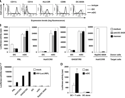
cell-cell interactions underlying mDC-enhanced HIV transmis-sion, the efficiencies of HIVtrans-infection mediated by iDCs and mDCs were compared and the role of C-type lectins in viral transmission was examined. Purified CD14⫹monocytes were used to generate iDCs, and the maturation of iDCs was achieved by LPS treatment (34). The phenotypes of iDCs and mDCs were confirmed by immunostaining of cell surface markers. As expected, iDCs and mDCs uniformly expressed high levels of CD11c, and they were nearly negative for CD14 at day 7 of differentiation (Fig. 1A). HLA-DR and CD86 expression levels were significantly increased in mDCs relative to iDCs, indicating efficient DC maturation, while the surface expression of DC-SIGN was decreased in mDCs (Fig. 1A).
To quantify HIV transmission efficiency mediated by iDCs and mDCs, a single-cycle luciferase reporter HIV was used. The virus was pseudotyped separately with R5- or X4-tropic HIV Env. Various types of target cells were used in HIV transmission assays, including activated autologous PBLs, the human CD4⫹T-cell line Hut/CCR5 (57), and the human osteosarcoma cell line GHOST/R5, and were engineered to express HIV receptors (12). When DCs were pulsed with small amounts of R5-tropic HIV and cocultured separately with dif-ferent types of target cells, mDCs were 3-fold (P ⬍ 0.05), 18-fold (P⬍0.001), and 8-fold (P⬍0.01) more effective than iDCs in transmitting HIV infection to activated PBLs, Hut/ CCR5 cells, and GHOST/R5 cells, respectively (Fig. 1B). Sim-ilar results were observed in independent experiments using autologous DCs and PBLs derived from four different donors and using HIV pseudotyped with different R5-tropic Env pro-teins (data not shown).
Preincubation of iDCs with cocktails of DC-SIGN MAbs reduced HIV transmission to various types of target cells by 33% to 54% (P⬍0.05) (Fig. 1B), a finding consistent with our previous results (49, 57). Similarly, blockade of iDCs with mannan, an inhibitor of mannose-binding C-type lectins, de-creased HIV transmission by 22% to 56% (P⬍0.05). How-ever, DC-SIGN MAbs and mannan had no effect on HIV transmission mediated by mDCs (Fig. 1B). These results sug-gest that mDC-enhanced HIV transmission is independent of
on November 8, 2019 by guest
http://jvi.asm.org/
C-type lectins, which partly contribute to iDC-mediated HIV transmission. To confirm the effective function of DC-SIGN MAbs and mannan for neutralizing HIV transmission, Raji/ DC-SIGN cells (55), a human B-cell line engineered high levels of DC-SIGN expression, were used as a positive control. Consistent with our previous results (53, 56, 57), HIV trans-mission to Hut/CCR5 cells by Raji/DC-SIGN cells was abol-ished by DC-SIGN MAbs and mannan (Fig. 1B).
The HIV infection observed in cocultures was a direct result of DC-mediatedtrans-infection of target cells, as no viral in-fection was detected at 2 days postinin-fection (dpi) in DCs and Raji/DC-SIGN cells that were pulsed with small amounts of
[image:3.585.46.539.70.456.2]HIV (Fig. 1C). HIV infection in GHOST/R5 cells was signif-icantly higher (149-fold and 6-fold) than that in PBLs and Hut/CCR5 cells (Fig. 1C), consistent with the increased HIV infection observed in coculture assays (Fig. 1B). In addition, mDCs were threefold (P ⬍0.05) more potent than iDCs in stimulating X4-tropic HIV trans-infection of CD4⫹ T cells (Fig. 1D). Together, these data suggest that unique mDC-HIV interactions may account for enhanced HIV transmission to target cells. HIV pseudotyped with R5-tropic-Env was used in the following assays, given that the infection and transmission rate of X4-tropic HIV in DCs is significantly lower than that of R5-tropic HIV (20, 23, 38, 49).
FIG. 1. Mature DCs enhance HIV transmission to different types of target cells independently of C-type lectins. (A) Surface markers of iDCs and mDCs. Monocyte-derived iDCs were cultured with LPS to generate mDCs. Cell surface markers were stained with specific MAbs or isotype-matched IgG controls and analyzed by flow cytometry. The histogram peaks of CD11c staining on iDCs and mDCs were overlapped. (B) Enhanced HIV transmission by mDCs is independent of C-type lectins. DCs and Raji/DC-SIGN cells were preincubated separately with medium, anti-DC-SIGN cocktails, and mannan prior to HIV incubation, as described previously (57). Raji/DC-SIGN cells, iDCs, and mDCs were pulsed separately with single-cycle, luciferase reporter R5-tropic HIV-Luc/JRFL (multiplicity of infection [MOI], 0.2), washed, and cocultured separately with autologous PBLs, the CD4⫹T-cell line Hut/CCR5, and the HIV indicator cells GHOST/R5. HIV infection was determined after 2 days by measuring the luciferase activity. (C) No detectable HIVcis-infection in DCs and Raji/DC-SIGN cells. Cells were infected with HIV-Luc/JRFL (MOI, 0.2), and viral infection was determined at 2 dpi. (D) mDCs enhance transmission of HIV pseudotyped with X4-tropic Env (HXB2). Transmission of HIV-Luc/HXB2 with DCs as donors and Hut/CCR5 cells as targets was performed as described for panel B. The asterisk indicates a significant difference (P⬍0.05) compared with iDCs. The data show the means⫾standard deviations of triplicate samples. One representative experiment out of four is shown. cps, counts per second.
on November 8, 2019 by guest
http://jvi.asm.org/
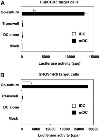
DC-target cell contact is required for efficient HIV
trans-mission mediated by iDCs and mDCs.To evaluate whether
mDC-enhanced HIV transmission requires cell-cell contact, HIV transmissions to different types of target cells by iDCs and mDCs were compared with transwell culture plates. HIV-pulsed DCs were separated from Hut/CCR5 or GHOST/R5 target cells by the use of transwell culture plates with perme-able membranes (56). The transwell membranes (pore size, 3
m) are permeable for HIV but not for DCs or target cells (data not shown). Compared with DC-alone controls, HIV infection was enhanced 32-fold or 168-fold when iDCs or mDCs, respectively, were in cocultures with Hut/CCR5 target cells (Fig. 2A). Similarly, compared with DC-alone controls, HIV infection was enhanced 66-fold or 400-fold when iDCs or mDCs, respectively, were in cocultures with GHOST/R5 target cells (Fig. 2B). When iDCs and mDCs were separated from target cells by the permeable membranes, HIV trans-mission decreased to background levels (Fig. 2). Similar results were obtained when transwell plates with membrane pore sizes of 0.4 m were used (data not shown). Thus, DC-target cell contact is required for efficient DC-mediated HIV transmission.
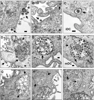
Enhanced HIV endocytosis and distinct viral trafficking in
mDCs relative to iDCs.To visualize HIV trafficking and
inter-actions with iDCs and mDCs, AT-2-inactivated HIV was used for electron microscopy assays. AT-2-inactivated HIV is con-formationally authentic and interacts with DCs similarly to
infectious HIV (19, 48). Using electron microscopy, previous studies have investigated interactions of AT-2-inactivated sim-ian immunodeficiency virus and human DCs or macaque DCs (19, 48). After a 1.5-h HIV exposure, cell surface-associated HIV and a few internalized viral particles in clathrin-coated vesicles were observed in iDCs (Fig. 3A, B, and C). By con-trast, in addition to the surface-associated HIV, numerous intact HIV particles were observed within intracellular endo-cytic compartments in mDCs (Fig. 3D, E, and F). No HIV particles were observed in controls without HIV incubation (not shown).
Given the complexity of DC membranes, to confirm that the HIV-containing compartments in mDCs were truly endocy-tosed structures rather than cell surface invaginations, RR was used during fixation as a membrane-impermeable dye. RR binds to carbohydrate moieties on the cell surface (32) and readily penetrates membrane invaginations due to its small size (14). Fixation of DCs at 4°C prevents the internalization of RR. Upon postfixation, RR forms an electron-dense precipi-tate that can be visualized by electron microscopy. This method has been used to study HIV entry and assembly in macrophages (15, 33, 51). Results showed that mDC surfaces and the invaginations were strongly labeled with RR, as ex-pected, while the membranes of the HIV-containing vesicles were largely resistant to RR staining (Fig. 3G, H, and I). These results confirm that significant amounts of HIV were internal-ized in the intracellular compartments in mDCs.
mDCs are more potent than iDCs in protecting HIV from
proteolysis. The above viral trafficking studies indicated that
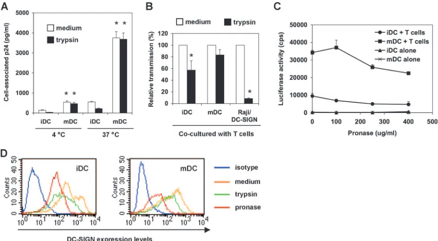
mDCs facilitated HIV internalization and altered HIV local-ization compared with iDCs. To determine whether viral traf-ficking contributes to DC-mediated HIV transmission, iDCs and mDCs were compared for binding and internalization of HIV and viral protection from proteolysis.
To measure HIV binding and internalization, iDCs and mDCs were pulsed separately with small amounts of infectious HIV for 2 h, and DC-associated HIV p24 was quantified. To test the proteolysis sensitivity of DC-associated HIV, DCs were treated with trypsin after the HIV incubation. At 4°C, HIV binding to mDCs was nearly fourfold (P⬍0.01) higher than that to iDCs (Fig. 4A). Trypsin treatment reduced iDC-associated HIV to background levels, indicating that virus mainly remained on the iDC surface upon binding at 4°C, while the trypsin treatment only slightly decreased mDC-bound HIV, by 13%. HIV internalization of mDCs at 37°C was sevenfold greater (P⬍ 0.01) than that of iDCs and was sev-enfold greater (P⬍0.01) than HIV binding to mDCs at 4°C. Nearly 50% iDC-associated HIV at 37°C was sensitive to pro-teolysis, whereas the mDC-associated HIV was completely re-sistant to trypsin treatment (Fig. 4A). These results suggest that increased HIV internalization and protection by mDCs may contribute to enhanced efficiency of viral transmission.
[image:4.585.78.249.69.305.2]To detect whether DCs protect HIV from protease treat-ment and further transfer HIV to T cells, HIV-pulsed iDCs and mDCs were treated with trypsin (250g/ml) before cocul-ture with Hut/CCR5 cells. Compared with medium controls, the average results of four independent experiments revealed that trypsin treatment reduced iDC-mediated HIV transmis-sion by 43%⫾16% (P⬍0.05), while a 17%⫾9% decrease in viral transmission was observed in trypsin-treated mDCs (Fig.
FIG. 2. DC-target cell contact is required for efficient HIV trans-mission mediated by iDCs and mDCs. Transtrans-mission of HIV-Luc/JRFL with iDCs and mDCs as donors and (A) Hut/CCR5 cells or (B) GHOST/R5 cells as targets was performed as described in the legend to Fig. 1B. Transwell culture plates with membrane pore sizes of 3m were used to separate DCs and target cells (Transwell). HIV infection was determined after 2 dpi by measuring the luciferase ac-tivity. The data show the means⫾standard deviations of triplicate samples. One representative experiment out of three is shown. cps, counts per second.
on November 8, 2019 by guest
http://jvi.asm.org/
4B). These data suggest that mDCs are more potent at pro-tecting internalized and surface-bound HIV from protease digestion. As a control, trypsin treatment of HIV-pulsed Raji/DC-SIGN cells under the same conditions significantly reduced viral transmission, by 91% (P⬍0.001) (Fig. 4B). We have carefully optimized the trypsin treatment conditions to ensure cell viability and sufficient HIV cleavage on cell surfaces (49); however, the trypsin treatment might not completely cleave DC surface-bound HIV given the complexity of DC membranes.
A recent study proposed that trypsin might be less potent than pronase at removing DC surface-bound HIV, although no comparison data were shown (11). To further demonstrate that DC-mediated HIV transmission is partially resistant to pro-teolysis, HIV-pulsed DCs were treated with pronase and then cocultured with Hut/CCR5 cells. HIV transmission gradually decreased when DCs were treated with increasing
[image:5.585.111.473.68.452.2]concentra-tions of pronase (Fig. 4C). Interestingly, pronase treatment (250g/ml) reduced iDC- and mDC-mediated HIV transmis-sion by 48% (P⬍0.05) and 24%, respectively. These data were comparable to those of trypsin treatment at the same concen-tration, suggesting that both trypsin and pronase may effi-ciently strip surface HIV from DCs. Furthermore, when DCs were treated with 400g/ml of pronase, HIV transmission by iDCs and mDCs was decreased by 51% (P⬍0.05) and 34%, respectively (Fig. 4C), indicating that mDC-associated HIV is more resistant to proteolysis. Together, these data suggest that DC-mediated HIV transmission may involve recycling of the internalized viruses in addition to DC surface-bound HIV. To confirm the effective proteolysis function, DC-SIGN on DC surfaces was stained after separate treatments with trypsin or pronase. Results showed that both trypsin and pronase treat-ments efficiently reduced surface DC-SIGN levels on iDCs and mDCs (Fig. 4D), which may also partially contribute to the
FIG. 3. Enhanced HIV endocytosis and distinct viral trafficking in mDCs relative to iDCs. DCs were exposed to AT-2-inactivated R5 HIV for 1.5 h, washed thoroughly, fixed, and prepared for electron microscopy. (A, B, and C) Cell surface-bound HIV and internalized viral particles in iDCs. (D, E, and F) HIV internalization is significantly enhanced in mDCs. Arrows indicate DC surface-associated HIV particles or intracellular compartments that trapped intact HIV particles. (G, H, and I) RR labeling of mDC plasma membranes. (I) Higher-magnification image of panel H (a partial area). The open arrows indicate RR-labeled mDC plasma membranes, and the black arrows point to HIV-containing compartments and HIV particles that were not labeled with RR. Scale bars, 0.1m (A to F), 0.2m (G, I), and 0.5m (H).
on November 8, 2019 by guest
http://jvi.asm.org/
decreased efficiency of iDC-mediated HIV transmission after protease treatment.
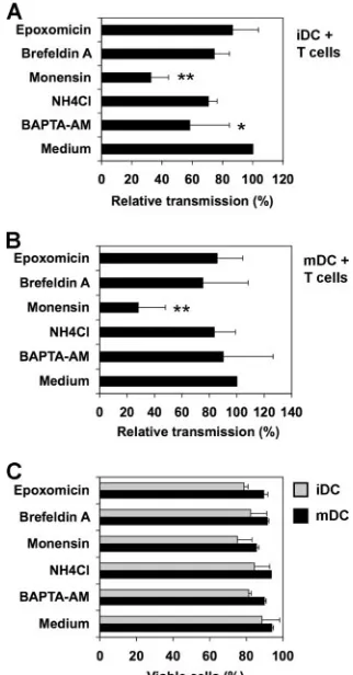
Effects of trafficking inhibitors on DC-mediated viral
trans-mission. To quantify the effect of HIV trafficking on
DC-mediated viral transmission, iDCs and mDCs were examined for their efficiencies in supporting HIVtrans-infection after treatment with various trafficking inhibitors. DCs were incu-bated separately with various inhibitors for 30 min and pulsed with small amounts of HIV in the presence of the inhibitors for 2 h. To avoid the effect of inhibitors on HIV infection of target cells, HIV-pulsed DCs were washed and cocultured with Hut/ CCR5 cells for 2 days in the absence of inhibitors. Various trafficking inhibitors included an intracellular Ca2⫹chelator,
BAPTA-AM, which can eliminate exosome secretion (43); NH4Cl, a weak base that neutralizes acidic endomembrane compartments (1); monensin, a polyether antibiotic that dis-rupts the structure of the Golgi apparatus and inhibits vesicu-lar transport in eukaryotic cells (18); brefeldin A, a macrocyclic lactone that inhibits small GTP-binding proteins and induces the rapid redistribution of the Golgi apparatus into the endo-plasmic reticulum (28); and epoxomicin, a potent and selective proteasome inhibitor (35). Medium that contained dissolvent was used as a control.
The average results of four independent experiments re-vealed that monensin significantly reduced iDC- and mDC-mediated HIV transmission, by 68% and 72% (P ⬍ 0.01
[Dunnett’s multiple comparison test]), respectively (Fig. 5A and B). These data suggest potential involvement of the Golgi apparatus or vesicular transport of HIV in DC-mediated HIV transmission. BAPTA-AM blocked 42% of iDC-mediated HIV transmission (P⬍ 0.05) (Fig. 5A), indicating that HIV transmission by iDCs is partially dependent on C-type lectins. As a control, monensin treatment did not significantly change DC-associated HIV p24 (91% to 119%, relative to medium controls). While BAPTA-AM treatment reduced iDC-associ-ated HIV p24 by 43%, it had no effect on mDC-associiDC-associ-ated HIV p24. Some inhibitor-dependent effects (such as NH4Cl) on cellular trafficking events are reversible after removal of the inhibitors. Thus, potential effects of other inhibitors on HIV
trans-infection could not be ruled out. No significant decrease in DC viability was observed after inhibitor treatment and 3 days in culture. The viability of inhibitor-treated DCs remained 76% to 94%, compared with the 88% to 94% viability of the medium controls (Fig. 5C). Together, these data suggest that intracellular trafficking inhibitors may disrupt DC-mediated viral transmission.
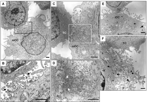
mDCs efficiently concentrate HIV at virological synapses.
[image:6.585.75.508.68.306.2]To visualize the formation of virological synapses between DCs and CD4⫹ T cells, an electron microscopy-based assay was developed. After a 1.5-h exposure of AT-2-inactivated HIV to iDCs and mDCs, DCs were washed thoroughly and cocultured with Hut/CCR5 cells for 1 h to allow DC–T-cell interactions
FIG. 4. mDCs are more potent than iDCs in protecting HIV from proteolysis. (A) mDCs enhance HIV binding and internalization. DCs were incubated with HIV-Luc/JRFL (30 ng of p24) at 4°C or 37°C for 2 h, washed and treated with trypsin or medium, and then lysed for HIV p24 quantification. Asterisks indicate significant differences (P⬍0.01) compared with iDCs at the same temperature. (B) DCs protect captured HIV from trypsin cleavage. HIV-pulsed iDCs, mDCs, and Raji/DC-SIGN cells were separately treated with trypsin before coculture with Hut/CCR5 target cells. Transmission of HIV-Luc/JRFL to Hut/CCR5 target cells was performed as described in the legend to Fig. 1B. The average results of four independent experiments are shown. Values for medium controls were set at 100%. Asterisks indicate significant differences (P⬍0.05) between trypsin-treated samples and medium controls. (C) DCs protect captured HIV from pronase cleavage. HIV-pulsed iDCs and mDCs were treated with increasing concentrations of pronase before coculture with Hut/CCR5 target cells. The data show the means⫾standard deviations of triplicate samples. One representative experiment out of two is shown. cps, counts per second. (D) Decreased surface DC-SIGN levels on DCs after protease treatment. DCs were stained for surface DC-SIGN after separate treatments with trypsin or pronase and analyzed by flow cytometry.
Medium treatment was used as a control.
on November 8, 2019 by guest
http://jvi.asm.org/
and viral transfer. Upon contact with CD4⫹T cells, a large amount of intact HIV particles were concentrated and polar-ized at mDC–T-cell synapses (Fig. 6A to D). Interestingly, membrane continuity between an HIV-containing compart-ment and the plasma membrane of an mDC was observed at an mDC–T-cell synapse (Fig. 6C and D). Although it is difficult to conclude that HIV was redistributed from the intracellular compartments following T-cell contact, the membrane conti-nuity with concentrated HIV particles could not be observed in HIV-pulsed mDCs without T-cell coculture (Fig. 3D to I). Large amounts of intact HIV particles were easily observed at numerous mDC–T-cell synapses; by contrast, the virological synapses formed between iDCs and T cells were not readily found (Fig. 6E and F and data not shown). A few intact HIV-like particles were observed at iDC–T-cell junctions (Fig. 6E and F). These results suggest that mDCs are more efficient
than iDCs in concentrating captured HIV at virological syn-apses.
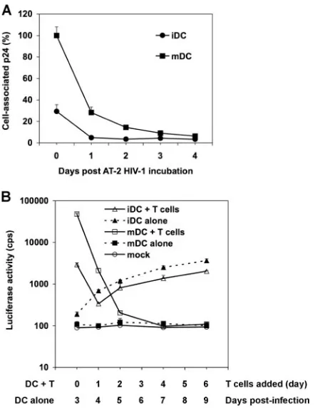
Intracellular HIV degradation and time course viral
trans-mission mediated by DCs.To compare intracellular HIV
deg-radation in iDCs and mDCs, DCs were exposed to small amounts of AT-2-inactivated HIV and trypsinized, and DC-associated HIV p24 from aliquots was measured daily for 4 days. The mDC-associated p24 was 3.4-fold higher than that of iDCs at day 0 (Fig. 7A). After 1 day in culture, iDC- and mDC-associated HIV was rapidly degraded, by 83% and 72%, respectively. After 3 days, almost no HIV p24 was detectable in iDCs and mDCs (Fig. 7A), suggesting that there is no long-term retention of HIV in DCs. These data are in agreement with previous studies showing that most incoming HIV is de-graded in DCs within 24 h (36, 37, 48).
To determine whether the small proportions of HIV re-tained in DCs can mediate long-termtrans-infection of CD4⫹ T cells, time course viral transmission by iDCs and mDCs was examined. Use of single-cycle HIV in this assay had the ad-vantage of avoiding viral transmission of progeny viruses that replicate incis-infected DCs. At 0, 1, 2, 4, and 6 dpi, aliquots of HIV-pulsed iDCs and mDCs were separately cocultured with Hut/CCR5 cells for an additional 3 days to quantify viral transmission. In parallel, HIV-infected DC-alone controls were harvested at 3, 4, 5, 7, and 9 dpi to measurecis-infection. HIV trans-infection mediated by mDCs was 16-fold (P ⬍
0.001) higher than that by iDCs when DCs were cocultured with Hut/CCR5 cells at 0 dpi (Fig. 7B). After 1 dpi, viral transmission mediated by iDCs and mDCs decreased by 9- and 23-fold (P⬍0.001), respectively, while mDCs were still 6-fold more potent than iDCs at enhancing viral transmission. At 2 dpi, mDC-mediated HIV transmission further decreased 10-fold, to almost basal level. Of note, in the above-described transmission assays (Fig. 1, 2, 4, and 5), HIV-pulsed DCs alone did not become detectably infected at 2 to 2.5 dpi, whereas an increasing HIV infection in iDCs became detectable after 3 to 4 dpi during the time course. The increased HIV infection in iDCs correlated with iDC-mediated viral transmission. Consis-tently, no HIV infection was detected in mDCs (Fig. 7B). HIV infection in iDC-alone samples was around twofold higher than that in iDC–T-cell cocultures at 4 dpi to 9 dpi, a result that was likely due to T-cell-induced DC maturation (Fig. 7B). Together, these results suggest that most incoming HIV is rapidly degraded by iDCs and mDCs and that there is no viral retention-mediated long-term transmission.
DISCUSSION
Understanding HIV-host cell interactions and defining the mechanisms of DC-mediated virus transmission are essential for developing effective strategies to combat HIV infection (54). Here we have compared the efficiency and mechanisms of HIV transmission mediated by iDCs and mDCs by using sin-gle-cycle HIV quantification assays. We have found that mDCs significantly facilitate HIV endocytosis and efficiently concen-trate HIV at virological synapses, which is likely to contribute to mDC-enhanced HIV transmission, at least in part. mDCs were more efficient than iDCs in transmitting HIV to various types of target cells independently of C-type lectins. Moreover, DC-target cell contact was required for efficient HIV-1
FIG. 5. Effects of trafficking inhibitors on DC-mediated viral trans-mission. iDCs (A) and mDCs (B) were incubated separately with various inhibitors for 0.5 h and pulsed with HIV-Luc/JRFL in the presence of the inhibitors for 2 h at 37°C. DCs were washed and cocultured with Hut/CCR5 cells for 2.5 days. Medium that contained dissolvent was used as a control. The average relative transmission of four independent experiments using DCs from different donors is shown (medium controls were set at 100%). Asterisks indicate signif-icant differences (*,P⬍0.05;**,P⬍0.01) compared with medium controls. (C) Viability of inhibitor-treated DCs after 3 days in culture. DCs were incubated separately with various inhibitors at 37°C for 2.5 h, washed, and cultured 3 days before staining with 7-amino-actinomycin D. Stained DCs were analyzed by flow cytometry. The average DC viability of three independent experiments is shown (me-dium controls were set at 100%).
on November 8, 2019 by guest
http://jvi.asm.org/
[image:7.585.82.243.69.377.2]mission mediated by iDCs and mDCs. These results suggest that HIV may exploit mDCs to efficiently spread viral infection in lymphoid tissues, which are the major resources for HIV replication (17).
The enhanced efficiency of HIV transmission by LPS-in-duced mDCs has potential clinical implications for HIV patho-genesis. A recent finding indicated that significantly increased plasma LPS levels in HIV-infected humans correlate with AIDS progression and systemic immune activation. The in-creased plasma LPS levels may result from microbial translo-cation through a breach in the integrity of the mucosal barrier in the gut (7). Indeed, LPS can induce mouse DC maturation in vivo (16). Although the LPS concentration that we used (10 ng/ml) for in vitro DC maturation is about 50-fold higher than that found in the plasma of HIV-infected patients (7), it is conceivable that increased LPS in HIV-infected individuals may induce DC maturation and potently stimulate HIV dis-semination in vivo. In addition, HIV coinfection with other
sexually transmitted pathogens can increase inflammatory stimulations at the mucosae (44), which may directly activate DCs in vivo and promote HIV spread. Further studies using myeloid DCs, plasmacytoid DCs, or Langerhans cells from HIV-infected individuals may be required to test this hypo-thesis.
[image:8.585.42.542.67.414.2]Our data suggest that both cell surface-bound and internal-ized HIV contributes to DC-mediated viral transmission. In contrast, a recent study indicates that DC-mediated HIVtrans -infection mainly derives from DC surface-bound virions (11). Although it is difficult to directly compare these results owing to the different approaches used in the studies, the dynamic recycling of internalized HIV to DC surfaces may also mediate HIVtrans-infection, which should be an important consider-ation. It has been shown that HIV trafficking to the infectious synapse between LPS-induced mDCs and CD4⫹T cells occurs via a tetraspanin-sorting pathway (21). HIV is internalized into endocytic compartments in LPS-induced mDCs, which are
FIG. 6. mDCs efficiently concentrate HIV at virological synapses. After a 1.5-h exposure to AT-2-inactivated R5 HIV, iDCs and mDCs were washed and cocultured separately with Hut/CCR5 cells for 1 h, fixed, and prepared for electron microscopy. Hut/CCR5 cells exhibit more condensed chromatins; DCs show typical surface dendrites, less-condensed chromatin, and electron-dense lysosome-like granules. (A) Large amount of intact HIV particles concentrated at the mDC–T-cell junction. (B) Higher-magnification images of the boxed areas from panel A. Black arrows indicate HIV particles that were concentrated at the synapses. (C) HIV particles concentrated at the mDC–T-cell junction. Membrane continuity was observed between an HIV-containing compartment and the plasma membrane of an mDC. (D) Higher-magnification images of the boxed areas from panel C. White arrows indicate HIV particles that were concentrated at the mDC–T-cell synapses. (E) Fewer HIV-like particles were observed at the iDC–T-cell junction. (F) A number of intact HIV-like particles were observed at the iDC–T-cell junction. Black arrows indicate HIV-like particles at the synapses. TC, Hut/CCR5 cells; scale bars, 1m (A to E) and 0.5m (F).
on November 8, 2019 by guest
http://jvi.asm.org/
nonconventional, nonlysosomal vesicles (21). Upon contact with T cells, internalized HIV in mDCs redistributes to form infectious synapses (21, 48). Together, these results support a model in which intracellular HIV trafficking contributes to HIV transmission mediated by DCs, particularly to mDC-en-hanced viral transmission. Although endocytosis of HIV by DCs may not occur at 4°C, we have observed that the invagi-nations of mDC plasma membranes were strongly labeled with RR at 4°C. We observed that trypsin treatment only slightly decreased mDC-bound HIV at 4°C, by 13%, which might be due to viral protection by the invaginations of mDC plasma membranes.
We found that monensin, an intracellular trafficking inhibi-tor, significantly blocked iDC- and mDC-mediated HIV trans-mission to CD4⫹ T cells. In addition to inhibiting vesicular transport in eukaryotic cells, monensin can also disrupt the structure of the Golgi apparatus and glycoprotein synthesis
(18, 39). In our experiments, monensin was washed away after the 2.5-h incubation with DCs, and no significant cytotoxic effects on DCs were observed after 3 days in culture. There-fore, it is unlikely that the reduced HIV transmission by mon-ensin was mainly due to disrupted protein synthesis, although the possibility cannot be ruled out. Monensin is used as an antiprotozoal, antibacterial, or antifungal agent and as a growth promoter in veterinary medicine (9). It might be inter-esting to further explore whether monensin can be used as an antiviral agent against HIV transmission in vivo.
Previous results (34, 42) and the present study indicate that DC–T-cell contact is required for efficient HIVtrans-infection mediated by iDCs and mDCs. The exocytosis of HIV-associ-ated exosomes also can play a role in iDC-mediHIV-associ-ated HIV
trans-infection (52), but it may not be an efficient pathway in mDC-enhanced HIV transmission given that iDCs produce more exosomes than do mDCs (45). Although cell-free su-pernatants from single-cycle HIV-pulsed mDCs were posi-tive for HIV Gag p24, they failed to initiate HIV infection in GHOST/R5 cells or Hut/CCR5 cells (data not shown). Nevertheless, the efficiency of exosome-mediated trans -in-fection by mDCs remains to be confirmed with replication-competent HIV.
Increased ICAM-1 expression on mDCs has been shown to correlate with mDC-enhanced HIV transmission (42). This is possibly due to stronger DC–T-cell interactions through ICAM-1 binding to T-cell-expressed LFA-1 (for “leukocyte function-associated molecule 1”) (25, 42). Despite the lack of expression of any identified ICAM ligands, such as LFA-1, CD11b/CD18, and CD11c, GHOST/R5 cells efficiently sup-ported mDC-enhanced HIV transmission (Fig. 2B and data not shown). Moreover, ICAM-1 MAb blockade of DCs, GHOST/R5 cells, or both did not significantly affect HIV transmission mediated by iDCs or mDCs (data not shown). Therefore, ICAM-1 may not be the only cellular factor that contributes to mDC-enhanced efficiency of HIVtrans -infec-tion. Cell-type-dependent HIV trafficking may play a role in mDC-enhanced viral transmission, at least in part.
Our results indicate that HIV capture by iDCs is less effi-cient than that by mDCs; thus, the differences in viral trans-mission efficiencies and virological synapses between iDCs and mDCs may only reflect the low levels of viral capture by iDCs. HIV entry in DCs can occur through endocytosis and viral receptor-mediated fusion, while productive HIV replication requires viral fusion (8, 23, 38). To visualize viral interaction with DCs, high concentrations of AT-2-inactivated HIV were used in a previous study (2 to 3g of p24/106DCs) (48) and in our electron microscopy assays (2 g of p24/6 ⫻ 105 DCs). Given that AT-2-inactivated HIV can mediate viral fusion with cell membranes (41), the majority of iDC-associated HIV par-ticles may undergo fusion, uncoating, or degradation processes in iDCs or in cocultured T cells. Therefore, intact HIV parti-cles could not be easily observed in iDC–T-cell cocultures by electron microscopy (Fig. 6E and F and data not shown).
[image:9.585.54.275.67.353.2]It has been shown that HIV fusion to DCs decreases as cells mature (10). The entry of HIV into LPS-induced mDCs seemed to be primarily through endocytosis. The large intra-cellular compartments that confined numerous HIV particles in mDCs (Fig. 3D to I) appeared morphologically similar to macropinocytosis-mediated HIV entry in macrophages and
FIG. 7. Intracellular HIV degradation and time course viral trans-mission mediated by DCs. (A) HIV degradation in DCs. DCs (7.5⫻ 105) were incubated with AT-2-inactivated R5-tropic HIV (50 ng of
p24), washed, and treated with trypsin. Aliquots of DCs were cultured, and DC-associated HIV p24 was measured daily. The p24 result (3,897 pg/ml) for mDCs at day 0 was set at 100%, and relative results are shown. (B) Time course HIV transmission by DCs. Transmission of HIV-Luc/JRFL (multiplicity of infection, 0.2) with iDCs and mDCs as donors and Hut/CCR5 cells as targets was performed as described in the legend to Fig. 1B. At 0, 1, 2, 4, and 6 dpi, aliquots of HIV-pulsed iDCs and mDCs were cocultured separately with Hut/CCR5 cells for an additional 3 days. In parallel, HIV infection of DC-alone controls was determined by measuring the luciferase activity at 3, 4, 5, 7, and 9 dpi. Mock controls of iDCs and mDCs without HIV infection were identical. All data are the means⫾standard deviations of triplicate samples. One representative experiment out of three is shown. cps, counts per second.
on November 8, 2019 by guest
http://jvi.asm.org/
brain microvascular endothelia (30, 33). Activation of DCs can trigger extensive and prolonged macropinocytic activity, en-abling DCs to sample large volumes of the extracellular milieu for immune surveillance (13). Although mDC-associated HIV was rapidly degraded, by 72%, after 1 day, about 14% and 9% of HIV p24 remained at day 2 and 3 in mDCs, respectively (Fig. 7A). Due to the high capacity in enhancing HIVtrans -infection by mDCs, these intracellularly retained viruses could represent an important HIV reservoir in vivo.
Cellular restriction factors that block productive HIV infec-tion in DCs may reflect the intrinsic antiviral immunity of the antigen-presenting cells. It has been suggested that reduced viral replication in mDCs is due to a block in reverse transcrip-tion (23), postintegratranscrip-tion blocks at the transcriptranscrip-tional level (3), and decreased viral fusion (10). It has been recently reported that APOBEC3G and APOBEC3F (for “apolipoprotein B mRNA-editing enzyme, catalytic polypeptide-like 3G and 3F”) mediate the postentry block to HIV replication in DCs (40). However, when the efficiency and mechanisms of HIV infec-tion and transmission between different subsets of DCs are compared, it is extremely important to consider different ap-proaches to DC generation and different stimuli for DC mat-uration (54). Using replication-competent and single-cycle HIV, we have found that HIV infection and transmission are functionally distinct from different subsets of mDCs induced by various stimuli (Dong et al. and L. Wu, unpublished results). Further understanding of the regulation of antiretroviral im-munity in DCs may provide new insights into more effective interventions against HIV infection and dissemination medi-ated by DCs.
ACKNOWLEDGMENTS
We thank T. Zahrt and J. Barbieri for critical reading of the manu-script. We thank M. Emerman, V. KewalRamani, and J. Lifson for the kind gift of reagents and C. Wells for expert assistance with electron microscopy. IL-2 was obtained from M. Gately (Hoffmann-La Roche Inc.) through the AIDS Research and Reference Reagent Program, NIAID, NIH.
This work was supported by grants to L.W. from the NIH (R01-AI068493) and the Research Affairs Committee of the Medical Col-lege of Wisconsin.
REFERENCES
1.Aiken, C.1997. Pseudotyping human immunodeficiency virus type 1 (HIV-1) by the glycoprotein of vesicular stomatitis virus targets HIV-1 entry to an endocytic pathway and suppresses both the requirement for Nef and the sensitivity to cyclosporin A. J. Virol.71:5871–5877.
2.Arrighi, J. F., M. Pion, E. Garcia, J. M. Escola, Y. van Kooyk, T. B. Geijtenbeek, and V. Piguet.2004. DC-SIGN-mediated infectious synapse formation enhances X4 HIV-1 transmission from dendritic cells to T cells. J. Exp. Med.200:1279–1288.
3.Bakri, Y., C. Schiffer, V. Zennou, P. Charneau, E. Kahn, A. Benjouad, J. C. Gluckman, and B. Canque.2001. The maturation of dendritic cells results in postintegration inhibition of HIV-1 replication. J. Immunol.166:3780–3788. 4.Banchereau, J., and R. M. Steinman.1998. Dendritic cells and the control of
immunity. Nature392:245–252.
5.Baribaud, F., S. Pohlmann, G. Leslie, F. Mortari, and R. W. Doms.2002. Quantitative expression and virus transmission analysis of DC-SIGN on monocyte-derived dendritic cells. J. Virol.76:9135–9142.
6.Boggiano, C., N. Manel, and D. R. Littman.2007. Dendritic cell-mediated
trans-enhancement of human immunodeficiency virus type 1 infectivity is independent of DC-SIGN. J. Virol.81:2519–2523.
7.Brenchley, J. M., D. A. Price, T. W. Schacker, T. E. Asher, G. Silvestri, S. Rao, Z. Kazzaz, E. Bornstein, O. Lambotte, D. Altmann, B. R. Blazar, B. Rodriguez, L. Teixeira-Johnson, A. Landay, J. N. Martin, F. M. Hecht, L. J. Picker, M. M. Lederman, S. G. Deeks, and D. C. Douek.2006. Microbial translocation is a cause of systemic immune activation in chronic HIV in-fection. Nat. Med.12:1365–1371.
8.Burleigh, L., P.-Y. Lozach, C. Schiffer, I. Staropoli, V. Pezo, F. Porrot, B. Canque, J.-L. Virelizier, F. Arenzana-Seisdedos, and A. Amara.2006. Infec-tion of dendritic cells (DCs), not DC-SIGN-mediated internalizaInfec-tion of human immunodeficiency virus, is required for long-term transfer of virus to T cells. J. Virol.80:2949–2957.
9.Butaye, P., L. A. Devriese, and F. Haesebrouck.2003. Antimicrobial growth promoters used in animal feed: effects of less well known antibiotics on gram-positive bacteria. Clin. Microbiol. Rev.16:175–188.
10.Cavrois, M., J. Neidleman, J. F. Kreisberg, D. Fenard, C. Callebaut, and W. C. Greene.2006. Human immunodeficiency virus fusion to dendritic cells declines as cells mature. J. Virol.80:1992–1999.
11.Cavrois, M., J. Neidleman, J. F. Kreisberg, and W. C. Greene.2007. In vitro derived dendritic cells trans-infect CD4 T cells primarily with surface-bound HIV-1 virions. PLoS Pathog.3:e4.
12.Cecilia, D., V. N. KewalRamani, J. O’Leary, B. Volsky, P. Nyambi, S. Burda, S. Xu, D. R. Littman, and S. Zolla-Pazner.1998. Neutralization profiles of primary human immunodeficiency virus type 1 isolates in the context of coreceptor usage. J. Virol.72:6988–6996.
13.Conner, S. D., and S. L. Schmid.2003. Regulated portals of entry into the cell. Nature422:37–44.
14.Damke, H., T. Baba, D. E. Warnock, and S. L. Schmid.1994. Induction of mutant dynamin specifically blocks endocytic coated vesicle formation. J. Cell Biol.127:915–934.
15.Deneka, M., A. Pelchen-Matthews, R. Byland, E. Ruiz-Mateos, and M. Marsh.2007. In macrophages, HIV-1 assembles into an intracellular plasma membrane domain containing the tetraspanins CD81, CD9, and CD53. J. Cell Biol.177:329–341.
16.De Smedt, T., B. Pajak, E. Muraille, L. Lespagnard, E. Heinen, P. De Baetselier, J. Urbain, O. Leo, and M. Moser.1996. Regulation of dendritic cell numbers and maturation by lipopolysaccharide in vivo. J. Exp. Med. 184:1413–1424.
17.Douek, D. C., L. J. Picker, and R. A. Koup.2003. T cell dynamics in HIV-1 infection. Annu. Rev. Immunol.21:265–304.
18.Fliesler, S. J., and S. F. Basinger.1987. Monensin stimulates glycerolipid incorporation into rod outer segment membranes. J. Biol. Chem.262:17516– 17523.
19.Frank, I., M. Piatak, Jr., H. Stoessel, N. Romani, D. Bonnyay, J. D. Lifson, and M. Pope.2002. Infectious and whole inactivated simian immunodefi-ciency viruses interact similarly with primate dendritic cells (DCs): differen-tial intracellular fate of virions in mature and immature DCs. J. Virol. 76:2936–2951.
20.Ganesh, L., K. Leung, K. Lore, R. Levin, A. Panet, O. Schwartz, R. A. Koup, and G. J. Nabel.2004. Infection of specific dendritic cells by CCR5-tropic human immunodeficiency virus type 1 promotes cell-mediated transmission of virus resistant to broadly neutralizing antibodies. J. Virol.78:11980–11987. 21.Garcia, E., M. Pion, A. Pelchen-Matthews, L. Collinson, J. F. Arrighi, G. Blot, F. Leuba, J. M. Escola, N. Demaurex, M. Marsh, and V. Piguet.2005. HIV-1 trafficking to the dendritic cell–T-cell infectious synapse uses a path-way of tetraspanin sorting to the immunological synapse. Traffic6:488–501. 22.Geijtenbeek, T. B., D. S. Kwon, R. Torensma, S. J. van Vliet, G. C. van Duijnhoven, J. Middel, I. L. Cornelissen, H. S. Nottet, V. N. KewalRamani, D. R. Littman, C. G. Figdor, and Y. van Kooyk.2000. DC-SIGN, a dendritic cell-specific HIV-1-binding protein that enhances trans-infection of T cells. Cell100:587–597.
23.Granelli-Piperno, A., E. Delgado, V. Finkel, W. Paxton, and R. M. Steinman. 1998. Immature dendritic cells selectively replicate macrophagetropic (M-tropic) human immunodeficiency virus type 1, while mature cells efficiently transmit both M- and T-tropic virus to T cells. J. Virol.72:2733–2737. 24.Granelli-Piperno, A., A. Pritsker, M. Pack, I. Shimeliovich, J. F. Arrighi,
C. G. Park, C. Trumpfheller, V. Piguet, T. M. Moran, and R. M. Steinman. 2005. Dendritic cell-specific intercellular adhesion molecule 3-grabbing non-integrin/CD209 is abundant on macrophages in the normal human lymph node and is not required for dendritic cell stimulation of the mixed leukocyte reaction. J. Immunol.175:4265–4273.
25.Gummuluru, S., V. N. KewalRamani, and M. Emerman.2002. Dendritic cell-mediated viral transfer to T cells is required for human immunodefi-ciency virus type 1 persistence in the face of rapid cell turnover. J. Virol. 76:10692–10701.
26.Gummuluru, S., M. Rogel, L. Stamatatos, and M. Emerman.2003. Binding of human immunodeficiency virus type 1 to immature dendritic cells can occur independently of DC-SIGN and mannose binding C-type lectin recep-tors via a cholesterol-dependent pathway. J. Virol.77:12865–12874. 27.Izquierdo-Useros, N., J. Blanco, I. Erkizia, M. T. Ferna´ndez-Figueras, F. E.
Borra`s, M. Naranjo-Go´mez, M. Bofill, L. Ruiz, B. Clotet, and J. Martinez-Picado.2007. Maturation of blood-derived dendritic cells enhances human immunodeficiency virus type 1 capture and transmission. J. Virol.81:7559– 7570.
28.Klausner, R. D., J. G. Donaldson, and J. Lippincott-Schwartz.1992. Brefeldin A: insights into the control of membrane traffic and organelle structure. J. Cell Biol.116:1071–1080.
29.Kwon, D. S., G. Gregorio, N. Bitton, W. A. Hendrickson, and D. R. Littman.
on November 8, 2019 by guest
http://jvi.asm.org/
2002. DC-SIGN-mediated internalization of HIV is required for trans-en-hancement of T cell infection. Immunity16:135–144.
30.Liu, N. Q., A. S. Lossinsky, W. Popik, X. Li, C. Gujuluva, B. Kriederman, J. Roberts, T. Pushkarsky, M. Bukrinsky, M. Witte, M. Weinand, and M. Fiala. 2002. Human immunodeficiency virus type 1 enters brain microvascular endothelia by macropinocytosis dependent on lipid rafts and the mitogen-activated protein kinase signaling pathway. J. Virol.76:6689–6700. 31.Lore, K., A. Smed-Sorensen, J. Vasudevan, J. R. Mascola, and R. A. Koup.
2005. Myeloid and plasmacytoid dendritic cells transfer HIV-1 preferentially to antigen-specific CD4⫹T cells. J. Exp. Med.201:2023–2033.
32.Luft, J. H.1971. Ruthenium red and violet. I. Chemistry, purification, meth-ods of use for electron microscopy and mechanism of action. Anat. Rec. 171:347–368.
33.Marechal, V., M. C. Prevost, C. Petit, E. Perret, J. M. Heard, and O. Schwartz.2001. Human immunodeficiency virus type 1 entry into macro-phages mediated by macropinocytosis. J. Virol.75:11166–11177.
34.McDonald, D., L. Wu, S. M. Bohks, V. N. KewalRamani, D. Unutmaz, and T. J. Hope.2003. Recruitment of HIV and its receptors to dendritic cell-T cell junctions. Science300:1295–1297.
35.Meng, L., R. Mohan, B. H. Kwok, M. Elofsson, N. Sin, and C. M. Crews. 1999. Epoxomicin, a potent and selective proteasome inhibitor, exhibits in vivo antiinflammatory activity. Proc. Natl. Acad. Sci. USA96:10403–10408. 36.Moris, A., C. Nobile, F. Buseyne, F. Porrot, J. P. Abastado, and O. Schwartz. 2004. DC-SIGN promotes exogenous MHC-I-restricted HIV-1 antigen presentation. Blood103:2648–2654.
37.Moris, A., A. Pajot, F. Blanchet, F. Guivel-Benhassine, M. Salcedo, and O. Schwartz.2006. Dendritic cells and HIV-specific CD4⫹T cells: HIV antigen presentation, T-cell activation, and viral transfer. Blood108:1643–1651. 38.Nobile, C., C. Petit, A. Moris, K. Skrabal, J. P. Abastado, F. Mammano, and
O. Schwartz.2005. Covert human immunodeficiency virus replication in dendritic cells and in DC-SIGN-expressing cells promotes long-term trans-mission to lymphocytes. J. Virol.79:5386–5399.
39.Pal, R., R. C. Gallo, and M. G. Sarngadharan.1988. Processing of the structural proteins of human immunodeficiency virus type 1 in the presence of monensin and cerulenin. Proc. Natl. Acad. Sci. USA85:9283–9286. 40.Pion, M., A. Granelli-Piperno, B. Mangeat, R. Stalder, R. Correa, R. M.
Steinman, and V. Piguet.2006. APOBEC3G/3F mediates intrinsic resistance of monocyte-derived dendritic cells to HIV-1 infection. J. Exp. Med.203: 2887–2893.
41.Rossio, J. L., M. T. Esser, K. Suryanarayana, D. K. Schneider, J. W. Bess, Jr., G. M. Vasquez, T. A. Wiltrout, E. Chertova, M. K. Grimes, Q. Sattentau, L. O. Arthur, L. E. Henderson, and J. D. Lifson.1998. Inactivation of human immunodeficiency virus type 1 infectivity with preservation of conforma-tional and funcconforma-tional integrity of virion surface proteins. J. Virol.72:7992– 8001.
42.Sanders, R. W., E. C. de Jong, C. E. Baldwin, J. H. Schuitemaker, M. L. Kapsenberg, and B. Berkhout.2002. Differential transmission of human immunodeficiency virus type 1 by distinct subsets of effector dendritic cells. J. Virol.76:7812–7821.
43.Savina, A., M. Furlan, M. Vidal, and M. I. Colombo.2003. Exosome release
is regulated by a calcium-dependent mechanism in K562 cells. J. Biol. Chem. 278:20083–20090.
44.Shattock, R. J., and J. P. Moore.2003. Inhibiting sexual transmission of HIV-1 infection. Nat. Rev. Microbiol.1:25–34.
45.Thery, C., A. Regnault, J. Garin, J. Wolfers, L. Zitvogel, P. Ricciardi-Castagnoli, G. Raposo, and S. Amigorena.1999. Molecular characterization of dendritic cell-derived exosomes. Selective accumulation of the heat shock protein hsc73. J. Cell Biol.147:599–610.
46.Trumpfheller, C., C. G. Park, J. Finke, R. M. Steinman, and A. Granelli-Piperno.2003. Cell type-dependent retention and transmission of HIV-1 by DC-SIGN. Int. Immunol.15:289–298.
47.Tsunetsugu-Yokota, Y., S. Yasuda, A. Sugimoto, T. Yagi, M. Azuma, H. Yagita, K. Akagawa, and T. Takemori.1997. Efficient virus transmission from dendritic cells to CD4⫹T cells in response to antigen depends on close contact through adhesion molecules. Virology239:259–268.
48.Turville, S. G., J. J. Santos, I. Frank, P. U. Cameron, J. Wilkinson, M. Miranda-Saksena, J. Dable, H. Stossel, N. Romani, M. Piatak, Jr., J. D. Lifson, M. Pope, and A. L. Cunningham.2004. Immunodeficiency virus uptake, turnover, and 2-phase transfer in human dendritic cells. Blood103: 2170–2179.
49.Wang, J. H., A. M. Janas, W. J. Olson, V. N. Kewalramani, and L. Wu.2007. CD4 coexpression regulates DC-SIGN-mediated transmission of human im-munodeficiency virus type 1. J. Virol.81:2497–2507.
50.Weissman, D., Y. Li, J. M. Orenstein, and A. S. Fauci.1995. Both a precursor and a mature population of dendritic cells can bind HIV. However, only the mature population that expresses CD80 can pass infection to unstimulated CD4⫹T cells. J. Immunol.155:4111–4117.
51.Welsch, S., O. T. Keppler, A. Habermann, I. Allespach, J. Krijnse-Locker, and H. G. Krausslich.2007. HIV-1 buds predominantly at the plasma mem-brane of primary human macrophages. PLoS Pathog.3:e36.
52.Wiley, R. D., and S. Gummuluru.2006. Immature dendritic cell-derived exosomes can mediate HIV-1 trans infection. Proc. Natl. Acad. Sci. USA 103:738–743.
53.Wu, L., A. A. Bashirova, T. D. Martin, L. Villamide, E. Mehlhop, A. O. Chertov, D. Unutmaz, M. Pope, M. Carrington, and V. N. KewalRamani. 2002. Rhesus macaque dendritic cells efficiently transmit primate lentiviruses independently of DC-SIGN. Proc. Natl. Acad. Sci. USA99:1568–1573. 54.Wu, L., and V. N. KewalRamani.2006. Dendritic-cell interactions with HIV:
infection and viral dissemination. Nat. Rev. Immunol.6:859–868. 55.Wu, L., T. D. Martin, M. Carrington, and V. N. KewalRamani.2004. Raji B
cells, misidentified as THP-1 cells, stimulate DC-SIGN-mediated HIV trans-mission. Virology318:17–23.
56.Wu, L., T. D. Martin, Y. C. Han, S. K. Breun, and V. N. KewalRamani.2004. Trans-dominant cellular inhibition of DC-SIGN-mediated HIV-1 transmis-sion. Retrovirology1:14.
57.Wu, L., T. D. Martin, R. Vazeux, D. Unutmaz, and V. N. KewalRamani.2002. Functional evaluation of SIGN monoclonal antibodies reveals DC-SIGN interactions with ICAM-3 do not promote human immunodeficiency virus type 1 transmission. J. Virol.76:5905–5914.
58.Yamashita, M., and M. Emerman.2004. Capsid is a dominant determinant of retrovirus infectivity in nondividing cells. J. Virol.78:5670–5678.