0022-538X/07/$08.00⫹0 doi:10.1128/JVI.00432-07
Copyright © 2007, American Society for Microbiology. All Rights Reserved.
Type- and Subcomplex-Specific Neutralizing Antibodies against
Domain III of Dengue Virus Type 2 Envelope Protein
Recognize Adjacent Epitopes
䌤
Soila Sukupolvi-Petty,
1† S. Kyle Austin,
2† Whitney E. Purtha,
2Theodore Oliphant,
3Grant E. Nybakken,
2Jacob J. Schlesinger,
4John T. Roehrig,
5Gregory D. Gromowski,
6Alan D. Barrett,
6Daved H. Fremont,
2and Michael S. Diamond
1,2,3*
Departments of Medicine,1Pathology & Immunology,2and Molecular Microbiology,3Washington University School of Medicine,
St. Louis, Missouri; Department of Medicine, University of Rochester, Rochester, New York4; Centers for Disease Control and
Prevention, Fort Collins, Colorado5; and Center for Biodefense and Emerging Infectious Diseases, Sealy Center for
Vaccine Development, Institute for Human Infections and Immunity, and Department of Pathology,
University of Texas Medical Branch, Galveston, Texas6
Received 28 February 2007/Accepted 30 August 2007
Neutralization of flaviviruses in vivo correlates with the development of an antibody response against the viral envelope (E) protein. Previous studies demonstrated that monoclonal antibodies (MAbs) against an epitope on the lateral ridge of domain III (DIII) of the West Nile virus (WNV) E protein strongly protect against infection in animals. Based on X-ray crystallography and sequence analysis, an analogous type-specific neutralizing epitope for individual serotypes of the related flavivirus dengue virus (DENV) was hypothesized. Using yeast surface display of DIII variants, we defined contact residues of a panel of type-specific, subcomplex-specific, and cross-reactive MAbs that recognize DIII of DENV type 2 (DENV-2) and have different neutralizing potentials. Type-specific MAbs with neutralizing activity against DENV-2 localized to a sequence-unique epitope on the lateral ridge of DIII, centered at the FG loop near residues E383 and P384, analogous in position to that observed with WNV-specific strongly neutralizing MAbs. Subcomplex-specific MAbs that bound some but not all DENV serotypes and neutralized DENV-2 infection recognized an adjacent epitope centered on the connecting A strand of DIII at residues K305, K307, and K310. In contrast, several MAbs that had poor neutralizing activity against DENV-2 and cross-reacted with all DENV serotypes and other flaviviruses recognized an epitope with residues in the AB loop of DIII, a conserved region that is predicted to have limited accessibility on the mature virion. Overall, our experiments define adjacent and structurally distinct epitopes on DIII of DENV-2 which elicit type-specific, subcomplex-specific, and cross-reactive antibodies with different neutralizing potentials.
Dengue fever (DF), the most prevalent arthropod-borne viral illness in humans, is caused by dengue virus (DENV). The four serotypes of DENV are transmitted to humans primarily
by the mosquitoesAedes aegyptiandAedes albopictus. DENV is
a member of theFlaviviridaefamily and is related to the viruses
that cause yellow fever and the Japanese, St. Louis, and West Nile encephalitides (8). Infection by DENV causes a spectrum of clinical disease, ranging from an acute, debilitating, self-limited febrile illness (DF) to a life-threatening hemorrhagic and capillary leak syndrome (dengue hemorrhagic fever/den-gue shock syndrome). At present, no approved antiviral treat-ment or vaccine is available, and therapy is supportive in na-ture. DENV causes an estimated 25 to 100 million cases of DF and 250,000 cases of dengue hemorrhagic fever per year world-wide, with 2.5 billion people at risk for infection (27, 48).
DENV is an enveloped virus with a single-stranded, positive-sense RNA genome (11). The 10.7-kilobase genome is trans-lated as a single polyprotein, which is then cleaved into three
structural proteins (C, prM/M, and E) and seven nonstructural (NS) proteins (NS1, NS2A, NS2B, NS3, NS4A, NS4B, and NS5) by virus- and host-encoded proteases. The 500-Å DENV mature virion has a well-organized outer protein shell, a 50-Å-thick lipid membrane bilayer, and a less-defined inner nu-cleocapsid core (37, 79). The icosahedral scaffold consists of 180 E and 180 M protein monomers arranged in a repeating
pattern that lacks the predicted T⫽3 quasisymmetry (37, 78).
The immature virion, which lacks cleavage of the prM protein, has a rough surface with 60 spikes (79), whereas the mature virion has a smooth surface. X-ray crystallographic analyses of the soluble ectodomains of the E proteins from tick-borne encephalitis virus and DENV demonstrated a dimeric assem-bly, with each subunit containing three domains (46, 59, 60). Domain III (DIII), which adopts an immunoglobulin-like fold, is believed by some to contain a cell surface receptor recogni-tion site (3, 60, 74, 77). Recent structural results detailing the postfusion trimeric conformation of DENV type 2 (DENV-2) and tick-borne encephalitis virus E proteins has prompted a new model for type II viral fusion (7, 47). In the postfusion trimer, there is a reorganized E protein domain structure, with significant exposure of the hydrophobic fusion peptide in DII (47).
The majority of flavivirus-neutralizing antibodies recognize the structural E protein, although some also bind to the prM/M
pro-* Corresponding author. Mailing address: Division of Infectious Dis-eases, Department of Medicine, Washington University School of Medi-cine, Campus Box 8051, 660 S. Euclid Ave., St. Louis, MO 63110. Phone: (314) 362-2842. Fax: (314) 362-9230. E-mail: [email protected].
† S.S.-P. and S.K.A. contributed equally to the manuscript. 䌤Published ahead of print on 19 September 2007.
12816
on November 8, 2019 by guest
http://jvi.asm.org/
tein (16, 21, 58, 73). Serotype-specific epitopes elicit antibodies with the strongest neutralizing activities (62, 63), and protection in animals by antibodies correlates with neutralizing activity in vitro (6, 20, 25, 43, 53, 63). Based on epitope mapping data, many type-specific neutralizing antibodies against individual flaviviruses localize to DIII (1, 2, 10, 18, 39, 53, 61, 65, 66, 76), whereas neutralizing monoclonal antibodies (MAbs) that cross-react with other flaviviruses localize primarily to DII, near the fusion peptide (17, 23, 24, 54, 61, 68). Alteration of specific residues in DIII results in the loss of binding of neutralizing MAbs (2, 12, 32, 39, 40, 49). Recently, our group, along with others, localized individ-ual contact residues of a large panel of anti-West Nile virus (anti-WNV) MAbs and defined a dominant neutralizing epitope on the lateral ridge of DIII (2, 53, 64). Crystallographic analysis indicated that a strongly neutralizing, DIII-specific anti-WNV MAb engaged four discontinuous segments, including the N-ter-minal linker region (residues 302 to 309) and three strand-con-necting loops, namely, BC (residues 330 to 333), DE (residues 365 to 368), and FG (residues 389 to 391), which together form a single concave surface patch (51). Comparison of available WNV sequences revealed nearly complete conservation of the structur-ally defined epitope. However, sequence analysis of other flavivi-ruses revealed diversity in the four segments, with notable varia-tion even between DENV serotypes. Interestingly, other groups have also identified individual flavivirus-specific neutralizing an-tibodies that localize to an analogous DIII binding region (32, 74, 75). Based on the coincident mapping of our and other neutral-izing MAbs, we predicted that this structural epitope, although specific for individual flaviviruses, would have a dominant role in neutralization of all flaviviruses (51, 52).
In this study, we mapped the contact residues of a panel of anti-DENV-2, DIII-specific MAbs with distinct neutralizing po-tentials. Type-specific MAbs with the strongest neutralizing activ-ities against DENV-2 localized to an epitope on the lateral ridge of DIII that was analogous in location to that seen with neutral-izing WNV MAbs. Subcomplex-specific neutralneutral-izing MAbs that recognized several serotypes of DENV bound an adjacent epitope centered on the A strand of DIII. In contrast, several poorly neutralizing MAbs recognized conserved flavivirus resi-dues within the AB loop that appear to have limited accessibility on the mature virion. Overall, in contrast to previous studies with WNV DIII, our data suggest the existence of two structurally distinct neutralizing epitopes on DIII of DENV-2 E protein, with a type-specific epitope on the lateral ridge of DIII centered at the unique FG loop and a subcomplex-specific epitope that binds the more conserved A strand.
MATERIALS AND METHODS
Cells and viruses.Vero, BHK21-15, and Raji-DC-SIGN-R cells were main-tained at 37°C in a 5% CO2incubator in Dulbecco’s modified essential medium
or RPMI 1640 supplemented with 10% fetal bovine serum (Omega Scientific Inc., Tarzana, CA), 1% penicillin G, and 1% streptomycin. The DENV strains used were 16007 (DENV-1), 16681 (DENV-2), 16652 (DENV-3), and H241 (DENV-4). The viruses were amplified in C6/36 cells according to established protocols (19). Plaque reduction neutralization titer (PRNT) assays were per-formed on BHK21-15 cells with individual MAbs and quantitated as previously described (53).
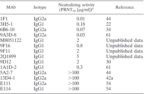
MAbs.The MAbs used in this study are presented in Table 1 and were purified from hybridoma culture supernatants or mouse ascites fluid by using an NAB protein A spin purification kit (Pierce, Rockford, IL). For sorting experiments, MAbs were labeled with Alexa Fluor 647 or Alexa Fluor 488, using a MAb labeling kit (Molecular Probes, Invitrogen, Carlsbad, CA).
Expression of DENV-2 E protein DIII in yeast.The DNA fragment encoding amino acid residues 294 to 409 (DIII) of the DENV-2 E protein was amplified from a DENV-2 strain 16681 infectious cDNA clone (35) by PCR, with BamHI and XhoI sites added at the 5⬘and 3⬘ends, respectively. The PCR products were digested and cloned as downstream fusions to Aga2 and Xpress epitope tag genes in the yeast surface display vector pYD1 (Invitrogen, Carlsbad, CA), under the control of an upstream GAL1 promoter. These constructs were transformed intoSaccharomyces cerevisiaestrain EBY100 (4, 5), using an S.c.EasyComp transformation kit (Invitrogen, Carlsbad, CA), to generate yeast that expressed DENV-2 DIII. Individual yeast colonies were grown to logarithmic phase at 30°C in tryptophan-free yeast medium containing 2% glucose. Fusion protein expres-sion was induced on the surface by growing yeast cells for an additional 48 h in tryptophan-free medium containing 2% galactose at 20°C. Yeast cells were harvested, washed with phosphate-buffered saline (PBS) supplemented with bo-vine serum albumin (1 mg/ml), and stained with 50l of diluted MAbs. Purified antibodies were used at a concentration of 50g/ml, and MAbs from ascites fluid were used at a 1:100 dilution. After a 30-min incubation on ice, yeast cells were washed in PBS with bovine serum albumin and then stained with a goat anti-mouse immunoglobulin G (IgG) secondary antibody conjugated to Alexa Fluor 647 (Molecular Probes, Invitrogen, Carlsbad, CA). After fixation with 1% para-formaldehyde in PBS, yeast cells were analyzed on a FACSCalibur flow cytom-eter (Becton Dickinson, Franklin Lakes, NJ).
Library construction and screening.To generate a random mutant library, DENV-2 DIII was mutated by error-prone PCR, using a GeneMorph II random mutagenesis kit (Stratagene). The mutant library was ligated into the pYD1 vector and transformed into XL2-Blue ultracompetent cells (Stratagene, La Jolla, CA). The colonies were pooled and transformed into yeast cells as de-scribed above.
For each individual MAb, the DENV-2 DIII mutant library was screened according to a previously described protocol (53). To identify yeasts that had selectively lost binding to a given MAb, the library was initially stained with this antibody conjugated to Alexa Fluor 647 for 30 min on ice. To control for the surface expression of DENV-2 DIII, yeast cells were subsequently stained for 30 min on ice with an Alexa Fluor 488-conjugated oligoclonal antibody that was derived from a pool of individual MAbs (1F1, 1A1D-2, 6B6-10, 9A3D-8, 13D4-1, and 5A2-7). After being stained, yeast cells were subjected to flow cytometry, and the population that was single MAb negative but oligoclonal antibody positive was identified. After three or four rounds of sorting, yeast cells were plated and individual colonies were tested for binding to individual MAbs by flow cytometry. For individual clones that had lost only the desired MAb binding epitope, the pYD1-DV2 DIII plasmid was recovered using a Zymoprep yeast miniprep kit (Zymo Research, Orange, CA). The plasmid was transformed into XL1-Blue competent cells (Stratagene, La Jolla, CA), purified using a QIAprep spin mini-prep kit (QIAGEN), and sequenced. In some cases, DENV-2 DIII variants with multiple mutations were isolated. To determine which mutation conferred the phenotype, single independent mutations were engineered by site-directed mu-tagenesis, using a QuikChange II mutagenesis kit (Stratagene, La Jolla, CA), or by using splice-overlap PCR (45).
[image:2.585.301.542.80.237.2]MAb staining of DENV-infected cells.Raji-DC-SIGN-R cells were infected with DENV-1, DENV-2, DENV-3, or DENV-4 at a multiplicity of infection of TABLE 1. Characteristics of DIII-specific MAbs used in this study
MAb Isotype Neutralizing activity (PRNT50[g/ml])a
Reference
1F1 IgG2a 0.01 44
3H5-1 IgG1 0.18 22
6B6-10 IgG2a 0.07 34
9A3D-8 IgG2a 0.03 61
M8051122 IgG1 2 Unpublished data
9F16 IgG1 0.8 Unpublished data
9F11 IgG1 2 Unpublished data
2Q1899 IgG1 5 Unpublished data
9D12 IgG1 2 30
1A1D-2 IgG1 0.3 61
5A2-7 IgG2a ⬎100 44
13D4-1 IgG2a ⬎100 42a
E111 IgG2a ⬎100 54
E114 IgG1 ⬎100 54
aDetermined by PRNT assay on BHK21 cells with 102PFU of DENV-2.
on November 8, 2019 by guest
http://jvi.asm.org/
0.5 or 1. Depending on the strain, cells were harvested at 72 or 96 h, washed three times in PBS, and fixed in PBS with 1% paraformaldehyde. Cells were then washed twice in PBS and permeabilized in Hanks’ balanced salt solution (Cell-gro, Herndon, VA) containing 10 mM HEPES (pH 7.3), 0.1% saponin (Sigma, St. Louis, MO), and 0.02% NaN3(HHSN). MAbs were bound to permeabilized
virus-infected cells for 30 min on ice, washed three times in HHSN, and resus-pended in 50l of a 1/500 dilution of Alexa Fluor-conjugated anti-mouse IgG (Molecular Probes, Invitrogen, Carlsbad, CA). After 15 min, cells were again washed in HHSN three times, fixed in PBS with 1% paraformaldehyde, and processed by flow cytometry.
Mapping of mutations onto the DENV-2 DIII crystal structure and virion.
Figures were prepared using the atomic coordinates of DENV-2 E (Research Collaboratory for Structural Bioinformatics [RCSB] accession number 1OAN) and the WNV E DIII/E16 Fab complex (RCSB accession number 1ZTX), using the programs Ribbons (9), MOLEMAN2 (36), Insight II (Accelrys, San Diego, CA), and PyMol (http://www.pymol.org). The representations of the DENV-2 virion were generated using the atomic coordinates and transformation matrices found in RCSB entry 1THD.
RESULTS
Strongly neutralizing MAbs against WNV localize to an epitope (see Fig. 2A) composed of the loops from three
-strands and an N-terminal region on the lateral ridge of DIII
(2, 14, 51, 53, 64, 74). Sequence analysis indicated that this neutralizing epitope is highly conserved among WNV strains but divergent from those of other flaviviruses (38, 51). To test whether the analogous epitope on DIII of DENV-2 elicited strongly neutralizing antibodies, we screened a panel of 40 MAbs obtained from colleagues for immunoreactivity with DENV-2 strain 16681. Twenty-one of 40 MAbs exhibited sig-nificant reactivity with DENV-2-infected cells or purified re-combinant soluble DENV-2 E protein derived from insect cells (data not shown). Of the MAbs that recognized DENV-2-infected cells, 14 bound strongly to yeast cells displaying DENV-2 DIII (data not shown). Of these, four had moderate
(50% PRNT [PRNT50], 1 to 100 g/ml) and six had strong
(PRNT50⬍1g/ml) neutralizing activities against strain 16681
in a standard PRNT assay (Table 1). Four others had weak or
no appreciable inhibitory activity (PRNT50,⬎100g/ml).
No-tably, five of the six strongly neutralizing MAbs were type specific and showed no significant cross-reactivity with DENV-1-, DENV-3-, or DENV-4-infected cells (Table 2).
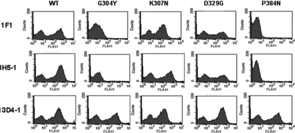
To map the amino acid contact residues of the DIII-specific DENV-2 MAbs with distinct neutralizing properties, we ap-plied a high-throughput strategy that previously mapped 167 WNV E protein-specific MAbs (53, 54). Error-prone PCR mutagenesis introduced random point mutations within DIII
of DENV-2 E. A library of⬃3.5⫻105variants was pooled and
used to create a mutant yeast expression library. Individual screens were performed to identify DIII mutants that lost binding selectively to strongly neutralizing MAbs. To eliminate mutants that abolished surface expression of DIII, yeast cells were stained sequentially with an Alexa Fluor 647-conjugated individual MAb and an Alexa Fluor 488-conjugated oligo-clonal antibody derived from a pool of individual MAbs. After several rounds of cell sorting, yeast cells that selectively lost expression of an individual MAb epitope but retained expres-sion of DIII on the surface were identified. Multiple indepen-dent yeast clones that selectively lost binding of individual MAbs were subjected to plasmid recovery and sequencing. Subsequently, mutant DIII expressed on the yeast surface was tested for MAb reactivity against the remainder of the panel of MAbs by flow cytometry (Fig. 1 and data not shown).
From each screen, we recovered 6 to 20 independent mutants that lost binding for an individual MAb. After sequencing them, we discovered that some of these mutants contained multiple mutations within the DIII region. In such cases, single mutations were engineered separately by site-directed mutagenesis to iden-tify the change that caused the phenotype. Type-specific MAbs that localized to DIII and neutralized DENV-2 strongly (1F1, 3H5-1, 6B6-10, 9A3D-8, and 9F16) showed markedly reduced
binding (⬎80% reduction) when residues G304 (five of five
MAbs), E383 (three of five MAbs), and P384 (four of five MAbs) were altered (Fig. 1 and Table 3). Similarly, type-specific MAbs
that neutralized moderately (PRNT50of 2 to 5g/ml; M8051122,
[image:3.585.43.543.81.243.2]9F11, and 2Q1899) also showed decreased binding when these three residues were changed. G304, E383, and P384 are located on adjacent loops and form a contiguous patch on the solvent-exposed surface at the lateral ridge of DIII (Fig. 2). As observed for DIII-specific neutralizing MAbs against WNV (53), the type-specific neutralizing DENV-2 MAbs bound overlapping epitopes.
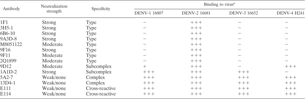
TABLE 2. Binding of MAbs to Raji-DC-SIGN-R cells infected with DENV-1, -2, -3, and -4
Antibody Neutralization
strength Specificity
Binding to virusa
DENV-1 16007 DENV-2 16681 DENV-3 16652 DENV-4 H241
1F1 Strong Type ⫺ ⫹⫹⫹ ⫺ ⫺
3H5-1 Strong Type ⫺ ⫹⫹⫹ ⫺ ⫺
6B6-10 Strong Type ⫺ ⫹⫹⫹ ⫺ ⫺
9A3D-8 Strong Type ⫺ ⫹⫹⫹ ⫺ ⫺
M8051122 Moderate Type ⫺ ⫹⫹⫹ ⫺ ⫺
9F16 Strong Type ⫺ ⫹⫹⫹ ⫺ ⫺
9F11 Moderate Type ⫺ ⫹⫹⫹ ⫺ ⫺
2Q1899 Moderate Type ⫺ ⫹⫹⫹ ⫺ ⫺
9D12 Moderate Subcomplex ⫹ ⫹⫹⫹ ⫺ ⫹⫹⫹
1A1D-2 Strong Subcomplex ⫹⫹⫹ ⫹⫹⫹ ⫹⫹⫹ ⫺
5A2-7 Weak/none Complex ⫹⫹⫹ ⫹⫹⫹ ⫹⫹⫹ ⫹⫹⫹
13D4-1 Weak/none Complex ⫹⫹⫹ ⫹⫹⫹ ⫹⫹⫹ ⫹⫹⫹
E111 Weak/none Cross-reactive ⫹⫹⫹ ⫹⫹⫹ ⫹⫹⫹ ⫹⫹⫹
E114 Weak/none Cross-reactive ⫹⫹⫹ ⫹⫹⫹ ⫹⫹⫹ ⫹⫹⫹
a⫹⫹⫹
, strong binding (40 to 100% compared to control) to infected cells;⫹, weak binding (15 to 40% compared to control) to infected cells;⫺, no appreciable binding detected.
on November 8, 2019 by guest
http://jvi.asm.org/
Consistent with this, the following additional point mutations caused significant loss of binding of individual type-specific neu-tralizing MAbs tested: T303Y (1F1), K307E (9A3D-8), E327R (9A3D-8), D329R (6B6-10), G330D (1F1 and 6B6-10), and S331Y (6B6-10). Mutation of G304, which appears to comprise part of a type-specific neutralizing epitope on DENV-2 DIII, also affected binding of both subcomplex-specific MAbs (9D12 and 1A1D-2) and one non- or weakly neutralizing MAb (E114). Be-cause binding of several MAbs of different classes was affected by the G304 mutation, we cannot absolutely exclude perturbations of the structure of this mutant DIII. Against this, we did observe relatively wild-type levels of binding of two other nonneutralizing MAbs (13D4-1 and E111) to this mutant.
In our panel of MAbs, we also identified two neutralizing MAbs (9D12 and 1A1D-2) that reacted with additional DENV serotypes. These subcomplex-specific MAbs showed moderate and strong inhibitory activities against DENV-2 infection, re-spectively (Tables 1 and 2). These MAbs, however, bound DIII somewhat distinctly. 1A1D-2, which binds DENV-1, DENV-2, and DENV-3, retained relatively normal binding with E383G and P384N mutations but showed markedly reduced binding with mutations in residues K305, K307, and K310. In compar-ison, 9D12, which binds DENV-2 and DENV-4 and weakly binds DENV-1, retained binding with the E383G and P384A mutations but had markedly reduced binding with K305E, K307E, K310E, and P384N mutations (Table 3).
Also in our panel were four MAbs (5A2-7, 13D4-1, E111, and E114) that had little or no neutralizing activity (Table 1). This group of MAbs recognized all four serotypes of DENV (Table 2). Two of them, E111 and E114, were cross-reactive and also recognized yeast expressing WNV DIII (data not shown). Independent yeast sorting for loss-of-binding mutations was performed with three of the four MAbs. Several mutations that specifically reduced binding
of the poorly neutralizing MAbs but had little effect on most strongly neutralizing MAbs were identified. For example, H317Y mutation specifically reduced binding of the 5A2-7, 13D4-1, and E111 MAbs (Table 3), and a DIII variant with two mutations (T315G and S331Y) also diminished binding of these MAbs but did not affect any neutralizing MAbs, with the exception of 6B6-10. Interestingly, the T315G sin-gle mutant only modestly decreased 5A2-7 (71% reduction) binding, and the S331Y single mutant had no effect on binding of 5A2-7, 13D4-1, or E111.
To visualize spatially the different recognition patterns of MAbs that strongly, moderately, and weakly neutralized DENV-2 infection, we docked the loss-of-binding mutations
(⬍20% of wild-type binding) defined by the yeast assay onto
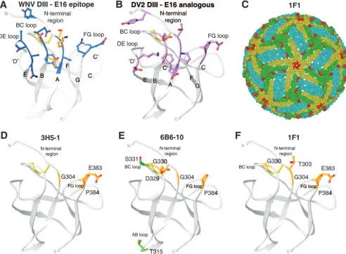
[image:4.585.47.541.69.292.2]the existing crystallographic structure of DENV-2 DIII, the prefusion DENV-2 E protein dimer structure (46), and the pseudoatomic model of the mature DENV-2 virion (37). The DIII structures were compared to the previously defined crys-tallographic epitope (16 contact residues) of E16 on the lateral ridge of DIII of WNV (51) (Fig. 2A and B). Type-specific MAbs with the strongest neutralizing activities (3H5-1, 6B6-10, and 1F1) localized to amino acids on the analogous lateral ridge of DIII (Fig. 2D, E, and F). Nonetheless, the yeast mapping did suggest some subtle differences. Whereas the lateral ridge epitope of the strongly neutralizing anti-WNV MAb E16 was centered on the N-terminal region and the BC loop (K307, T330, and T332 of WNV), strongly neutralizing type-specific anti-DENV-2 MAbs localized more to the FG loop (E383 and P384 of DENV-2). The E383 and P384 resi-dues in the FG loop are highly conserved among DENV-2 isolates but are not present in DENV-1, DENV-3, or DENV-4 strains (Fig. 3). The epitope of type-specific neutralizing MAbs against DENV-2 followed the same exposure pattern on the virion as that previously identified for E16 (34, 51): two of the
FIG. 1. Flow cytometry histograms of loss-of-function DIII variants (G304Y, K307N, D329G, and P384N) selected by yeast surface display after being sorted with MAbs. Representative histograms are shown for MAbs 1F1, 3H5-1, and 13D4-1 with wild-type DIII (WT) and each of the DIII mutants. Data shown are representative of three independent experiments.
on November 8, 2019 by guest
http://jvi.asm.org/
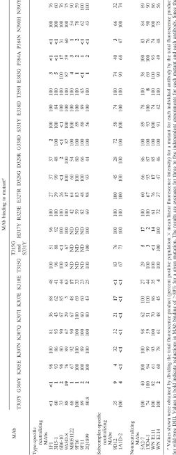
TABLE 3. Summary of MAb binding to DENV-2 DIII mutants expressed on the surfaces of yeast cells MAb MAb binding to mutant a T303Y G304Y K305E K307N K307Q K307I K307E K310E T315G
T315G and S331Y
H317Y R323E E327R D329G D329R G330D S331Y E338D T359I E383G P384A P384N N390H N390Y Type-specific neutralizing MAbs 1F1 <1 <1 98 100 81 36 88 48 100 51 96 100 27 37 37 2 100 100 100 3< 1 < 1 100 76 3H5-1 60 1 93 86 93 43 62 41 96 88 92 100 39 99 48 100 100 84 100 5< 1 < 1 100 60 6B6-10 33 2 98 80 90 67 85 44 100 <1 100 100 26 <1 2 <1 <1 100 100 100 17 31 100 76 9A3D-8 88 19 76 89 67 29 5 63 83 67 100 100 17 100 100 87 100 100 100 87 59 60 100 75 M8051122 68 1 67 92 90 67 48 17 ND ND ND 42 64 60 54 100 100 100 100 43 1 54 90 9F16 100 1 100 100 100 100 69 33 ND ND ND 59 83 100 80 100 89 100 100 12 2 78 59 9F11 89 2 100 89 100 60 60 23 ND ND ND 82 48 100 66 100 88 89 63 <1 1 <1 62 100 2Q1899 80.8 2 100 100 100 80 43 25 100 100 100 69 98 93 44 100 56 100 100 12 < 1 43 100 Subcomplex-specific neutralizing MAbs 9D12 35 94 32 32 21 17 <1 83 76 100 100 100 45 28 72 58 74 100 74 40 3 66 32 1A1D-2 100 4< 1 < 1 < 1 < 1 < 1 < 1 67 73 100 100 100 100 100 100 100 100 100 90 68 47 100 74 Nonneutralizing MAbs 5A2-7 100 40 100 100 98 62 100 27 29 51 7 100 60 66 86 100 89 78 100 38 100 83 84 89 13D4-1 74 100 94 99 59 51 100 44 100 32 100 67 93 87 100 93 100 8 69 100 76 90 90 WN E111 100 92 61 93 100 70 86 35 100 <1 14 61 76 17 85 100 100 74 100 100 93 74 100 86 WN E114 100 2 60 78 61 48 45 4 100 100 100 72 37 47 46 100 91 42 100 90 49 48 75 56 aValues shown were obtained by dividing the total fluorescence product (percent positive population ⫻ mean linear fluorescence intensity) for a mutant for each individual antibody by the total fluorescence product for wild-type DIII. Values in bold indicate reductions in MAb binding of ⬎ 80% for a given mutation. The results are averages for three to five independent experiments for each mutant and each antibody. Since the G304Y mutant had slightly less surface expression on yeast cells, the values for this mutant only were normalized to the strongest binding antibody fo r that mutant ⫻ 100. ND, not determined.
on November 8, 2019 by guest
http://jvi.asm.org/
[image:5.585.168.421.71.725.2]three DIIIs per icosahedral asymmetric unit were exposed, with steric hindrance noted at the fivefold clustered DIII (Fig. 2C).
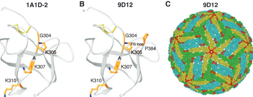
Subcomplex-specific MAbs (1A1D-2 and 9D12) that strongly and moderately neutralized DENV-2 infection recognized a flanking epitope (Fig. 4A and B). This epitope was centered on amino acids K305, K307, and K310 on the A strand. Consistent with the limited cross-reactivity of these subcomplex-specific MAbs, K310 is completely conserved among all four DENV se-rotypes, whereas K305 and K307 are conserved in DENV-4 and DENV-1 strains, respectively (Fig. 3). Although the 1A1D-2 and 9D12 MAb epitopes are reasonably exposed in all three symmetry environments on the mature virion (Fig. 4C and data not shown), some differences with the type-specific lateral ridge epitope were apparent, as follows: the 9D12 epitope appears predominantly exposed on the fivefold clustered DIII, and thus, one could spec-ulate that a full complement of 180 Fabs could bind DENV-2 at saturation.
Several of the poorly neutralizing, cross-reactive MAbs (5A2-7, 13D4-1, and E111) mapped to an amino acid residue (e.g., H317) that was localized to the back side of DIII in the AB loop (Fig. 5A and B). Consistent with the cross-reactive nature of these MAbs, the entire AB loop sequence (E314, T315, Q316, H317, G318, and T319) is completely conserved among all four DENV serotypes. Moreover, the H317, G318, and T319 residues are present in virtually all WNV isolates and the H317 and T319 amino acids are conserved among flavivi-ruses (Fig. 3 and data not shown). Structurally, the AB loop has limited exposure on the surface of the E protein dimer and faces inward toward the nucleocapsid in all three symmetry environments of the mature virion (Fig. 5C and Fig. 6).
DISCUSSION
[image:6.585.45.537.70.432.2]The goal of this study was to characterize epitopes on DIII recognized by potent neutralizing antibodies against DENV-2.
FIG. 2. DIII lateral ridge antibody epitope. The structures of WNV and DENV-2 DIIIs are shown, with identification of binding sites of type-specific neutralizing antibodies. (A) Structure of WNV E16 neutralizing epitope determined by X-ray crystallography (16 residues in blue) or by using yeast display mapping (4 residues in orange). (B) Structure of DENV-2 DIII, with the corresponding 16 amino acids of the WNV E16 neutralizing antibody epitope highlighted. (C) Yeast display epitope residues (red) for the 1F1 MAb were mapped onto the pseudoatomic model of the mature DENV-2 virion (37). Virions are depicted as 2.0-Å-radius C-␣atoms and are colored according to their E protein symmetry relationships, i.e., twofold (cyan), threefold (green), or fivefold (yellow) symmetry. (D to F) Structure of DENV DIII, with amino acid residues that significantly affect binding of type-specific neutralizing MAbs 3H5-1 (D), 6B6-10 (E), and 1F1 (F) marked in orange. The DIII disulfide bond is depicted in yellow.
on November 8, 2019 by guest
http://jvi.asm.org/
Previous studies had established a dominant type-specific epitope for eliciting protective antibodies in vitro and in vivo against the related flavivirus WNV. Here we tested a panel of MAbs against DIII of the DENV-2 E protein. Type-specific strongly neutralizing MAbs mapped to an analogous epitope centered on the FG loop of the lateral ridge region on DIII of DENV-2. Subcomplex-specific strongly neutralizing MAbs lo-calized to a flanking epitope that was centered on three lysine residues in the A strand of DIII.
Extensive MAb competition binding studies have been per-formed by several groups to identify distinct antigenic and functional determinants on DENV-2 (18, 22, 29, 30, 61). Po-tently inhibitory type-specific MAbs were localized to domain B, which is now called DIII, based on structural analysis of the
[image:7.585.44.542.67.218.2]domain organization of flavivirus E proteins (46, 60). None-theless, amino acid contact residues of few neutralizing MAbs that react with DENV-2 have been established. For these (3H5-1, 4E11, G8D11, and 4G2), precise mapping data were obtained by analyzing neutralization escape mutants (39) and by differential recognition of chimeric DENV variants (32), site-specific DENV-2 mutants (17, 67), and E protein peptide sequences (44, 70, 72). We used a forward genetic strategy, error-prone PCR mutagenesis of DIII of DENV-2 E protein, and expression on yeast cells to map antibody contact residues in a nonbiased manner. By having a panel of DIII MAbs with differing neutralization potentials, we minimized the possibility that mutations would grossly affect folding. The validity of the yeast approach for identifying critical contact residues was
FIG. 3. DIII amino acid sequence alignment. The sequence and secondary structure of DIII from the DENV-2 (strain 16681) E protein are aligned with those for DENV-1 (strain 16007), DENV-3 (strain 16652), DENV-4 (strain 1036), and WNV (New York 1999). The secondary structure of DENV-2 E DIII residues 294 to 395 (RCSB entry 1OAN) was predicted by DSSP (33). The results of yeast surface display epitope mapping are highlighted, with DENV-2 residues recognized primarily by type-specific MAbs colored magenta, subcomplex-specific residues colored green, and cross-reactive residues colored orange. The residues contacted by E16 on WNV DIII, as determined by crystallography, are colored blue (51), with black asterisks denoting residues identified by yeast display (53). Colored asterisks denote DENV-2 residues that are recognized by multiple classes of antibodies. For example, G304Y mutation resulted in a loss of binding of all type- and subcomplex-specific MAbs and a single cross-reactive MAb.
FIG. 4. DIII A-strand epitope. The structure of DENV-2 DIII is shown, with identification of binding sites of subcomplex-specific neutralizing antibodies. (A and B) Structure of DENV DIII, with amino acid residues that significantly affect binding of subcomplex-specific neutralizing MAbs 1A1D-2 (A) and 9D12 (B) marked in orange. (C) Yeast display epitope residues (red) for the 9D12 MAb were mapped onto the pseudoatomic model of the mature DENV-2 virion. Virions are depicted as described in the legend to Fig. 2.
on November 8, 2019 by guest
http://jvi.asm.org/
[image:7.585.54.540.498.685.2]confirmed by X-ray crystallographic studies that resolved the structural interface between DIII and a neutralizing anti-WNV Fab fragment (51). Of the DENV-2 MAbs that have been reported to contact specific amino acid residues in DIII, only the type-specific MAb 3H5-1 was available for our analysis. Prior fine mapping studies suggested that 3H5-1 recognized either a Glu-Pro-Gly motif centered at amino acids 383, 384, and 385 (32) or a linear peptide encompassing amino acids 386
to 397 (32, 72). Our yeast mapping experiments confirmed an essential role for residues E383 and P384 but also suggested an additional important contact residue (G304) located on an adjacent strand.
DIII of the E protein adopts an immunoglobulin-like fold (46, 60) that is significantly exposed on the surface of the mature virion (50, 78). The lateral ridge epitope on DIII was previously defined by X-ray crystallographic and nuclear
[image:8.585.52.540.70.246.2]mag-FIG. 5. DIII AB loop epitope. The structure of DENV-2 DIII is shown, with identification of binding sites of poorly neutralizing antibodies. (A and B) Structure of DENV DIII, with amino acid residues that significantly affect binding of the poorly neutralizing MAbs E111 (A) and 13D4-1 (B) marked in orange. (C) Yeast display epitope residues (red) for the 13D4-1 MAb were mapped onto the pseudoatomic model of the mature DENV-2 virion. Virions are depicted as described in the legend to Fig. 2. Note that the 13D4-1 epitope is poorly accessible on the virion compared to the 1F1 (Fig. 2C) and 9D12 (Fig. 4C) epitopes.
FIG. 6. Mapping of MAb epitopes onto the DENV-2 E protein dimer. The yeast display epitope residues (in red) for the type-specific 1F1 (A), subcomplex-specific 9D12 (B), and cross-reactive 13D4-1 (C) MAbs were rendered on the crystal structure of the DENV-2 E protein dimer (46) and are shown in side view, with the bottom side facing the viral lipid membrane.
on November 8, 2019 by guest
http://jvi.asm.org/
[image:8.585.135.448.428.694.2]netic resonance studies of Fab-DIII complexes of WNV and Japanese encephalitis virus and encompasses four discontinu-ous loops (51, 74, 75). In our study, we found six different strongly neutralizing MAbs against DENV-2 that localized to two overlapping structural epitopes on the lateral ridge and adjacent A strand of DIII.
For flaviviruses, virus type-specific epitopes generally elicit the most potent neutralizing antibodies (2, 28, 42, 53, 56, 63, 64). Of the DIII-specific MAbs against DENV-2 in our panel, in general, the ones with the strongest neutralizing activities were type specific and localized to the lateral ridge of DIII centered at the FG loop, near residues E383 and P384. None-theless, some of the type-specific neutralizing MAbs inhibited virus infectivity less strongly, although they recognized similar residues. Although further biophysical studies are needed, the affinities of binding of MAbs for a given DENV-2 DIII epitope may correlate with relative occupancy and may predict the strength of neutralization. Such a result was observed with less strongly neutralizing MAbs that recognized the lateral ridge epitope of DIII of WNV (57).
The two subcomplex-specific (1A1D-2 [PRNT50, 0.3g/ml]
and 9D12 [PRNT50, 2g/ml]) MAbs that we tested had strong
and moderate neutralizing activities, respectively, consistent
with prior studies with the group-specific MAb 4E11 (PRNT50
values of 0.3 to 2.4 g/ml for DENV-1 to DENV-4) (71).
1A1D-2 and 9D12 bind a more conserved epitope among DENV serotypes that partially overlaps with the type-specific lateral ridge neutralizing epitope but is centered on the A strand at residues K305, K307, and K310. Notably, several of these amino acids were recently identified as contact residues for the MAb 4E11, which neutralizes all four DENV serotypes (41, 70). Such broadly neutralizing subcomplex- or group-spe-cific MAbs may have potential for development as antibody-based therapeutics against all serotypes of DENV.
An epitope map analysis of the different DENV-2 MAbs at the amino acid sequence level begins to explain their serotype-specific properties. Type-serotype-specific neutralizing MAbs against DENV-2 are centered on amino acids E383 and P384 in the FG loop, which are conserved among DENV-2 isolates but divergent in all other DENV serotypes. Subcomplex-specific neutralizing MAbs that bind some but not all DENV serotypes recognize a distinct epitope centered on three A-strand lysines of DIII. K310 is completely conserved among all four DENV serotypes, whereas K305 and K307 are conserved in DENV-4 and DENV-1 strains, respectively. The distinct binding speci-ficities of 9D12 and 1A1D-2 for other DENV serotypes likely reflect differences in interactions with specific lysines on the A strand and the divergence of other additional contact residues in the FG loop and the G strand. Cross-reactive poorly neu-tralizing MAbs localized to the AB loop on DIII. Their lack of neutralizing activity is probably explained by the limited sur-face exposure of the AB loop. As seen for the DIII-specific poorly neutralizing anti-WNV MAb E9, a relative lack of ac-cessibility directly affects the stoichiometry of binding, such that a threshold for antibody neutralization is rarely reached (57). The cross-reactivity of these AB loop MAbs with all DENV serotypes and distantly related flaviviruses is reason-ably explained by sequence conservation: amino acids 314 to 319 are completely conserved among all four DENV serotypes, and residues 317 and 319 (which include the critical H317
amino acid) are conserved in virtually all flaviviruses. This analysis has implications for the development of novel DIII epitope-based diagnostic reagents for polyclonal antibodies, as mutation of AB loop sequences could abolish serotype and flavivirus nonneutralizing cross-reactive epitopes, making DIII-based immunologic assays more predictive of serotype-specific neutralizing antibodies.
Although the sequence of the lateral ridge epitope of DIII is variable among flaviviruses, the majority of mutations that abolish binding of virus-specific strongly neutralizing antibod-ies map there (2, 10, 12, 18, 53, 61, 63, 64, 67, 75), suggesting the existence of a type-specific neutralizing epitope for flavivi-ruses on DIII. It is important, however, that these epitope maps were derived using murine MAbs. Whether the human antibody response recognizes the same or different epitopes has not yet been determined in detail. Only 8% (4 of 51 antibodies) and 0% (0 of 11 antibodies) of human single-chain antibodies that were isolated from phage display libraries of WNV-infected or naı¨ve patients reacted with DIII (26, 69). These results suggest that the human antibody repertoire against flaviviruses may actually be directed away from these DIII neutralizing epitopes and toward the inherently less neu-tralizing immunodominant epitopes on DI and DII (55). Since our results suggest that strongly neutralizing antibodies against DIII of DENV-2 map to the lateral ridge and A-strand epitopes, flavivirus DIII-based vaccines (13, 15, 31) that inten-tionally skew the humoral response to these epitopes and away from the cross-reactive, poorly accessible epitope in the AB loop could elicit a greater protective response.
ACKNOWLEDGMENTS
We thank C. Nelson for help with structural analysis of DENV E proteins, members of our laboratories for critical reviews of the manu-script, and R. Putnak for providing several of the MAbs used in this study.
This work was supported by the Pediatric Dengue Vaccine Initiative (M.S.D., D.H.F., J.T.R., J.J.S., and A.D.B.) and by NIH grants AI061373 (M.S.D.) and U54 AI057160 (Midwest Regional Center of Excellence for Biodefense and Emerging Infectious Diseases Re-search).
REFERENCES
1.Beasley, D. W., and J. G. Aaskov.2001. Epitopes on the dengue 1 virus envelope protein recognized by neutralizing IgM monoclonal antibodies. Virology279:447–458.
2.Beasley, D. W., and A. D. Barrett. 2002. Identification of neutralizing epitopes within structural domain III of the West Nile virus envelope pro-tein. J. Virol.76:13097–13100.
3.Bhardwaj, S., M. Holbrook, R. E. Shope, A. D. Barrett, and S. J. Watowich.
2001. Biophysical characterization and vector-specific antagonist activity of domain III of the tick-borne flavivirus envelope protein. J. Virol.75:4002– 4007.
4.Boder, E. T., and K. D. Wittrup.1998. Optimal screening of surface-dis-played polypeptide libraries. Biotechnol. Prog.14:55–62.
5.Boder, E. T., and K. D. Wittrup.1997. Yeast surface display for screening combinatorial polypeptide libraries. Nat. Biotechnol.15:553–557. 6.Brandriss, M. W., J. J. Schlesinger, E. E. Walsh, and M. Briselli.1986.
Lethal 17D yellow fever encephalitis in mice. I. Passive protection by mono-clonal antibodies to the envelope proteins of 17D yellow fever and dengue 2 viruses. J. Gen. Virol.67:229–234.
7.Bressanelli, S., K. Stiasny, S. L. Allison, E. A. Stura, S. Duquerroy, J. Lescar, F. X. Heinz, and F. A. Rey.2004. Structure of a flavivirus envelope glyco-protein in its low-pH-induced membrane fusion conformation. EMBO J.
23:728–738.
8.Burke, D. S., and T. P. Monath.2001. Flaviviruses, p. 1043–1125.InD. M. Knipe, P. M. Howley, D. E. Griffin, R. A. Lamb, M. A. Martin, B. Roizman, and S. E. Straus (ed.), Fields virology, 4th ed., vol. 1. Lippincott Williams & Wilkins, Philadelphia, PA.
on November 8, 2019 by guest
http://jvi.asm.org/
9.Carson, M.1987. Ribbon models of macromolecules. J. Mol. Graphics
5:103–106.
10.Cecilia, D., and E. A. Gould.1991. Nucleotide changes responsible for loss of neuroinvasiveness in Japanese encephalitis virus neutralization-resistant mu-tants. Virology181:70–77.
11.Chambers, T. J., C. S. Hahn, R. Galler, and C. M. Rice.1990. Flavivirus genome organization, expression, and replication. Annu. Rev. Microbiol.
44:649–688.
12.Chambers, T. J., M. Halevy, A. Nestorowicz, C. M. Rice, and S. Lustig.1998. West Nile virus envelope proteins: nucleotide sequence analysis of strains differing in mouse neuroinvasiveness. J. Gen. Virol.79:2375–2380. 13.Chen, S., M. Yu, T. Jiang, Y. Deng, C. Qin, and E. Qin.2007. Induction of
tetravalent protective immunity against four dengue serotypes by the tandem domain III of the envelope protein. DNA Cell Biol.26:361–367. 14.Choi, K. S., J. J. Nah, Y. J. Ko, Y. J. Kim, and Y. S. Joo.2007. The DE loop
of the domain III of the envelope protein appears to be associated with West Nile virus neutralization. Virus Res.123:216–218.
15.Chu, J. H., C. C. Chiang, and M. L. Ng.2007. Immunization of flavivirus West Nile recombinant envelope domain III protein induced specific im-mune response and protection against West Nile virus infection. J. Immunol.
178:2699–2705.
16.Colombage, G., R. Hall, M. Pavy, and M. Lobigs.1998. DNA-based and alphavirus-vectored immunisation with prM and E proteins elicits long-lived and protective immunity against the flavivirus, Murray Valley encephalitis virus. Virology250:151–163.
17.Crill, W. D., and G. J. Chang.2004. Localization and characterization of flavivirus envelope glycoprotein cross-reactive epitopes. J. Virol.78:13975– 13986.
18.Crill, W. D., and J. T. Roehrig.2001. Monoclonal antibodies that bind to domain III of dengue virus E glycoprotein are the most efficient blockers of virus adsorption to Vero cells. J. Virol.75:7769–7773.
19.Diamond, M. S., D. Edgil, T. G. Roberts, B. Lu, and E. Harris.2000. Infection of human cells by dengue virus is modulated by different cell types and viral strains. J. Virol.74:7814–7823.
20.Diamond, M. S., E. Sitati, L. Friend, B. Shrestha, S. Higgs, and M. Engle.
2003. Induced IgM protects against lethal West Nile virus infection. J. Exp. Med.198:1–11.
21.Falconar, A. K. 1999. Identification of an epitope on the dengue virus membrane (M) protein defined by cross-protective monoclonal antibodies: design of an improved epitope sequence based on common determinants present in both envelope (E and M) proteins. Arch. Virol.144:2313–2330. 22.Gentry, M. K., E. A. Henchal, J. M. McCown, W. E. Brandt, and J. M.
Dalrymple.1982. Identification of distinct antigenic determinants on den-gue-2 virus using monoclonal antibodies. Am. J. Trop. Med. Hyg.31:548– 555.
23.Goncalvez, A. P., R. Men, C. Wernly, R. H. Purcell, and C. J. Lai.2004. Chimpanzee Fab fragments and a derived humanized immunoglobulin G1 antibody that efficiently cross-neutralize dengue type 1 and type 2 viruses. J. Virol.78:12910–12918.
24.Goncalvez, A. P., R. H. Purcell, and C. J. Lai.2004. Epitope determinants of a chimpanzee Fab antibody that efficiently cross-neutralizes dengue type 1 and type 2 viruses map to inside and in close proximity to fusion loop of the dengue type 2 virus envelope glycoprotein. J. Virol.78:12919–12928. 25.Gould, E. A., A. Buckley, A. D. Barrett, and N. Cammack.1986. Neutralizing
(54K) and non-neutralizing (54K and 48K) monoclonal antibodies against structural and non-structural yellow fever virus proteins confer immunity in mice. J. Gen. Virol.67:591–595.
26.Gould, L. H., J. Sui, H. Foellmer, T. Oliphant, T. Wang, M. Ledizet, A. Murakami, K. Noonan, C. Lambeth, K. Kar, J. F. Anderson, A. M. de Silva, M. S. Diamond, R. A. Koski, W. A. Marasco, and E. Fikrig.2005. Protective and therapeutic capacity of human single-chain Fv-Fc fusion proteins against West Nile virus. J. Virol.79:14606–14613.
27.Halstead, S. B.1988. Pathogenesis of dengue: challenges to molecular biol-ogy. Science239:476–481.
28.Heinz, F. X.1986. Epitope mapping of flavivirus glycoproteins. Adv. Virus Res.31:103–168.
29.Henchal, E. A., M. K. Gentry, J. M. McCown, and W. E. Brandt.1982. Dengue virus-specific and flavivirus group determinants identified with monoclonal antibodies by indirect immunofluorescence. Am. J. Trop. Med. Hyg.31:830–836.
30.Henchal, E. A., J. M. McCown, D. S. Burke, M. C. Seguin, and W. E. Brandt.
1985. Epitopic analysis of antigenic determinants on the surface of dengue-2 virions using monoclonal antibodies. Am. J. Trop. Med. Hyg.34:162–169. 31.Hermida, L., L. Bernardo, J. Martin, M. Alvarez, I. Prado, C. Lopez, L.
Sierra Bde, R. Martinez, R. Rodriguez, A. Zulueta, A. B. Perez, L. Lazo, D. Rosario, G. Guillen, and M. G. Guzman.2006. A recombinant fusion protein containing the domain III of the dengue-2 envelope protein is immunogenic and protective in nonhuman primates. Vaccine24:3165–3171.
32.Hiramatsu, K., M. Tadano, R. Men, and C. J. Lai.1996. Mutational analysis of a neutralization epitope on the dengue type 2 virus (DEN2) envelope protein: monoclonal antibody resistant DEN2/DEN4 chimeras exhibit re-duced mouse neurovirulence. Virology224:437–445.
33.Kabsch, W., and C. Sander.1983. Dictionary of protein secondary structure: pattern recognition of hydrogen-bonded and geometrical features. Biopoly-mers22:2577–2637.
34.Kauffman, B., G. Nybakken, P. R. Chipman, W. Zhang, D. H. Fremont, M. S. Diamond, R. J. Kuhn, and M. G. Rossmann. 2006. West Nile virus in complex with a neutralizing monoclonal antibody. Proc. Natl. Acad. Sci. USA103:12400–12404.
35.Kinney, R. M., S. Butrapet, G. J. Chang, K. R. Tsuchiya, J. T. Roehrig, N. Bhamarapravati, and D. J. Gubler.1997. Construction of infectious cDNA clones for dengue 2 virus: strain 16681 and its attenuated vaccine derivative, strain PDK-53. Virology230:300–308.
36.Kleywegt, G. J.1997. Validation of protein models from Calpha coordinates alone. J. Mol. Biol.273:371–376.
37.Kuhn, R. J., W. Zhang, M. G. Rossmann, S. V. Pletnev, J. Corver, E. Lenches, C. T. Jones, S. Mukhopadhyay, P. R. Chipman, E. G. Strauss, T. S. Baker, and J. H. Strauss.2002. Structure of dengue virus: implications for flavivirus organization, maturation, and fusion. Cell108:717–725. 38.Li, L., A. D. Barrett, and D. W. Beasley.2005. Differential expression of
domain III neutralizing epitopes on the envelope proteins of West Nile virus strains. Virology335:99–105.
39.Lin, B., C. R. Parrish, J. M. Murray, and P. J. Wright.1994. Localization of a neutralizing epitope on the envelope protein of dengue virus type 2. Virology202:885–890.
40.Lin, C. W., and S. C. Wu.2003. A functional epitope determinant on domain III of the Japanese encephalitis virus envelope protein interacted with neu-tralizing-antibody combining sites. J. Virol.77:2600–2606.
41.Lisova, O., F. Hardy, V. Petit, and H. Bedouelle.2007. Mapping to com-pleteness and transplantation of a group-specific, discontinuous, neutralizing epitope in the envelope protein of dengue virus. J. Gen. Virol.88:2387–2397. 42.Mandl, C. W., F. Guirakhoo, H. Holzmann, F. X. Heinz, and C. Kunz.1989. Antigenic structure of the flavivirus envelope protein E at the molecular level, using tick-borne encephalitis virus as a model. J. Virol.63:564–571. 42a.Mason, P. W., M. U. Zu¨gel, A. R. Semproni, M. J. Fournier, and T. L.
Mason.1990. The antigenic structure of dengue type 1 virus envelope and NS1 proteins expressed inEscherichia coli. J. Gen. Virol.71:2107–2114. 43.Mathews, J. H., and J. T. Roehrig.1984. Elucidation of the topography and
determination of the protective epitopes on the E glycoprotein of Saint Louis encephalitis virus by passive transfer with monoclonal antibodies. J. Immu-nol.132:1533–1537.
44.Megret, F., J. P. Hugnot, A. Falconar, M. K. Gentry, D. M. Morens, J. M. Murray, J. J. Schlesinger, P. J. Wright, P. Young, M. H. Van Regenmortel, et al.1992. Use of recombinant fusion proteins and monoclonal antibodies to define linear and discontinuous antigenic sites on the dengue virus enve-lope glycoprotein. Virology187:480–491.
45.Misulovin, Z., X. W. Yang, W. Yu, N. Heintz, and E. Meffre.2001. A rapid method for targeted modification and screening of recombinant bacterial artificial chromosome. J. Immunol. Methods257:99–105.
46.Modis, Y., S. Ogata, D. Clements, and S. C. Harrison.2003. A ligand-binding pocket in the dengue virus envelope glycoprotein. Proc. Natl. Acad. Sci. USA
100:6986–6991.
47.Modis, Y., S. Ogata, D. Clements, and S. C. Harrison.2004. Structure of the dengue virus envelope protein after membrane fusion. Nature427:313–319. 48.Monath, T. P.1994. Dengue: the risk to developed and developing countries.
Proc. Natl. Acad. Sci. USA91:2395–2400.
49.Morita, K., M. Tadano, S. Nakaji, K. Kosai, E. G. Mathenge, B. D. Pandey, F. Hasebe, S. Inoue, and A. Igarashi.2001. Locus of a virus neutralization epitope on the Japanese encephalitis virus envelope protein determined by use of long PCR-based region-specific random mutagenesis. Virology287:
417–426.
50.Mukhopadhyay, S., B. S. Kim, P. R. Chipman, M. G. Rossmann, and R. J. Kuhn.2003. Structure of West Nile virus. Science302:248.
51.Nybakken, G., T. Oliphant, S. Johnson, S. Burke, M. S. Diamond, and D. H. Fremont.2005. Structural basis for neutralization of a therapeutic antibody against West Nile virus. Nature437:764–769.
52.Nybakken, G. E., C. A. Nelson, B. R. Chen, M. S. Diamond, and D. H. Fremont.2006. Crystal structure of the West Nile virus envelope glycopro-tein. J. Virol.80:11467–11474.
53.Oliphant, T., M. Engle, G. Nybakken, C. Doane, S. Johnson, L. Huang, S. Gorlatov, E. Mehlhop, A. Marri, K. M. Chung, G. D. Ebel, L. D. Kramer, D. H. Fremont, and M. S. Diamond.2005. Development of a humanized monoclonal antibody with therapeutic potential against West Nile virus. Nat. Med.11:522–530.
54.Oliphant, T., G. Nybakken, M. Engle, Q. Xu, C. A. Nelson, S. Sukupolvi-Petty, A. Marri, B. Lachmi, U. Olshevsky, D. H. Fremont, T. C. Pierson, and M. S. Diamond.
2006. Determinants of West Nile virus envelope protein domain I and II antibody recognition and neutralization. J. Virol.80:12149–12159.
55.Oliphant, T., G. E. Nybakken, S. K. Austin, Q. Xu, J. Bramson, M. Loeb, M. Throsby, D. H. Fremont, T. C. Pierson, and M. S. Diamond.2007. Induction of epitope-specific neutralizing antibodies against West Nile virus. J. Virol.
81:11828–11839.
56.Peiris, J. S. M., J. S. Porterfield, and J. T. Roehrig.1982. Monoclonal antibodies against the flavivirus West Nile. J. Gen. Virol.58:283–289.
on November 8, 2019 by guest
http://jvi.asm.org/
57.Pierson, T. C., Q. Xu, S. Nelson, T. Oliphant, G. E. Nybakken, D. H. Fremont, and M. S. Diamond.2007. The stoichiometry of antibody-mediated neutralization and enhancement of West Nile virus infection. Cell Host Microbe1:135–145.
58.Pincus, S., P. W. Mason, E. Konishi, B. A. Fonseca, R. E. Shope, C. M. Rice, and E. Paoletti.1992. Recombinant vaccinia virus producing the prM and E proteins of yellow fever virus protects mice from lethal yellow fever enceph-alitis. Virology187:290–297.
59.Rey, F. A.2003. Dengue virus envelope glycoprotein structure: new insight into its interactions during viral entry. Proc. Natl. Acad. Sci. USA100:6899– 6901.
60.Rey, F. A., F. X. Heinz, C. Mandl, C. Kunz, and S. C. Harrison.1995. The envelope glycoprotein from tick-borne encephalitis virus at 2 angstrom resolution. Nature375:291–298.
61.Roehrig, J. T., R. A. Bolin, and R. G. Kelly.1998. Monoclonal antibody mapping of the envelope glycoprotein of the dengue 2 virus, Jamaica. Vi-rology246:317–328.
62.Roehrig, J. T., J. H. Mathews, and D. W. Trent. 1983. Identification of epitopes on the E glycoprotein of Saint Louis encephalitis virus using mono-clonal antibodies. Virology128:118–126.
63.Roehrig, J. T., L. A. Staudinger, A. R. Hunt, J. H. Mathews, and C. D. Blair.
2001. Antibody prophylaxis and therapy for flaviviral encephalitis infections. Ann. N. Y. Acad. Sci.951:286–297.
64.Sanchez, M. D., T. C. Pierson, D. McAllister, S. L. Hanna, B. A. Puffer, L. E. Valentine, M. M. Murtadha, J. A. Hoxie, and R. W. Doms.2005. Charac-terization of neutralizing antibodies to West Nile virus. Virology336:70–82. 65.Schlesinger, J. J., S. Chapman, A. Nestorowicz, C. M. Rice, T. E. Ginocchio, and T. J. Chambers.1996. Replication of yellow fever virus in the mouse central nervous system: comparison of neuroadapted and non-neuroadapted virus and partial sequence analysis of the neuroadapted strain. J. Gen. Virol.
77:1277–1285.
66.Seif, S. A., K. Morita, S. Matsuo, F. Hasebe, and A. Igarashi.1995. Finer mapping of neutralizing epitope(s) on the C-terminal of Japanese enceph-alitis virus E-protein expressed in recombinant Escherichia coli system. Vac-cine13:1515–1521.
67.Serafin, I. L., and J. G. Aaskov.2001. Identification of epitopes on the envelope (E) protein of dengue 2 and dengue 3 viruses using monoclonal antibodies. Arch. Virol.146:2469–2479.
68.Stiasny, K., S. Kiermayr, H. Holzmann, and F. X. Heinz.2006. Cryptic properties of a cluster of dominant flavivirus cross-reactive antigenic sites. J. Virol.80:9557–9568.
69.Throsby, M., C. Geuijen, J. Goudsmit, A. Q. Bakker, J. Korimbocus, R. A. Kramer, M. Clijsters-van der Horst, M. de Jong, M. Jongeneelen, S. Thijsse, R. Smit, T. J. Visser, N. Bijl, W. E. Marissen, M. Loeb, D. J. Kelvin, W. Preiser, J. ter Meulen, and J. de Kruif.2006. Isolation and characterization of human monoclonal antibodies from individuals infected with West Nile virus. J. Virol.80:6982–6992.
70.Thullier, P., C. Demangel, H. Bedouelle, F. Megret, A. Jouan, V. Deubel, J. C. Mazie, and P. Lafaye.2001. Mapping of a dengue virus neutralizing epitope critical for the infectivity of all serotypes: insight into the neutral-ization mechanism. J. Gen. Virol.82:1885–1892.
71.Thullier, P., P. Lafaye, F. Megret, V. Deubel, A. Jouan, and J. C. Mazie.
1999. A recombinant Fab neutralizes dengue virus in vitro. J. Biotechnol.
69:183–190.
72.Trirawatanapong, T., B. Chandran, R. Putnak, and R. Padmanabhan.1992. Mapping of a region of dengue virus type-2 glycoprotein required for binding by a neutralizing monoclonal antibody. Gene116:139–150.
73.Vazquez, S., M. G. Guzman, G. Guillen, G. Chinea, A. B. Perez, M. Pupo, R. Rodriguez, O. Reyes, H. E. Garay, I. Delgado, G. Garcia, and M. Alvarez.
2002. Immune response to synthetic peptides of dengue prM protein. Vac-cine20:1823–1830.
74.Volk, D. E., D. W. Beasley, D. A. Kallick, M. R. Holbrook, A. D. Barrett, and D. G. Gorenstein.2004. Solution structure and antibody binding studies of the envelope protein domain III from the New York strain of West Nile virus. J. Biol. Chem.279:38755–38761.
75.Wu, K. P., C. W. Wu, Y. P. Tsao, T. W. Kuo, Y. C. Lou, C. W. Lin, S. C. Wu, and J. W. Cheng. 2003. Structural basis of a flavivirus recognized by its neutralizing antibody: solution structure of the domain III of the Japanese encephalitis virus envelope protein. J. Biol. Chem.278:46007–46013. 76.Wu, S. C., W. C. Lian, L. C. Hsu, and M. Y. Liau.1997. Japanese encephalitis
virus antigenic variants with characteristic differences in neutralization re-sistance and mouse virulence. Virus Res.51:173–181.
77.Yu, S., A. Wuu, R. Basu, M. R. Holbrook, A. D. Barrett, and J. C. Lee.2004. Solution structure and structural dynamics of envelope protein domain III of mosquito- and tick-borne flaviviruses. Biochemistry43:9168–9176. 78.Zhang, W., P. R. Chipman, J. Corver, P. R. Johnson, Y. Zhang, S. Mukhopadhyay,
T. S. Baker, J. H. Strauss, M. G. Rossmann, and R. J. Kuhn.2003. Visualization of membrane protein domains by cryo-electron microscopy of dengue virus. Nat. Struct. Biol.10:907–912.
79.Zhang, Y., J. Corver, P. R. Chipman, W. Zhang, S. V. Pletnev, D. Sedlak, T. S. Baker, J. H. Strauss, R. J. Kuhn, and M. G. Rossmann.2003. Struc-tures of immature flavivirus particles. EMBO J.22:2604–2613.