0022-538X/10/$12.00 doi:10.1128/JVI.01423-10
Copyright © 2010, American Society for Microbiology. All Rights Reserved.
Conserved and Variable Features of Gag Structure and
Arrangement in Immature Retrovirus Particles
䌤
†
Alex de Marco,
1Norman E. Davey,
1Pavel Ulbrich,
2Judith M. Phillips,
3Vanda Lux,
4James D. Riches,
1Tibor Fuzik,
2Tomas Ruml,
2Hans-Georg Kra
¨usslich,
4Volker M. Vogt,
3and John A. G. Briggs
1*
Structural and Computational Biology Unit, European Molecular Biology Laboratory, Meyerhofstrasse 1, 69117 Heidelberg,
Germany1; Department of Biochemistry and Microbiology and Center of Applied Genomics, Institute of
Chemical Technology, Prague, Technicka 3, 166 28, Prague, Czech Republic2; Department of
Molecular Biology and Genetics, Cornell University, Ithaca, New York 148533; and
Department of Infectious Diseases, Virology, University Heidelberg,
Im Neuenheimer Feld 324, 69120 Heidelberg, Germany4
Received 8 July 2010/Accepted 21 August 2010
The assembly of retroviruses is driven by oligomerization of the Gag polyprotein. We have used cryo-electron
tomography together with subtomogram averaging to describe the three-dimensional structure of in vitro
-assembled Gag particles from human immunodeficiency virus, Mason-Pfizer monkey virus, and Rous sarcoma virus. These represent three different retroviral genera: the lentiviruses, betaretroviruses and alpharetrovi-ruses. Comparison of the three structures reveals the features of the supramolecular organization of Gag that are conserved between genera and therefore reflect general principles of Gag-Gag interactions and the features that are specific to certain genera. All three Gag proteins assemble to form approximately spherical hexameric lattices with irregular defects. In all three genera, the N-terminal domain of CA is arranged in hexameric rings around large holes. Where the rings meet, 2-fold densities, assigned to the C-terminal domain of CA, extend between adjacent rings, and link together at the 6-fold symmetry axis with a density, which extends toward the center of the particle into the nucleic acid layer. Although this general arrangement is conserved, differences can be seen throughout the CA and spacer peptide regions. These differences can be related to sequence differences among the genera. We conclude that the arrangement of the structural domains of CA is well conserved across genera, whereas the relationship between CA, the spacer peptide region, and the nucleic acid is more specific to each genus.
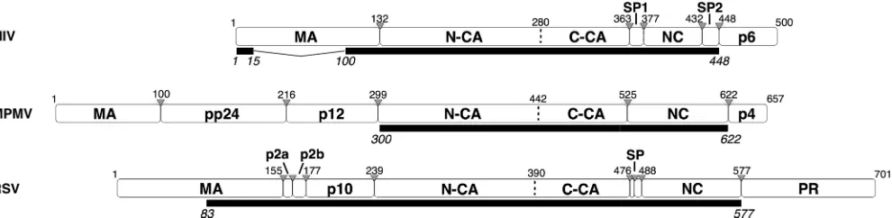
Retrovirus assembly is driven by the oligomerization of Gag, a multidomain protein, including an N-terminal membrane binding domain (MA), a two-domain structural component (CA), and an RNA binding domain (NC). The Gag proteins of all orthoretroviruses, including the alpha-, beta-, and lentiret-roviruses discussed here, share this conserved modular archi-tecture (Fig. 1). Despite very weak sequence conservation, the tertiary structures of MA, CA, and NC are conserved among retroviruses. Outside these conserved domains the Gag pro-teins of different retroviruses exhibit substantial variability. Other domains may be present or absent, and the length and sequence of linker peptides may also vary (12) (Fig. 1).
Oligomerization of Gag in an infected cell leads to the for-mation of roughly spherical immature virus particles, where Gag is arranged in a radial fashion with the N-terminal MA domain associated with a surrounding lipid bilayer, and the more C-terminal NC pointing toward the center of the particle (15, 44, 46). Subsequent multiple cleavages of Gag by the viral protease lead to a rearrangement of the virus. NC and the
RNA condense in the center of the particle, CA assembles into a capsid or shell around the nucleoprotein, and MA remains associated with the viral membrane. This proteolytic matura-tion is required to generate an infectious virion (2). In contrast to the mature CA lattice, which has been extensively studied (11, 16, 36), the Gag lattice in immature particles is incom-pletely understood.
Gag itself contains all of the necessary determinants for particle assembly. For example, the expression of Gag alone in an insect cell expression system is sufficient to generate virus-like particles (3, 17, 22, 38). Retroviral Gag proteins also can be assembledin vitroin the presence of nucleic acids to form spherical particles (9, 19, 39, 43, 47). The arrangement of Gag within thesein vitro-assembled Gag particles is indistinguish-able from that found in immature virus particles (6), and thein
vitroassembly systems have proved valuable for unraveling the
principles of virus assembly (18, 28, 29, 39). Multiple layers of interaction promote the assembly of Gag in vivo, including MA-membrane-MA interactions, CA-CA interactions, and NC-RNA-NC interactions. An extensive body of literature has explored which regions of Gag are required for assembly and which can be replaced or deleted without compromising as-sembly. MA-membrane-MA interactions contribute but are not essential. NC-RNA-NC interactions appear to function to nonspecifically link Gag molecules together and can be re-placed bothin vivoand in vitroby other interaction domains such as leucine zippers (4, 13, 20, 32, 48). The C-terminal
* Corresponding author. Mailing address: Structural and Computa-tional Biology Unit, European Molecular Biology Laboratory, Meyer-hofstrasse 1, 69117 Heidelberg, Germany. Phone: 49 6221 387 8482. Fax: 49 6221 387 8519. E-mail: [email protected].
† Supplemental material for this article may be found at http://jvi .asm.org/.
䌤Published ahead of print on 1 September 2010.
11729
on November 8, 2019 by guest
http://jvi.asm.org/
domain of CA (referred to here as C-CA) and the stretch of amino acids immediately following this domain (termed the spacer peptide [SP] region) are critical for assembly and sen-sitive to mutation (1, 22, 27, 30).
We set out to understand how the substantial sequence variation among Gag proteins in different retroviruses is man-ifested in structural differences in the immature Gag lattice. To do this, we studied three retroviruses from different genera: the lentivirus human immunodeficiency virus type 1 (HIV-1), the betaretrovirus Mason-Pfizer monkey virus (M-PMV), and the alpharetrovirus Rous sarcoma virus (RSV). These retroviruses are those for whichin vitro assembly was first established and has been most extensively studied (6, 19, 24, 28, 29, 35, 43, 47).
The domain structures of the three retroviruses differ most substantially upstream of CA. Both M-PMV and RSV have domains located between MA and CA that are absent in HIV (Fig. 1). In M-PMV there are 198 residues forming the pp24 and p12 domains; in RSV there are 84 residues forming the p2a, p2b, and p10 domains. The three retroviruses have differ-ent requiremdiffer-ents for regions upstream of CA during assembly. The C-terminal 25 residues of p10 are essential for proper immature RSV assembly, bothin vitroand in vivo, and these residues are inferred to interact directly with N-CA to stabilize the hexamer by forming contacts between adjacent N-CA do-mains (35). An equivalent assembly domain has not been de-scribed for other retroviruses. Within M-PMV p12 is the so-called internal scaffolding domain that is not essential for assembly in vitro (43) but is required for particle assembly when the precursor is expressed under the control of the M-PMV promoter (41). It is a key domain for the membrane-independent assembly of immature capsids (40).
In HIV, five residues upstream of CA must be present for assembly of immature virus-like spherical particles in vitro, although larger upstream extensions, including part of MA, are required for efficient assembly of regular particles, both for HIV and RSV. For HIV, if the entire MA domain is included,
in vitro assembly requires the presence of inositol penta- or
hexakis phosphate (8). If no sequences upstream of CA are present, thein vitroparticles in both of these viruses adopt a mature-type tubular morphology (10, 18). It has been hypoth-esized that cleavage at the N terminus of N-CA during matu-ration leads to the N-terminal residues of CA folding back into the N-CA structure to form a-hairpin. The-hairpin is
im-portant for assembly of the mature CA lattice, whereas its absence is important for immature assembly (23, 42). These requirements explain why, in HIV and RSV, immature Gag lattice-like structures are formed only if regions upstream of CA are present (18). In M-PMV, an immature Gag lattice can be produced when the regions upstream of CA are deleted if this is combined with mutations (such as deleting the initial proline of CA), which prevent-hairpin formation (43).
During maturation, HIV and RSV Gag proteins are cleaved twice between CA and NC to release a small peptide called SP1 or SP. In RSV the most N-terminal of these two cleavages can occur at one of two possible positions such that the re-leased peptide is either 9 or 12 amino acids long (33). In M-PMV only one cleavage occurs between CA and NC, and no short peptide is produced. The region between the final helix of CA and the Zn fingers has been proposed to adopt a helical bundle architecture in HIV and RSV based on bioinformatic prediction, on mutational analysis, and on structural studies (1, 22, 27, 45). In all three viruses, C-CA and the residues imme-diately downstream are critical for assembly and are sensitive to mutation. C-CA contains the major homology region, a group of residues that are highly conserved across the retrovi-ruses.
Cryo-electron tomography (cET) studies of immature virus particles (6, 45) have resolved the electron density of the HIV Gag lattice in three dimensions at low resolution. Using these methods, we have also described the three-dimensional archi-tecture ofin vitro-assembled HIV Gag particles (6). In imma-ture viruses and in vitro-assembled particles, Gag is seen to adopt an 8 nm hexameric lattice, as was predicted from previ-ous Fourier analysis of two-dimensional images (7, 46). The hexameric lattice is interrupted by irregularly shaped holes and cracks in the lattice (6, 45). A similar observation has been made using AFM ofin vitro-assembled particles of M-PMV Gag (26). These holes and cracks allow an otherwise planar hexameric lattice to form the surface of an approximately spherical particle.
[image:2.585.47.542.69.190.2]The radial positions of the MA, CA, and NC domains had been assigned previously from cryo-electron micrographs (44, 46). Based on these assignments and the shape of the density, the position and relative orientations of CA domains can be modeled into the low-resolution structure of the HIV lattice (6, 45). Density ascribed to the N-terminal domain of CA (N-CA) forms rings around large holes at the 6-fold symmetry
FIG. 1. Modular architecture of the full-length Gag proteins of HIV, M-PMV, and RSV. White rectangles illustrate Gag polyprotein cleavage products. The extent of the constructs used in the electron microscopic analysis is specified under each protein as a black rectangle. Gray triangles specify cleavage sites. Residue numbers are counted from the beginning of Gag.
on November 8, 2019 by guest
http://jvi.asm.org/
positions in the lattice. Below this layer, at the expected radius of the C-CA, are 2-fold densities, interpreted as corresponding to dimers of C-CA. These densities are linked by rodlike den-sities, which descend into the NC-nucleic acid layer.
HIV is the only retrovirus for which the arrangement of Gag in the immature particle has been described in three dimen-sions. Prior to this work, important open questions were there-fore: which features of the arrangement of Gag are conserved between genera and therefore reflect general principles of Gag-Gag interactions, and which features are specific to cer-tain genera? We have applied subtomogram averaging of cryo-electron tomograms to generate reconstructions of in vitro -assembled Gag particles from HIV, M-PMV, and RSV. These allow identification of the general and variable features of the arrangement of Gag and the architecture of immature retro-viruses.
MATERIALS AND METHODS
Sample preparation.Gag proteins were expressed in Escherichia coliand purified. Immature virus-like Gag particles were prepared byin vitroassembly of proteins with nucleic acid as described previously for HIV (19), M-PMV (26), or RSV (35). The constructs used are illustrated in Fig. 1. The nucleic acid was a 50-nucleotide (nt) single-stranded DNA (RSV), a 73-nt single-stranded DNA (HIV), or MS2 phage RNA (M-MPV). The assembled particles were kept at 4°C for at most 6 days until flash frozen in liquid ethane for analysis.
Electron microscopy and image processing.In vitro-assembled particles of HIV, M-PMV, and RSV Gag were mixed with 10-nm gold beads, deposited on C-flat Holey carbon grids, and vitrified by plunge freezing in liquid ethane. Tilt series were collected on an FEI Tecnai F30 Polara transmission electron micro-scope with Gatan GIF 2002 post column energy filter and a 2kx2k Multiscan charge-coupled device camera. The data collection was performed at 300 kV using the University of California at San Francisco (UCSF) software package and tomograms reconstructed using the IMOD software package (25) as described previously (6). Tilt series were typically collected between⫺60° and⫹60° with an angular increment of 3° and a total electron dose of⬃70 e/Å2
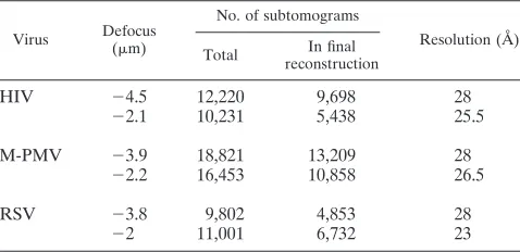
. Subtomogram averaging was carried out by using a six-dimensional search (14) in a reference-free manner as described previously (6). Independent reconstructions of half datasets were used to verify the reliability of the structures (see Results) and combined to produce the final reconstruction. Only subtomograms giving a cross correlation with the reference of greater than the mean cross correlation were included in the final reconstruction. The final reconstruction was filtered to the resolution to which the two half datasets are comparable, according to the Fourier shell correlation (see Fig. S3 in the supplemental material). The datasets are summarized in Table 1.
Lattice map representations were generated by using Amira (Visage Imaging), together with the EM Package (37). A hexagon is placed at the final aligned position of each tomogram and colored according to the cross-correlation value between the subtomogram and the average. Tomograms aligned to an inappro-priate radial position were excluded based on the radii distribution of the set of 20 nearest subtomograms surrounding each subtomogram. If the radius of the selected subtomogram was in the first or in the fourth quartile of the distribution,
the subtomogram was excluded. Only hexagons with a cross-correlation over a defined threshold are displayed. Structure interpretation and fitting were carried out using UCSF Chimera (34), which was also used to produce structure figures. The angles plotted in Fig. 4B for each hexamer are 180° minus the mean angle between the 6-fold axis of a hexamer and the 6-fold axes of its immediate neighbors.
Bioinformatics.To establish conserved retroviral features present or absent in our proteins of interest, 12 Gag protein sequences of the 27 International Com-mittee on Taxonomy of Viruses (ICTV) reference species from these genera were chosen as follows. From the 27 ICTV reference species, those with incom-plete Gag sequences or from defective viruses were not considered. To avoid overweighting the importance of very similar viruses, only one representative sequence was chosen from groups of viruses with sequence similarity in Gag greater than 70%, giving 12 sequences. These sequences were aligned by using MAAFT (21). Conserved features are those identified in⬎75% of the se-quences.
The SP1 region is hypothesized to form an alpha-helix based on bioinformatic prediction, mutational analysis, and structural studies (1, 22, 27). A helical wheel representation of the 18 residues after the CA adjacent cleavage site was created to examine the helical hydrophobic moment of the region for the three viruses of interest. The complete region between the final helix of CA and the first zinc finger of NC was modeled in three dimensions as an alpha-helix using chimera (see Fig. S4 in the supplemental material).
RESULTS AND DISCUSSION
We describe here the local and global architecture of the Gag lattice inin vitro-assembled, immature Gag particles for three retroviral genera, with the aim of identifying general features of Gag assembly and those features which vary among genera.In vitro-assembled Gag particles were produced using expression vectors and assembly conditions that have previ-ously been described (18, 35, 43). Each Gag protein represents approximately the minimal size protein for whichin vitro as-sembly is efficient and correct and can occur without other additions (see the introduction). As a consequence, domains upstream of the conserved CA domains were variably trun-cated or absent (constructs are illustrated in Fig. 1).
The structure of the Gag lattice inin vitro-assembled
imma-ture particles.Vitrified,in vitro-assembled Gag particles were
[image:3.585.44.283.81.197.2]imaged by using cryo-electron tomography. Initial data collec-tion was carried out at a defocus of approximately⫺4m. In order to assess the reliability of the reconstructions, for each type ofin vitro-assembled Gag particle, the data set was split into two halves and two completely independent reference-free subtomogram averaging reconstructions were carried out. A second round of data collection was carried out with a defocus of approximately⫺2m. Again, the data set for each type of particle was split into two halves, and each half was recon-structed using a different reconstruction from the⫺4m data set as a starting model. Sections through these independent reconstructions are shown in Fig. S1 in the supplemental ma-terial. No significant differences were observed between inde-pendent reconstructions of particles assembled from the same Gag protein. The independent reconstructions were compared to one another by using Fourier Shell Correlation and were comparable to a resolution of 26.5 Å (see Fig. S2 in the sup-plemental material). This comparison of two independent re-constructions verified the robustness of the reconstruction pro-cess and allowed us to assess the resolution to which the reconstructions could be reliably interpreted. The two inde-pendent reconstructions were therefore aligned, averaged, and filtered to 26.5 Å to generate final reconstructions (Fig. 2A). The same process was carried out within vitro-assembled Gag
TABLE 1. Subtomogram data sets used in this study
Virus Defocus (m)
No. of subtomograms
Resolution (Å) Total In final
reconstruction
HIV ⫺4.5 12,220 9,698 28
⫺2.1 10,231 5,438 25.5
M-PMV ⫺3.9 18,821 13,209 28
⫺2.2 16,453 10,858 26.5
RSV ⫺3.8 9,802 4,853 28
⫺2 11,001 6,732 23
on November 8, 2019 by guest
http://jvi.asm.org/
particles from each of the three different retroviruses. Isosur-face representations of the reconstructions were generated (Fig. 2B) and colored for illustrative purposes.
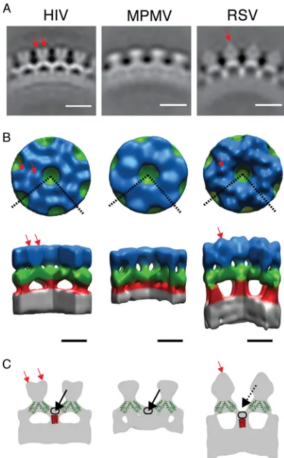
The three different reconstructions exhibit substantial simi-larity. An outer density (blue in Fig. 2B) is arranged in hex-americ rings around large holes. Where these rings meet, 2-fold densities (green in Fig. 2B) extend between adjacent 6-fold rings. Six of these 2-fold densities converge at each 6-fold axis, beneath the holes in the hexameric rings. Here they connect with a density (red in Fig. 2B), which descends further toward the center of the particle. This density meets the NC-nucleic acid layer (gray in Fig. 2B), which is not ordered in a
hexameric manner. Despite these similarities, structural differ-ences are seen at all radii of the Gag layer.
Outermost in the particles (blue in Fig. 2B) are the N ter-mini of the Gag-derived proteins, where the three constructs used differ substantially (Fig. 1). The M-PMV construct begins directly with the N-CA domain, lacking the N-terminal proline residue. Accordingly, the M-PMV reconstruction shows the least featured outer density (blue in Fig. 2B), which can be presumed to represent N-CA.
The HIV construct includes the first 15 and last 33 residues of MA. This protein has been found to efficiently assemblein
vitro(18). The reconstruction shows small protrusions around
the holes at the 6-fold axis (red arrows in Fig. 2B and C). The extra MA residues may form these densities. Alternatively, the presence of part of MA may lead to a more rigid N-CA lattice than present in M-PMV, allowing individual N-CA domains to be more clearly resolved. In this case, the small protrusions could also represent part of N-CA.
The RSV construct includes p10, p2, and about one-half of MA, which together total 156 amino acid residues upstream of the CA domain. The reconstruction shows large extra densities above the 2-fold axes of N-CA (red arrow in Fig. 2A to C, visible as prominent caplike densities in Fig. 2A). The densities are positioned above the 2-fold axes of the lattice, where they link between adjacent hexamers. These densities could include the N-terminal part of p10 and perhaps sequences further upstream in the p2 and/or MA domains. In this model, each of the caplike densities would be formed from two p10 domains, one from each hexamer, which by linking adjacent hexamers would stabilize the hexameric lattice.
Moving toward the center of the reconstructions, the next layer can be assigned to the C-CA domain as described previ-ously (6, 44–46) (green in Fig. 2B). If the crystal structure of the C-CA dimer (PDB: 3ds2) is superimposed onto the HIV density (as in reference 6 or Fig. 2C), the density occupied by the crystal structure can be assigned to the ordered part of the C-CA domain. The shape of this density is similar in all three reconstructions and, considering the high level of conservation of retroviral CA domains, the same crystal structure has also been superimposed on the RSV and M-PMV reconstructions for illustrative purposes. The density around the 6-fold axis that is not occupied by the crystal structure can be assigned to residues downstream of the final CA helix. In HIV and M-PMV the top of this region forms a density below the hexag-onal hole created by the N-CA domains, at the center of the C-CA ring, forming the floor of the hole in the ring (black arrows in Fig. 2C). The position of this floor makes it most likely that it is formed from the residues immediately down-stream of the final helix of C-CA. In RSV the floor is farther toward the center of the particle, making the hexagonal hole deeper (compare the positions of the solid and dashed black arrows for HIV and RSV in Fig. 2C).
[image:4.585.59.263.68.397.2]The lower floor in the center of the C-CA hexamer hole in the RSV reconstruction suggests a difference in the arrange-ment of the residues immediately downstream of C-CA and in their interactions with the structured domain of C-CA. This suggests that the arrangements of C-CA, and the dimensions of the hexamer, which are well conserved across the retroviruses, do not require a specific conserved arrangement of the amino acid sequences that comprise the floor or a specific conserved
FIG. 2. Structure of thein vitro-assembled Gag particles. (A) Ra-dial section through the average structures. The density is white. The scale bar is 10 nm. (B) Surface rendering of the average structures. The color scheme follows an approximate radial division of the structure according to the different domains of Gag as previously assigned (6, 44, 45, 46): gray is the NC-nucleic acid layer, red is the SP1 region, green is the C-CA region, and blue is the N-CA. The dashed lines on the top view represent the section cut out in the side view. Red arrows indicate densities protruding from the N-CA density (see the text). Scale bar, 5 nm. (C) Sections through the isosurface representations (gray), illus-trating features referred to in the text. The crystal structure of the CA dimer (PDB: 3ds2) (green) is superimposed on the sections indicated previously (6). Red rods indicate possible positions of a hypothetical 24-Å six-helix bundle. Red arrows indicate densities protruding from the N-CA density. Black arrows indicate density forming the floor of the hole in the C-CA ring. Black rings indicate possible positions of the residues between the final helix in the C-CA and the six-helix bundle. For explanations, see the text.
on November 8, 2019 by guest
http://jvi.asm.org/
interaction with the downstream structure present on the 6-fold axis. Instead, the conserved arrangement of C-CA is presumably predominantly mediated by interactions between CA dimers around the 6-fold axis.
The region between C-CA and the NC-nucleic acid layer is strikingly different in the three viruses. In RSV a strong rodlike feature descends from below C-CA to the NC-nucleic acid layer along the 6-fold axis (red in Fig. 2B). In HIV a similar feature is seen, but as described above the density begins within the C-CA layer, where it forms the floor of the hole at the 6-fold axis in the C-CA lattice. In M-PMV the floor is present, but there is no clear rodlike density between C-CA and the NC-nucleic acid layer.
Comparing structural differences with sequence differences.
A sequence alignment of the C-CA-NC region highlights fea-tures conserved across multiple orthoretroviruses (see Materi-als and Methods and see Fig. S3 in the supplemental material). The alignment for the three viruses of interest is shown in Fig. 3A. This alignment identifies a number of previously described conserved features, the major homology region (MHR), nu-cleotide-binding Zn finger motifs, a CA-adjacent cleavage site, and a conserved cysteine pair. Most strikingly conserved is the MHR, which is known to be present in all orthoretroviruses. A pair of well-conserved cysteine residues, which approach close to one another between helices 3 and 4 of C-CA in the HIV CA crystal structure, is present in betaretroviruses,
lentivi-FIG. 3. Comparisons of the Gag sequences from HIV, M-PMV, and RSV. (A) Multiple alignment of Gag C-CA-NC for RSV, M-PMV, and HIV. Regions of interest are boxed, and colored circles indicate the coloring of these regions in panels B and C. Residues conserved across⬎75% of viruses in an extended alignment, including 12 viral species (see Fig. S3 in the supplemental material), are colored using ClustalX coloring; residues conserved across all species are specified by an asterisk. Orange triangles denote cleavage sites. Basic residues adjacent to the NC are underlined. A dashed red box surrounds an RKK motif unique to M-PMV C-CA. The position of helices from the solved structures for RSV (PDB: 1d1d) and HIV (PDB: 3ds2) are shown under the alignment for illustrative purposes. Residues are numbered from the beginning of Gag. (B) HIV C-CA (PDB: 3ds2) with residues colored to indicate the positions of regions of interest: yellow, MHR; purple, NAN; orange, paired cysteine residues; and red, RKK motif (see the text and panels A and C). (C) Table of regions of interest defined in panel A; the colors correspond to the coloring in the multiple alignment (A) and HIV structure (A). Consensus signifies genera in which the feature is found (␣signifies alpha,signifies beta,␦signifies delta, and L signifies lentiretroviruses). There is currently no solved structure for the CA region in M-PMV, the final helix of M-PMV is defined assuming structural homology to HIV (PDB: 3ds2). (D) Helical wheel representation of the 18 residues after the CA adjacent cleavage site for the proteins of interest.
on November 8, 2019 by guest
http://jvi.asm.org/
ruses, and deltaretroviruses but absent in the alpharetrovi-ruses. Not previously described is a conserved NAN motif in the turn between helices 2 and 3, which is also absent in the alpharetroviruses. These regions are highlighted on the struc-ture of the C-CA dimer from HIV-1 in Fig. 3B, and summa-rized in Fig. 3C.
The conserved cysteine pair and NAN turn motif within C-CA, which are absent in RSV but present in HIV and M-PMV, are located away from the dimer axis, in a position that would lie close to the hole at the 6-fold axis (Fig. 3B). This is the region where the position of the floor differs between the retroviruses. We speculate that the cysteine pair and NAN sequence motifs may contribute to the organization of this region in HIV and M-PMV.
A stretch of amino acids roughly corresponding to the HIV-1 SP1 has been suggested to have helical propensity based upon computational prediction, mutational analysis, and nu-clear magnetic resonance (NMR) (1, 31). It has been proposed that this region assembles into a six-helix bundle in the imma-ture HIV-1 Gag lattice (45). We examined this proposal by considering the helical hydrophobic moment of this region (Fig. 3D and see Fig. S4 in the supplemental material). Both HIV and RSV show a 16-residue stretch, starting from the residue immediately upstream of the cleavage site and running into SP1 or SP, with a strong helical hydrophobic moment, a finding consistent with the assembly of this region into a six-helix bundle held together by hydrophobic side chains in its center. M-PMV does not show a strong helical hydrophobic moment. The calculated hydrophobic face in RSV is slightly larger than that in HIV. In both HIV and RSV, the 16-residue alpha-helical bundle would be a maximum of ⬃24 Å long depending on the angle between the helices and the axis of the bundle. A 24-Å bundle is superimposed onto the structures in Fig. 2C for illustrative purposes.
The resolution of the reconstructions is not sufficient to resolve the secondary structural details, which would be nec-essary to describe the structure of the CA-NC linker region unambiguously, and multiple models are therefore consistent with the observed structures. In one such model, the region between the final helix of C-CA and the beginning of the proposed helical bundle is arranged to form the floor density in HIV (outlined by black ring in Fig. 2C), while the rodlike structure is formed by the proposed six-helix bundle. Below this region, basic residues close to the Zn fingers are involved in binding nucleic acid. In RSV, the region between the final helix of C-CA and the start of the helical bundle is not held in the floor position. It instead forms the top part of the rodlike structure (outlined by black ring in Fig. 2C). In the model, the lower part of the rod would then be formed by the six-helix bundle. In M-PMV the residues after the final helix of CA also form a floorlike structure as for HIV. The rodlike structure is not visible in M-PMV, and the nucleic acid directly abuts C-CA. This observation can be interpreted in two ways. Either the helical bundle is absent or the helical bundle is present despite the absence of a strong helical hydrophobic moment, but the rodlike density is obscured because it is buried in the nucleic acid layer. Presently available data do not allow these two possibilities to be distinguished.
Although we attribute the differences in tomographic den-sity features in the NC region to differences in amino acid
sequences among the three viruses studied, we cannot entirely exclude a role for the type of nucleic acid used to promote assembly. Whereas M-PMV particles were formed with RNA, the HIV and RSV particles were formed with DNA oligonu-cleotides. These respective nucleic acids were found previously to promote assembly in the most robust manner. We think it is unlikely that the HIV and RSV lattices are affected by the use of DNA since reconstructions of immature HIV particles (6, 45), which contain RNA, show the same arrangement of the NC layer.
The observation that the density interpreted to be the NC-nucleic acid layer directly abuts the base of the CA dimer in M-PMV suggests a possible direct interaction between the two. If the sequence of the C-CA domain of M-PMV is mapped onto the crystal structure of the HIV dimer, a basic RKK motif present in M-PMV but absent in RSV and HIV (see the dashed red boxes in Fig. 3A and the red area in Fig. 3B) is exposed on the underside of each monomer. These basic res-idues provide a potentially potent binding site for nucleic acid, and may explain the proximity of the nucleic acid layer to C-CA in the M-PMV structure. We hypothesize that such an interaction may be sufficient to pull the nucleic acid layer toward the CA layer, even if the Zn fingers are held at a distance from the CA layer by a rodlike structure, leading to the rod becoming buried within the nucleic acid layer.
Global arrangement of Gag in the in vitro-assembled Gag
particles.The global arrangement of the Gag shell within an
individualin vitro-assembled particle can be visualized by plac-ing a hexamer at the position and orientation to which each subtomogram has converged during the alignment and recon-struction procedure (6). Two example particles for each genus are illustrated in Fig. 4A. The relative positions and orienta-tions of the placed hexamers are not random but instead are consistent with a hexameric lattice. All three types of particles are assembled from a single assembled hexameric lattice with irregularly shaped defects, indicating that this pattern is a con-served outcome of retroviral Gag protein assembly.
The mean curvature of the particles is not the same in all cases. Similar to previous observations using conventional cryo-EM (5, 47)in vitro-assembled HIV Gag particles had a diameter of (94 ⫾ 4 nm, n ⫽ 12), measured to the C-CA domain, which is similar to the diameter of M-PMV particles (90⫾7 nm,n⫽12) and significantly larger than RSV particles (60⫾3 nm,n⫽12). The differing diameters are reflected in the different curvatures of the lattice visible in Fig. 4A. This change in size reflects changes in the angle between adjacent hexamers, which in HIV and M-PMV are approximately 170° and 169°, compared to 163° in RSV (Fig. 4B). It is tempting to speculate that the tighter curvature in RSV may result from a difference in the relative orientations of the C-CA domains around the 6-fold axis, facilitated by the structural differences observed in the CA-SP1 region. This speculation is supported by the observation that, upon in vitro assembly of chimeric HIV-RSV Gag-derived proteins, the main determinant for particle size was CA (3). RSV CA was found to induce the formation of smaller, more tightly curved particles than HIV-CA in these chimeric proteins.
Although less dramatic than the differences between the genera, there is also variability in the angle between hexamers within each genus, when different particles are compared and
on November 8, 2019 by guest
http://jvi.asm.org/
also within individual particles. This variability is manifested in the formation of particles with slightly varying diameters and with deviations from a perfect spherical shape. Flexibility in the angle between the hexamers is a necessary basis for the for-mation of heterogeneous particles, a known feature of all ret-roviruses.
Summary. The comparison of the immature Gag lattice
from three retroviral genera highlights the general features of particle architecture. All three of the particles assemble from a single hexameric lattice with irregular defects. The arrange-ment of the two CA domains is strikingly conserved between different genera, whereas the angles between adjacent hexam-ers appear to vary, changing the degree of curvature and thus the particle size. Outside of the CA domains, sequence differ-ences among the three genera are manifested as structural differences. Based on mutational data and chimeric Gag pro-teins containing foreign domains, the C-CA-SP region seems to contain the critical assembly determinants for the immature
Gag shell. Nevertheless, the inferred structure of this region, and its relationship to CA and the NC-nucleic acid layer, ap-pear to be specific to each genus.
ACKNOWLEDGMENTS
We thank Pasi Laurinma¨ki and Sarah Butcher for sharing unpub-lished data.
This study was supported by the grants from the Deutsche For-schungsgemeinschaft within Schwerpunktprogramm 1175 to J.A.G.B. and H.-G.K., from the European Union (FP6 LSHP-CT-2007-036793; HIV PI resistance) to J.A.G.B. and H.-G.K., from the Czech Ministry of Education (1M6837805002 and MSM 6046137305) to T.R., and from the U.S. Public Health Service (CA20081) to V.M.V.
P.U., J.M.P., and V.L. preparedin vitro-assembled Gag particles, with contributions from T.F. A.D.M. and J.D.R. collected electron microscopy data. A.D.M. and J.A.G.B. analyzed data. N.E.D. contrib-uted bioinformatics analyses. J.A.G.B., V.M.V., H.-G.K., and P.U. conceived the experiments. A.D.M. and J.A.G.B. wrote the paper with help from N.E.D., P.U., T.R., H.-G.K., and V.M.V. N.E.D. and A.D.M. prepared figures.
REFERENCES
1.Accola, M. A., S. Hoglund, and H. G. Gottlinger.1998. A putative alpha-helical structure which overlaps the capsid-p2 boundary in the human im-munodeficiency virus type 1 Gag precursor is crucial for viral particle assem-bly. J. Virol.72:2072–2078.
2.Adamson, C. S., and E. O. Freed.2007. Human immunodeficiency virus type 1 assembly, release, and maturation. Adv. Pharmacol.55:347–387. 3.Ako-Adjei, D., M. C. Johnson, and V. M. Vogt.2005. The retroviral capsid
domain dictates virion size, morphology, and coassembly of gag into virus-like particles. J. Virol.79:13463–13472.
4.Borsetti, A., A. Ohagen, and H. G. Gottlinger.1998. The C-terminal half of the human immunodeficiency virus type 1 Gag precursor is sufficient for efficient particle assembly. J. Virol.72:9313–9317.
5.Briggs, J. A., M. C. Johnson, M. N. Simon, S. D. Fuller, and V. M. Vogt.2006. Cryo-electron microscopy reveals conserved and divergent features of gag packing in immature particles of Rous sarcoma virus and human immuno-deficiency virus. J. Mol. Biol.355:157–168.
6.Briggs, J. A., J. D. Riches, B. Glass, V. Bartonova, G. Zanetti, and H. G. Krausslich.2009. Structure and assembly of immature HIV. Proc. Natl. Acad. Sci. U. S. A.106:11090–11095.
7.Briggs, J. A., M. N. Simon, I. Gross, H. G. Krausslich, S. D. Fuller, V. M. Vogt, and M. C. Johnson.2004. The stoichiometry of Gag protein in HIV-1. Nat. Struct. Mol. Biol.11:672–675.
8.Campbell, S., R. J. Fisher, E. M. Towler, S. Fox, H. J. Issaq, T. Wolfe, L. R. Phillips, and A. Rein.2001. Modulation of HIV-like particle assembly in vitro by inositol phosphates. Proc. Natl. Acad. Sci. U. S. A.98:10875–10879. 9.Campbell, S., and V. M. Vogt.1997. In vitro assembly of virus-like particles with Rous sarcoma virus Gag deletion mutants: identification of the p10 domain as a morphological determinant in the formation of spherical par-ticles. J. Virol.71:4425–4435.
10.Campbell, S., and V. M. Vogt.1995. Self-assembly in vitro of purified CA-NC proteins from Rous sarcoma virus and human immunodeficiency virus type 1. J. Virol.69:6487–6497.
11.Cardone, G., J. G. Purdy, N. Cheng, R. C. Craven, and A. C. Steven.2009. Visualization of a missing link in retrovirus capsid assembly. Nature457: 694–698.
12.Coffin, J. M., S. H. Huges, and H. E. Varmus.1997. Retroviruses. Cold Spring Harbor Laboratory Press, Cold Spring Harbor, NY.
13.Crist, R. M., S. A. Datta, A. G. Stephen, F. Soheilian, J. Mirro, R. J. Fisher, K. Nagashima, and A. Rein.2009. Assembly properties of human immuno-deficiency virus type 1 Gag-leucine zipper chimeras: implications for retro-virus assembly. J. Virol.83:2216–2225.
14.Forster, F., O. Medalia, N. Zauberman, W. Baumeister, and D. Fass.2005. Retrovirus envelope protein complex structure in situ studied by cryo-elec-tron tomography. Proc. Natl. Acad. Sci. U. S. A.102:4729–4734. 15.Fuller, S. D., T. Wilk, B. E. Gowen, H. G. Krausslich, and V. M. Vogt.1997.
Cryo-electron microscopy reveals ordered domains in the immature HIV-1 particle. Curr. Biol.7:729–738.
16.Ganser-Pornillos, B. K., A. Cheng, and M. Yeager.2007. Structure of full-length HIV-1 CA: a model for the mature capsid lattice. Cell131:70–79. 17.Gheysen, D., E. Jacobs, F. de Foresta, C. Thiriart, M. Francotte, D. Thines,
and M. De Wilde.1989. Assembly and release of HIV-1 precursor Pr55gag virus-like particles from recombinant baculovirus-infected insect cells. Cell 59:103–112.
18.Gross, I., H. Hohenberg, C. Huckhagel, and H. G. Krausslich.1998. N-terminal extension of human immunodeficiency virus capsid protein converts FIG. 4. Global arrangement of Gag lattice in the particles.
(A) Global lattice maps ofin vitro-assembled Gag particles. The cen-ters of each hexameric unit cell are marked with hexamers, which are colored according to cross correlation on a scale from low (red) to high (green). Higher cross-correlation values indicate that the subtomo-gram is more similar to the average structure. The cross-correlation range in the each map has been set between the minimum and the maximum cross-correlation value present in the map. Maps are shown in perspective, such that hexamers on the rear surface of the particle appear smaller and fainter. Two representative particles are shown for each genus. (B) Scatter plot showing the distribution of the angles between each hexamer and its neighbors (see Materials and Methods). The mean value for each particle is marked in red. For each construct, 12 particles have been analyzed.
on November 8, 2019 by guest
http://jvi.asm.org/
the in vitro assembly phenotype from tubular to spherical particles. J. Virol. 72:4798–4810.
19.Gross, I., H. Hohenberg, and H. G. Krausslich.1997. In vitro assembly properties of purified bacterially expressed capsid proteins of human immu-nodeficiency virus. Eur. J. Biochem.249:592–600.
20.Johnson, M. C., H. M. Scobie, Y. M. Ma, and V. M. Vogt.2002. Nucleic acid-independent retrovirus assembly can be driven by dimerization. J. Virol. 76:11177–11185.
21.Katoh, K., K. Misawa, K. Kuma, and T. Miyata.2002. MAFFT: a novel method for rapid multiple sequence alignment based on fast Fourier trans-form. Nucleic Acids Res.30:3059–3066.
22.Keller, P. W., M. C. Johnson, and V. M. Vogt.2008. Mutations in the spacer peptide and adjoining sequences in Rous sarcoma virus Gag lead to tubular budding. J. Virol.82:6788–6797.
23.Kingston, R. L., T. Fitzon-Ostendorp, E. Z. Eisenmesser, G. W. Schatz, V. M. Vogt, C. B. Post, and M. G. Rossmann.2000. Structure and self-association of the Rous sarcoma virus capsid protein. Structure8:617–628. 24.Klikova, M., S. S. Rhee, E. Hunter, and T. Ruml.1995. Efficient in vivo and
in vitro assembly of retroviral capsids from Gag precursor proteins expressed in bacteria. J. Virol.69:1093–1098.
25.Kremer, J. R., D. N. Mastronarde, and J. R. McIntosh.1996. Computer visualization of three-dimensional image data using IMOD. J. Struct. Biol. 116:71–76.
26.Kuznetsov, Y. G., P. Ulbrich, S. Haubova, T. Ruml, and A. McPherson.2007. Atomic force microscopy investigation of Mason-Pfizer monkey virus and human immunodeficiency virus type 1 reassembled particles. Virology360: 434–446.
27.Liang, C., J. Hu, R. S. Russell, A. Roldan, L. Kleiman, and M. A. Wainberg. 2002. Characterization of a putative alpha-helix across the capsid-SP1 boundary that is critical for the multimerization of human immunodeficiency virus type 1 gag. J. Virol.76:11729–11737.
28.Ma, Y. M., and V. M. Vogt.2004. Nucleic acid binding-induced Gag dimer-ization in the assembly of Rous sarcoma virus particles in vitro. J. Virol. 78:52–60.
29.Ma, Y. M., and V. M. Vogt.2002. Rous sarcoma virus Gag protein-oligonu-cleotide interaction suggests a critical role for protein dimer formation in assembly. J. Virol.76:5452–5462.
30.Melamed, D., M. Mark-Danieli, M. Kenan-Eichler, O. Kraus, A. Castiel, N. Laham, T. Pupko, F. Glaser, N. Ben-Tal, and E. Bacharach.2004. The conserved carboxy terminus of the capsid domain of human immunodefi-ciency virus type 1 Gag protein is important for virion assembly and release. J. Virol.78:9675–9688.
31.Morellet, N., S. Druillennec, C. Lenoir, S. Bouaziz, and B. P. Roques.2005. Helical structure determined by NMR of the HIV-1 (345-392) Gag se-quence, surrounding p2: implications for particle assembly and RNA pack-aging. Protein Sci.14:375–386.
32.Muriaux, D., S. Costes, K. Nagashima, J. Mirro, E. Cho, S. Lockett, and A. Rein.2004. Role of murine leukemia virus nucleocapsid protein in virus assembly. J. Virol.78:12378–12385.
33.Pepinsky, R. B., R. J. Mattaliano, and V. M. Vogt. 1986. Structure and
processing of the p2 region of avian sarcoma and leukemia virus gag pre-cursor polyproteins. J. Virol.58:50–58.
34.Pettersen, E. F., T. D. Goddard, C. C. Huang, G. S. Couch, D. M. Greenblatt, E. C. Meng, and T. E. Ferrin.2004. UCSF Chimera: a visualization system for exploratory research and analysis. J. Comput. Chem.25:1605–1612. 35.Phillips, J. M., P. S. Murray, D. Murray, and V. M. Vogt.2008. A molecular
switch required for retrovirus assembly participates in the hexagonal imma-ture lattice. EMBO J.27:1411–1420.
36.Pornillos, O., B. K. Ganser-Pornillos, B. N. Kelly, Y. Hua, F. G. Whitby, C. D. Stout, W. I. Sundquist, C. P. Hill, and M. Yeager.2009. X-ray struc-tures of the hexameric building block of the HIV capsid. Cell137:1282–1292. 37.Pruggnaller, S., M. Mayr, and A. S. Frangakis.2008. A visualization and segmentation toolbox for electron microscopy. J. Struct. Biol.164:161–165. 38.Royer, M., S. S. Hong, B. Gay, M. Cerutti, and P. Boulanger.1992. Expres-sion and extracellular release of human immunodeficiency virus type 1 Gag precursors by recombinant baculovirus-infected cells. J. Virol.66:3230–3235. 39.Rumlova-Klikova, M., E. Hunter, M. V. Nermut, I. Pichova, and T. Ruml. 2000. Analysis of Mason-Pfizer monkey virus Gag domains required for capsid assembly in bacteria: role of the N-terminal proline residue of CA in directing particle shape. J. Virol.74:8452–8459.
40.Sakalian, M., and E. Hunter.1999. Separate assembly and transport do-mains within the Gag precursor of Mason-Pfizer monkey virus. J. Virol. 73:8073–8082.
41.Sommerfelt, M. A., S. S. Rhee, and E. Hunter.1992. Importance of p12 protein in Mason-Pfizer monkey virus assembly and infectivity. J. Virol. 66:7005–7011.
42.Tang, C., Y. Ndassa, and M. F. Summers.2002. Structure of the N-terminal 283-residue fragment of the immature HIV-1 Gag polyprotein. Nat. Struct. Biol.9:537–543.
43.Ulbrich, P., S. Haubova, M. V. Nermut, E. Hunter, M. Rumlova, and T. Ruml.2006. Distinct roles for nucleic acid in in vitro assembly of purified Mason-Pfizer monkey virus CANC proteins. J. Virol.80:7089–7099. 44.Wilk, T., I. Gross, B. E. Gowen, T. Rutten, F. de Haas, R. Welker, H. G.
Krausslich, P. Boulanger, and S. D. Fuller.2001. Organization of immature human immunodeficiency virus type 1. J. Virol.75:759–771.
45.Wright, E. R., J. B. Schooler, H. J. Ding, C. Kieffer, C. Fillmore, W. I. Sundquist, and G. J. Jensen.2007. Electron cryotomography of immature HIV-1 virions reveals the structure of the CA and SP1 Gag shells. EMBO J. 26:2218–2226.
46.Yeager, M., E. M. Wilson-Kubalek, S. G. Weiner, P. O. Brown, and A. Rein. 1998. Supramolecular organization of immature and mature murine leuke-mia virus revealed by electron cryo-microscopy: implications for retroviral assembly mechanisms. Proc. Natl. Acad. Sci. U. S. A.95:7299–7304. 47.Yu, F., S. M. Joshi, Y. M. Ma, R. L. Kingston, M. N. Simon, and V. M. Vogt.
2001. Characterization of Rous sarcoma virus Gag particles assembled in vitro. J. Virol.75:2753–2764.
48.Zhang, Y., and E. Barklis.1997. Effects of nucleocapsid mutations on human immunodeficiency virus assembly and RNA encapsidation. J. Virol.71:6765– 6776.