TO COMPARE THE EASE OF PERFORMING
TRANSNASAL FLEXIBLE LARYNGOSCOPY
USING DIFFERENT TOPICAL PREPARATIONS
AND METHODS OF APPLICATION
\
A DISSERTATION SUBMITTED IN PARTIAL FULFILMENT OF M.S BRANCH IV OTORHINOLARYNGOLOGY EXAMINATION OF THE TAMIL NADU DR.
TO COMPARE THE EASE OF PERFORMING
TRANSNASAL FLEXIBLE LARYNGOSCOPY
USING DIFFERENT TOPICAL PREPARATIONS
AND METHODS OF APPLICATION
DEPARTMENT OF OTORHINOLARYNGOLOGY
CHRISTIAN MEDICAL COLLEGE
VELLORE
CERTIFICATE
I declare that this dissertation entitled “To compare the ease of performing transnasal flexible laryngoscopy using different topical preparations and
methods of application’’ submitted towards partial fulfilment of the requirements of the Tamil Nadu Dr. M.G.R.Medical University for the MS Branch IV, Otorhinolaryngology examination to be conducted in May 2018, is the bonafide work done by me, and due acknowledgements have been made in text to all materials used.
Dr. Asha K Joy
Postgraduate Student (M S Otorhinolaryngology) Register Number: 221514351
DEPARTMENT OF OTORHINOLARYNGOLOGY
CHRISTIAN MEDICAL COLLEGE
VELLORE
CERTIFICATE
This is to certify that the dissertation entitled “To compare the ease of performing transnasal flexible laryngoscopy using different topical preparations and
methods of application’’ is a bonafide original work of Dr. Asha K Joy, submitted in partial fulfilment of the rules and regulations for the M S Branch IV, Otorhinolaryngology examination of The Tamil Nadu Dr. M.G.R. Medical University to be held in May, 2018.
Dr. Anna B. Pulimood Dr. Rita Ruby Anbuselvi A.
Principal Professor and Head,
DEPARTMENT OF OTORHINOLARYNGOLOGY
CHRISTIAN MEDICAL COLLEGE
VELLORE
CERTIFICATE
This is to certify that the dissertation entitled “To compare the ease of performing transnasal flexible laryngoscopy using different topical preparations and
methods of application’’ is a bonafide original work of Dr. Asha K Joy, submitted in partial fulfilment of the rules and regulations for the M.S. Branch IV, Otorhinolaryngology examination of The Tamil Nadu Dr. M.G.R. Medical University to be held in May, 2018.
Dr. Suma Susan Mathews
Professor and Guide,
Department of Otorhinolaryngology, Christian Medical College,
ACKNOWLEDGEMENTS
I would like to thank God Almighty for being with me each step of the way and helping
me to finish this dissertation.
I wish to express my sincere gratitude to my guide Dr. Suma Susan Mathews,
Professor, Department of Otorhinolaryngology, Christian Medical College and
Hospital, Vellore, for all her hard work, wisdom, motivation, expert guidance and
encouragement without which this dissertation would not have been possible. I could
not have imagined having a better advisor and mentor for my dissertation.
I am grateful to Dr. Rita Ruby Anbuselvi A., Professor and Head of
Otorhinolaryngology, Christian Medical College and Hospital, Vellore and co-guide
for the thesis, for her constant support and encouragement in carrying out this study.
I am extremely grateful to Dr. Ajay Philip from the Department of
Otorhinolaryngology for his valuable help, advice and guidance in this study.
I am very thankful to all the endoscopy room Nurses and the receptionist, Mrs.Hepsy
without whose help the study could not have been completed.
I would like to thank Dr. B.Antonisamy and Ms. Hepsy Chelliah from the Department
of Biostatistics for patiently understanding and helping me with the analysis of the
data.
I am thankful to our PG coordinator, Dr. Lalee Varghese for conducting timely
I am grateful to all my friends and colleagues from the Department of
Otorhinolaryngology for helping me in collecting the samples and making the study a
reality.
I would like to thank all the patients who agreed to be a part of this study.
I would also like to thank the Fluid Research Committee, CMC Hospital for granting
me permission for conducting this study.
A special thanks to my parents, my husband Mr. Cyrus Kallupurackal and my son
Master Nathan C. Thomas for their love, concern and support throughout the work on
CONTENTS
INTRODUCTION ... 1
AIMS AND OBJECTIVES ... 2
REVIEW OF LITERATURE ... 3
HISTORY OF LARYNGOLOGY ... 3
VISUALIZATION OF THE LARYNX ... 13
INDIRECT MIRROR LARYNGOSCOPY ... 14
DIRECT RIGID LARYNGOSCOPY ... 16
FLEXIBLE NASOPHARYNGOLARYNGOSCOPY (NPL SCOPY) ... 19
The physics of fiberoptic endoscopy ... 21
Endoscopic anatomy ... 25
Indications of NPL scopy ... 29
Contraindications of flexible laryngoscopy ... 46
Positioning... 47
Technique ... 47
Cleaning and disinfection of endoscopes ... 54
Complications of flexible laryngoscopy ... 55
NASAL PREPARATION PRIOR TO ENDOSCOPY ... 55
Local anaesthetics: ... 57
Sympathomimetics ... 63
TOPICAL AGENTS IN FLEXIBLE NPL SCOPY ... 63
MATERIALS AND METHODS ... 65
RESULTS ... 70
DISCUSSION ... 90
LIMITATIONS ... 95
CONCLUSION ... 96
BIBLIOGRAPHY ... 97
APPENDIX ... i
INFORMATION SHEET ... i
1
INTRODUCTION
The field of laryngology developed because of man’s inherent desire to understand the body’s internal mechanism which helpsin communicating with his fellow being.
Prior to the development of endoscopes, the visualization techniques for the larynx included an indirect mirror laryngoscopy (IDL) and rigid direct laryngoscopy. Though IDL is less expensive and easily available, the technique required masterly skill along with patient compliance providing at times an inadequate view of the larynx. Rigid laryngoscopy often required general anaesthesia and was very uncomfortable for the patient if done under local anaesthesia. The introduction of flexible laryngoscopy provided an excellent view of the larynx in normal anatomic position and was well tolerated by the patient. Learning the skill to perform a flexible laryngoscopy therefore became essential and is now included in the armamentarium of a practicing Otorhinolaryngologist.
Many agents have been used to facilitate a smooth examination. Studies have compared different preparations to placebo and none(1–5), but it remains unclear which is the best to use, if at all required. Further, there have been no Indian studies looking at the efficiency of various topical agents to facilitate flexible nasopharyngolaryngoscopy.
2
AIMS AND OBJECTIVES
AIM OF THE STUDY:
The aim of our study was to compare the efficacy of topical agents in relieving pain and discomfort during the flexible fibreoptic nasopharyngolaryngoscopy (NPL) and improving introduction and advancement of the endoscope, thus facilitating the completion of the procedure.
OBJECTIVES OF THE STUDY:
To assess the effectiveness of the topical agents used during NPL scopy in :
1. Relieving pain and discomfort during the procedure;
2. Improving introduction and advancement of the endoscope;
3. Facilitating the chance of complete examination. This study had four intervention arms:
i) 10% lignocaine spray ii) 2% lignocaine gel
3
REVIEW OF LITERATURE
HISTORY OF LARYNGOLOGY:
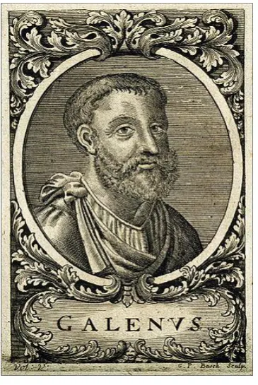
[image:18.595.208.391.370.643.2]The earliest studies in Laryngology date back to the Greek civilization where Hippocrates (400 B.C.) advocated the concept of minimally invasive procedures and proposed that the epiglottis prevents food from entering the airway.(6) .It was in the Roman era that dental mirrors were used to explore the oral cavity. Galen of Pergamon in the second century AD, despite the stiff Roman resistance in conducting cadaveric dissections, performed vivisection and named the epiglottis and recurrent laryngeal nerve(6,7).
4
Julio Casserius ,an Italain , student of Hieronymus Fabriciusgave a detailed description of the anatomy of the larynx of mammals including humans in his book “The Anatomy of Voice and Hearing”(6,8).
The 19th and 20th centuries saw a major boom in technology with the advent of
microscopes, endoscopes and stroboscopes with subsequent advancements and improvisations in operative interventions and techniques in the field of laryngology.
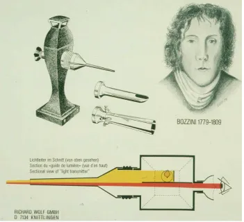
[image:19.595.124.476.396.716.2]Philip Bozzini in 1807 used an external light source (candle-light) channelledby mirrors, placed in an instrument called the Lichtleiter or light conductor. He did not observe the larynx, but his efforts did advance the entire field of endoscopy and he is regarded as the father of minimally invasive surgery.(6,9).
5
The early efforts at visualizing the larynx were all variations of the indirect laryngoscopy using a set of mirrors to reflect back the image to the observer while using an external light source. Benjamin Babington is credited with first developing the “laryngoscope” in 1829, an indirect laryngoscope called glottoscope, which used direct sunlight and mirrors(6,10). Fredrich Hoffman, Liston and Avery in the mid 1800s used concave mirrors which are classically associated with the otolaryngologist. Manuel Garcia, a musician, is oftentimes credited with viewing his vocal cords; described vocal fold motion and generation of voice from the vocal folds in his book ”Observations on the Human Voice”(6).
6
With the discovery of the light bulb by Thomas Edison in 1879 and topical
anaesthesia for the larynx in 1884, many hurdles in the examination of larynx were eliminated. Johann Czermak was the first to use artificial light source and a curved mirror in 1888(6,8).
In the middle of the 19th century, laryngology was a field which was practised by
physicians and neurologists; Jacob Solis-Cohen, a general surgeon, helped in making it a surgical speciality(6).
The term endoscopy was coined by Antonin Desormeaux, a urologist, in 1853(6,11) Direct laryngoscopy, bronchoscopy and oesophagoscopy developed in parallel with each other through sharing of technical advancements. Kussmaul studied the concept of sword swallowers and applied it to view the oesophagus using Desormeaux
7
urethroscope; thus performing the first direct oesophagoscopy in 1868. Alfred Kirsten is credited with developing the first true direct laryngoscope in 1895 and he named it “autoscope”(7,8).
Gustav Killian started using rigid scopes for exploring the airway and he was the first to use a 9mm tube beyond the carina. He also coined the term “bronchoscopy” and introduced the supine position. He developed the inverted V laryngoscope blade to better view the anterior commissure and introduced laryngeal suspension that advanced bimanual surgery and allowed the surgeon to draw diagrams to illustrate the findings(6,8).
8
Chevalier Jackson developed a practical distal lighting method for endoscopic equipment in 1905, by using a side channel (tube within a tube concept). He also introduced distal suction for endoscopes(6,10).
Fig 6: Gustav Killian performing suspension laryngoscopy, Adapted from Laryngeal Imaging, Kendall et al.
9
Yankauer in 1910 developed a laryngoscope wide enough for binocular vision and started using magnification for laryngeal surgeries.
Documentation of findings of laryngeal pathology remained a challenge in early laryngology history. Laryngoscopic photography was introduced by Stein in collaboration with Czermak, and the technique was perfected by French(10). However, the images lacked optimal resolution, were difficult to obtain and required cumbersome equipment and exception skill (citation), thus providing great scope for improvement.
The binocular microscope was introduced by the Zeiss Optical Company in 1953 and in 1960, Scalco described the use of the microscope with the Lynch suspension
10
laryngoscope(6,12). Harold Hopkins invented the rod lens optical system in 1959 wherein a glass rod was used to replace the air interspace between lenses of the telescopes at that time. The new design provided a wider viewing angle and absorbed less light during image transmission(6,13). Karl Storz after observing the prototype for the flexible fibreglass gastroscope, began developing the idea of coupling a fiber optic light source with rigid endoscopes. In 1965, Storz partnered with Hopkins to develop the Storz-Hopkins telescope which had unmatched illumination and superb image resolution(6,13)
11
John Tyndall in 1870, was the first to describe the optical properties of glass rods, which permitted light to be guided. His work lay the foundations of flexible endoscopy. Braid and Hansell incorporated the light guiding properties of glass rods into plastic or glass fibres. These fibres were stretched to make them thin, long and flexible. In 1920, Heinrich Lamm, a medical student, suggested that optical fibre bundles might be utilized in flexible scopes(6,10). The era of flexible endoscopy began with Edward Benedict’s introduction of the flexible gastroscope in 1933 in the U.S. (6,14). The early flexible scopes were flexible only distally to allow accommodation around the patient’s internal anatomy and the optical systems were composed of a sophisticated series of lenses inside the scope which provided very poor image quality.
With further advancements in technology, glass-clad fibres were introduced in which each fibre had a protective coating with a lower refractive index which decreased the interface between fibres, thereby decreasing the attenuation of the image as it travels down the fibre. These technologies led to the development of the completely flexible fiber optic gastroscope in 1957, which was 0.5 inch in diameter and was invented by Hirschowitz and Curtiss(6). From then on, fibreoptic instruments have been developed for gastrointestinal applications first, and then the technology has been modified for use in the larynx and trachea.
12
was much better tolerated by the patient. In 1968, Swashima and Hirose were the first to describe a flexible transnasal endoscope(6,15). This marked the transition of the field of laryngology from mirror-based indirect laryngoscopy to flexible laryngopharyngoscopy; enabling office based laryngology to be practiced. The first intervention to move to office settings was injection laryngoplasty.
13
VISUALIZATION OF THE LARYNX:
Improved understanding of vocal fold physiology and sound production along with the advancements in technology have significantly improved the otolaryngologists’ ability to visualize the larynx. The examination involves a careful evaluation of the anatomic and physiologic aspects of the nasopharynx, pharynx, base of tongue and larynx.
The ideal laryngeal examination technique should assess the function of each intrinsic laryngeal muscle and the integrity of the cricoarytenoid joint, along with evaluating the phonatory function of the larynx. The examination should be non-invasive, easy to perform, inexpensive, quick and cause minimal discomfort/risk to the patient. The test should allow the patient to perform a full range of laryngeal function, allow video recording of the findings and provide means of intervention if needed.(18)
As no ideal examination method exists till date, the laryngeal examination technique should be tailored according to the patient’s condition and compliance. It is therefore important to recognize the advantages and limitations of the various techniques and to choose the appropriate equipment and method to aid in optimal diagnosis. The three commonly used techniques for diagnosis are:
i) Indirect mirror laryngoscopy ii) Direct rigid laryngoscopy
14
INDIRECT MIRROR LARYNGOSCOPY:
The laryngeal mirror was invented by Garcia, a Spanish singing teacher in 1855; Turk and Czermak popularized its medical uses in the 1800s(8,19)
An indirect laryngoscopy requires a laryngeal mirror, external light source, 4-by-4 inch gauze pad and antifog solution.(20)
The patient is seated opposite the examiner and at a slightly elevated level with legs uncrossed. He is asked to lean forward slightly and protrude the tongue. The mirror can be warmed to just above body temperature or dipped in antifog solution or smeared to the patient’s inner cheek or tongue to coat it with saliva, to prevent fogging. (18)
15
The advantages of this method are that it is inexpensive, and allows a rapid, gross examination of the larynx and pharynx(21) . This method is severely limited in comparison to the newer methods, especially as a sole examination technique for patients with dysphonia. The disadvantage is that video documentation is not possible. A large base of tongue, soft palate or a hyper-reflexive gag which prevent an adequate examinationin 5% to 10% patients (21). Moreover, the examination is not performed in a normal physiologic phonation position as the tongue is extended and larynx is elevated.The patient is required to produce a higher pitch voice for optimal visualisation of the vocal cord(21).
16
DIRECT RIGID LARYNGOSCOPY(22,23):
This involves direct visualization of the larynx with the help of a laryngoscope and a light source for illumination. Some of the different types of scopes available are:
i) Abrahamson-Dedo scope: For venturi type ventilation ii) Dedo: Anterior commissure scope
iii) Ossoff: Posterior commissure scope
iv) Holinger: Hour-glass anterior commissure scope v) Jako: Laser scope
vi) Zeitels: Wide-angle scope
Fig 11: The viewing (left to right) and distal ends of Jackson, Hollinger, Zeitels and Bouchayer laryngoscopes, Adapted from Cummings
17
Technique:
The patient is prepared and anaesthesized according to standard protocol. The position is supine with head extended and neck flexed in order to open the laryngeal inlet also known as the sniffing position.
The eye should be covered and dental guard be placed.The largest possible laryngoscope is selected and after adequate lubrication, is inserted into the oral cavity with care to protect the lips from injury with the non-dominant hand.
Midline is maintained while advancing the laryngoscope to identify the base of tongue, valeculla, epiglottis, posterior pharyngeal wall and arytenoids and finally the vocal cords. The larynx is fully examined by moving the laryngoscope and the larynx. The subglottis, bilateral pyriform sinus and post-cricoid region should be visulaized
18
Fig 13: Direct laryngoscopy- technique, Adapted from An Atlas of Head & Neck Surgery, Vol 1 , 4th edition
19
Complications that may be encountered are(23,24)
i. Mucosal injury to lips/angle of mouth
ii. Dental injury
iii. Mucosal injury to tonsillar pillars and tonsillo-lingual sulcus iv. Subcutaneous emphysema of neck
The advantage lies in the fact that the equipment is less expensive than a flexible laryngoscope and is readily available, however the inability to perform a dynamic assessment of vocal cord limits its application(21).
FLEXIBLE NASOPHARYNGOLARYNGOSCOPY (NPL SCOPY):
The flexible laryngoscopy negates the limitations of a rigid laryngoscopy by allowing dynamic assessment of the vocal cords and its neighbouring structures, along with the added advantage of not requiring general anaesthesia. Though the instrument is costly, it is user friendly and can be easily used in an office-setting.
Fiberoptic nasopharyngolaryngoscopes, can be selected in diameters ranging from 1.9 mm to 6 mm and in adult and child lengths.
20
The proximal end of the endoscope has an eyepiece, an angulation control lever, instrument port, focusing ring and light source adapter.
Fig 15: Adult fibreoptic nasopharyngoscope with 4-mm distal diameter, 2-way articulation and video-recording capabilities, Adapted from Medscape
21
The term fibreoptic applies to a system which transmits light and images through thin fibers of optical glass by total internal reflection.
The physics of fiberoptic endoscopy(25)
When light travels through a low density medium such as air and strikes the surface of a high density medium like optical glass, a part of gets reflected back into air and a part of it crosses the interface and gets refracted in the glass. This causes a decrease in the velocity and change in direction of the refracted light.
As shown in the figure above, when incident angle is less than critical angle, part of light is reflected and part of it refracted. When the incident angle is equal to critical angle, part of light beam is reflected and part of it passes along the air glass interface. When the incident angle becomes greater than the critical angle, all light is reflected back at the interface. Thus, when light travels from a high density medium (glass) to a low density medium (air) all of it gets reflected at the interface when the incident angle exceeds the critical angle.
22
This is the concept utilized in a single optical fiber glass, wherein the refracted beam travels along the total length of the fiber by total internal reflection.
By coating the fibers with a thin layer of low index glass, the loss of light at the points of contact with other fibers is avoided, thereby enabling them to be placed in bundles. The fiber bundles are created by arranging them parallel to each other in the horizontal and vertical position, and with the ends of each fiber in exactly the same corresponding position at opposite ends of the bundle. This allows each end to form an image plane that would transmit the image from one end to the other.
23
By placing an objective lens at one end, the image gets focussed onto the image plane; and by placing an eyepiece at the other end, the image gets transferred from the image plane to the eye. The image transmission would remain perfect even if the optical fiber bundle is bent in any direction or contour.
Figure 19: Enlarged view ofOptical glass fiber bundle, Adapted from Wallace FJ. Fiber Optic Endoscopy. J Urol. 1963 Sep;90(3):324–34
24
The fibers used to form these bundles are 0.0005 inches in diameter and each of these image transmitting bundles have around 250,000 fibers. The light transmission through these bundles is very efficient, with approximately 52% light getting transmitted through a 0.225 inch diameter, 30cm long bundle.
The ability of these fiberoptic bundles to transmit high intensity light around corners and through irregular contours has been utilized in developing endoscopes to view the internal anatomy of the human body which is otherwise difficult to view. The optical fibers are made from high refractive index glass and are coated with a low refractive index glass. They are then drawn in the molten state to the desired diameter and arranged as explained earlier. The ends of the stacked bundle are polished. When attached to a high intensity light source they can transmit images of pristine quality.
The structures viewed during a routine flexible nasopharyngolaryngoscopy are:
1. Nose: Nasal turbinates and septum
2. Nasopharynx: Eustachian tube orifice, fossa of Rosenmuller and the nasopharyngeal walls.
3. Oropharynx: soft palate, uvula and base of tongue, vallecula
4. Larynx: epiglottis, aryepiglottic folds, arytenoids, false cords &true cords 5. Hypopharynx: pyriform sinus and post cricoid space
25
Endoscopic anatomy:
The flexible endoscope is introduced via the nostrils into the nasal cavity.
When introducing the scope, first the septum should be examined for any spurs or deviations. The turbinates and meatii on the lateral wall of the nose are examined for hypertrophy or any lesions. Any obstruction to the posterior choana should be noted.
The pharynx(10,26):
It is divided into three parts- the nasopharynx, the oropharynx and the hypopharynx.
26
carcinoma nasopharynx. Adenoid tissue is mostly seen in the posterior wall of the nasopharynx in children and young adults.
The soft palate forms the boundary between the nasopharynx and oropharynx. Endoscopically, the soft palate is seen as a smooth structure sloping away from the nasal floor. If the patient is asked to say “k” the posterior surface of soft palate is pulled superiorly, making it almost horizontal so as to close the nasopharynx.Nowadays rigid nasal endoscopes are preferred for detailed examination of the nose and nasopharynx and flexible NPL is used only when rigid endoscopy cannot be done.
The anterolateral walls of the oropharynx are formed by the tonsils and its pillars. The anterior pillar is not seen in a transnasal endoscopy; but the posterior pillars can be seen. The anterior boundary is formed by the base of tongue and the posterior boundary is formed by the mucosa overlying the constrictor muscles. The mucosa of the base of tongue can appear irregular because due to the lingual tonsils.
The vallecula is an area often obscured by the epiglottis and is better visualised by asking the patient to protrude his tongue. It is bounded laterally by the lateral glossoepiglottic folds and has a median ridge of mucosa called the median glossoepiglottic fold. Small to medium sized inclusion cysts referred to as vallecular cysts may be seen here. The lingual surface of the epiglottis which is often hidden can be visualized at this juncture.
27
post cricoid area and posterior pharyngeal walls. The pyriform sinuses end inferiorly at the level of the cricopharyngeus muscle, which serves as the valve at the superior end of the oesophagus. The inferior pharyngeal constrictors and the mucosa overlying it form the lateral and posterior pharyngeal wall.
The common variations that maybe encountered are prominent internal carotid arteries, lymphoid follicles, greater cornua of hyoid and the thyroid. The carotids might occasionally be seen as pulsatile masses posterolaterally or in the posterior pharyngeal wall. Lymphoid follicles are seen as 2-5mm, pinkish masses on the posterior pharyngeal wall. The cornua of the hyoid or thyroid becomes prominent when the patient is asked to perform Valsalva manoeuvre.
28
The larynx(10,26):
It is divided into the supraglottis, the glottis, and the subglottis.
The medial boundary of the hypopharynx forms the lateral boundary of the supraglottis. The main endoscopic features of the supraglottis are the epiglottis in the midline and the aryepiglottic folds. These folds extend downward from the epiglottis to the arytenoid. It is referred to as the marginal zone as it is a common boundary between the hypopharynx. The epiglottis is a slightly curved structure, with its concave surface facing the larynx; and thus it is called the laryngeal surface of the epiglottis. Laterally the aryepiglottic folds connect it to the arytenoids. The inferior free edges of the supraglottic structures are known as the false vocal folds. The laryngeal ventricles separate the false vocal folds from the true cords. The posterior supraglottis houses the paired arytenoid cartilages and the small corniculate and cuneiform cartilages which cap them. There are two prominences on the body of the arytenoids- the muscular process and the vocal process. The muscular process is situated laterally and is not seen endoscopically; however, the vocal process, which forms the posterior attachment of the vocal fold, can be seen.
29
should be done. The infraglottic surface of the vocal fold extends laterally corresponding to the underlying the conus elasticus.
The subglottis is the part of the larynx inferior to the true cords. The superior boundary has been a topic of debate, but a commonly used rule states that it lies 5 mm below the vocal fold anteriorly and 10mm posteriorly, which corresponds to the lamina of the cricoid posteriorly and its smaller size anteriorly. Posteriorly the subglottis extends to the cricotracheal junction. The subglottis is the area of the larynx with the lowest prevalence of pathology; the common being stenotic lesions, malignancy, or inflammation. To examine this area, patient co-operation is of utmost importance. Care must be taken to not touch any mucosa, which can trigger cough or gag. The scope is advanced towards the anterior commissure and then the adjustable portion of the scope is directed posteroinferiorly.
Indications of NPL scopy:
The indications of flexible nasal endoscopy can be
- Diagnostic - Therapeutic
Diagnostic indications
i) Assessment of nose and nasopharyngeal pathologies
30
ii) Evaluation of velopharyngeal insufficiency:
When the patient counts aloud or repeats a specific phrase, the nasopharyngoscope can be used to assess the pattern of velopharyngeal closure(28). Recent studies suggest that the use of nasopharyngoscopy in addition to conventional speech therapy may help to improve velopharyngeal closure during articulation.
iii) Assessment of vocal cords for hoarseness
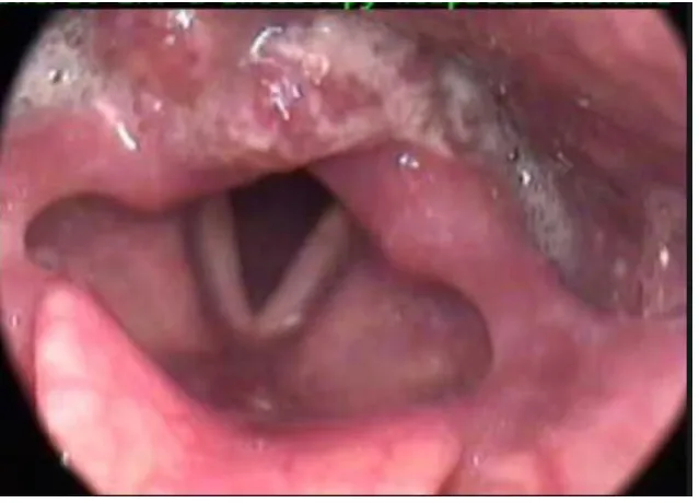
Hoarseness can be the presenting complaint in a number of conditions of the larynx. The common pathology that might be seen during flexible endoscopy are vocal fold nodules, polyps, papillomas, malignancy, Reinke’s oedema, sulcus vocalis etc.(10,23)
31
Fig 23: Left Vocal cord haemorrhagic polyp, Adapted from Cummings Otolaryngology Head and Neck Surgery, 6th edition
32
Fig 25: Reinke’s edema, Adapted from Google Images
33
iv) Airway assessment:
It can also be used in evaluation of patients with difficult airway: in cervicofacial trauma, uncontrolled epistaxis and suspected foreign body ingestion. It may also assist in the evaluation of the severity of angioedema (29).
v) To obtain endoscopy guided biopsies(30):
The flexible endoscope with a working channel is used. After obtaining a good view of the lesion of interest, a 1.8mm flexible cup biopsy forceps is passed through the working channel by the assistant till the forceps is visible 1-2 cm beyond the tip of the endoscope. The forceps are opened and the scope is advanced till the forceps can come into contact with the lesion. The forceps are then closed and removed from the scope. Multiple biopsies
34
can be taken by leaving the scope at the same position till adequate sampling is obtained. The procedure is usually well tolerated by patients. The common complications are bleeding and occasionally injury to normal vocal fold tissue.
vi) To detect lesions in the upper aerodigestive tract(23)
Malignant and benign conditions of the nasopharynx, oropharynx, hypopharynx and larynx can be detected and documentation of the same can be done.
35
Fig 29: Malignancy Supraglottis, Adapted from Cummings Otolaryngology Head and Neck Surgery, 6th edition
36
vii) Functional endoscopic evaluation of swallowing
[image:51.595.168.488.71.301.2]Fiberoptic endoscopic evaluation of swallowing (FEES) has emerged as a comparable alternative to video fluoroscopy and modified barium swallow in the evaluation of dysphagia(31). It was first described by Langmore et al in 1988(32,33). The procedure is ideally done with the patient in seated position, unreclined and awake. By visualizing the pharynx during swallowing, the patient can be assessed for delays in swallowing, laryngeal penetration, aspiration, and pharyngeal residue after swallowing. The use of topical nasal anesthesia is currently controversial for this procedure owing to concern for the risk of desensitizing the pharyngeal or laryngopharyngeal mucosa, which would thereby skew results. However, current American Speech-Language-Hearing Association (ASHA) guidelines allow physicians to choose its use(32). The equipment needed is a flexible
37
laryngoscope with a halogen or xenon light source. A complete examination involves examination of the anatomy at rest and during movement, the accumulated oropharyngeal secretion level and bolus flow of various liquids and solids during swallowing. After noting the anatomy or rather any pathology in the anatomy, the pharyngeal squeeze manoeuvre is performed(34). This involves asking the patient to make high-pitched, strained voice, preferably in a raising crescendo. In a normal person, this will result in recruitment of the pharyngeal constrictor muscles. This serves as a reasonable surrogate for pharyngeal motor strength(35,36). Before presentation of any food, any amount of secretion in the laryngopharynx should be observed and quantified as follows(37):
After this, food is presented to the patient. Food colouring can be used but is not mandatory(34,38). Initially water or ice chips are given, if the patient does well on these then pureed consistencies and crackers or pretzels are presented. The presence of aspiration or penetration should be noted. Any vallecular, pharyngeal wall or pyriform sinus residue also should be noted.
38
FEES is limited by the fact that the oral phase of swallowing is not assessed and in this regard the modified barium swallow is superior to it(34).
Sensory testing can be combined with FEES and it is then known as flexible endoscopic evaluation of swallowing with sensory testing (FEESST). It was first described by Aviv et al(32,39). The basic concept of this test is that stimulation of the supraglottis (with a small puff of air) elicits the laryngeal adductor reflex (LAR); which is a brisk closure of the larynx and can be easily identified. The response is noted on both sides and is quantified at various levels of air-puff pressure.(34)
viii) Evaluation of obstructive sleep apnea
The Müller manoeuvre can help identify patients who could benefit from surgery. The patient should forcefully inspire against a closed nose and mouth, thereby evoking retropalatal collapse. However, these findings are not clearly correlated with the need for surgical intervention, and the subject remains controversial(40,41).
Sleep nasendoscopy was first described by Croft and Pringle in 1991. Keziran and Hohenhorst changed it to “ drug-induced sleep endoscopy” (DISE)(42). The three key features of this method are:
- Use of pharmacologic agents to achieve sedation.
- Target depth of sedation as approximating natural sleep as possible. - Endoscopic evaluation of upper airway during this period.
39
stimuli at normal conversational volume. The drugs commonly used for the procedure are Propofol, midazolam, dexmedetomidine or a combination of Propofol and midazolam.
The endoscopic examination is mainly an assessment of the pharynx, which is achieved by moving the scope a number of times to evaluate the entire length of the pharynx during cycles of airway obstruction and normal breathing. The VOTE classification is used as standard for DISE grading(42). This involves grading the level of obstruction at the 4 major structures which cause airway obstruction in most patients: Velum(palate), oropharyngeal lateral walls including tonsils, tongue and epiglottis. The level of obstruction is graded as:
- None: No vibration of involved structure and <50% airway narrowing as compared to normal breathing.
- Partial: Vibration and 50-75% airway narrowing - Complete: Obstruction and >75% airway narrowing ix) Stroboscopy(43):
Oertel developed the first stroboscope in 1895, but the procedure gained wideapread acceptance only in the later part of the 20th century.
40
generating a series of still images of the vocal fold at different points of the glottic cycle. These images are then fused into an apparent continuous sequence and can be observed by the examiner(44).
x) Narrow band imaging:
[image:55.595.125.510.162.444.2]Squamous dysplasia and carcinoma in situ of the upper aerodigestive tract is very difficult to detect by routine endoscopic examination. Narrow band imaging is a novel technique for visualizing mucosal surfaces using narrow bands or selected portions of the light spectrum. In general the long wavelengths(red) diffuse deeper, whereas the short wavelengths (blue) diffuse superficially. Also, the blue spectrum corresponds to the peak
41
absorption of hemoglobin, thus any abnormal superficial blood vessels or angiogenesis will appear darker and more obvious. The procedure can be easily done in the out-patient setting ,using appropriate light filters in conjunction with flexible laryngoscopy(45,46).
xi) Optical coherence tomography- OCT(47–49):
[image:56.595.133.545.187.433.2]It is an innovation in the field of optical imaging technique which provides high resolution images (~10 to 20um), cross-sectional images of microscopic subsurface structures. The images are generated by measuring the intensity of the scattered back light after probing the tissue with a low-power, near-infrared source of light. Interferometric methods are used to detect light that gets reflected back from tissue, which is used to create a 2-dimensional image. The image formed is similar to a vertical histologic section. The OCT system can be used to scan tissue in a linear, transverse or radial fashion and can be easily interfaced with endoscopes, catheters, laproscopes etc. It provides information about the thickness of the
42
epithelium, integrity of basement membrane and the structure of lamina propria. This can be very useful in the detection of early glottic malignancies and premalignant conditions. It can also differentiate scar tissue and inflammation from malignant conditions. Thus, directed biopsies can be obtained.
[image:57.595.235.418.222.539.2].
43
Therapeutic indications:
i) Office based injection laryngoplasty:
44
ii) Percutaneous tracheostomy:
45
A 16-gauge Teflon introducer needle is now inserted between the first and second or second and third tracheal rings. The needle is withdrawn and a J-tipped guidewire is advanced through the outer sheath of the introducer. Serial dilation is then performed. The tracheostomy tube which is prefitted with an appropriately sized loading dilator is threaded over the guidewire and inserted into the trachea. The whole procedure is visualized via an endoscope and reduces technical difficulties of the procedureand complications like accidental extubation, false passage creation, pneumothorax, pneumomediastinum. The disadvantage is that endoscopy adds extra cost, time and can cause difficulty in ventilating patient and CO2 retention.(56)
iii) Fiberoptic intubation(57):
46
Contraindications of flexible laryngoscopy:
Nasopharyngoscopy is considered a very safe procedure with few contraindications and complications in experienced hands.
[image:61.595.200.471.71.282.2]Relative contraindications include coagulopathies that might result in significant bleeding if even minor trauma occurs during the procedure. In the setting of craniofacial trauma, the benefits should be carefully weighed against the risks of inadvertent intracranial instrumentation and exacerbation of nasopharyngeal injuries. Epiglottitis can also be considered as a relative contraindication in view of impending airway compromise(20).
47
Positioning:
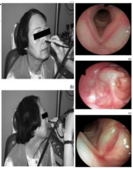
All patients being examined should be seated with the head extended forward and the neck flexed on the chest (sniffing position) to bring the pharynx and larynx into alignment. A headrest should be used to stabilize the head. Orientation of the distal tip of the endoscope can be controlled with the index finger or thumb through a switch. The dominant hand should hold the scope while the non-dominant hand can be used to stabilize the scope by resting it on the patient’s nose or cheek.
The examination can also be done with the patient in supine position. If this position is used, the clinician should be standing at the head end of the patient, a little to left of midline with similar endoscopic control as described above. In this position, however, the visualization of structures caudal to the nasopharynx is poor because the tongue falls back.
Technique(20):
48
The patient is asked to breathe through the nose, to separate the palate from the posterior nasal wall and enable passage of the scope into the oropharynx. The scope is directed downwards to give a panoramic view of the oropharynx. The palatine tonsils can be seen laterally along the palatopharyngeal arches and the base of tongue and the lingual tonsil can be seen in the midline. The scope is passed further down to view the epiglottis and the vallecula. When directing the tip of the scope posteriorly, the laryngeal surface of epiglottis, arytenoids, aryepiglottic folds, false vocal folds, true vocal cords, and pyriform sinus are visualized. The true cords appear white and taut; any change in the mucosal colour or any superficial irregularities should be noted.
With deep breathing the cords are abducted and the glottis remains open; the patient can be asked to sniff or inspire deeply through the nose to cause maximal vocal cord abduction so as to allow optimal laryngeal examination(58). To assess the function and movement of the cords and the arytenoid cartilages, the patient is asked to say “e” or “ah”. The subglottis can also be usually seen just below the true cords. The scope should not be passed through the true cords as this can elicit laryngospasm. The hypopharyngeal region should also be examined closely to rule out any abnormalities.
There are many maneuvers that have been described to help in better visualization of the above structures and facilitate the advancement of the scope. These are:
49
- For visualization of hypopharynx:
i) The head torsion technique(59):
The patient’s head is turned to the side which needs to be observed in detail. This gives a clear view of the inferior surface of the vocal fold and the ventricles of the same side and a better visualization of the pyriform sinus of the opposite side.
i) Valsalva manoeuvre(60):
[image:64.595.189.406.265.541.2]The patient is asked to blow with their mouth closed. This provides better view of the pyriform sinus and post-cricoid area.
50
ii) Trumpet manoeuvre(61,62)
The patient is asked to keep his index finger between his lips and blow as hard as possible, as though trying to inflate a balloon. This causes the larynx to get elevated and the pyriform sinus and post-cricoid areas to be better visualized. iii) Anterior neck traction(44,62)
While performing the endoscopic examination, anterior neck skin traction is given and the patient is asked to perform Valsalva. This enables better visualization of the hypopharynx.
iv) Modified Killian method(63,64):
[image:65.595.198.398.382.576.2]This was first described in 2014.
51
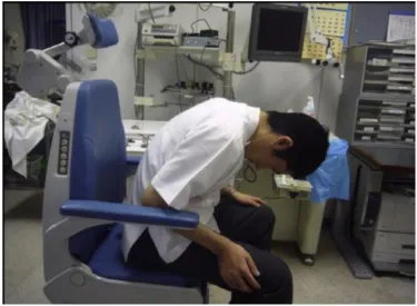
- The patients neck is bent forward and the chin depressed far enough for the patient to look at his umbilicus. This is the Killian position.
- In the modified Killian position, the patient is further bent forward from this position, as if bowing.
[image:66.595.128.504.207.482.2]- With the patient in this position, the head is turned to either side and is asked to perform Valsalva manoeuvre. This combination of modified Killian position, head torsion and Valsalva manoeuvre constitutes the modified Killian method. Fig 40: The modified Killian position, Adapted fromSakai A, Okami K,
52
[image:67.595.103.498.108.408.2]-- This method has shown to give a clearer view of the entire circumference of the hypopharyngeal space and the cervical oesophageal entry; thus becoming an indispensable technique in identifying lesions of the hypopharynx.
53
- For visualization of subglottis(65)
After advancement of the scope till just above the glottis, the patient is asked to place his chin on the chest, while continuing to breathe through the nose. When done correctly, this provides clear view of the lateral and posterior subglottis. Fig 42: Fiberoptic findings with (A) normal position, (B) with Valsalva manoeuvre and (C) with the MK method; Adapted fromSakai A, Okami K, Sugimoto R, Ebisumoto K, Yamamoto H, Maki D, et al. A new technique to expose the
54
Cleaning and disinfection of endoscopes(66):
After use, the endoscopes must be thoroughly cleaned with water and detergent. Air is expelled from all channels and the scope is bathed in 2% glutaraldehyde solution. This is noncorrosive to rubbers, plastics and metals and destroys bacteria and viruses within 4 minutes. The disinfection time is 20 mins at 20 0 centigrade. Other agents that
[image:69.595.108.394.75.336.2]can be employed for disinfection are alcohol, chlorine dioxide, peracetic acid and Cidex OPA(0.55% ortho-phthaldehyde).(67,68) Cidex OPA is being used as an
55
alternative agent to 2% glutaraldehyde as it is fast acting High-level disinfectant and does not cause respiratory irritation when compared to glutaraldehyde(68)
Complications of flexible laryngoscopy:
The complications of the procedure are uncommon, the usual ones being tearing, coughing and occasional epistaxis. Rarely there can be transient laryngospasm, vasovagal syncope and residual foreign body sensation. The incidences of desaturation and laryngospasm are more common in the pediatric population(69).
NASAL PREPARATION PRIOR TO ENDOSCOPY
56
There are however no clear guidelines as to which mode of application is the best or whether any anaesthetic is needed at all(2,3).
Pain (which is a noxious stimuli) anywhere in the body is detectedby the free nerve endings of primary afferent C type fibers and Aδ fibers. The inflammatory mediators (like serotonin, bradykinins, cytokines, prostaglandins etc.), which are released from damaged tissue, can stimulate these fibers directly and cause reduction in the activation threshold of these fibers, thereby causing reduced tolerance to pain.
[image:71.595.72.478.393.712.2]These fibers then synapse with secondary afferent neurons in the dorsal horn of the spinal cord, and the sensation are finally carried by the spinothalamic tract and spinoreticular tract to the somatosensory cortex in the brain.
57
The agents commonly used for this purpose are:
Local anaesthetics:
These are drugs that bind reversibly to specific receptor site in the pore of the sodium channels located in the nerves, thereby blocking ion movement in the pore. When the agent is applied in appropriate concentrations, it can act on every part of the nervous system, irrespective of the type of nerve fiber. It causes both motor and sensory paralysis in the area innervated by the nerve. The effect of the drug is reversible with recovery of nerve function, without any damage to nerve fibers, when used in the clinically recommended dose.
These drugs have an aromatic and an amine group separated by an intermediate group. Based on this they are divided into two groups:
a. Esters- these have an ester link between the intermediate group and the aromatic portion. Examples are: Tetracaine, Benzocaine, Procaine
58
Mechanism of action:
These drugs block conduction by preventing the large transient increase in
[image:73.595.174.391.81.302.2]permeability to sodium ions of excitable membranes, which normally occurs by a slight depolarization of the membrane. These agents interact directly with voltage gated Na+ channels to block conduction across these reversibly.
Fig 46: Mechanism of action of local anaesthetics, Adapted from Google images.
59
The action is dependent on the type of nerve fiber and frequency of stimulation. Small unmyelinated C fibers (which mediate pain), autonomic fibers and small myelinated Aδ fibers get blocked before the larger myelinated Aγ, Aβ and Aα fibers (which mediate touch, position, pressure and motor information). Also, a resting nerve is much less sensitive to the action of local anaesthetic than a nerve which is repeatedly stimulated; thus, higher frequency of stimulation and a higher positive resting membrane potential cause a greater degree of anaesthetic blockage.
Prolongation of action by using vasoconstrictors:
The duration of action of local anaesthetics is directly proportional to the time of contact with the nerve fiber. Catecholamines which act on α-adrenergic receptors cause vasoconstriction, thereby reducing absorption of local anaesthetics and prolonging their duration of action. So, in clinical practice a vasoconstrictor, mostly adrenaline, is added to the local anaesthetics. The only disadvantage of this is that the systemic side effects of the local anesthetics is increased because of deposition in skeletal beds (β2 adrenergic action causes dilation of vascular bed in skeletal muscles).
Side effects of local anaesthetics:
These effects are due to systemic absorption of these agents, and the danger of such reactions is proportional to the concentration of drug in systemic circulation.
60
stages of intoxication, there is CNS depression and death due to respiratory failure.
b. CVS: These agents cause decrease in excitability, conduction rate and force of contraction, usually due to accidental direct intravascular injection, especially if adrenaline is also present. The CVS effects usually occur after high systemic concentrations are obtained and after the CNS effects are produced. Rarely, lower doses of some agents can cause sudden cardiovascular collapse and death, probably due to sudden onset of ventricular fibrillation or action on pacemaker.
c. Smooth muscle: These drugs depress contractions in vascular, bronchial and gastrointestinal smooth muscles.
Hypersensitivity: Some people are hypersensitive to local anaesthetics and have allergic dermatitis or a typical asthma attack. The ester group of local anaesthetics is known to be more notorious in causing hypersensitivity; and also the addition of vasoconstrictor increases allergic reactions due to addition of sulfite as an antioxidant.
Metabolism of local anaesthetics:
61
between rates of absorption and elimination, the rate of degradation of the drugs becomes very important in determining the clinical safety of these agents.
The commonly used local anaesthetics in otolaryngology clinic:
1. Lignocaine or Lidocaine:
This agent has intermediate to low potency, an onset of action of about 90 seconds and duration of action of 45-60 minutes. The topical preparations available are:
i. 1%, 2% and 4% solutions
ii. 2% viscous oral gel
iii. 2% aqueous based gel
iv. 4% and 10% spray
The maximum dose allowed without causing systemic toxicity is 3-5 mg/kg and topical preparations for office-based otolaryngology clinics rarely exceed the toxic dose and cause adverse reactions(72,73). To prevent potential lignocaine toxicity, the pharmacology and safety profile of the drug cannot be overlooked.
62
death due to respiratory failure. The CVS manifestations include reduced contractility, conduction rate and excitability of cardiac muscles.
If an overdose/toxicity is detected the procedure should be discontinued, and pulse oximetry and hemodynamic monitoring should be initiated. Early supplementation of oxygen could prevent acidosis and hypoxia, and retard progression towards seizure and hemodynamic collapse(74). ICU support might be necessary in cases of severe toxicity.
Methemoglobinemia is a condition in which hemoglobin cannot bind to oxygen, because of its oxidation from ferrous state to the ferric state. It’s a rare complication of the use of lignocaine and has been reported in bronchoscopy procedures, when lignocaine was combined with benzocaine(75). The symptoms include anxiety, headache, fatigue, coma and sometimes death. When the levels of methemoglobin is less than 30%, spontaneous recovery can be obtained in about 24 hours by removing the offending agent and administering oxygen. For severe cases, methylene blue at a dose of 1-2 mg/kg administered slowly intravenously is the treatment of choice.
Tetracaine:
It is a highly potent local anaesthetic, which has a slow onset of action of around 10 minutes, and duration of action between 60 to 120 minutes.
63
Sympathomimetics
Xylometazoline/ Oxymetazoline:
These are sympathomimetic drugs which act on alpha 1A-adrenoceptor agonists and cause vasoconstriction of cutaneous microvasculature. The onset of action is within seconds and the duration of action lasts for about 6 hours. The common side effects are burning, stinging, sneezing, dryness and sometimes headache. Rebound congestion is the undesired side effect which occurs due to prolonged use of the drug and leads to a condition called rhinitis medicamentosa.
TOPICAL AGENTS IN FLEXIBLE NPL SCOPY
There are no clear guidelines for the use of topical anaesthetic drugs for performing NPL scopy. Types of preparation which have been traditionally used include
lidocaine, phenylephrine and lidocaine, oxymetazoline, xylometazoline and cocaine. Some of these were found to cause lessdiscomfort than others, some had side effects and some others were more expensive. Any method whichreduces discomfort will help in the examination, diagnosis and treatment of patients’ disorders.
Studies have compared different preparations to placebo and to no treatment (1–5), but it remains unclear which is the best preparation to use, if any. There can also be a trade-off between using agents that may be beneficial to the patient in terms of
reducing pain and discomfort but, for example gels and pastes, may obscure the image for the endoscopist(71).
64
between the groups(1). Georgalas et al compared the effect of co-phenylcaine and saline and found no significant difference between the two groups. They also reported a worse taste scores for the co-phenylcaine(4). Leder et al compared tetracaine,
ephedrine and saline and found no difference in the outcome measure(5). Frosh et al compared lidocaine, saline and no treatment and demonstrated a significantly higher pain scores in the lignocaine group(2). Sadek et al in 2001 compared lignocaine/ phenylephrine, xylometazoline, lignocaine and no treatment. This study did not demonstrate any difference in the pain scores but reported a reduced level of general unpleasantness when using vasoconstrictor(76).
The study by Lennox et al and Smith et al compared co-phenylcaine and cocaine and did not find any difference in the pain scores(77,78). As these studies did not make a comparison against placebo or no treatment, a conclusion on the benefit of topical agents cannot be drawn.
65
MATERIALS AND METHODS
Study design:
A randomized control open label study
Study population:
Patients who attended the ENT out-patient department and for whom a flexible nasopharyngolaryngoscopy was ordered for evaluation of their ENT complaints.
Inclusion criteria:
1. Patients requiring a transnasal flexible nasopharyngolaryngoscopy
2. Age > 18 years
3. Patients who could complete the post-procedure questionnaire following the examination
Exclusion criteria:
1. Any known bleeding disorders
2. Pregnancy
66
4. History of a previous nasopharyngolaryngoscopy- to reduce bias on objective assessment.
Pilot study:
A pilot study was done to assess the feasibility of the study and to calculate sample size. In the pilot, 10 patients were included in each arm. The results of the pilot study are summarized in the table below.
Agent Mean
Lignocaine spray 2.6
Lignocaine gel 2.9
Pledget 2.5
Aqueous gel 3.0
Study period:
67
Ethics Committee Approval:
Once the study proposal was made, it was put forward to the Institutional Research Board. After obtaining the approval from the ethics committee (IRB Min No. 9801), the study was initiated in August,2016.
Statistics:
Sample size calculation:
Using pilot study data with a maximum difference of 1 in mean pain score between any 2 groups with a standard deviation of 2.1, 80% power and 5% α ; the sample size was calculated as 95 in each group.
Study recruitment:
Patients who had to undergo a nasopharyngolaryngoscopy were included in the study based on the inclusion and exclusion criteria mentioned above. The patients who met the inclusion criteria were randomly allocated to one of the four groups:
i. 10% lignocaine spray
ii. 2% lignocaine gel
iii. 4% lignocaine solution with 0.05% xylometazoline (1:1) pledgets
iv. Water based aqueous gel
68
in each nostril. In the gel group, 1-2 ml of the gel was applied on the scope prior to scopy.
Then, the endoscopy was performed and at the end of examination, the patient and the examiner were given separate questionnaires to fill up. (Appendix page iv and v)
[image:83.595.99.481.256.664.2]The methodology algorithm was as follows:
69
Statistical analysis:
Categorical variables were summarized using counts and percentages. Quantitative variables were summarized using mean (SD) for normally distributed variables and median (IQR) for skewed variables. Chi square test was used to compare the
proportions between categorical variables. One way analysis of variance was used to compare the mean between more than two groups for the normally distributed
variables and for skewed variables Kruskalwallis H test was used. The Independent t-test was used to compare the means between two groups for normally distributed continuous variables and the Mann-Whitney U test was used for skewed
70
RESULTS
This prospective randomised open label study was conducted in the department of ENT in Christian Medical college, Vellore. Patients who had to undergo a flexible nasopharyngolaryngoscopy for the first time were eligible to participate in the study and were randomly allotted into the four study groups, if they fulfilled the inclusion criteria. A total of 376 patients were recruited into the study from the period between August,2016 to August,2017. The baseline characteristics of the study population and the statistical analysis are described below.
BASELINE CHARACTERISTICS OF STUDY POPULATION
Randomization
71
The 4 missing were due to loss of randomization envelopes.
Age
The mean age (± SD) of the patients was 42.8(13.6). The youngest patient was 18 years and oldest patient was 80 years. The age distribution of the population was as follows:
94
94 95
93
4
AGENT
[image:86.595.69.516.72.378.2]LIG SPRAY LIG GEL PLEDGET AQ GEL MISSING
72
Sex distribution:
Among the total of 376 patients who were recruited, 211 were males and 165 were females.
33
141
128
67
7
0 20 40 60 80 100 120 140 160
[image:87.595.72.509.71.324.2]<25 26-40 41-55 56-70 >70
73
The sex distribution in all the different groups of intervention was as follows: 211, 56%
165, 44%
Male Female
60
51
47
53
34
43
48
40
0 10 20 30 40 50 60 70
LIG SPRAY LIG GEL PLEDGET AQ GEL
[image:88.595.135.458.62.318.2]MALE FEMALE
Fig 50: Sex distribution of study population
[image:88.595.72.473.368.661.2]74
STATISCAL DATA ANALYSIS
After the endoscopy was performed, all patients were given a questionnaire and asked to grade the symptoms on a 10 point visual analogue scale where 1 was when the least symptoms were experienced and 10 the most symptoms were experienced.
1. Overall pain/ discomfort
The mean(±SD) for the overall pain/discomfort was 2.98(1.06) in the lignocaine spray group, 2.89(1.45) in lignocaine gel group, 2.8(0.91) in the otrivin-lignocaine pledget group and 3.03(1.17) in the aqueous gel group. The p value was 0.527; thus, there was no statistically significant difference between the groups.
2.98
2.89
2.8
3.03
2.65 2.7 2.75 2.8 2.85 2.9 2.95 3 3.05
lig spray lig gel pledget aqueous gel
[image:89.595.107.483.416.754.2]discomfort/ overall pain
75
2. Gender vs overall pain/discomfort
There was found to be no significant difference in the overall pain/discomfort scores in males and females (p=0.8481).
2.895 2.9 2.905 2.91 2.915 2.92 2.925 2.93 2.935 2.94 2.945
male female
pain/discomfort
[image:90.595.105.549.219.694.2]pain/discomfort
76
3. Burning/ stinging after application of agent
As the data was not normally distributed, the median and interquartile range (25th to 75th percentile) for each of the groups is mentioned in the table below.
The p value was 0.0001, thus saying that the burning is significantly higher in the lignocaine spray group when compared to the other groups.
MEDIAN IQR(0.25,0.75)
LIG SPRAY 2.00 1.00, 2.00
LIG GEL 1.00 1.00, 1.00
PLEDGET 1.00 1.00,2.00
AQ GEL 1.00 1.00, 1.00
4. Nose pain
As the data was not normally distributed, the median and interquartile range (25th to 75th percentile) for each of the groups is mentioned in the table below.
[image:91.595.68.468.282.515.2]The p value was 0.4875; hence, there was no statistically significant difference in the scores between the 4 groups.
77
MEDIAN IQR(0.25,0.75)
LIG SPRAY 1.00 1.00, 2.00
LIG GEL 1.00 1.00, 2.00
PLEDGET 1.00 1.00,2.00
AQ GEL 1.00 1.00, 2.00
5. Throat pain
As the data was not normally distributed, the median and interquartile range (25th to 75th percentile) for each of the groups is mentioned in the table below.
The p value was 0.7839; hence, there was no statistically significant difference in the scores between the 4 groups.
MEDIAN IQR(0.25,0.75)
LIG SPRAY 1.00 1.00, 1.00
LIG GEL 1.00 1.00, 1.00
PLEDGET 1.00 1.00,1.00
AQ GEL 1.00 1.00, 1.00
Table 2: Nose pain in the various groups
78
6. Gagging/ nausea sensation
As the data was not normally distributed, the median and interquartile range (25th to 75th percentile) for each of the groups is mentioned in the table below. The p value was 0.3185; hence, there was no statistically significant difference in the scores between the 4 groups.
MEDIAN IQR(0.25,0.75)
LIG SPRAY 1.00 1.00, 2.00
LIG GEL 1.00 1.00, 1.00
PLEDGET 1.00 1.00,1.00
AQ GEL 1.00 1.00, 1.00
7. Unpleasant taste
As the data was not normally distributed, the median and interquartile range (25th to 75th percentile) for each of the groups is mentioned in the table below.
The p value was 0.0928; hence, there was no statistically significant difference in the scores between the 4 groups.
MEDIAN IQR(0.25,0.75)
LIG SPRAY 1.00 1.00, 1.00
LIG GEL 1.00 1.00, 1.00
PLEDGET 1.00 1.00,1.00
AQ GEL 1.00 1.00, 1.00
Table 4: Gagging scores in the various groups