0022-538X/09/$12.00 doi:10.1128/JVI.01676-09
Copyright © 2009, American Society for Microbiology. All Rights Reserved.
Interaction of ICP34.5 with Beclin 1 Modulates Herpes Simplex Virus
Type 1 Pathogenesis through Control of CD4
⫹
T-Cell Responses
䌤
David A. Leib,
1,2* Diane E. Alexander,
1Douglas Cox,
1Jiyi Yin,
1and Thomas A. Ferguson
1Departments of Ophthalmology and Visual Sciences1and Molecular Microbiology,2Washington University School of Medicine,
St. Louis, Missouri 63110
Received 10 August 2009/Accepted 9 September 2009
Autophagy is an important component of host innate and adaptive immunity to viruses. It is critical for the degradation of intracellular pathogens and for promoting antigen presentation. Herpes simplex virus type 1 (HSV-1) infection induces an autophagy response, but this response is antagonized by the HSV-1 neuroviru-lence gene product, ICP34.5. This is due, in part, to its interaction with the essential autophagy protein Beclin 1 (Atg6) via the Beclin-binding domain (BBD) of ICP34.5. Using a recombinant virus lacking the BBD, we examined pathogenesis and immune responses using mouse models of infection. The BBD-deficient virus (⌬68H) replicated equivalently to its marker-rescued counterpart (⌬68HR) at early times but was cleared more rapidly than⌬68HR from all tissues at late times following corneal infection. In addition, the infection of the cornea with⌬68H induced less ocular disease than⌬68HR. These results suggested that⌬68H was attenuated due to its failure to control adaptive rather than innate immunity. In support of this idea,⌬68H stimulated a significantly stronger CD4ⴙ T-cell-mediated delayed-type hypersensitivity response and resulted in signifi-cantly more production of gamma interferon and interleukin-2 from HSV-specific CD4ⴙT cells than⌬68HR. Taken together, these data suggest a role for the BBD of ICP34.5 in precluding autophagy-mediated class II antigen presentation, thereby enhancing the virulence and pathogenesis of HSV-1.
Autophagy is a conserved cellular pathway that eliminates defective proteins and organelles, prevents abnormal protein aggregate accumulation, and removes intracellular pathogens (11, 22, 32, 56). This process begins with the formation and elongation of a double membrane that fuses to form an auto-phagosome. The cytoplasmic contents are nonspecifically se-questered inside the autophagosome and then are degraded once the autophagosome fuses with the lysosome. Autophagy is upregulated during starvation, growth factor withdrawal, hypoxia, and infection (10). Following metabolic stress, auto-phagy can generate metabolic precursors that can be recycled for the de novo synthesis of proteins. The autophagic pathway has important roles in development, immune defense, apopto-sis, tumor suppression, and the prevention of neuronal degen-eration (reviewed in references 21 and 31).
Autophagy is not limited to the degradation of self proteins; it also can engulf and break down invading microorganisms in a process termed xenophagy (26). Xenophagy can limit the replication of pathogens (3, 4, 29, 34, 42), but some infectious agents can exploit autophagy to enhance their replication (2, 18, 38). There are pathogens that actively inhibit autophagy through interaction with the essential autophagy-promoting protein, Beclin 1 (24, 35). Beclin 1 is the mammalian homolog of yeast Atg6 and is required for the formation of the auto-phagosome membrane through its interaction with VPS34, a class III phosphatidylinositol 3-kinase (19, 27). Autophagy/ xenophagy is also an important process for the adaptive
im-mune response to infection through the delivery of antigens for major histocompatibility complex class I and II (MHC-I and -II) presentation (7, 9, 12, 36, 45). The inhibition of autophagy by pathogens therefore would serve to block CD4⫹and CD8⫹ cell responses and allow pathogens to remain underrecognized by the adaptive immune response.
The interferon (IFN)-inducible double-strand RNA-induc-ible protein kinase (PKR) pathway is required for virus- and starvation-induced autophagy (50). The PKR-mediated induc-tion of autophagy requires the phosphorylainduc-tion of the transla-tion initiatransla-tion factor eIF2␣(50). Herpes simplex virus type 1 (HSV-1) inhibits autophagy through at least two domains and the activities of the late protein ICP34.5, its C-terminal medi-ation of dephosphorylmedi-ation of eIF2␣, and its N-terminal bind-ing to Beclin 1 (5, 16, 35). HSV-1 strains lackbind-ing ICP34.5 show significant attenuation in vivo, increased sensitivity to IFN (33), an inability to counteract the PKR-induced phosphoryla-tion of eIF2␣, and the induction of the generalized shutoff of protein synthesis in infected cells. These activities originally were ascribed solely to ICP34.5’s ability to recruit PP1␣and redirect its activity to dephosphorylate eIF2␣to counteract the general shutoff of protein synthesis mediated by PKR (17). This function is mediated by the C-terminal domain of ICP34.5 that contains homology to the growth arrest and DNA damage 34 (GADD34) gene (6). Independently of the role of ICP34.5 in countering the PKR-induced antiviral state, viruses lacking ICP34.5 also exhibit altered patterns of autophagy. This man-ifests as increased long-lived protein degradation, the in-creased formation of autophagosomes, inin-creased autophagic vacuole volume density, and the enhanced xenophagic degra-dation of virions (35, 50, 51). Such alterations in the autophagy pathway now can be ascribed to ICP34.5’s ability to bind Beclin 1 in addition to its mediation of eIF2␣dephosphorylation (35).
* Corresponding author. Present address: Department of Microbi-ology and ImmunMicrobi-ology, Dartmouth-Hitchcock Medical Center, 630E Borwell Bldg., HB 7556, 1 Medical Center Dr., Lebanon, NH 03756. Phone: (603) 650-8616. Fax: (603) 650-6223. E-mail: David.A.Leib @Dartmouth.edu.
䌤Published ahead of print on 14 October 2009.
12164
on November 8, 2019 by guest
http://jvi.asm.org/
The deletion of the Beclin 1-binding domain (BBD) of ICP34.5 renders HSV-1 less able to regulate autophagosome forma-tion, and viruses lacking this domain are neuroattenuated (35). To determine the impact of Beclin 1-ICP34.5 interactions on the pathogenesis of HSV-1 during infection, we examined the ability of BBD-deficient virus to replicate, cause disease, and stimulate an immune response in mice. We determined that mice infected with HSV-1 lacking the ICP34.5 BBD were able to clear virus more efficiently than mice infected with wild-type virus. We observed the significantly enhanced stimulation of CD4⫹T cells by the virus lacking the BBD compared to that of mice infected with wild-type virus. These data suggest that Beclin 1 binding and the inhibition of autophagy by ICP34.5 are important for HSV-1 pathogenesis through its ability to suppress autophagy and to dampen the activation of CD4⫹T cells.
MATERIALS AND METHODS
Cells and viruses.African green monkey kidney (Vero) cells were propagated as previously described (40). Viruses used in this study were in the background of strain 17syn⫹. The ICP34.5 Beclin 1-binding mutant (⌬68H) and its marker-rescued virus (⌬68HR) have been described previously (1).
Animal procedures.In all experiments, male and female 6- to 8-week-old strain 129 Ev/Sv mice and C57BL/6 (Charles River Laboratories, Wilmington, MA) or congenicRag1⫺/⫺andIrf3⫺/⫺mice (kindly provided by Michael
Dia-mond with permission from Tadatsugu Tanaguchi) were used (43, 47). For neurovirulence experiments, anesthetized mice were infected intracranially with 20l of virus at the indicated dose (49). These mice were monitored daily for survival for 21 days. For corneal infections, corneas were bilaterally scarified with a 25-gauge needle and inoculated with 2⫻106PFU of virus in a volume of 5l
as previously described (40). Eye swab material was collected and assayed for virus by standard plaque assay as previously described (25). Trigeminal ganglia and 6-mm biopsy specimen punches of periocular skin were removed and placed in 1.5-ml tubes containing 1-mm-diameter glass beads and 1 ml of medium. Biopsy punches of periocular skin also were taken for hematoxylin and eosin staining for histopathology. Trigeminal ganglia and periocular skin homogenates were prepared by freezing and thawing samples, mechanically disrupting them in a Mini-Beadbeater-8 (Biospec Products, Bartlesville, OK), and sonicating them. Brain homogenates were prepared by freezing and thawing samples, shaking them in 4 ml of medium with 3-mm-diameter glass beads, and clarifying them at 5,000⫻gfor 5 min. The titers of homogenates were determined by plaque assay on Vero cells, and the amount of virus was expressed as the PFU per milliliter of tissue homogenate. Mice also were observed and scored for disease and weighed for the indicated time period. Scoring was done in a semiquantitative fashion on a scale of 0 to 4, with a score of 0 being no disease and 4 being death (46). Disease scores represent a composite score reflecting keratitis, blepharitis, neu-rological symptoms, and general health, such as fur ruffling and lethargy.
Latency establishment and reactivation assays.Ocular infections were per-formed as described above, except with 2⫻104PFU/eye of the indicated virus.
To assay for the establishment of latency, mice were sacrificed 28 days postin-fection and their trigeminal ganglia were removed. DNA was isolated using a DNeasy tissue preparation kit (Qiagen, Valencia, CA). The number of latent genomes per trigeminal ganglia was determine by real-time PCR essentially as described previously (48). Briefly, a 70-bp fragment of the thymidine kinase (tk) gene was amplified from trigeminal ganglia DNA and 10-fold dilutions of HSV-1 bacterial artificial chromosome (BAC) DNA (17-49 BAC) (14). BAC DNA was used simply to generate a standard curve to determine the number of genome copies per trigeminal ganglion, since BAC DNA models the episomal, latent genome. To control for the total DNA content of each sample, the single-copy mouse adipsin gene was amplified in each sample along with dilutions of mouse genomic DNA to generate a standard curve. The values for the tk copy number were normalized to the lowest value of the mouse adipsin copy number to yield the normalized genome copy per ganglion, which was expressed on a log scale. For the reactivation assay, mice were sacrificed 28 days postinfection, and their trigeminal ganglia were removed, bisected, and explanted onto Vero cell mono-layers essentially as described previously (25). For 7 days after explant, 100-l samples of culture supernatant were sampled and replated on a fresh monolayer of Vero cells and monitored for the presence of infectious virus.
Measurement of CD4 T-cell responses, T-cell depletion, and cytokine expres-sion.For the measurement of delayed-type hypersensitivity (DTH), mice were infected on the cornea with⌬68H or⌬68HR as described above. Seven days later, recipient mice were challenged in the right footpad with 1⫻106PFU of
UV-inactivated⌬68HR (15) and in the left foot with culture medium (0.033 ml per injection). Twenty-four hours later, footpad thickness was determined with a micrometer by a masked observer. DTH responses were recorded as the differ-ence between the right and left footpads and expressed as micrometers⫾ standard errors of the means (SEM). The depletion of CD4⫹and CD8⫹T cells was performed using anti-CD4 (GK1.5) and anti-CD8 (clone 2.43) antibodies purified from hybridoma supernatants. Mice were depleted of CD4⫹or CD8⫹ cells by three daily 100-g doses of anti-CD8 beginning 4 days prior to viral infection. The efficiency of depletion was determined to be⬎99.9% for both CD4⫹and CD8⫹T cells by flow cytometry on isolated spleen cells at the end of the experiment.
For the measurement of CD4⫹T-cell IFN-␥and interleukin-2 (IL-2) produc-tion, mice were infected with⌬68H or⌬68HR via the cornea as described above. Seven days later spleen and draining lymph nodes were harvested, and the CD4⫹ T cells were purified using a CD4⫹T-cell enrichment kit (Stem Cell Technolo-gies, Vancouver, British Columbia, Canada). CD4⫹T cells (4⫻106/ml) were
mixed with an equal volume of 4⫻105
/ml dendritic cells (DC) in complete medium (RPMI 1640, 10% FBS, 5⫻10⫺5M 2-mercaptoethanol), and 0.2 ml of
the mixture was added per well to a 96-well tissue culture plate for a final ratio of 10:1 (T cell:DC). Each well then received 2l⌬68HR antigens. Antigens for the T-cell cytokine assays were prepared as described previously (57). Briefly, antigens were extracted from Vero cells 10 h after⌬68HR infection at a multi-plicity of infection of 4. Extracts underwent three freeze-thaw cycles, two 2-min homogenization cycles using UV inactivation, and clarification by centrifugation at 200⫻g. Supernatants were harvested at 48 h, and IFN-␥and IL-2 concen-trations were determined by using a Bio-Rad Bioplex 200.
RESULTS
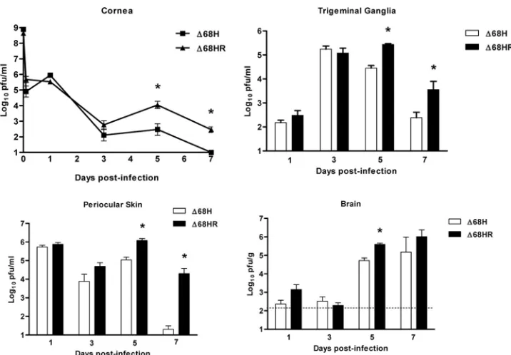
Interaction between ICP34.5 and Beclin 1 is important for efficient viral replication in peripheral tissues at late times following corneal infection.Previous studies showed that the BBD of ICP34.5 was necessary for wild-type neurovirulence following intracerebral infection (35), but it was completely dispensable for wild-type replication in primary MEFs (1). Furthermore, the ability of ICP34.5 lacking the BBD to block eIF2␣phosphorylation was equivalent to that of the wild type (D. E. Alexander and D. A. Leib, unpublished data). This demonstrated that the dephosphorylation-mediating activity of the C-terminal GADD34 homologous domain of ICP34.5 was unaffected by the deletion of the BBD. The role of the BBD in promoting viral replication and pathogenesis following periph-eral infection, however, has not been tested. We therefore examined the growth and pathogenesis of the BBD-deleted virus,⌬68H, and its marker-rescued virus,⌬68HR, following corneal infection. Viral titers in eye swab material (cornea), trigeminal ganglia, periocular skin, and brain were determined at various times postinfection (Fig. 1). The replication of both viruses was comparable in all tissues at days 1 and 3 postinfec-tion, but by days 5 and 7 the titers of⌬68H were significantly lower than those of⌬68HR. This was especially true in the periocular skin, perhaps reflecting a higher efficacy of inflam-mation-mediated immune clearance from this tissue compared to that of corneas and the nervous system. The replication of ⌬68HR in peripheral tissues was not significantly different from that of wild-type strain 17 at any time postinfection (data not shown). Thus, Beclin 1 binding by ICP34.5 contributes to the persistence of HSV-1 replication following corneal inocu-lation at late times postinfection.
Interaction between ICP34.5 and Beclin 1 results in greater virulence and disease following corneal infection.Replication
VOL. 83, 2009 BECLIN 1 BINDING AND HSV-1 PATHOGENESIS 12165
on November 8, 2019 by guest
http://jvi.asm.org/
differences between⌬68H and ⌬68HR were reflected in the course of clinical disease and the overall health of the infected animals. Clinical disease and weight loss were greater for ⌬68HR than⌬68H at days 5 and 6 postinfection (Fig. 2A). Compared to the status of mock-infected mice (Fig. 2B) on day 5, severe inflammation was observed in the periocular skin in mice infected with⌬68HR (Fig. 2C). The skin had multiple foci of ulcerative dermatitis and the epidermis was either ne-crotic or lost, with associated serum exudate and cellular debris containing degenerate neutrophils. In the underlying dermis there was infiltration by neutrophils and macrophages. In the deeper portion of the dermis, there was diffuse infiltration by neutrophils, lymphocytes, and macrophages, with moderate edema. In general, while qualitatively similar pathology was seen following infection with⌬68H, it was much less severe (Fig. 2D). In addition, none of the mice infected with⌬68HR survived beyond 7 days, while 91% of mice infected with⌬68H survived (data not shown). Taken together, these data provide further evidence that interaction with Beclin 1 contributes to the efficient replication and pathogenesis of HSV-1.
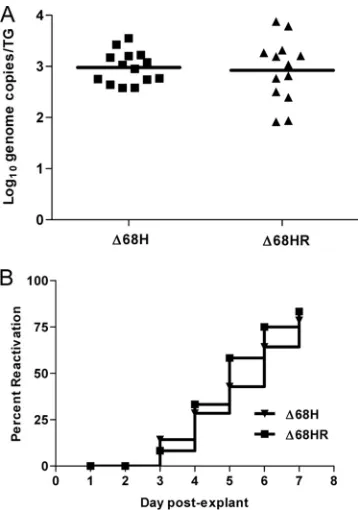
Establishment of latency and reactivation does not require interaction of ICP34.5 with Beclin 1.Decreased growth in the cornea observed for⌬68H may limit the amount of virus that can traffic to the trigeminal ganglia and establish a latent in-fection. Decreased growth in the trigeminal ganglia may fur-ther limit the ability of the virus to establish latency and to reactivate. Consequently, we evaluated the ability of⌬68H and ⌬68HR to establish a latent infection by harvesting trigeminal ganglia 28 days following corneal infection and quantifying the number of latent genomes via real-time PCR. Despite growth
defects in the cornea and trigeminal ganglia at times beyond day 5,⌬68H and⌬68HR established latency equivalently (Fig. 3A). In addition, following the explant cocultivation of latently infected trigeminal ganglia, both the ICP34.5 Beclin 1-binding mutant (⌬68H) and its marker-rescued virus (⌬68HR) reacti-vated comparably, as determined by the presence of infectious virus (Fig. 3B). Thus, the BBD of ICP34.5 is dispensable for the establishment of, and reactivation from, latency.
IRF3 deficiency in mice does not restore neurovirulence to
⌬68H.Viruses lacking the ICP34.5 BBD are neuroattenutated in terms of lethality and replication following the intracranial infection of mice (35 and data not shown). It also has been shown that a domain of ICP34.5 that partially overlaps with the BBD is necessary for the disruption of the interaction between IFN regulatory factor 3 (IRF3) and TANK-binding kinase 1 (TBK1) (55). This TBK1-interacting domain of ICP34.5 thereby is able to prevent the induction of IFN-stimulated antiviral genes. We wished, therefore, to address the possibility that the neuroattenuation of ⌬68H is due to its inability to control TBK1-mediated signaling rather than its inability to control autophagy and autophagy-related pathways. To accom-plish this, we intracerebrally infected mice lacking IRF3 (Irf3⫺/⫺) and mice lacking MyD88 (Fig. 4 and data not shown).
The prediction was that if ablated TBK1 binding was the ex-planation for the attenuation of⌬68H, then virulence should be completely restored in mice lacking these innate response mediators.
[image:3.585.112.470.65.314.2]Wild-type C57BL/6 mice and congenic Irf3⫺/⫺ mice were intracranially infected with⌬68H and⌬68HR. Similar to our findings with wild-type 129 Ev/Sv mice (35), the infection of
FIG. 1. Beclin 1 binding is important for efficient HSV-1 growth following corneal infection. 129 Ev/Sv mice were infected with 2⫻106PFU/eye of⌬68H or⌬68HR on bilaterally scarified corneas. Mice were sacrificed, and tear film (cornea), trigeminal ganglia, periocular skin, and brains were harvested on days 1, 3, 5, and 7 postinfection. Data are expressed as the geometric mean PFU/milliliter of homogenized tissue⫾SEM for six samples per experimental group in each of two independent experiments. The limit of detection of these assay was 10 PFU/ml for corneas, trigeminal ganglia, and periocular skin and 100 PFU/ml, as indicated by the horizontal dashed line, for the assay of brain homogenates. Asterisks indicateP⬍0.05 (Student’sttest).
on November 8, 2019 by guest
http://jvi.asm.org/
wild-type C56BL/6 mice with⌬68H resulted in significantly less mortality than that of mice infected with⌬68HR (P⬍0.0001 by log-rank test) (Fig. 4A). Consistently with previously de-scribed mouse strain differences in susceptibility to HSV-1 infection, C57BL/6 mice are more resistant to fatal HSV-1 infection than 129 Ev/Sv mice (20). Interestingly, the neuro-virulence of⌬68H was not restored to that of⌬68HR following the infection of IRF3-deficient mice, with differences between the survival ofIrf3⫺/⫺and control mice remaining significant (P⫽0.0046, log-rank test) (Fig. 4B). In fact, there also was no significant increase in the mortality ofIrf3⫺/⫺mice compared to that of wild-type mice infected with either⌬68H or⌬68HR. Consistent with these findings, the infection ofMyd88⫺/⫺mice also revealed no difference in pathogenesis between⌬68H and ⌬68HR (data not shown). Taken together, these data suggest that innate immunity regulated by the MyD88-independent (IRF3) and MyD88-dependent pathways do not account for the difference in neurovirulence between⌬68H and ⌬68HR. Furthermore, an inability to interfere with IRF3-mediated sig-naling does not account for the attenuation of⌬68H.
Virulence of⌬68H is restored inRag1ⴚ/ⴚ
mice.The differ-ence in the replication between⌬68H and⌬68HR is observed at later time points (5 to 7 days), suggesting that the control of the immune response to HSV-1 by the BBD involves adaptive immunity. The inability of IRF3 and Myd88 deficiency to re-store virulence to⌬68H further supported this hypothesis (Fig. 4 and data not shown). We addressed this initially by examin-ingRag1⫺/⫺mice, which lack functional T and B cells (44, 47).
Mice were infected intracerebrally with⌬68H and⌬68HR, and
lethality was monitored (Fig. 5). These studies show that the neurovirulence of⌬68H was completely restored following the infection of Rag1⫺/⫺mice, with no significant difference
be-tween the lethality of mice infected with⌬68H and ⌬68HR (P⫽0.1147 by log-rank test) (Fig. 6). This suggests that the attenuation of ⌬68H is due to its inability to overcome the adaptive, rather than the innate, immune response.
⌬68H stimulates stronger CD4ⴙ T-cell responses than
⌬68HR.The restoration of virulence to⌬68H inRag1⫺/⫺mice
(Fig. 5), coupled with observations by others that autophagy plays a pivotal role in promoting the endogenous MHC-II processing of antigens for presentation to CD4⫹T cells (7, 9, 36), provided a rationale for further examining BBD and adap-tive immunity. Recent work has demonstrated a role for ICP34.5 in the control of autophagy and of MHC-I-restricted antigen presentation (12), but its role in controlling MHC-II-restricted responses has not been addressed. Given that CD4⫹ T-cell responses are important for immunity to HSV-1 (23, 41, 52), we wished to address this directly by measuring the T-cell responses in DTH assays following corneal infection with ⌬68H and⌬68HR (Fig. 6A).⌬68H-infected mice exhibited a significantly greater (P⬍0.001) DTH response to HSV anti-gens than ⌬68HR-infected mice. The depletion of CD4⫹ T cells (⫺CD4) eliminated this response in⌬68H-infected mice, while the removal of CD8⫹T cells (⫺CD8) had no effect. In addition, CD4⫹ T cells from mice infected with ⌬68H pro-duced significantly more IFN-␥ (Fig. 6B) and interleukin-2 (IL-2) (Fig. 6C) when stimulated in vitro with HSV-1 antigens than CD4⫹ T cells from ⌬68HR-infected mice. Taken
to-FIG. 2. Decreased disease and weight loss are observed in mice infected with⌬68H compared to those of mice infected with⌬68HR. 129 Ev/Sv mice were infected with 2⫻106PFU/eye of either⌬68H or⌬68HR on bilaterally scarified corneas. (A) Animals were scored for periocular disease and overall clinical disease from days 2 to 10 postinfection, along with a change in weight. Disease data are expressed as the geometric mean disease score, or percent change in body weight,⫾SEM for five mice per experimental group in two independent experiments. Asterisks indicateP⬍0.05 by Student’sttest. (B to D) Representative hematoxylin- and eosin-stained histology sections of the periocular skin of mice 5 days following mock infection (B) or infection with⌬68HR (C) or⌬68H (D). Magnification,⫻200.
VOL. 83, 2009 BECLIN 1 BINDING AND HSV-1 PATHOGENESIS 12167
on November 8, 2019 by guest
http://jvi.asm.org/
[image:4.585.81.500.68.326.2]gether, these data suggest that the BBD of ICP34.5 is impor-tant for HSV-1 pathogenesis through its suppression of auto-phagy and subsequent dampening of the MHC-II-restricted stimulation of CD4⫹T cells.
DISCUSSION
In this study, we have further dissected the role of ICP34.5’s BBD in the pathogenesis of HSV-1. Our previous work deter-mined that this domain was important for neurovirulence fol-lowing intracerebral infection. From the standpoint of patho-genesis and immunity, however, the corneal model of HSV-1 infection is both more physiologically relevant to pathogenesis in humans and revealing of mechanisms of immunity. The reductions of⌬68H replication in the cornea, trigeminal gan-glia, and periocular skin were consistent with previously ob-served reductions in brain titers (35). The unexpected obser-vation in this study was the inability of BBD-deficient⌬68H to control the adaptive immune response. This is likely to be true for ICP34.5-null viruses and explain the mechanism behind the previous observation of increased MHC-II expression in cul-tured cells infected with an ICP34.5 null virus relative to that
of wild-type virus (53). Our observations also lend further rationale to the deletion of ICP34.5 from live-attenuated HSV vaccines, not only to reduce neurovirulence but also to pro-mote antigenicity and the stimulation of a stronger adaptive response. Interestingly, however, there was no alteration of the establishment of or reactivation from the latency of ⌬68H compared to that of⌬68HR. This demonstrates that the BBD and its control of autophagy do not significantly impact retro-grade or anteroretro-grade spread or the reactivation of HSV-1.
Previous work has demonstrated the activity of a specific domain of ICP34.5 (amino acids 72 to 104) that binds to TBK1 and disrupts its functional interaction with IRF3 (55). This domain serves to prevent the induction of IFN and IFN-stim-ulated genes and overlaps partially with the BBD that spans residues 68 to 87 (35). Data in this study show that the viru-lence of⌬68H is not increased inIrf3⫺/⫺mice relative to that of control mice. Thus, the BBD-deleted ICP34.5 still may be able to interact with TBK1, and the residues between 87 and 104 are necessary and sufficient for this interaction. Experi-ments are in progress to test this idea, but importantly, our data show that the attenuation of⌬68H is due to its inability to bind Beclin 1 rather than its inability to bind TBK1. IRF3 is a pivotal component in the development and amplification of the type 1 IFN response, and mice lacking IRF3 show increased susceptibility to virus infections (8). Our experiments with
Irf3⫺/⫺mice also provide evidence that innate immunity is not a major component in the attenuation of⌬68H.
[image:5.585.73.252.67.322.2]Previous work showed that BBD is dispensable for wild-type
FIG. 3.⌬68H and⌬68HR establish latency and reactivate equally. 129 Ev/Sv mice were infected with 2 ⫻ 104PFU/eye of ⌬68H or
[image:5.585.327.510.67.336.2]⌬68HR on bilaterally scarified corneas. Following the establishment of latency, mice were sacrificed and trigeminal ganglia were harvested. (A) Real-time PCR for the HSV-1 tk gene was performed on DNA isolated from trigeminal ganglia of latently infected mice. All samples were normalized to the single-copy mouse adipsin gene. Data are reported as the geometric means of the number of genome copies per trigeminal ganglion⫾SEM for 12 or 14 trigeminal ganglia per exper-imental group. (B) Trigeminal ganglia were bisected and cocultured on a monolayer of Vero cells. Supernatants were removed daily for 7 days after explant and added to fresh Vero monolayers. The results were recorded as the percentage of wells positive for reactivated virus for 12 or 14 trigeminal ganglia per experimental group in two independent experiments.
FIG. 4. Deletion of IRF-3 does not restore neurovirulence to an ICP34.5 Beclin 1-binding mutant. (A and B) Survival of C57BL/6 wild-type (A) and congenic Irf3⫺/⫺ (B) mice infected intracranially with 1⫻102PFU of the indicated virus. The number of animals was
n⫽10 for each experimental group in two independent experiments.
on November 8, 2019 by guest
http://jvi.asm.org/
replication in cultured murine fibroblasts and neurons (1). The current work, however, shows that BBD mutants are compro-mised in their ability to grow in epithelial and neuronal tissues in vivo. This supports the hypothesis that the BBD-mediated inhibition of autophagy impacts viral replication only in the context of an intact adaptive immune system. Alternatively, this may reflect other differences between cultured cells and in vivo infection. The relatively late clearance of ⌬68H from infected mice and its attenuation inIrf3⫺/⫺mice, however, are consistent with a role for adaptive immunity rather than direct xenophagic degradation in the enhanced clearance of⌬68H. Autophagy plays a role in the adaptive immune response by delivering antigens for MHC-II presentation (7, 9). Since it has been shown that viral proteins from Epstein-Barr virus and influenza virus can be presented on MHC-II molecules via autophagy, it is likely that HSV-1 also presents antigens to CD4⫹T cells via the autophagy pathway (36, 45). The signif-icantly increased CD4⫹ T-cell activation observed in ⌬ 68H-infected mice is strong evidence that a major role of BBD is to downregulate antigen presentation to CD4⫹T cells. Further evidence for this is that the virulence of⌬68H was restored in
Rag1⫺/⫺mice, which lack B and T cells. The restoration of
virulence to⌬68H also has been observed in mice lacking PKR (35). We previously had ascribed this to the autophagic deg-radation of HSV-1 being PKR dependent (51) and that the
Pkr⫺/⫺mice therefore were unable to mount a direct
[image:6.585.70.259.65.357.2]xenoph-agic attack. This conclusion currently is being reevaluated to
FIG. 5. Deletion ofRag1 restores neurovirulence to an ICP34.5 Beclin 1-binding mutant. (A and B) Survival of C57BL/6 wild-type (A) and congenicRag1⫺/⫺(B) mice infected intracranially with 1⫻102 PFU of the indicated virus. The number of animals wasn⫽20 for each experimental group in two independent experiments.
FIG. 6. CD4⫹T-cell responses to⌬68H and⌬68HR. (A) For the measurement of DTH response, C57BL/6 mice were infected via the cornea with⌬68H or⌬68HR. Seven days later, recipient mice were challenged in the right footpad with 1⫻106PFU of UV-inactivated
⌬68HR and in the left foot with cell culture medium. Twenty-four hours later, footpad thickness was determined with a micrometer by a masked observer. DTH responses were recorded as the difference between the right and left footpads and are expressed as microme-ters⫾SEM. For the measurement of cytokine production, C57BL/6 mice were infected on the cornea with⌬68H or⌬68HR. Seven days later, spleen and draining lymph nodes were harvested, and the CD4⫹ T cells were purified. CD4⫹T cells were mixed with DC at a ratio of 10:1 and stimulated with HSV-1 antigens. Supernatants were harvested at 48 h, and IFN-␥(B) and IL-2 (C) concentrations were determined by Bioplex 200 (Bio-Rad).
VOL. 83, 2009 BECLIN 1 BINDING AND HSV-1 PATHOGENESIS 12169
on November 8, 2019 by guest
http://jvi.asm.org/
[image:6.585.297.537.78.584.2]test whether T-cell responses are diminished in HSV-infected PKR-deficient mice. We also currently are clarifying whether other components of the adaptive immune system can be coun-tered by BBD, since autophagy plays a role in other aspects of the adaptive immune response, such as T-cell survival and proliferation and B lymphocyte development (30, 39). Recent work also has shown a novel process of membrane-derived autophagy to be important for delivering antigens for presen-tation on MHC-I molecules, and that this process also is in-hibited by ICP34.5 (12). This work was performed using an ICP34.5-null virus, and it will be of interest to study the specific role of BBD in inhibiting MHC-I-restricted T-cell activation.
The mechanism by which ICP34.5 inhibits Beclin 1-mediated autophagy is unknown, but the interaction between these pro-teins may provide some clues. ICP34.5 does not interact with the N-terminal domain of Beclin 1 (35). This region of Beclin 1 contains aBcl-2-interacting domain, suggesting that HSV-1 and gammaherpesviruses have evolved different mechanisms to target Beclin 1-mediated autophagy (28, 37). Second, given that ICP34.5 binds the C-terminal region of Beclin 1, which contains the evolutionarily conserved VPS34 binding domain, ICP34.5 may block or compete for VPS34 binding to Beclin 1. The conserved domain of Beclin 1 is required for autophagy function; therefore, ICP34.5 interaction with this region may prevent or limit Beclin 1-mediated autophagy (13).
It is clear that ICP34.5 has multiple functions that promote efficient viral replication and pathogenesis. However, PP1␣ binding and the blockade of translational arrest is the domi-nant function. This idea is supported by previous work showing that ICP34.5-null viruses remain attenuated in SCID mice (54). Thus, counteracting the innate immune response may be a critical function of ICP34.5 independent of BBD. This is exemplified further by an ICP34.5 mutant that is unable to bind PP1␣ but retains the BBD (D. Alexander, unpublished results). Following infection, this mutant undergoes the com-plete shutoff of protein synthesis, and in vitro and in vivo growth phenotypes are indistinguishable from those of an ICP34.5-null virus. Furthermore, ICP34.5-null mutants are at-tenuated to a much greater degree than that of the ICP34.5 Beclin 1-binding mutant. Notwithstanding the dominance of the PP1␣binding function of ICP34.5, it is clear that Beclin 1 binding is sufficient to control autophagy and to modulate host immunity, and that this domain significantly impacts the fitness of HSV-1. Our future studies will elucidate the precise mech-anisms by which BBD interfaces with the cell and, in turn, with the immune system.
ACKNOWLEDGMENTS
This work was supported by NIH grants to D. Leib (EY09083), to T. A. Ferguson (EY06765 and EY015570), and to the Department of Ophthalmology and Visual Sciences Core Grant (EY02687) from the National Eye Institute. D. Alexander also was supported by a research training grant in the visual sciences (NIH EY013360). Support from Research to Prevent Blindness to the Department of Ophthalmology and Visual Sciences, a Macular Vision Research Foundation award to T. A. Ferguson, and a Senior Scientific Investigator Award to D. A. Leib are gratefully acknowledged.
We thank Belinda McMahon and Suellen Greco for assistance with the histopathology and Michael Diamond and Tadatsugu Tanaguchi for the provision of theIrf3⫺/⫺mice.
REFERENCES
1.Alexander, D. E., S. L. Ward, N. Mizushima, B. Levine, and D. A. Leib.2007. Analysis of the role of autophagy in replication of herpes simplex virus in cell culture. J. Virol.81:12128–12134.
2.Amer, A. O., and M. S. Swanson.2005. Autophagy is an immediate macro-phage response to Legionella pneumophila. Cell Microbiol.7:765–778. 3.Andrade, R. M., M. Wessendarp, M. J. Gubbels, B. Striepen, and C. S.
Subauste.2006. CD40 induces macrophage anti-Toxoplasma gondii activity by triggering autophagy-dependent fusion of pathogen-containing vacuoles and lysosomes. J. Clin. Investig.116:2366–2377.
4.Birmingham, C. L., A. C. Smith, M. A. Bakowski, T. Yoshimori, and J. H. Brumell.2006. Autophagy controls Salmonella infection in response to dam-age to the Salmonella-containing vacuole. J. Biol. Chem.281:11374–11383. 5.Chou, J., J. J. Chen, M. Gross, and B. Roizman.1995. Association of a Mr
90,000 phosphoprotein with protein kinase PKR in cells exhibiting enhanced phosphorylation of translation initiation factor eIF-2 alpha and premature shutoff of protein synthesis after infection with gamma 134.5- mutants of herpes simplex virus 1. Proc. Natl. Acad. Sci. USA92:10516–10520. 6.Chou, J., and B. Roizman.1994. Herpes simplex virus 1␥134.5 gene function,
which blocks the host response to infection, maps in the homologous domain of the genes expressed during growth arrest and DNA damage. Proc. Natl. Acad. Sci. USA91:5247–5251.
7.Crotzer, V. L., and J. S. Blum.2005. Autophagy and intracellular surveil-lance: modulating MHC class II antigen presentation with stress. Proc. Natl. Acad. Sci. USA102:7779–7780.
8.Daffis, S., M. A. Samuel, B. C. Keller, M. Gale, Jr., and M. S. Diamond.2007. Cell-specific IRF-3 responses protect against West Nile virus infection by interferon-dependent and -independent mechanisms. PLoS. Pathog.3:e106. 9.Dengjel, J., O. Schoor, R. Fischer, M. Reich, M. Kraus, M. Muller, K. Kreymborg, F. Altenberend, J. Brandenburg, H. Kalbacher, R. Brock, C. Driessen, H. G. Rammensee, and S. Stevanovic.2005. Autophagy promotes MHC class II presentation of peptides from intracellular source proteins. Proc. Natl. Acad. Sci. USA102:7922–7927.
10.Deretic, V., and B. Levine.2009. Autophagy, immunity, and microbial adap-tations. Cell Host Microbe5:527–549.
11.Dunn, W. A., Jr.1994. Autophagy and related mechanisms of lysosome-mediated protein degradation. Trends Cell Biol.4:139–143.
12.English, L., M. Chemali, J. Duron, C. Rondeau, A. Laplante, D. Gingras, D. Alexander, D. Leib, C. Norbury, R. Lippe, and M. Desjardins.2009. Auto-phagy enhances the presentation of endogenous viral antigens on MHC class I molecules during HSV-1 infection. Nat. Immunol.10:480–487. 13.Furuya, N., J. Yu, M. Byfield, S. Pattingre, and B. Levine.2005. The
evolu-tionarily conserved domain of Beclin 1 is required for Vps34 binding, auto-phagy and tumor suppressor function. Autoauto-phagy1:46–52.
14.Gierasch, W. W., D. L. Zimmerman, S. L. Ward, T. K. Vanheyningen, J. D. Romine, and D. A. Leib.2006. Construction and characterization of bacterial artificial chromosomes containing HSV-1 strains 17 and KOS. J. Virol. Methods135:197–206.
15.Griffith, T. S., X. Yu, J. M. Herndon, D. R. Green, and T. A. Ferguson.1996. CD95-induced apoptosis of lymphocytes in an immune privileged site in-duces immunological tolerance. Immunity5:7–16.
16.He, B., J. Chou, D. A. Liebermann, B. Hoffman, and B. Roizman.1996. The carboxyl terminus of the murine MyD116 gene substitutes for the corre-sponding domain of the␥134.5 gene of herpes simplex virus to preclude the
premature shutoff of total protein synthesis in infected human cells. J. Virol.
70:84–90.
17.He, B., M. Gross, and B. Roizman.1997. The␥134.5 protein of herpes
simplex virus 1 complexes with protein phosphatase 1alpha to dephosphor-ylate the alpha subunit of the eukaryotic translation initiation factor 2 and preclude the shutoff of protein synthesis by double-stranded RNA-activated protein kinase. Proc. Natl. Acad. Sci. USA94:843–848.
18.Jackson, W. T., T. H. Giddings, Jr., M. P. Taylor, S. Mulinyawe, M. Rabin-ovitch, R. R. Kopito, and K. Kirkegaard.2005. Subversion of cellular auto-phagosomal machinery by RNA viruses. PLoS Biol.3:e156.
19.Kihara, A., Y. Kabeya, Y. Ohsumi, and T. Yoshimori.2001. Beclin-phophati-dylinositol 3-kinase complex functions at the trans-Golgi network. EMBO Rep.2:330–335.
20.Kirchner, H., M. Kochen, K. Munk, H. M. Hirt, S. E. Mergenhagen, and D. L. Rosenstreich.1978. Differences in susceptibility to herpes simplex virus infection of inbred strains of mice. IARC Sci. Publ.1978:783–788. 21.Klionsky, D. J.2007. Autophagy: from phenomenology to molecular
under-standing in less than a decade. Nat. Rev. Mol. Cell Biol.8:931–937. 22.Klionsky, D. J., and S. D. Emr.2000. Autophagy as a regulated pathway of
cellular degradation. Science290:1717–1721.
23.Koelle, D. M., S. N. Reymond, H. Chen, W. W. Kwok, C. McClurkan, T. Gyaltsong, E. W. Petersdorf, W. Rotkis, A. R. Talley, and D. A. Harrison.
2000. Tegument-specific, virus-reactive CD4 T cells localize to the cornea in herpes simplex virus interstitial keratitis in humans. J. Virol. 74:10930– 10938.
24.Kyei, G. B., C. Dinkins, A. S. Davis, E. Roberts, S. B. Singh, C. Dong, L. Wu, E. Kominami, T. Ueno, A. Yamamoto, M. Federico, A. Panganiban, I.
on November 8, 2019 by guest
http://jvi.asm.org/
Vergne, and V. Deretic.2009. Autophagy pathway intersects with HIV-1 biosynthesis and regulates viral yields in macrophages. J. Cell Biol.186:255– 268.
25.Leib, D. A., D. M. Coen, C. L. Bogard, K. A. Hicks, D. R. Yager, D. M. Knipe, K. L. Tyler, and P. A. Schaffer.1989. Immediate-early regulatory gene mutants define different stages in the establishment and reactivation of herpes simplex virus latency. J. Virol.63:759–768.
26.Levine, B.2005. Eating oneself and uninvited guests: autophagy-related pathways in cellular defense. Cell120:159–162.
27.Liang, X. H., S. Jackson, M. Seaman, K. Brown, B. Kempkes, H. Hibshoosh, and B. Levine.1999. Induction of autophagy and inhibition of tumorigenesis by beclin 1. Nature402:672–676.
28.Liang, X. H., L. K. Kleeman, H. H. Jiang, G. Gordon, J. E. Goldman, G. Berry, B. Herman, and B. Levine.1998. Protection against fatal Sindbis virus encephalitis by Beclin, a novel Bcl-2-interacting protein. J. Virol.72:8586– 8596.
29.Ling, Y. M., M. H. Shaw, C. Ayala, I. Coppens, G. A. Taylor, D. J. Ferguson, and G. S. Yap.2006. Vacuolar and plasma membrane stripping and auto-phagic elimination of Toxoplasma gondii in primed effector macrophages. J. Exp. Med.203:2063–2071.
30.Miller, B. C., Z. Zhao, L. M. Stephenson, K. Cadwell, H. H. Pua, H. K. Lee, N. N. Mizushima, A. Iwasaki, Y. W. He, W. Swat, and H. W. Virgin.2007. The autophagy gene ATG5 plays an essential role in B lymphocyte devel-opment. Autophagy4:309–314.
31.Mizushima, N., and D. J. Klionsky.2007. Protein turnover via autophagy: implications for metabolism. Annu. Rev. Nutr.27:19–40.
32.Mortimore, G. E., and A. R. Poso.1987. Intracellular protein catabolism and its control during nutrient deprivation and supply. Annu. Rev. Nutr.7:539– 564.
33.Mulvey, M., V. Camarena, and I. Mohr.2004. Full resistance of herpes simplex virus type 1-infected primary human cells to alpha interferon re-quires both the Us11 and␥134.5 gene products. J. Virol.78:10193–10196.
34.Nakagawa, I., A. Amano, N. Mizushima, A. Yamamoto, H. Yamaguchi, T. Kamimoto, A. Nara, J. Funao, M. Nakata, K. Tsuda, S. Hamada, and T. Yoshimori.2004. Autophagy defends cells against invading group A Strep-tococcus. Science306:1037–1040.
35.Orvedahl, A., D. Alexander, Z. Talloczy, Q. Sun, Y. Wei, W. Zhang, D. Burns, D. A. Leib, and B. Levine.2007. HSV-1 ICP34.5 confers neurovirulence by targeting the Beclin 1 autophagy protein. Cell Host Microbe1:23–35. 36.Paludan, C., D. Schmid, M. Landthaler, M. Vockerodt, D. Kube, T. Tuschl,
and C. Munz.2005. Endogenous MHC class II processing of a viral nuclear antigen after autophagy. Science307:593–596.
37.Pattingre, S., A. Tassa, X. Qu, R. Garuti, X. H. Liang, N. Mizushima, M. Packer, M. D. Schneider, and B. Levine.2005. Bcl-2 antiapoptotic proteins inhibit Beclin 1-dependent autophagy. Cell122:927.
38.Prentice, E., W. G. Jerome, T. Yoshimori, N. Mizushima, and M. R. Denison.
2004. Coronavirus replication complex formation utilizes components of cellular autophagy. J. Biol. Chem.279:10136–10141.
39.Pua, H. H., I. Dzhagalov, M. Chuck, N. Mizushima, and Y. W. He.2007. A critical role for the autophagy gene Atg5 in T cell survival and proliferation. J. Exp. Med.204:25–31.
40.Rader, K. A., C. E. Ackland-Berglund, J. K. Miller, J. S. Pepose, and D. A. Leib.1993. In vivo characterization of site-directed mutations in the
pro-moter of the herpes simplex virus type 1 latency-associated transcripts. J. Gen. Virol.74:1859–1869.
41.Rajasagi, N. K., S. H. Kassim, C. M. Kollias, X. Zhao, R. Chervenak, and S. R. Jennings.2009. CD4⫹T cells are required for the priming of CD8⫹T cells following infection with herpes simplex virus type 1. J. Virol.83:5256– 5268.
42.Rich, K. A., C. Burkett, and P. Webster.2003. Cytoplasmic bacteria can be targets for autophagy. Cell Microbiol.5:455–468.
43.Sato, M., H. Suemori, N. Hata, M. Asagiri, K. Ogasawara, K. Nakao, T. Nakaya, M. Katsuki, S. Noguchi, N. Tanaka, and T. Taniguchi.2000. Dis-tinct and essential roles of transcription factors IRF-3 and IRF-7 in response to viruses for IFN-alpha/beta gene induction. Immunity13:539–548. 44.Schatz, D. G., M. A. Oettinger, and D. Baltimore.1989. The V(D)J.
recom-bination activating gene, RAG-1. Cell59:1035–1048.
45.Schmid, D., M. Pypaert, and C. Munz.2007. Antigen-loading compartments for major histocompatibility complex class II molecules continuously receive input from autophagosomes. Immunity26:79–92.
46.Smith, T. J., C. E. Ackland-Berglund, and D. A. Leib.2000. Herpes simplex virus virion host shutoff (vhs) activity alters periocular disease in mice. J. Virol.74:3598–3604.
47.Spanopoulou, E., C. A. Roman, L. M. Corcoran, M. S. Schlissel, D. P. Silver, D. Nemazee, M. C. Nussenzweig, S. A. Shinton, R. R. Hardy, and D. Balti-more.1994. Functional immunoglobulin transgenes guide ordered B-cell differentiation in Rag-1-deficient mice. Genes Dev.8:1030–1042. 48.Strand, S. S., and D. A. Leib.2004. Role of the VP16-binding domain of vhs
in viral growth, host shutoff activity, and pathogenesis. J. Virol.78:13562– 13572.
49.Strelow, L. I., and D. A. Leib.1995. Role of the virion host shutoff (vhs) of herpes simplex virus type 1 in latency and pathogenesis. J. Virol.69:6779– 6786.
50.Tallo´czy, Z., W. Jiang, H. W. T. Virgin, D. A. Leib, D. Scheuner, R. J. Kaufman, E. L. Eskelinen, and B. Levine.2002. Regulation of starvation-and virus-induced autophagy by the eIF2alpha kinase signaling pathway. Proc. Natl. Acad. Sci. USA99:190–195.
51.Tallo´czy, Z., H. W. I. Virgin, and B. Levine.2006. PKR-dependent autoph-agic degradation of herpes simplex virus type 1. Autophagy2:24–29. 52.Thomas, J., S. Gangappa, S. Kanangat, and B. T. Rouse.1997. On the
essential involvement of neutrophils in the immunopathologic disease: her-petic stromal keratitis. J. Immunol.158:1383–1391.
53.Trgovcich, J., D. Johnson, and B. Roizman.2002. Cell surface major histo-compatibility complex class II proteins are regulated by the products of the ␥134.5 and UL41 genes of herpes simplex virus 1. J. Virol.76:6974–6986.
54.Valyi-Nagy, T., M. U. Fareed, J. S. O’Keefe, R. M. Gesser, A. R. MacLean, S. M. Brown, J. G. Spivack, and N. W. Fraser.1994. The herpes simplex virus type 1 strain 17⫹gamma 34.5 deletion mutant 1716 is avirulent in SCID mice. J. Gen. Virol.75:2059–2063.
55.Verpooten, D., Y. Ma, S. Hou, Z. Yan, and B. He.2009. Control of TANK-binding kinase 1-mediated signaling by the␥134.5 protein of herpes simplex
virus 1. J. Biol. Chem.284:1097–1105.
56.Virgin, H. W., and B. Levine.2009. Autophagy genes in immunity. Nat. Immunol.10:461–470.
57.Xu, M., A. J. Lepisto, and R. L. Hendricks.2004. CD154 signaling regulates the Th1 response to herpes simplex virus-1 and inflammation in infected corneas. J. Immunol.173:1232–1239.
VOL. 83, 2009 BECLIN 1 BINDING AND HSV-1 PATHOGENESIS 12171