Observational study of the effect of beach- chair position on
cerebral blood flow in patients undergoing shoulder surgery
A Thesis submitted to the Tamil Nadu Dr. MGR Medical University in
partial fulfillment of the degree MD ANAESTHESIA.
By
Dr Jesudoss A
Christian Medical College and Hospital, Vellore,
Tamil Nadu - 632004
CERTIFICATE
This is to certify that the dissertation titled“Observational study of the effect of beach- chair position on cerebral blood flow in patients undergoing shoulder
surgery” is a bonafide work of Dr. Jesudoss A in partial fulfillment of the requirements for the MD Anesthesia (final) examination of the Tamil Nadu
Dr. MGR medical university to be conducted in April 2017.
Signature:
Dr. Sajan Philip George,
Guide and Head of The Department, Department of Anesthesiology, Christian Medical College, Dr. Anna Pulimood
Principal,
DECLARATION
I hereby declare that this dissertation titled“Observational study of the effect of beach- char position on cerebral blood flow in patients undergoing shoulder
surgery”was prepared by me in partial fulfillment of the regulations for the award of the degree of MD ANAESTHESIA of the Tamil Nadu Dr.MGR Medical
University, Chennai. This has not formed the basis for the award of any degree to me before and I have not submitted this to any other university previously.
Jesudoss A
ACKNOWLEDGEMENTS
I acknowledge God, for all His grace and guidance.
Dr. Sajan Philip George for all that he has taught me.
Dr. Melvin Alex Abraham and colleagues in the department of Anaesthesiology, CMC Vellore and all my teachers, for making this study and this course a reality.
ABSTRACT
TITLE: “Observational study of the effect of beach- chair position on cerebral blood flow in patients undergoing shoulder surgery”
DEPARTMENT: Anesthesiology
NAME OF THE CANDIDATE: Dr. Jesudoss A
DEGREE & SUBJECT: MD Anaesthesia
NAME OF THE GUIDE: Dr. Sajan Philip George
Background:
Methods:
An observational study was conducted in the department of Anesthesia in the
Christian Medical College, Vellore from January 2015 to June 2016. All
consenting patients who underwent shoulder surgeries in sitting position were
included in the study according to the inclusion and exclusion criteria. Standard
general anesthesia was given to all patients. In addition to pulse oximetry and
electrocardiogram, invasive arterial blood pressure was monitored. Baseline value
of middle cerebral artery blood flow velocity is measured by transcranial Doppler
before and after induction in supine. After patient is positioned in beach chair
position arterial blood pressure was read at the level of the tragus. Doppler was
repeated in whenever there was hypotension and after treating it. The
percentage reduction in mean arterial blood pressure and the middle cerebral
artery blood flow velocity were compared.
Results:
Twenty patients were recruited for the study. The mean age was 38 years. There
were 15% ASA II patients and 85% were ASA I. The reduction in the mean arterial
Except for the 6 patients who were overzealously treated after hypotension, an average
of 73.41mmHg of the mean arterial pressure was required to maintain the middle
cerebral artery blood flow velocity, thereby cerebral perfusion.
Conclusion:
There are statistically significant changes in mean arterial pressure and middle
cerebral artery blood flow velocity. It was concluded that an average of 73.41mmHg is
the minimum mean arterial pressure required to maintain cerebral perfusion.
Keywords: beach – chair, sitting, transcranial Doppler, middle cerebral artery,
CONTENTS
TITLE PAGE NO:
1) Introduction 10
2) Aims and objectives 13
3) Review of literature 15
4) Methods 68
5) Results 71
6) Discussion 85
7) Conclusions and limitations 92
8) Bibliography 95
9) Annexures 98
Proforma 99
Consent form 100
Information sheet 102
INTRODUCTION
The surgeries of the shoulder joint can be done in lateral position or in
sitting (modified beach chair) position. Surgeons prefer to operate mostly in sitting position because it gives better view of both the anterior and posterior parts of the shoulder region. Both the positions have advantages and disadvantages but
hypotension and cerebral hypoperfusion are serious complications of sitting position. The effects of posture combined with the effects of anesthesia exacerbates hypotension and cerebral hypoperfusion.
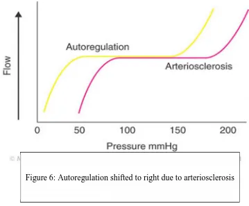
Though brain can autoregulate its flow and perfusion, beyond a certain limit autoregulation does not work. Traditionally within the range of mean arterial
pressure between 50 and 150 mmHg, autoregualtion seems to work effectively but the range cannot be the same for all individuals. Since the cerebral perfusion
One of the methods of measuring the cerebral blood flow is by transcranial Doppler. The middle cerebral artery blood flow velocity (measured by the
AIMS AND OBJECTIVES
AIMS
To observe the effect sitting position on cerebral blood flow in patients undergoing shoulder surgery.
OBJECTIVES
Primary objective:
To compare the cerebral blood flow with invasive blood pressure in supine and beach chair positions in patients undergoing shoulder surgery
Secondary objective:
Cerebral Blood Flow
Brain is a highly perfused organ, receiving approximately 14% of cardiac output. (54ml/ 100g / minute). The total flow to the whole brain is approximately 750 ml.
Autoregulation
Brain utilizes 20% of the oxygen need of the body, even though it is 2% of the total body weight. Brain requires glucose to provide ATP which is the fuel for its (i) basal metabolism i.e. for maintaining the trans-membrane ionic gradient (ii) for maintaining cellular integrity and (iii) synaptic transmission. Since there is no substrate reserve in the central nervous system, it cannot sustain anaerobic metabolism for more than a few minutes.
Auto regulatory responses maintain the internal milieu of the central nervous system. Flow-metabolism coupling and active vasomotion are the two clinically distinct processes involved in auto regulation.(3)
Flow Metabolism Coupling
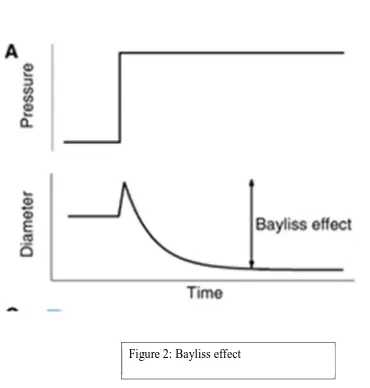
Bayliss effect (myogenic response)
[image:19.612.68.438.304.692.2]The smooth muscle cells of large arteries and smaller arterioles constrict in response to increased pressure and dilate in response to decreased pressure. It is a critical component of these vessels and more prominent in the cerebral vasculature.
Regulation of CBF
1. Cellular mechanisms of cerebral vasomotion a. Nitric Oxide (NO)
Studies show that Nitric Oxide may be involved in the basal cerebro-vascular tone by activating guanylatecyclase. It may also act through calcitonin gene related peptide and ATP-K Channels and thromboxane A2 which is a vasoconstrictor.
Nitric Oxide also plays a role in the vasodilatory response to the changes in perfusion pressure and hypercapnia.(3)
b. Vasoactive Peptides
c. Potassium Channels
K-ATP: ATP sensitive potassium channels are responsible for dilation response to hypotension, hypercapnia, acidosis and hypoxia.
K-CA: Calcium activated potassium channels are responsible for basal cerebrovascular tone.(3)
d. Prostaglandins
Prostaglandins play a significant role in the neonatal cerebral blood flow than that of the adult. Pg E2 and Pg I2 are the dilators and
Pg F2alpha and thromboxane A2 are the constrictors in the cerebral circulation that mediate hypercapneic vasodilation indirectly.(3)
e. Endothelin
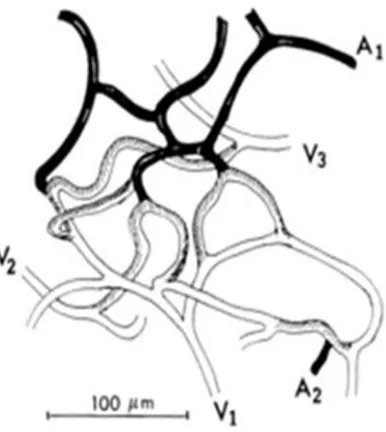
2. Cerebral Micro Circulation
Highly tortuous capillary beds (increased path length) increases the transit time of red blood cells even though blood flow velocity is greater than other tissues.
[image:22.612.200.393.402.618.2](i) Pressure regulation:
Cerebral blood flow is explained by Hagan-Poisseuille - Laminar flow equation. It states that there is a direct relationship between the flow, the caliber of cerebral vessels and the cerebral perfusion pressure (CPP).
π- Mathematical constant
ΔP - Pressure gradient (CPP)
r –Radius or caliber of blood vessel
μ - Dynamic viscosity of blood
l - Length of the blood vessel.
Hence any increase in CPP or cerebral vasodilation will lead to increase in cerebral blood flow.
MAP: Mean arterial pressure
ICP: Intra cranial pressure
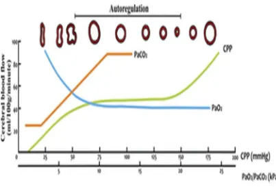
Over a large range of mean arterial pressure the cerebral blood flow remains unchanged. The lower range of auto regulation is 50 mm Hg and the upper limit is 150mm Hg. Beyond these limits the flow is pressure dependent or pressure passive. The flow increases with increase and reduces with decrease in mean arterial pressure. It is also to be noted that even within the auto-regulatory range, a rapidly changing mean blood pressure will cause transient change in cerebral blood flow. (6, 7)
(ii) Venous physiology
Since large amount of cerebral blood volume is in the veins, any change in the venous diameter, can cause increase in the intra cranial blood volume and hence the intra cranial pressure. (3,8)
Monroe Kellie doctrine states that “in the setting of a non-distensible cranial vault,
the volume of blood, CSF and brain tissue must be in equilibrium”.(8)
(iii) Rheologic factors
Some animal and human studies show that inverse relationship exists between the CBF and hematocrit. Hematocrit influences the viscosity of blood. Viscosity also determines the circulatory resistance as per the Hagen–Poisseuille law.
It states that,
R= 8 l µ / r4, where l is length of conduct, µ is blood viscosity, r is radius of the vessel.
4. Metabolic & Chemical influences
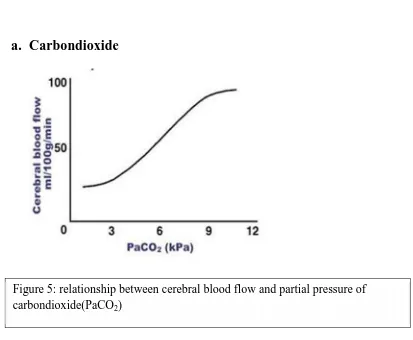
a. Carbondioxide
[image:27.612.97.510.100.440.2]Within physiological range, for every 1 mm Hg increase or decrease in partial pressure of carbondioxide in artery, there will be 3% increase or decrease in cerebral blood flow in a linear relationship. At normotension, there is a nearly linear response of cerebral blood flow at a PaCO2 between 20 and 80 mm Hg. As PaCO2 approaches extremes the linear response fades. This response is highly reproducible one.(3)
In a normal brain, cerebral blood volume is a 5 millilitre per 100g of brain and if PaCO2 ranges from 25-70 mm Hg, cerebral blood volume changes about 0.049 millilitre per 100g for every 1-3 mmHg change in PaCO2. For the brain tissue in an adult(which is about 1400g), there can be around 20 ml change in total cerebral blood volume if the PaCO2values from of 25 to 55 mm Hg.(6)
Arteriolar tone set by the systemic arterial blood pressure, modulates the effect of PaCO2 on cerebral blood flow. It is blunted by moderate hypotension and is abolished by severe hypotension. Conversely, carbondioxide tension modifies pressure auto-regulation by widening the auto-regulatory plateau from hypercapnia to hypocapnia.(3)
b. Oxygen
c. Temperature
Hypothermia reduces the rate of energy utilization by both electro physiological functions as well as the basal metabolism associated with the maintenance of cellular integrity. For every 1o C reduction of temperature, there is 7% reduction in CMRO2 (Cerebral metabolic rate for O2). Because the cerebral blood flow is closely coupled with metabolism, there is a parallel decrease in cerebral blood flow with hypothermia – induced reduction in CMRO2. Auto-regulation and carbondioxide reactivity are well preserved in moderate hypothermia.(3)
5. Neurogenic influences
cerebral vessels and the cortical pial vessels, mediate vasodilation through substance P, neutrokinin A and CGRP. Ischaemia stimulates these fibres causing vasodilation.
6. Clinical Consideration
a. Hypertensive Patient:
[image:30.612.124.483.424.721.2]Although this has some protective effect against “break-through” caused by
surpassing the upper limit of auto-regulation, it occurs at the expenses of lower limit. The sclerotic changes in the vessels and the shift in auto-regulatory response caused by hypertension are modified by treatment with antihypertensives for a long time.
CO2reactivity in this group is no different from the normotensive population. This finding underscores the probable difference in mechanisms between carbondioxide induced and blood pressure induced cerebral vasomotion.
b. Elderly Patient:
Some elderly patients who had postural hypotension were found to suffer from cerebral ischemia with very small decrease in blood pressure, which would not have otherwise occurred. This shows that ageing affects cerebral auto-regulation.(10)
as increases of cerebrovascular resistance with advancing age. Neuronal atrophy and in part cerebral arteriosclerosis play a role in reduction of grey matter flow with advancing age were most evident in the middle cerebral arterial distribution. The association of risk factors on enhances the reduction in flow.(11)
There is also variation of auto-regulation in elderly population. Some can tolerate a greater fall in mean arterial pressure whereas, even a minor fall in mean arterial pressure leads to cerebral ischemia.
In a study conducted in elderly healthy subjects done by quantitative T2 mapping MRI, it was found that there is reduction of cerebral blood flow between 0.5% and 0.7% per year with a high range of individual variation.(12)
7. Pharmacology
a. Intravenous anesthetic agents
b. Volatile anesthetics
They have intrinsic vasodilatory property and cause a dose dependent reduction in systemic blood pressure and modify cerebral auto-regulation. They reduce CMRO2 in a dose dependent manner but at a slower rate. Certain studies have shown that at for isoflurane at a minimum alveolar concentration of 1.1 there is 19% increase in cerebral blood flow when the arterial blood pressure is kept within normal limits. Cerebral metabolic rate is reduced by about 45%. Recent studies have shown that in human, sevoflurane as well as desflurane reduce the cerebral blood flow significantly (compared to the cerebral blood flow in non-anesthetized patients). At 1.0 MAC concentrations, sevoflurance and desflurane decreased cerebral blood flow by 38% and 22% and CMR by 39% & 35% respectively.
CO2reactivity is well maintained with volatile agents.(6)
8. Pathological changes (Cerebral ischemia)
evidence of ischemia begins to appear only when cerebral blood flow has fallen to 20 ml/100g/min. When it drops to 15 ml/100g/min, the cortical EEG becomes isoelectric. Potentially irreversible membrane failure (elevated extra cellular potassium and loss of the direct cortical response) rapidly occurs only when cerebral blood flow is reduced to about 6 ml/ 100g/min.(6)
The ancient idea of ischemic insult and injury was that lysis of the neurons was restricted to the duration of ischemia and during the initial reperfusion stage. Recent data shows that neuronal lysis after an ischemic insult is an active process in which neurons continue to die for a prolonged duration after the trigger of ischemic insult. The severity of the ischemic insult determines the degree of the neuronal lysis.(6)
Cerebral neuroprotection and anesthetics
1. Barbiturates
Barbiturate therapy has been mainly useful in preventing neuronal injury due to hypoxia and ischemia. The neuroprotective effects of barbiturates are mainly by reducing the oxygen demand, increasing the oxygen delivery and by inhibiting the damaging pathological neuronal pathways. The exact mechanism of action is not completely known but the proposed mechanisms are reduction in cerebral metabolism (CMRO2) by improving regional cerebral blood flow;
suppression of seizures; reduction in the intracranial pressure; loss of thermo –
2. Volatile anesthetics:
Isoflurane is a powerful suppressant of cerebral metabolic rate in the cortex of cerebrum. There have been reports of protective effects (EEG evidence) in humans. It has been recently observed that neuroprotective efficacy of isoflurane is not lasting. When evaluated for neuronal lysis 2 days after ischemic insult isoflurane anesthesia is found to reduce the injury vigorously. But it was found on that after 14 days reduction in injury was much evident. This indicates that neuronal injury sustains into the post-ischemic recovery time and that the neuroprotection may not continue for a long period after the insult. (6)
Similarly sevoflurane also reduces ischemic injury but the efficacy of the
sevoflurane does vary from halothane. Desflurane’ role in neuroprotection is also
SHOULDER SURGERY
Positions for shoulder surgery
For shoulder surgery the selection of an appropriate position of the patient and its maintenance throughout the procedure is of major importance. The surgeon must support the part and work in such a way that he would gain easy access to the structures he wants to expose.(14)
[image:37.612.127.514.435.668.2]1. Lateral Position
As shown in the figure patient should be rolled into lateral position on to adjacent lateral positioning posts. A vacuum / bean bag can also be used. An axillary roll should be placed and care taken to ensure optimum ventilation and prevent traction injuries to brachial plexus.(15)
Advantages
When compared to sitting position the advantages of lateral position are better visualization and instrument access for some surgeries. There is decreased risk of reduced cerebral perfusion.
Injuries
In the dependent arm (in the lateral position) there can be injury to the brachial
plexus. It is due to the stretching of the upper plexus caused by the patient’s body
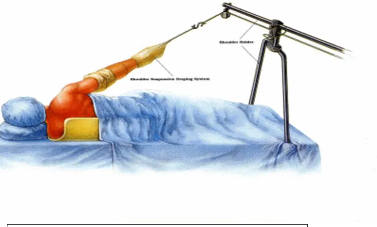
weight compressing the arm which is abducted and internally rotated. Brachial plexus could get injured in the non-dependent side also. The non-dependent arm is usually suspended on the ether screen above the head. A modification of this position, in which the arm is held by a suspension toward the ceiling is used during shoulder procedures.(17)
Radial nerve palsy of the dependent arm is another serious injury which can happen in this position. For non-shoulder surgery incorrect positioning, positioning on a hard surface like arm boards without appropriate padding or a combination of both can cause compression of the radial nerve at the bend of the elbow.(18)
Peroneal nerve injury can happen when the fibular head is compressed against the operating table directly. Bony prominences such as greater trochanter, fibular head and ankle of the dependent side are to be padded properly. Saphenous nerve injury is prevented by placing pillows between the knees and ankles. Avoid excessive flexion or extension at the hip to prevent lumbar plexus or sciatic nerve injury.
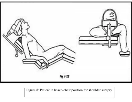
2. Sitting or beach chair position
[image:41.612.71.523.163.514.2]It has been found that supine position for shoulder surgery was uncomfortable and does not provide adequate access to superior posterior and lateral aspects of shoulder. The uncomfortable posture for surgeons leads to mental & physical fatigue.
In 1925, surgeons used sitting position for doing shoulder arthrodesis in children
affected by anterior poliomyelitis (for the first time in St. Louis unit of Shriners’
Hospital for crippled children) They found that only in this position they could palpate bony landmarks and secure optimum position for function.(14)
As shown in the picture, the beach chair (sitting) position is used for most of the anterior approaches of the shoulder surgeries. The torso and head are elevated to an angle of about 20-30oto horizontal. The legs are kept flexed at knee joints using a pillow to avoid over -stretching of the hamstrings. The legs are maintained little raised than the horizontal to increase the venous return. Sequential compression devices are placed on legs to prevent deep venous thrombosis.
Disadvantages
Postural hypotension, reduced cerebral blood flow, macroglossia, quadriplegia, injury to brachial plexus and sciatic nerve are the possibilities. Greater auricular nerve neuropraxia and incidents of ischemic optic neuropathy are also recorded. (21,22)
Advantages
Easy exposure of the surgical field.
Surgical Procedures
1. Open Surgery
Repairs for instability, subacromial decompression, acromioplasty, fracture fixation and arthroplasty are the most commonly performed open surgical procedures.
Approaches
(i) Anterior approach: where the incision starts just lateral to the tip of the coracoid upto the axillary crease.
2. Arthroscopic surgery
Both diagnostic and therapeutic procedures are done through arthroscopy. The main post sites are posterior and lateral. The posterior site is not covered when only interscelene block is given. Hence additional local anesthetic infiltration is required.
Effect of beach-chair position on cerebral blood flow during arthroscopic shoulder surgeries
and reduction in cardiac output. Venous return from the cerebral circulation is increased by inspiratory sub-atmospheric pressure during spontaneous ventilation, but this mechanism is nullified by positive-pressure ventilation. Obstruction of the internal jugular veins in the sitting position may also impede cerebral venous drainage, especially with unfavorable positions of the head and neck such as flexion of the head.
In a case series where the head elevation was 900to the horizontal the reduction in mean arterial pressure was around 30%. At the end of the procedure there was significant neurological dysfunction like hemispheric infarct, posterior circulation infarct, and even brain death in two patients.(22)
Generally cerebral perfusion pressure is auto-regulated adequately if the mean arterial pressure is between 50-150 mm Hg. It was Drummond who argued that the actual values must be more and the lower limit of auto-regulation should be calculated on a patient to patient basis derived from the individual’s resting mean
Anesthesia for Shoulder Surgeries
There are numerous approaches of anesthesia for shoulder surgery and each technique should be tailored to the surgical and patient requirement. For short procedures nerve block can be sufficient if the patient is willing. Longer procedure usually requires general anesthesia. In case of regional anesthesia, sedating the patient might lead to loss of airway. Hence regional with light sedation or formal general anesthesia can be used.(20)
REGIONAL ANESTHESIA
The shoulder region is supplied by nerves of cervical and brachial plexus. The cervical plexus supplies the skin over the clavicle, the shoulder tip, and first two intercostal spaces anteriorly via the superficial cervical plexus and supraclavicular nerves. Brachial plexus via the upper lateral cutaneous branch of axillary nerve supplies skin over deltoid muscle. The medial cutaneous nerve of the arm, intercosto-brachial nerve supply the medial side of the arm and axillary region.
The acromioclavicular joint and capsule of the glenohumeral joint are supplied by suprascapular nerve. The inferior aspect of the capsule and glenohumeral joint.is supplied by the axillary nerve (19)
Techniques of regional anesthesia for shoulder surgery
Interscalene block
stellate ganglion block, pneumothorax and inadvertent injection into epidural space or vertebral artery.(19)
Meier modification approach is used to insert perineural catheter along the interscalene groove. This is used to infuse local anesthetics using elastomeric ballon pumps or syringe pumps.
The advantages are we can totally avoid opioids and patient can be fully ambulant. We can give patient controlled analgesia through the perineural catheters. Some centers discharge the patient with catheter in situ and simple elastomeric pump.
General Anesthesia
Patients undergoing arthroscopic shoulder surgery may be awake, undergo conscious sedation (with regional blocks) or general anesthesia with either a supraglottic airway or tracheal tube. The anesthesia practice varies widely from country to country as well as between institutions.
Methods of monitoring cerebral blood flow
Hemispheric methods
(i) It means global measurements of cerebral blood flow. It is otherwise called as one-dimensional method. This was first described by Kety-Schmidt. They used nitrous oxide as the substrate to measure global cerebral blood flow, based on
Fick’s principle. This method has the disadvantage of being cumbersome and invasive because it requires retrograde catheterization of the jugular bulb and arterial blood sampling.
(ii) AVDO2the arterio-venous difference in oxygen content:
Two Dimensional Method
(i) Xenon Clearance
On the basis of Kety’s work, Lassen and Ingvar developed methods to determine
cortical regional cerebral blood flow. The radioactive tracer 133 xenon is injected into cerebral arterial supply and cerebral washout was followed using external scintillation counters placed over skull. This requires carotid artery puncture.
This has been modified into intravenous and inhalational xenon which are less invasive.(3)
(i) Thermal Clearance
(ii) Cold Xenon
This method is non-invasive and has high resolution. The disadvantages are its high cost and longer time taken to measure the blood flow.
(iii) Tomography
Tomography methods are relatively high cost, take several minutes to measure the cerebral blood flow and the scope of repeated measurement is limited.
(a) Xenon-enhanced computed tomography
Disadvantage:
(i) unfavourable signal / noise ratio
(ii) anesthetic effect of xenon due to high dose usage
(iii) Some studies show that xenon increases intracranial pressure when used beyond certain concentration.(3)
(b) Perfusion Tomography
(c) Position Emission Tomography
(d) Single Photon Emission
(iv) Perfusion Weighted Magnetic Resonance Imaging
This method provides a higher resolution (<1cm) three dimensional picture of cerebral blood flow taking several minutes to measure with limited possibility of repeated measurements. MRI resolution and the ability to correlate cerebral blood flow information with structural information could potentially make this “the gold standard”.
Doppler Methods
(i) Laser Doppler Flowmentry
TRANSCRANIAL DOPPLER (TCD)
TCD is a non-invasive technique used to measure blood flow, velocity in basal cerebral arteries. It was introduced in 1982 by Aaslid et al.(24). Basically a wave formed produced by the moving red blood cells is visualized by TCD.(25) Then the velocity of those cells is calculated by means of Doppler principle.
Advantages
Of the various mentioned methods of measurement of cerebral blood flow, the choice depends upon the local availability of equipment and expertise, cost, subject, derived anatomic resolution (one or two dimensional). A particularly important consideration is the ability to perform repeated measures in a given subject.(3)
Equipment
Pulsed wave Doppler
A transducer generates pulses of ultrasound which are sent into the patient and echoes are produced. These echoes return to the transducer and are detected and displayed as Doppler waveforms. TCD uses pulsed waved Doppler which allows the technologist to change the depth (in millimeters) and follow a vessel along its course. Each vessel is identified at certain depth. The middle cerebral artery and basilar artery are long vessels that are insonated at multiple depths along their course. Pulsed wave Doppler allows for direction of flow, either toward or away from the probe.
Frequency is the number of cycles a sound wave goes through in one
Sample volume is the length of the vessel in millimeters from where the
Doppler wave forms are obtained. It is usually from 6 to 8 mm.(25)
Intensity / Power is the amount of energy dissipated into the tissue which is
converted into heat. While doing transorbital window it is important to reduce the power and keep it between 10% and 25%. (according to the FDA limits) (24)
Positioning
All windows except transforamenal (suboccipital) window can be done in supine. For insonating basilar artery through suboccipital window patient will be either in prone or in lateral. If the patient needs to be in supine, thin neck is exposed by keeping a rolled towel under head and shoulder.(28)
Windows
[image:61.612.70.525.234.665.2]The areas in the head through which the ultrasound beam can travel easily are called acoustic windows.
Figure 9: a- TCD transcranial doppler probes
b- Transorbital window
c- Retromandibular window
(i) The Retro mandibular window
Otherwise called as submandibular window, it is situated below the angle of mandible. The probe is to be pointed superiorly and slightly medial to insonate extracranial part of the internal carotid artery.
(ii) The Transtemporal window
The probe is placed over the temporal area above the zygomatic arch and in front of the tragus. The entire window is divided into anterior, pre auricular and posterior regions which are to be scanned for the strongest possible signal. Here we can insonate middle, anterior and posterior cerebral artery.
(iii) The Tranforamenal window
The soft past below the bony cranium in the middle of the neck is palpated and the probe is placed in this region. This window is useful in insonating basilar and vertebral artery through foramen magnum. (25)
(iv) The Transorbital window
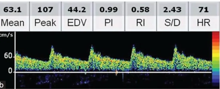
Waveform and variables
Figure 10: Transcranial Doppler waveform
Peak Systolic Velocity (cm/s)
It is the first peak on every TCD waveform for each pulse. Rapid upstroke rules out any stenotic lesion.
End-Diastolic Velocity (cm/s)
It is seen in all major intracranial arteries which will be between 20 and 50% of peak systolic velocity.
Mean Flow Velocity
Pulsatility index
It is the difference between systolic and diastolic flow velocities divided by the mean flow velocity. Pulsatility index value ranges from 0 to infinity. 0 means systolic and diastolic velocity are the same. In case of infinity mean velocity is 0. It is generally applied to vascular beds in skin and muscles where resistance and impedance to flow are high.
Resistivity index
67
The normal values of velocity in different arteries are given in the table below.
ARTERY WINDOW DEPTH
mm MEAN VELOCITY cm/ sec DIRECTION OF FLOW
ECICA Retromandibular 45–50 30 +/- 9 Away
MCA Transtemporal 30–65 50 +- 12 Toward
ACA Transtemporal 60–75 50 +/- 11 Away
PCA–P1 Transtemporal 60–70 39 +/- 10 Toward PCA–P2 Transtemporal 60–70 40 +/- 10 Away
OA Transorbital 45–55 21 +/- 5 Toward
Supracliniod ICA
Transorbital 65–80 41 +/- 11 Away
Parasellar ICA
Transorbital 65–80 47 +/- 14 Toward
VA Transforaminal 60–75 38 +/- 10 Away
BA Transforaminal 80–
120
41 +/- 10 Away
Patients admitted for shoulder surgery under Orthopaedics department will be chosen according to the inclusion and exclusion criteria.
Informed consent will be taken
Inside the operation room, with patient in supine position intravenous line will be secured. Before induction his/her radial artery will be cannulated under local anesthesia for invasive blood pressure measurement (MAP).
Transcranial doppler will be used to monitor middle cerebral artery blood velocity–Vmca (as surrogate for cerebral blood flow
monitoring) which will be done in the side opposite to the operating side.
Data will be collected pre-induction in supine (NIBP & IBP- (MAP), HR, SPO2, Vmca)
After preoxygenation patient will be induced with Fentanyl 2mcg/kg, Propofol 2-3mg/kg, will be relaxed and intubated, mechanically ventilated and maintained with isoflurane (MAC 0.9 - 1). ETCO2 will be maintained between 30 and 35mmHg.
fifteen minutes till the end of the surgery and post extubation in supine. Transcranial doppler will be done whenever there is a fall in blood pressure and after it has been corrected.
Blood pressure will be maintained MAP>60 mmHg in normotensive patients and MAP>70 mmHg in hypertensive patients.
DEMOGRAPHIC VARIABLES
[image:72.612.73.508.156.406.2]AGE DISTRIBUTION
Figure 11: Age distribution of the patients (n=20).
There were totally 20 subjects. The age of the patients ranged from 18 to 65 years with mean (standard deviation) as 38 (14) years. 60% patients are between 21 to 40 years of age. 10% patients are from the age of 18 to 20years.20% of the patients
0 1 2 3 4 5 6 7 AGE
GENDER DISTRIBUTION
[image:73.612.73.512.306.564.2]Out of the 20 patients recruited for the study only one was a female patient which is only 5% of the total.
Figure 12: Distribution of the patients based on their Gender (n = 20).
GENDER DISTRIBUTION
MALE FEMALE
ASA RISK DISTRIBUTION
As shown in figure 13, 3 patients were ASA risk II (which is 15%) and 18 were ASA I (85%). ASA III and above were excluded from the study. Out of three patients two of them were diabetics and one was both diabetic and asthmatic
patient. All of them were well controlled with oral hypoglycemic agents.
ASA
ASA I
ASA II
[image:74.612.74.510.291.546.2]PRIMARY OUTCOMES
[image:75.612.74.540.188.642.2]1: CHANGE IN MEAN ARTERIAL PRESSURE AFTER POSITION CHANGE (FROM SUPINE TO BEACH CHAIR)
Figure 14: Line graph to show the pattern of mean arterial pressure for individual patients 0 10 20 30 40 50 60 70 80 90 100 P1 P2 M AP
Change in MAP from supine to beach chair position
1 2 3 4 5 6 7 8 9 10
11 12 13 14 15 16 17 18 19 20
2: CHANGE IN MIDDLE CEREBRAL ARTERY BLOOD FLOW VELOCITY (VMCA) AFTER POSITION CHANGE (FROM SUPINE TO BEACH CHAIR)
0 10 20 30 40 50 60 70 80 90 P1 P2 VM CA
Change in VMCAfrom supine to beach chair position
1 2 3 4 5 6 7 8 9 10
11 12 13 14 15 16 17 18 19 20
[image:76.612.72.543.130.639.2]Fig 17: Distribution of VMCA percentage reduction from Baseline.
Variable Mean SD Min Max
MAP percentage reduction 25.02 9.89 0 41.6
VMCApercentage reduction 27.95 10.60 6.43 50.06
The percentage reduction in the mean arterial pressure ranges from 0 to 41.6% with the mean value of 25.02%. For one patient other than the initial drop in the blood pressure as soon as change in the position there was no further hypotension at all but still he had a minor drop in VMCA.
The paired samples t test was performed to test the significance of difference between supine and beach chair positions and found it was statistically significant (p < 0.001) for both mean arterial pressure (MAP) and middle cerebral artery blood flow velocity (VMCA)
COMPARING THE CHANGES IN MEAN ARTERIAL PRESSURE AND THE MCA BLOOD FLOW VELOCITY
The correlation between the MAP change from baseline and VMCA change from the baseline is performed using Pearson’s Correlation Coefficient and it is found
[image:80.612.90.533.290.632.2]CHANGES IN DIFFERENT ASA GROUPS
ASA Median IQR P value
1 27.9070 20.79–31.32 0.842
2 29.4118 14.28–41.57
The percentage reduction in MAP between different ASA group was analyzed. There is no significant difference in the fall in blood pressure between ASA I and ASA II patients.
ASA Median IQR Pvalue
1 27.1762 22.09–36.31 0.921
2 28.0936 18.49–33.49
[image:81.612.67.549.157.267.2]There is no significant difference noted between ASA I and ASAII patients in VMCA.
Table 3: MAP percentage reduction among different ASA groups
[image:81.612.65.549.486.631.2]SR. NO. AGE BASELINE MAP HYPOTENSION MAP TREATED MAP
1 20 95 64 75
2 65 82 66 77
3 18 70 53 65
4 24 78 53 65
5 60 82 66 75
6 55 87 60 76
7 55 91 64 84
8 34 85 60 70
9 23 93 62 72
10 33 82 61 73
11 28 86 59 70
12 45 86 62 73
[image:82.612.70.547.112.610.2]83
The above table shows the mean arterial pressure values in supine, during
hypotension in sitting position and after treatment with ephedrine or phenylephrine bolus.
Out of the 20 patients, the six patients who were overzealously treated during hypotension were excluded along with one patient who had no hypotension . Among the thirteen patients the MAP requirement was an average of 73.15mmHg (with a minimum of 65 mmHg and a maximum of 84mmHg) to keep the middle cerebral artery blood flow velocity around baseline. Hence can maintain adequate cerebral perfusion above 73.15mmHg.
Distribution among age groups
Serial No.
Age Percentage reduction of MAP
Percentage reduction of VMCA
MAP(mmHg) to maintain normal VMCA
1 65 19.51 27.17 77
2 52 14.28 33.49 75
3 60 19.51 35.35 75
4 55 31.03 40.31 76
[image:83.612.67.591.420.699.2]5 55 29.67 25.56 84
Table 6: Percentage reduction of MAP and VMCAin older age group
Serial No. Age Percentage
reduction of MAP
Percentage
reduction of VMCA
1 20 32.63 12.81
2 28 31.25 25.15
3 18 24.28 37.28
4 24 32.05 29.42
5 37 4.28 22.75
6 37 41.57 18.49
7 40 0 6.43
8 30 30 45.50
9 39 22.07 29.47
10 34 29.41 28.09
11 23 22.58 50.06
12 33 25.60 21.72
13 28 31.39 17.31
[image:84.612.68.549.113.690.2]Discussion:
This observational study was aimed at assessing the correlation between the
reduction in mean arterial pressure and cerebral blood flow in patients undergoing shoulder surgery in beach-chair position.
22 patients were enrolled in the study. Out of which 2 patients were excluded from the study due to poor trans-temporal Doppler window and wide swing in blood pressure recordings.
Mc Culloch et al(29) did a similar observational study in 19 patients .All the patients were more than 55 years of age and known cases of cardiovascular or cerebrovascular disease. All patients were given interscalene block for analgesia and standard intravenous induction and intubation was done. Anesthesia was
maintained with desflurane. They did controlled hypotension with remifentanil and phenylephrine infusion and monitored NIBP and invasive blood pressure and continuous middle cerebral artery blood flow velocity by transcranial Doppler.
In our study we included patients from age of 18 to 65 years, with a mean age of 38(standard deviation 14). 25% of the patients were more than 45 years and 75% of them were less than 45 years. None of our patients were given regional
anesthesia. We gave general anesthesia using standard intravenous induction and intubation. Anesthesia was maintained with isoflurane (miminum alveolar
concentration: 0.8–0.9) and 0.1 to 0.15mg /kg morphine was given for analgesia. We monitored invasive blood pressure and measured middle cerebral artery blood flow velocity by transcranial Doppler at various points namely before induction; after intubation in supine; then during hypotension in sitting position and after the hypotension was treated. We considered a 20% reduction in mean arterial pressure in order to treat it. In elderly patients we treated if mean arterial pressure was less than 70mmHg.
We noticed that the percentage reduction of mean arterial pressure varied from 0 to a maximum of 41.6%. The mean percentage reduction in mean arterial pressure was 25.02%.
The mean arterial pressure change was accompanied by reduction in middle cerebral artery blood flow velocity measured by transcranial Doppler
arterial pressure, middle cerebral artery blood flow velocity reduced by an average of 27.95%. The minimum drop was about 6.43% and a maximum of 50%.
It was found that one of our patients had very stable blood pressure throughout the procedure but had a minor drop in middle cerebral artery blood flow velocity.
We performed paired sample t test to test the significance of difference of mean arterial pressure and middle cerebral artery blood flow velocity in supine and beach-chair positions. It was statistically significant with p < 0.001.
It was also one of the objectives to analyze the correlation between the percentage reduction of mean arterial pressure and middle cerebral artery blood flow velocity.
We used Pearson’s correlation coefficient and found that there is no linear
correlation between the two variables.
The equation we derived is as follows,
Where a, b and c are constants and their values were found to be as, a = 2.934; b = 1.507; c = -0.04
VMCA: Percentage reduction of Middle cerebral artery blood flow velocity
MAP: Percentage reduction of Mean arterial pressure
To explain, there was a direct linear relationship between the percentage reduction of middle cerebral artery blood flow velocity and mean arterial pressure till 20%, following which they did not correlate directly.
While doing the study, we over-treatedsix patients’ blood pressure beyond the
baseline pressure, so we were not able to determine the lower limit of mean arterial pressure required to maintain cerebral blood flow near normal in those patients. Hence we excluded those values in our analysis. In the rest of the patients, in order to maintain a near normal middle cerebral artery blood flow velocity the average required mean arterial pressure was 73.1mmHg. This has inter-individual variation with a minimum value of 64mmHg and maximum value of 84mmHg.
We found variations in different age group also. We had 25% patients above 45 years and 75% were below 45 years. In patients above the age of 45 the average percentage reduction was 22.8% which was accompanied by a 32.38% reduction in middle cerebral artery blood flow velocity in sitting position.
Whereas, in the younger age group, the percentage reduction in mean arterial pressure was 23.78%, with a 26.47% reduction in middle cerebral artery blood flow velocity in sitting position.
We can say that, though the drop in the mean arterial pressure was similar in both the age groups, the fall in the cerebral blood flow was much larger in the older age group who would require a higher mean arterial pressure to maintain cerebral perfusion. The calculated average value of mean arterial pressure to maintain cerebral blood flow near the baseline was 77.4mmHg in the older age group whereas the average mean arterial pressure in all patients was 73.15mmHg.
In the study by McCulloch et all (29) all patients were known cases of
We found that there was no significant difference among the two ASA groups. None of our patients were hypertensives also. If we could have done the study in patients with higher ASA risk we might have obtained a significant difference among the ASA groups.
Conclusion:
This observational study in patients coming for shoulder surgery in sitting position showed correlation between mean arterial pressure and middle cerebral artery blood flow velocity
Mean arterial pressure can be taken as a surrogate, especially in sitting position, for cerebral perfusion thus necessitating active treatment in the event of hypotension.
We conclude that an average of 73.15mmHg is the minimum mean arterial pressure required to maintain cerebral perfusion in beach-chair position. In the older age group it is better to maintain the mean arterial pressure above
Limitations
1. Although the calculated sample size was 45, we were able to recruit only 20 patients. Hence the power of the study is reduced. The number of patients above the age of 45 was only 5.
2. We could not do a continuous transcranial Doppler monitoring.
BIBLIOGRAPHY
1. Marilyn J. Cipola. The Cerebral Circulation. Morgan and Claypool Life Sciences;
2. Kim. E. Barrett, Susan M. Barman, Scott Boitano, Heddwen Brooks.
Ganong’s Review of Medical Physiology. 24th ed. LANGE;
3. James E. Cottrell, William L. Young. Cottrell and Young’s Nueroanesthesia.
5th ed. MOSBY ELSEVIER;
4. Richard D. Hoge, Jeff Atkinson, Brad Gill, Gérard R. Crelier, Sean Marrett, and, G. Bruce Pike. Linear coupling between cerebral blood flow and oxygen consumption in activated human cortex. PNAS. 1999 Jun 14;96(16):9403–8. 5. Richard B Buxton and Lawrence R Frank. A Model for the Coupling Between
Cerebral Blood Flow and Oxygen Metabolism During Neural Stimulation. J Cereb BLOOD FLOW Metab. 1996 Jul 8;17:64–72.
6. Ronald D. Miller. Miller’s Anesthesia. seventh edition. ELSEVIER; 305-340 p.
7. Alifia Tameem, MBBS MD FRCA, Hari Krovvidi, MD FRCA Specialist Registrar, Queen Elizabeth Hospital Birmingham, Edgebaston, Birmingham, West Midlands, B15 2TH, et al. Cerebral physiology. BJA [Internet]. 2013 Mar 1; Available from:
http://ceaccp.oxfordjournals.org/content/early/2013/02/28/bjaceaccp.mkt001.f ull
8. Paul G. Barash, Bruce F. Cullen, Robert K. Stoelting, Michael K. Cahalan, M. Christine Stock. Clinical Anesthesia. 6th edition. Wolters Kluwer/ Lippincott Williams & Wilkins;
9. S. Strandgaard, J. Olesen, E. Skinhøj, N. A. Lassen. Autoregulation of Brain Circulation in Severe Arterial Hypertension. Br Med J. 1973 Mar 3;1.
10. L WOLLNER, S T McCARTHY, N D W SOPER, D J MACY. Failure of cerebral autoregulation as a cause of brain dysfuction in the elderly. Br Med J. 1979: 1.
Regional Cerebral Blood FlowStudies in Normal Subjects and Subjects With Risk Factors for Atherothrombotic Stroke. JAMA Neurol. 36(7).
12. Wagner M, Jurcoane A, Volz S, Magerkurth J, Zanella FE, Neumann-Haefelin T, et al. Age-related changes of cerebral autoregulation: new insights with
quantitative T2’-mapping and pulsed arterial spin-labeling MR imaging. AJNR Am J Neuroradiol. 2012 Dec;33(11):2081–7.
13. Kewal K. Jain. THE HANDBOOK OF NEUROANESTHESIA. New York: Humana press;
14. Leroy C. Abbott ; John B. deC. M. Saunders ; Helen Hagey ; Ellis W. Jones Jr. SURGICAL APPROACHES TO THE SHOULDER JOINT. J Bone Jt Surg. 1949 Apr;31(2):235–55.
15. Xinning Li, MD; Josef K. Eichinger, MD; Timothy Hartshorn, MD; Hanbing Zhou, MD; Elizabeth G. Matzkin, MD; Jon P. Warner, MD. A Comparison of the Lateral Decubitus and Beach-chair Positions for Shoulder Surgery:
Advantages and Complications. J Am Acad Orthop Surg. 2015 Jan 1; 16. Nicolas Bonnaig, MD, Steven Dailey, MD, and Michael Archdeacon, MD,
MSE. PROPER PATIENT POSITIONING AND COMPLICATION
PREVENTION IN ORTHOPAEDIC SURGERY. J Bone Jt Surg. 2014 Jul 2;96 A(13):1135–40.
17. Christopher J. Winfree, MD, David G. Kline, MD,. Surgical Neurology Intraoperative positioning injuries. ELSEVIER. 2005 Jan;63(1):5–16.
18. Sarah Gerken, MD.,. Preventing positioning injuries: An Anesthesiologist’s
perspectives.
19. Christina L. Beecroft, FRCA, FDS RCS, David M. Coventry, FRCA. Anaesthesia for shoulder surgery. Oxf J. CEACC pain(2008) 8(6):193–8. 20. Andre P. Boezart. Anesthesia and Orthopaedic Surgery. Mc Graw Hill
23. Darren J. Friedman, MD; Nata Z. Parnes, MD; Zachary Zimmer; Laurence D. Higgins, MD; Jon J.P. Warner, MD. Prevalence of Cerebrovascular Events During Shoulder Surgery and Association With Patient Position. Helio Orthop. 32(4).
24. L. K. Moppett, R. P. Mahajan. Transcranial doppler ultrasonography in anaesthesia and intensive care.
25. Nicoletto, Heather A; Burkman, Marilyn H. Transcranial Doppler Series Part II: Performing a Transcranial Doppler. Am J Electroneurodiagnostic Technol. 2009 Mar 1;49(1):14–27.
26. C C Bishop, S Powell, D Rutt and, N L Browse. Transcranial Doppler
measurement of middle cerebral artery blood flow velocity: a validation study. 27. F.J. Kirkham†, T.S. Padayachee‡, S. Parsons§, L.S. Seargeant†, F.R. House∣,,
R.G. Gosling‡. Transcranial measurement of blood velocities in the basal cerebral arteries using pulsed Doppler ultrasound: Velocity as an index of flow.
28. Bathala L, Mehndiratta MM, Sharma VK. Transcranial doppler: Technique and common findings (Part 1). Ann Indian Acad Neurol. 2013;16(2):174–9. 29. McCulloch TJ, Liyanagama K, Petchell J. Relative hypotension in the
PROFORMA
DATA COLLECTION SHEET
SERIAL NO: DATE:
HOSPITAL ID: AGE: SEX:
DIAGNOSIS:
PROPOSED SURGERY: ASA:
COMORBIDITIES MEDICATIONS
Informed Consent Form for Subjects
Informed Consent form to participate in a research study
Study Title:
Study Number: ____________
Subject’s Initials: __________________ Subject’s Name: _________________________________________
Date of Birth / Age: ___________________________
(Subject)
(i) I confirm that I have read and understood the information sheet dated ____________ for the above study and have had the opportunity to ask questions.
(ii) I understand that my participation in the study is voluntary and that I am free to withdraw at any time, without giving any reason, without my medical care or legal rights being affected.
(iv) I agree not to restrict the use of any data or results that arise from this study provided such a use is only for scientific purpose(s).
(v) I agree to take part in the above study.
(vi) I am aware of the Audio-visual recording of the Informed Consent.
(Click here for Audio Visual guidelines)
Signature (or Thumb impression) of the Subject/Legally Acceptable
INFORMATION SHEET
Research Title: The effect of beach chair position on cerebral blood flow in patients undergoing shoulder surgery
Information sheet Introduction
I am Dr. Jesudoss A, M.D post graduate with the Department of Anaesthesiology and I have ten months of experience in the department. I am doing a research on
measurement of blood flow to brain in patients undergoing shoulder surgery in sitting position (beach chair position). I am going to give information and invite you to be part of this research. You do not have to decide today whether or not you will participate in the research. Before you decide, you can talk to anyone you feel comfortable with about the research.
There may be some words that you do not understand. Please ask me to stop as we go through the information and I will take time to explain. If you have questions later, you can ask me or the anaesthetist on the day of surgery.
Purpose of the research: For patients who are planned for shoulder surgery, sitting position (or beach chair position) is chosen because of better visualization of surgical site. But this position can cause decrease in pressure in the blood vessels to brain and also blood flow to brain. If there occurs a significant reduction in blood flow brain it will affect the brain function. So we will treat blood pressure to bring back to normal. In order to prevent this I am going to do a Doppler scan (an ordinary ultrasound scan) of your head and also measure blood pressure by radial artery (blood vessel in the wrist) cannulation.
Participant selection: You have been invited to participate because you have surgery in your shoulder in sitting position.
Voluntary participation: your participation in this research is entirely voluntary. Whether you choose to participate or not, the management of anaesthesia and your safe recovery will not change. If you choose not to participate in this research project, you will be offered the same anaesthesia routinely given in this hospital for shoulder surgery.
anaesthesia i.e. by injecting a small amount of lignocaine in the skin to numb it. This might cause a little discomfort to you.
2. Then in the same position we will scan your head using Doppler scan (ultra sound).
3. Then anaesthesia will be given as per the standard protocol followed in this hospital. Surgery will be done in sitting position and if required the Doppler scan will be repeated during surgery also.
Side effects: commonly hematoma (collection of blood) and bleeding can happen due to the injection put in your blood vessel at the wrist. Temporary spasm of the artery, catheter-site- infection, ischemic damage, psuedoaneurysm formation are the other rare complications. We will take all precautions necessary to keep you safe.
Benefits: while measuring blood flow to your brain during the procedure, if there is any significant reduction we can treat immediately.
Reimbursements: Your will not be charged for the Doppler scan or for the injection that we put in your artery to measure blood pressure.
Confidentiality: Your name will not be mentioned anywhere neither the data sheet nor the final published study. Your data will bear a study number and the number will be used till analysis. The master sheet will have your study number.
Sharing the result: The result of this research is a property of Christian medical college and I’m entitled to publish it in a journal or present it in a conference.
Right to refuse or withdraw: You do not have to take part in this research if you do not wish to do so. You may also withdraw participating in this research even inside the operating room. It is your choice and all of your rights will be respected.
This proposal has been reviewed and approved by IRB, Christian Medical College, which is a committee whose task is to make sure that research participants are protected from harm. If you wish you can find more about the IRB.
Contact
1. Dr. Jesudoss A (Primary investigator) PG registrar,
Office: 0416 2282105
2. Dr. Melvin Alex Abraham (Co-investigator) Department of Anaesthesiology, CMC Vellore. Phone: +91 9894540049
Email ID: melvinalexabraham@gmail.com
3. Research office, II floor, Carman block, Christian medical college, Bagayam, Vellore 632 002.
Email:research@cmcvellore.ac.in Telephone: 04162284294
DATASHEET
P3_MAP P3_VMCA P1TOP3MAP P1TOP2MAP P1TOP2VMCA HOSPITAL NO SR.NO. 75 53.9 21.05263158 32.63157895 12.8113879 257050B 1 77 48.3 6.097560976 19.51219512 27.17622081 301722G 2
86 46.3 -7.5 31.25 25.15856237 229545G 3
65 47.6 7.142857143 24.28571429 37.28489484 256516G 4 65 37.9 16.66666667 32.05128205 29.42430704 257520G 5 75 34.5 -7.142857143 14.28571429 33.49875931 351721F 6 78 39.1 -11.42857143 4.285714286 22.75862069 213736G 7 75 22.5 8.536585366 19.51219512 35.35353535 623672A 8 109 39 -22.47191011 41.57303371 18.49529781 211054G 9
69 37.5 -15 0 6.432748538 202629G 10
76 53.4 12.64367816 31.03448276 40.31620553 344311G 11
80 47.5 0 30 45.50359712 872560F 12
83 39.4 -7.792207792 22.07792208 29.47368421 952972B 13 84 44.1 7.692307692 29.67032967 25.56818182 178569G 14 70 56.4 17.64705882 29.41176471 28.09364548 370908G 15 62 60.4 33.33333333 22.58064516 50.0635324 307808G 16 73 40.5 10.97560976 25.6097561 21.72897196 417998G 17 70 45.6 18.60465116 31.39534884 17.31958763 447661G 18 73 55.6 15.11627907 27.90697674 30.22071307 150762D 19