STUDY OF HOMOCYSTEINE LEVELS IN
PREECLAMPTIC WOMEN AND ITS
ASSOCIATION WITH THE SEVERITY
OF THE DISEASE
A CASE CONTROL STUDY
Dissertation submitted to
THE TAMILNADU
Dr.M.G.R MEDICAL UNIVERSITY
In partial fulfilment of the requirement
for the award of
M.S.DEGREE
–
BRANCH - II
OBSTETRICS & GYNAECOLOGY
GOVT. KILPAUK MEDICAL COLLEGE
KILPAUK, CHENNAI.
BONAFIDE CERTIFICATE
This is to certify that the dissertation entitled “ STUDY OF HOMOCYSTEINE LEVELS IN PREECLAMPTIC WOMEN AND
ITS ASSOCIATION WITH THE SEVERITY OF THE DISEASE” is the bonafide original work of DR.KOKILA.S under the guidance of PROF DR. M S. SORNAM MD., DGO., Department of Obstetrics and
Gynaecology, KMCH, Chennai in partial fulfilment of the requirements for MS Obstetrics and Gynaecology branch II examination of the Tamilnadu Dr.MGR Medical university to be held in April 2017 . The period of Postgraduate study and training from June 2014 to April 2017.
Prof .Dr.M.S.Sornam, MD., DGO.,
Professor of Obstetrics and Gynaecology, Kilpauk Medical College and Hospital Chennai–600010.
Prof .Dr.T.K.Shaanthy Gunasingh, MD., DGO., Professor and Head of the Department Obstetrics and Gynecology
Kilpauk Medical College and Hospital Chennai–600010.
Prof.Dr.R.Narayana Babu MD, DCH THE DEAN
DECLARATION
I solemnly declare that this dissertation “STUDY OF HOMOCYSTEINE LEVELS IN PREECLAMPTIC WOMEN AND ITS
ASSOCIATION WITH THE SEVERITY OF THE DISEASE” was
prepared by me at Government Kilpauk Medical College and Hospital, Chennai, under the guidance and supervision of Dr. M.S. SORNAM MD., DGO. Professor, Dept of Obstetrics and Gynaecology, Chennai.
This dissertation is submitted to The Tamilnadu Dr. M.G.R. Medical University, Chennai in partial fulfilment of the University regulations for the award of the degree of M.S. (Obstetrics and Gynaecology).
Place: Chennai
ACKNOWLEDGEMENT
I start this thesis in the name of Almighty God, the most beneficent and forgiving. I thank God for giving me the privilege to learn from the able teachers in my department.
I express my sincere thanks to PROF.DR.R.NARAYANA BABU M.D.,DCH.,Dean, Kilpauk Medical College for allowing me to conduct the
study using the available facilities.
I would like to express my sincere gratitude to my beloved Professor and Head of the Department of Obstetrics and Gynaecology Prof.Dr.T.K.SHAANTHY GUNASINGH M.D., D.G.O., for her guidance
and encouragement.
I convey my heartfelt gratitude and sincere thanks to my guide Dr.M.S.SORNAM,MD.,DGO., Professor , Department of Obstetrics and
Gynaecology, for her motivation, advice and valuable criticism which enable me to complete this work.
I am grateful to my Assistant Professors, colleagues and my friends for their advice and suggestions
.
My heartfelt thanks to my parents, my brothers and friends, who have been a constant source of encouragement and immense help, for instilling in me a sense of commitment and for their belief in me.
ABBREVIATIONS
DIVC
-
Disseminated Intravascular Coagulation
HELLP
-
Hemolysis, Elevated Liver enzymes, Low Platelets
HLA-G
-
Human Leucocyte Antigen G
IUGR
-
Intra Uterine Growth Restriction
IUD
-
Intra Uterine Death
MGA
-
Maternal Gestational Age
PLGF
-
Placental Like Growth factor
PPH
-
Post Partum Hemorrhage
SAM
-
S-Adenosyl Methionine
sENG
-
soluble Endoglin
sFlt
-
1 - soluble Fms-like tyrosine kinase 1
THFA
-
Tetra Hydro Folic Acid
INDEX
S.NO
TITLE
PAGE NO
1.
INTRODUCTION
12.
REVIEW OF LITERATURE
33.
AIM OF THE STUDY
364.
OBSERVATION AND RESULTS
415.
DISCUSSION
806.
CONCLUSION
837.
BIBLIOGRAPHY
8.
ANNEXURES
A) MASTER CHART
B) PROFORMA
INTRODUCTION
Preeclampsia is a pregnancy specific disorder which complicates about 3-10% of all nulliparous gestation[1]. The incidence is markedly influenced by race and ethnicity. Preeclampsia is the most common serious medical disorder of human pregnancy. It is characterised by the development of hypertension with proteinuria after 20 weeks of gestation .Edema is no longer consider as a diagnostic criteria but it is commonly associated with the normal pregnancy. The cause of Preeclampsia is not completely understood .various assumption based on numerous theories are being considered till date .
The etiology of preeclampsia is suggested to be abnormal placentation. The disorder is usually manifested after 20th week of gestation and the complications aggravate with the advancement of pregnancy and in the absence of proper patient management. Fetal complications of preeclampsia are directly related to gestational age and the severity of maternal disease and result in increased rates of preterm delivery, fetal growth restriction (FGR), placental abruption and perinatal death.
Hyperhomocysteinemia leads to endothelial dysfunction through various mechanisms. In addition to that homocysteine is also affect the fibrinolytic pathway which is contributing to the pathophysiology of preeclampsia.
REVIEW OF LITERATURE
PREECLAMPSIA
Preeclampsia is a hypertensive disorder of pregnancy associated with widespread vascular endothelial malfunction, clinically defined by hypertension and proteinuria occurring after 20 weeks of pregnancy . According to the new ACOG diagnostic criteria for preeclampsia , the diagnostic dependence on the proteinuria has been eliminated. The new onset of hypertension is associated with absence of proteinuria and the presence of thrombocytopenia, elevated liver enzymes, renal insufficiency, pulmonary edema and cerebral accident syndrome. Maternal disease due to preeclampsia is hypertension and proteinuria with or without multisystem involvement. Fetal disease due to preeclampsia includes are FGR, fetal hypoxia , oligohydramnios [4,5].
DEFINITION OF PREECLAMPSIA:
Hypertension in pregnancy is defined as
Systolic blood pressure of 140mmHg or higher
Diastolic blood pressure of 90mmHg or higher (Korotkoff V)
These measurements have to be confirmed on at least two occasions 4 to 6 hours apart.
The gold standard method for determining proteinuria is the 24-hour
urinary protein excretion.
Proteinuria is defined as
Excretion of more than 300mg of protein/24hr
Or a urinary protein-to-creatinine ratio of 30mg/mmol
Or if the above methods are unavailable, a protein value of about
30mg/dl (≥ 1+ on dipstick ) 2 random urine samples collected 4 to 6 hr
apart.
Urinary tract infection must be excluded before attributing the
proteinuria to preeclampsia.
Dipstick testing should be used only for screening purposes where
other methods are not available. In all pregnant women who present with
gestational hypertension the presence of proteinuria must be confirmed with a
protein-to- creatinine ratio[15,16] or preferably a 24-hour urine collection.
The new onset of hypertension and proteinuria after 20weeks gestation
are consider to be essential criteria to confirm the diagnosis of preeclampsia
presents with HELLP syndrome and in about 20 to 25% women who presents
directly with preeclampsia, either hypertension or proteinuria is absent.
Therefore with acceptance of the fact that the disease progress and worsening
can occur in the absence of proteinuria. The Society of Obstetric Medicine of Australia and New Zealand (SOMANZ) has widened these criteria and have
included the presence of other systemic manifestations irrespective of
SOCIETY OF OBSTETRIC MEDICINE OF AUSTRALIA AND
NEW ZEALAND DEFINITION OF PREECLAMPSIA
The onset of hypertension after 20weeks gestation is accompanied by
one or more of the following:
Renal involvement:
o Proteinuria ≥ 300mg/24 hr or urine spot protein/creatinine ratio
≥ 30 mg/mmol
o Serum or plasma creatinine≥ 0.09mmol/L or decreased urine output
( <500mL/24hr)
Hematological involvement:
o Thrombocytopenia
o Disseminated intravascular coagulation
o Hemolysis
Hepatic involvement:
o Abnormal liver function( SGOT and/or SGPT >50 IU/L, Elevated serum bilirubin >25IU/L
o Upper abdomen pain
Neurologic involvement:
o Convulsions(eclampsia)
o Hyperreflexia with sustained clonus
o Severe headache (persisting, atypical)
o Persistent visual disturbances (photopsia, scotomata, cortical
blindness, retinal vasospasm)
Hemiparesis/ Hemiplegia
o Pulmonary edema
o FGR and signs of fetal distress
INCIDENCE AND RISK FACTORS:
Preeclampsia incidence is about ranges 3 and 10 percent [1,12] in a
healthy nulliparous women. In case of nulliparous women, preeclampsia occurs
in milder form. The onset of preeclampsia in nulliparous women in near term
or intrapartum which accounts for 75% of cases and such a occurrence has
minimal risk of adverse outcomes. Whereas in case of women with a multiple
pregnancy,[24,25] chronic hypertension,[23,26] previous preeclampsia,[23,27]
pre gestational diabetes mellitus [23], and in those with pre existing
thrombophilias [28-30] preeclampsia occur in a very severe form.
Preeclampsia is considered as a disease occurring in primigravida
.Limited sperm exposure with the same partner prior to conception[31-36]
increase the risk of preeclampsia. There is a new factor in the recent days with
development in assisted reproductive technology has given way for multiple
challenges that affect the maternal immune system thereby, accounting for
RISK FACTORS FOR PREECLAMPSIA
Couple related risk factors:
Primi paternity and limited sperm exposure
Pregnancy after artificial insemination
Protective effect of partner change in case of previous preeclamptic pregnancy
Maternal or pregnancy related risk factors:
Advanced maternal age.
Multiple pregnancy, previous history of preeclampsia
Chronic hypertension and/or chronic renal disease.
Maternal connective tissue disorders (e.g., SLE)
Maternal chronic infections, maternal low birth weight
Obesity and insulin resistance and pre gestational diabetes mellitus
Previous history of thrombophilias, maternal susceptibility genes
History of preeclampsia in the family, hydropic degeneration of placenta
Gestational hypertension
It is defined as hypertension (BP 140/90) without proteinuria on at least two occasions, at least 6hours apart after 20th weeks of gestation in women known to be normotensive before pregnancy and before 20 weeks of gestation. Characteristics of this condition that differentiates it from preeclampsia is the absence of proteinuria and it is differentiated from chronic hypertension by onset of problem after 20weeks and the absence of hypertension before pregnancy. This condition can be associated with maternal obesity, multiple gestations, diabetes and previous history of preeclampsia. This condition is not benign and pregnancy outcomes in severe gestational hypertension is worse than in mild preeclampsia. The end result is about 15 to 20% patients with gestational hypertension is development of clinical syndrome of preeclampsia . Early gestational hypertension might be associated with preeclamptic pathology such as poor placentation and histologic evidence of placental ischemia and hemodynamic changes characterised by vasoconstriction and decreased cardiac output.
Diagnostic criteria of gestational hypertension:
1. Blood pressure ≥150/100mmhg
2. Gestational age less than 30weeks
3. Evidence of end organs damage increased serum creatinine,liver enzymes, LDH, thromocytopenia
4. Oligohydramnios 5. FGR
6. Abnormal uterine artery and /or umbilical artery Doppler velocimetry
ETIOPATHOGENESIS OF PREECLAMPSIA
Such compartmentalization is arbitrary as preeclampsia is a
continuous process. Preeclampsia is more likely a continuum of
VASCULAR ENDOTHELIAL ACTIVATION AND PRO AND
ANTIANGIOGENIC PROTEINS IN PREECLAMPSIA:
The endothelium is one of the key cells involved in the pathophysiology of preeclampsia which leads to an imbalance between the prostacyclin and thromboxane [40-42] and impairment of nitric oxide synthesis, a potent vasodilator. Endothelial cell activation leads to platelet activation and this activated platelets cause thrombosis of the spiral arteries which in turn causes a decreased uteroplacental blood. In preeclampsia, there is presence of impaired endothelial cell function and platelet aggregation prior to the onset of elevated thrombin and fibrin formation.[43-46].
The clinical syndrome of preeclampsia which occurs as a result of
placental ischemia is considered to be related to a protein known as Soluble
fms-like tyrosine kinase 1 (sFlt-1), which is produced by the placenta. It exerts
its actions by binding to vascular endothelial growth factor (VEGF) and it also
binds to placental like growth factor (PLGF). Hence elevated concentration of
sFlt-1 leads to reduced concentration of free VEGF & PLGF which in turn
causes impaired endothelial function[42]. The increased sFlt concentration of
corresponds to worsening of disease . Maynard et al.[47] exhibited that soluble
placental derived VEGF receptor (sFlt1) an antagonist of VEGF and PLGF is
not regulated. In case of preeclampsia which causes an elevated systemic sFlt1
that decreases after delivery [48,49,50] .The growth factor produced by the
deciduas are angiogenic in nature that induces PLGF. PLGF is needed for
the development of placenta in the early stages. Levine et al states that
maternal systemic hypertension is a result of the maternal response to sFlt
ANTI ANGIOGENIC PROTEINS
Soluble endoglin
Soluble endoglin (sEng) is a placental derived protein is a recent
parameter that sEng increases several weeks prior to the clinical onset of
preeclampsia. [52] .This protein causes decreased endothelial nitric
oxide-dependent vasodilatation. This leads to maternal hypertension and endothelial
dysfunction. Soluble endoglin is a co receptor for TGF-β. This receptor is
present over the surface of the endothelial cells and syncytiotrophoblasts.
Soluble endoglin (sEng) an angiogenic protein is present in the patients with preeclampsia. There is a positive correlation between the blood levels of
ANTI ANGIOGENIC PROTEINS
Soluble endoglin
Soluble endoglin (sEng) is a placental derived protein is a recent
parameter that sEng increases several weeks prior to the clinical onset of
preeclampsia. [52] .This protein causes decreased endothelial nitric
oxide-dependent vasodilatation. This leads to maternal hypertension and endothelial
dysfunction. Soluble endoglin is a co receptor for TGF-β. This receptor is
present over the surface of the endothelial cells and syncytiotrophoblasts.
sEng and the progress , worsening of the disease. The levels of sEng declines in the postpartum period. It is observed from the clinical studies that measurement of both sEng and sFLT-1 proteins are found to be a better predictor of preeclampsia than the measurement of either of the proteins. There are certain studies in which elevated sEng levels are observed especially in early onset of preeclampsia.
UTERINE VASCULAR CHANGES
The walls of the spiral arterioles is invaded by the interstitial and
endovascular trophoblasts .This migration transform the small masculoelastic
spiral arteries into large dilated, tortuous channels which are the source of
blood supply to the human placenta.
The so formed blood vessels in the uterus and the placenta has low
resistance, low-pressure, high flow system..The mechanism by which the spiral
arterioles are transformed in to the uteroplacental arteries is known as the
physiological changes during pregnancy.[38] In case of a normal pregnancy
the vascular changes induced by the trophoblast are seen in the intervillous
space and in the inner third of the myometrium where the spiral arterioles
originate from the radial arteries. The changes in the blood vessels takes place
in the two stages . First stage being “the conversion of the decidual segments
of the spiral arterioles by a wave of endovascular trophoblast migration in the
first trimester. and second stage being the involvement of myometrial
segments by a subsequent wave in the second trimester.”[38] .As a result of
these vascular changes around 100 to 150 spiral arterioles are transformed
into dilated , tortuous, and funnel-shaped vessels that communicate via
On the other hand , minimal change in the blood vessels are observed
after placentation in patients with preeclampsia or fetal growth restriction . In
these pregnancies the changes in the blood vessels takes place only in the
decidual portion and the myometrial portion of spiral arterioles does not
undergo any change .Thereby making the myometrial spiral arterioles sensitive
and responsive to the hormonal changes [38] .These changes in blood vessels
are also appreciated in a number of pregnancies associated with fetal growth
restriction[38,39,41] .Meekins and associates[4] have done studies which states
the invasion of trophoblast into the blood vessels is not an all-or-none
phenomenon in case of both normal as well as pregnancies complicated by
preeclampsia.
lacental cytotrophoblasts invade the uterine wall where they breach eins
and extensively remodel maternal spiral arterioles
IMMUNOLOGY OF PREECLAMPSIA
There are other hypothesis for preeclampsia syndrome like loss of maternal immune tolerance against the paternal derived placental and foetal antigenic tissues. Tolerance dysregulation also explains an increased risk when the paternal antigenic load is increased which is seen with two sets of paternal chromosomes- “a double dose” as observed in molar pregnancies and in trisomy 13.
It is perceived that preeclampsia is a result of non functioning of the maternal immune system which in turn fails to recognise the fetoplacental unit. Death of extra villous cytotrophoblast occur due to increased synthesis of immune cells which leads to elevated levels of TNF α .Women with
that there is a significant role of HLA in case of impaired spiral arteriole invasion. The natural killer cells secrete VEGF and placental growth factor which are responsible for the interactions between these cells and trophoblasts in a normal pregnancy. There is presence of elevated amounts of soluble fms like tyrosine kinase, an antagonist of VEGF, PLGF in preeclamptic women.[17,18].
Factors contributing to an enhanced immunologically mediated inflammatory reaction include the placental micro particles which are circulating in the maternal body. Maternal immune response syndrome is a condition in which there is oxidative injury of the placenta , 2ndstage of the two stage model hypothesis for preeclampsia. The syndrome occurs due to activation of immune system in the mother in response to the circulating placental micro particles. This has lead to the formation of a new hypothesis called the
placental
debris hypothesis of preeclampsia.[54]GENETIC CONFLICT HYPOTHESIS
Haig’s theory of genetic conflict states that [55] the genes in the fetus
are selected to increase the nutrient transfer to the fetus. In contrast to this the
genes from the mother are selected to reduce the amount of transfer and
maintain the optimal level. A similar conflict between the genes that are
obtained from the mother and the genes obtained from the father exists within
the fetal cells which is known as the genetic imprinting phenomenon.
The conflict hypothesis have been demonstrated that elevation of
maternal blood pressure due to placental factors (fetal genes), whereas
linkage studies have identified three preeclampsia loci showing significant
linkage: 2p12, 2p25 and 9p13.[57] . Oudejans et al.[58] have recently
published the importance of genomic imprinting .Oudejans et al has
Prediction of Preeclampsia
A well known of mechanism of pathophysiology of preeclampsia is impaired trophoblastic invasion of the spiral arteries which increases impedence of flow to uterine arteries. Several studies have shown that evidence of reduced uterine flow is associated with the development of preeclampsia.
Elevated level of second trimester β- human chorionic gonodotropin (Β
HCG) have been identified in patients at risk for hypertensive disorders during pregnancy. Another study demonstrated that in presence of a diastolic notch , the association of serum screening with alfa –feto-protein and human chorionic gonodotrophin , improve sensitivity and positive predivtive value to 91% and 41% respectively.
Inspite of the promising results heterogeneity between studies regarding gestational age at time of study of population has lead us to incline towards the combination of ultrasonographic and biochemical markers as screening procedure of preeclampsia.
Maternal echocardiography
In pregnancy, changes happen in cardiovascular system with initial vasodilatative adaptation of the maternal cardiovascular system which begins in 1st trimester as a result of invasion by spiral arteries by the trophoblast. Indeed remodelling in the spiral arteries contributes to 20% to 26% . Echo parameters of cardiac changes in pregnancy may be, in normotensive women an important of pregnancy complications and indicator of predisposition to cardiac diseases.
Provocative pressor tests
These are tests which assess blood pressure increase in response to a stimulus . They are cumbersome and time consuming . Sensitivities of all these tests range from 55to 70% with specificity of 85% . They include :
Angiotensin sensitivity test:
1.
Roll over test:
It measures the hypertensive response in women at 28 to 32 weeks who are resting in the left lateral decubitus position and then roll over to a supine position. A positive test is an elevation of 20 mmHG or more in blood pressure when patients roll over from the lateral to the supine position .
2.
Urinary calcium:
Several studies have demonstrated that preeclampsia is associated with hypocalciuria . An urinary calcium concentration equal to or less than 12 mg/ dl in a 24 hr collection has positive and negative predictive value of 85 and 91% respectively , for the diagnosis of preeclampsia .Determination of calcium creatinine ratio in a randomly obtained urine sample seems to be accurate as 24 hr collection .
New biochemical markers :
Several new biochemical markers associated with preeclampsia are:
Adrenomedulin Podocyturia
mRNA
Placental protein 13 PAPP –A
Activin A and inhibin A P- selectin
Fibronectin
Heat shock protein Fms like tyrosin kinase 1 Soluble endoglin
Cell free fetal DNA
Chronic hypertension in pregnancy
Chronic hypertension complicating pregnancy accounts for about 0.5-3% .Increase in incidence of obesity and a high maternal age in today’s world
is expected to increase the incidence of chronic hypertension complicating pregnancies..Hypertension before pregnancy or before 20th week of gestation on more than one occasion atleast 4-6hours apart or persistence of hypertension for 12weeks postpartum is defined as chronic hypertension.
It is very difficult to diagnose chronic hypertension in patients who attend the antenatal clinic for the first time after 16 weeks of pregnancy because there is a physiological fall in blood pressure after 16 weeks of gestation. This physiological fall in blood pressure might mask the original findings of elevated blood pressure and can be recorded as a normal blood pressure which in turn will eventually be increased in the third trimester. These women are more prone to develop gestational hypertension.
Classification of chronic hypertension :
1. Mild hypertension
2.Severe hypertension Systolic Bp 160/110mmHg or >160/110mmHg
Etiology of chronic hypertension:
In 90% of cases of chronic hypertension is due to essential hypertension and remaining 10% cases it is due to an underlying disorder .i.e.chronic renal disease, endocrine disorder,collagen vascular disease.
1.Renal Diseases
Renal parenchymal diseases such as glomerular nephritis,reflux nephropathy, adult polycytic kidney diseases.
Renovascular hypertension
2.Endocrine disorder
Diabetes with vascular involvement Thyrotoxicosis
Pheochromocytoma Primary aldosteronism Cushing syndrome
3.Collegen vascular disease
SLE
Scleroderma
4.Others
Aortic coarctation
Maternal and perinatal risk of chronic hypertension
There is an increased risk of development of superimposed preeclampsia and abruptio placentae in case of chronic hypertension complicating pregnancies.It observed that about 10 to 25% patients with mild hypertension develop preeclampsia . On the other hand, about 50% patients with severe chronic hypertension develops preeclampsia . The overall rate of superimposed preeclampsia is 25%. However, it is observed that certain factors contribute to the rate significantly and presence of hypertension for 4 years (31% versus 22%), history of preeclampsia in previous pregnancies (32% versus 23%), and in those who have a diastolic blood pressure is 100–110 mm Hg compared to who have a diastolic blood pressure is less than 100 mm Hg at baseline.
PREECLAMPSIA SUPERIMPOSED ON CHRONIC HYPERTENSION
Criteria for diagnosis:
A) New-onset proteinuria ≥ 300mg/24 hours in hypertensive women who
had no proteinuria before 20 weeks of gestation.
B)
A sudden increase in proteinuria or blood pressure or platelet count <100,000/μL in a woman with hypertension and proteinuria before 20
HOMOCYSTEINE
Homocysteine formed as a result of metabolism of the essential amino acid methionine is a sulphur containing amino acid.It is formed at the junction of metabolism of the amino acid methionine and synthesis of cysteine and taurine . Metabolism(fig 1) of homcystiene occurs via two biochemical pathways–
1. re-methylation,in which homocysteine is transformed into methionine , and
2. trans-sulfuration, in which homocysteine is transformed into cysteine and taurine.
HOMOCYSTEINE METABOLISM
Homocysteine , a sulphur containing amino acid does not form proteins. Metabolism of this amino acid is at the juncture of two metabolic pathways: remethylation and transsulfuration.
In remethylation reaction methionine is formed as a result of acceptance of a methyl group from N-5 methyltetrahydrofolate or betaine by homocysteine. The reaction with N-5 methyltetrahydrofolate takes place in all the body tissues and is vitamin B12 dependent .Whereas the reaction with betaine takes place only in liver and is not dependent on vitamin B12. S-adenosyl methionine(SAM) is formed when ATP activates a methionine. SAM is a universal methyl donor.A by-product of these methylation reactions is S-adenosyl homocysteine(SAH) which on hydrolysis regenerates homocyteine making it available for the next new cycle.
HYPERHOMOCYSTEINEMIA IN HUMANS:
CAUSES OF HYPERHOMOCYSTEINEMIA[59]
A) ENZYME DEFECTS:
1) Cystathionine synthase ( Homocystinuria-1)
2) Homocysteine methyl transferase( Homocystinuria-2) 3) Methyl THFA reductase deficiency ( Homocystinuria-3) 4) Cystathionase deficiency ( cystathioninuria)
B) ACQUIRED CAUSES
1) Vitamin B12, Folic acid and pyridoxine deficiency. 2) Hypothyroidism, chronic renal failure.
HOMOCYSTEINE IN NORMAL PREGNANCY
The normal serum homocysteine level in non pregnant adult female
range between 5 and 15 μmol/L. It exists in two forms. 75 to 80% exists as
protein bound form and 15-25 % exist as acid soluble free form [60]. Hemodilution, reduced levels of albumin and increase in the demand of the amino acid methionine by both the mother and the fetus during pregnancy leads to reduced amount of homocysteine . Of this the decrease in serum albumin level correlates well with the serum homocysteine level.
During normal pregnancy the homocysteine levels are significantly lower in all trimesters compared with non pregnant control values. Walker et al[61] in a study of normal pregnant women and non pregnant women, measured serum homocysteine level by chromatography and found that the lowest level of homocysteine was found during the second trimester and homocysteine rises in the third trimester. However this rise is still well below the normal non pregnant level.[61,62] The normal level of homocysteine level during normal pregnancy is:
First trimester- 3.9-7.3μmol/L
Second trimester-3.5-5.3 μmol/L
HOMOCYSTEINE IN PREECLAMPSIA AND OTHER
PREGNANCY RELATED DISORDERS:
Numerous studies have found an association between homocysteine and various pregnancy related disorders. Klai et al[63] assessed the genetic make up of patients with proven placental vasculopathies and found an association with MTHFRA1298C polymorphism which leads to hyperhomocysteinemia. Elevated homocysteine levels are associated with development of vasculopathy of the placenta. Many hypothesis have been proposed for the pathogenesis of homocysteine induced vasculopathy and endothelial dysfunction.
MECHANISMS BY WHICH HOMOCYSTEINE MAY INDUCE
VASCULAR INJURY AND ENDOTHELIAL DYSFUNCTION:
Increased amounts of monocyte chemoattractant protein -1 and interleukin 8 by the amino acid homocysteine , in turn leads to recruitment of leukocytes [64].
The thiolactone is a metabolite of homocysteine that combines with LDL cholesterol and produces aggregrates. These aggregates are then taken up by the circulating macrophages in the arterial intima and forms foam cells which in turn release lipid into the atherosclerotic plaques. [65].
This thiolactone also induces apoptosis in cultured trophoblasts thereby leading to placental dysfunction[65]
Homocysteine decreases the utero placental flow by increasing the smooth muscle proliferation and enhancing the collagen production.
Oxidation of reduced homocysteine leads to formation of free radicals which damage the endothelial cells due to oxidative stress resulting in endothelial cell activation [73,74].
The pro aggregatory effects of homocysteine and impaired endothelial function lead to marked platelet accumulation.
HYPERHOMOCYSTEINEMIA IN PREECLAMPSIA
REVIEW OF CLINICAL STUDIES:
Numerous studies have proved the association of hyperhomocysteinemia in preeclampsia.[82-89]. Similarly the level of homocysteine correlated with the severity of preeclampsia[86].Md.Mazammel Hoque et al[82] compared the homocysteine levels in healthy pregnant, preeclamptic and eclamptic pregnant women and found that homocysteine levels are significantly raised in preeclampsia .Mahal M et al[83] compared serum homocysteine and HDL levels in preeclamptic/ eclamptic mother and found that both HDL and homocysteine are increased in women with preeclampsia/eclampsia.
Singh Urmila et al[84] studied 90 women and found that homocysteine levels were significantly different between non preeclamptic and preeclamptic mothers . In that study, homocysteine levels correlated directly with the degree of hypertension. And he proved that maternal homocysteine levels in normal pregnant women were significanty lower than non pregnant levels.
Ingec et al[89] compared the homocysteine levels among mild and severe preeclampsia with eclampsia and concluded that homocysteine levels are significantly elevated in severe preeclampsia and eclampsia, with significant difference between mild and severe preeclamptic group. However in that study , it was concluded that there was not much difference between normal pregnancy and in mild preeclampsia. Stolkova et al[86] did a study on the distribution of homocysteine levels in preeclamptic women and found that there is an association between serum homocysteine levels and the severity of preeclampsia.
Makedos and Papanicollou et al[87] compared maternal homocysteine, folic acid and B12 levels among women with preeclampsia and found that hyperhomocysteinemia is associated with poor maternal and fetal outcome and found that hyperhomocysteinemia occurred in preeclampsia independent of maternal folate and B12 levels.of preeclampsia.
The Hordaland study[97] showed that women with Homocysteine levels
more than 15μ/L are more likely to suffer from pregnancy related
complications and are also associated with poor maternal and fetal outcome. Baksu et al [88] studied the homocysteine levels in pregnancies complicated with severe and non severe preeclampsia and found that at a homocysteine
level of above 15μmol/L the maternal and fetal morbidities increased and also proved that homocysteine levels differed significantly among the severe and non severe group.
HYPERHOMOCYSTEINEMIA IN OTHER PREGNANCY
Hyperhomocysteinemia is found to have some association in neural
tube defects.[90]
Hyperhomocysteinemia in amniotic fluid is associated with small for gestational babies.[91] 32
Hyperhomocysteinemia is an independent marker for low birth
weight.[92]
Hyperhomocysteinemia together with uterine artery Doppler could
predict IUGR[93] and hyperhomocysteinemia in preeclampsia is associated with IUGR.[87] The Hordaland homocysteine study[97] showed that there is an association between hyperhomocysteinemia and IUGR independent of preeclampsia.
Hyperhomocysteinemia in preeclampsia is inversely related toinsulin sensitivity.[94]
Hyperhomocysteinemia is an independent risk factor for the
development of placental abruption and infarction.[95,97]
Hyperhomocysteinemia is one of the causes for recurrent pregnancy
losses in the first trimester
STUDY OF HOMOCYSTEINE LEVELS IN PREECLAMPTIC WOMEN
AND ITS ASSOCIATION WITH THE SEVERITY OF THE DISEASE
AIMS &OBJECTIVES
1. To find the association of the homocysteine levels in Preeclampsia and normal pregnancy
2. To know the prediction cut off values of serum homocysteine levels and to find out the severity of preeclampsia.
BENEFITS OF THE STUDY
1) This study evaluates the significant role of homocysteine in preeclampsia.
2) To find out vascular complication early if hyperhomocysteinemia exists. 3) To know the fetal complications early and improve the maternal and
METHODOLOGY
1) All the patient attending AN OPD and Labour ward who are undergoing preeclamptic workup were included and compared to equal number of healthy normotensive pregnant women
2) Blood pressure was recorded (two measurements 6 hrs apart in the semi recumbent position)
3) All the patients underwent a detailed history taking and examination as mentioned in the proforma.
All these patients were subjected to blood investigations like Complete hemogram, urine routine, renal function tests ,liver function tests, serum fibrinogen and serum uric acid, they also had obstetrics ultra sonogram and fundoscopy, 24 hours urinary protein excretion was done for all patients included in the study. Urine culture were done for appropriate patients. Peripheral smear study was done for all patients to exclude megaloblastic anaemia, dimorphic anaemia and HELLP Serum folate and vitaminB12 levels could not be estimated because of limited resources. Hence the presence of megaloblastic or dimorphic anemia are taken as indirect indictors of folate or B12 deficiency and these patients were ruled out of the study. Once vitamin deficiencies were excluded, serum homocysteine level was estimated.
until analysis. Serum homocysteine was measured by fluorescence polarization immunoassay (FPIA) run on Abbott’s AxSYM machine using Abbott’s kit.
STUDY DESIGN :
Case control study
DURATION OF STUDY
-9
MONTHSPLACE OF STUDY
:Antenatal outpatient department, Labour ward and Antenatal ward , Dept of Obstetrics & Gynaecology , Govt Kilpauk medical college &Hospital , Chennai.
SAMPLE SIZE:
N=2X(4X4)(0.84+1.96)2/( Mean difference=3)2 = 27.80=30 each group n-sample size
r-ratio of cases to contols(=1)
Zβ-Represents the desired power of the study(for 80%-0.84)
Zα/2-Represents the desired level of statistical significance (for 0.05-1.96) s-statistical difference(σ-)
cases -30 control -30 2 2 /2 2
)
ifference
(
)
Z
(
)
1
(
d
Z
r
r
INCLUSION CRITERIA:
CASES:
1) Pregnant women with BP≥140/90mmHG (previously normotensive women) on atleast 2 occasions 6 hours apart.
2) Gestational age 28-40weeks (sure of gestational age by LMP or USG in 1st or early 2ndtrimester).
CONTROL:
Healthy pregnant normotensive women of gestational age of 28-40weeks.
EXCLUSION CRITERIA:
1)Chronic hypertension
2) Gestational Diabetes mellitus 3)Type 1&2 Diabetes mellitus 4) Connective tissue disorders 5) Multiple pregnancy
6) Liver diseases 7) Severe Anaemia 8)Smoking
9)Obesity
STATISTICAL ANALYSIS
OBSERVATION AND RESULT
[image:47.595.113.509.258.647.2]Correlation of variables with preeclampsia
TABLE -1
Maternal Age # preeclampsia
CrosstabPREECLAMP
SIA
NO
YES
Total
AGE
GROU
P
UP
TO
25
YEARS
Count
8
6
14
%
within
PREECLAMPSIA
26.7% 20.0% 23.3%
% of Total
13.3% 10.0% 23.3%
26 YEARS &
ABOVE
Count
22
24
46
%
within
PREECLAMPSIA
73.3% 80.0% 76.7%
% of Total
36.7% 40.0% 76.7%
Total
Count
30
30
60
%
within
PREECLAMPSIA
100.0% 100.0% 100.0%
Chi-Square Tests
Value df
Asymp.
Sig.
(2-sided)
Exact Sig.
(2-sided)
Exact Sig.
(1-sided)
Pearson
Chi-Square
.373
a1
.542
Continuity
Correction
b.093
1
.760
Likelihood
Ratio
.374
1
.541
Fisher's
Exact
Test
.761
.381
Linear-by-Linear
Association
.366
1
.545
No
of
Valid
Cases
60
TABLE -2
Gravida # preeclampsia
Crosstab
PREECLAMPSIA
NO
YES
Total
GRAVID
A
1
Count
6
16
22
%
within
PREECLAMPSIA
20.0%
53.3%
36.7%
% of Total
10.0%
26.7%
36.7%
2
Count
18
6
24
%
within
PREECLAMPSIA
60.0%
20.0%
40.0%
% of Total
30.0%
10.0%
40.0%
3
Count
6
8
14
%
within
PREECLAMPSIA
20.0%
26.7%
23.3%
% of Total
10.0%
13.3%
23.3%
Total Count
30
30
60
%
within
PREECLAMPSIA
100.0% 100.0% 100.0%
Among preeclamptic women, primigravida were 16, 2ndgravid were 6, 3rd gravida were 8. Among normotensive women, primigravida 6, 2nd gravida were 18, and 3rdgravida were 6. Pearson Chi-square -10.831 p=0.004 The degree of gravidity has significant relation to preeclampsia.
Chi-Square Tests
Value
df
Asymp. Sig.
(2-sided)
Pearson Chi-Square
10.831
a2
.004
Likelihood Ratio
11.282
2
.004
Linear-by-Linear Association
1.802
1
.180
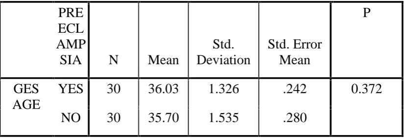
TABLE -3
Gestational age # preeclampsia
Group Statistics
PRE
ECL
AMP
SIA
N
Mean
Std.
Deviation
Std. Error
Mean
P
GES
AGE
YES
30
36.03
1.326
.242
0.372
NO
30
35.70
1.535
.280
TABLE-4
B MI # preeclampsia
Group Statistics
PRE
ECL
AMP
SIA N
Mean
Std.
Deviation
Std. Error
Mean
P
PRE PREG
BMI
YES 30
24.713 2.4710
.4511
NO
30
21.020 1.1941
.2180
0.000
TABLE-5
Serum Homocysteine # preeclampsia
Group Statistics
PRE
ECL
AMP
SIA
N
Mean
Std.
Deviation
Std. Error
Mean
P
SER
HOMOCYSTEI
NE
YES 30
16.923 2.3218
.4239
0.000
NO
30
9.647
1.4088
.2572
TABLE -6
Urine albumin # preeclampsia
Crosstab
PREECLAMPSI
A
NO
YES
Total
URI
ALBUMIN
1+
Count
0
1
1
%
within
PREECLAMPSIA
.0%
3.3%
1.7%
2+
Count
0
9
9
%
within
PREECLAMPSIA
.0%
30.0%
15.0%
3+
Count
0
3
3
%
within
PREECLAMPSIA
.0%
10.0%
5.0%
4+
Count
0
17
17
%
within
PREECLAMPSIA
.0%
56.7%
28.3%
NIL
Count
30
0
30
%
within
PREECLAMPSIA
100.0% .0%
50.0%
Total Count
30
30
60
%
within
PREECLAMPSIA
Pearson Chi-square 60.000 ,p= 0.000 which is very significant .Hence urine
albumin has very significant relation to preeclampsia.
Chi-Square Tests
Value
df
Asymp. Sig.
(2-sided)
Pearson Chi-Square
60.000
a4
.000
Likelihood Ratio
83.178
4
.000
TABLE-6
Serum Uric acid # preeclampsia
Group Statistics
PRE
ECL
AMP
SIA
N
Mean
Std.
Deviation
Std. Error
Mean
P
URIC
ACID
YES
30
6.82
.780
.142
0.000
NO
30
5.20
.459
.084
TABLE- 7
24 hr urinary protein # preeclampsia
Group Statistics
PRE
ECL
AM
PSI
A
N
Mean
Std.
Deviation
Std. Error
Mean
P
24HR
URI
PROTEINE
YES 30
395.10 107.310
19.592
NO 30
186.40 15.867
2.897
0.000
ROC CURVE FOR VARIABLES IN RELATION TO
PREECLAMPSIA
AREA UNDER CURVE- 0.998333
STATISTICAL SIGNIFICANCE P <0.0001
Hence serum Homocysteine is a statistically significant variable to predict preeclampsia with p value <0.0001
SER HOMOCYSTEINE
0
20
40
60
80
100
0
20
40
60
80
100
100-Specificity
S
e
n
s
it
iv
it
y
ROC CURVE
Variable
SER_HOMOCYSTEINE
SER HOMOCYSTEINE
Classification variable
PREECLAMPSIA
Sample size
60
Positive group :
PREECLAMPSIA = 1
30
Negative group :
PREECLAMPSIA = 0
30
Disease prevalence (%)
unknown
Standard Errora
0.00254
AREA UNDER THE ROC CURVE
Area under the ROC curve (AUC)
0.998333
Standard Error
a0.00254
95% Confidence interval
b0.937079
to
1.000000
z statistic
196.319
Significance level P (Area=0.5)
<0.0001
YOUDEN INDEX
Youden index J
1.0000
ROC CURVE FOR BLOOD UREA
AREA UNDER CURVE-0.894444
STATISTICAL SIGNIFICANCE P <0.0001
Hence blood urea is a statistically significant variable in relation to preeclampsia.
UREA
0 20 40 60 80 100
0 20 40 60 80 100
100-Specificity
S
e
n
s
it
iv
it
y
ROC curve
Variable
UREA
Classification variable
PREECLAMPSIA
Sample size
60
Positive group :
PREECLAMPSIA = 1
30
Negative group :
PREECLAMPSIA = 0
30
Disease prevalence (%)
unknown
Area under the ROC curve (AUC)
Area under the ROC curve (AUC)
0.894444
Standard Error
a0.0396
95% Confidence interval
b0.788001
to
0.958932
z statistic
9.959
Significance level P (Area=0.5)
<0.0001
Youden index
Youden index J
0.6333
ROC FOR SERUM CREATININE
AREA UNDER CURVE-0.827778
STATISTICAL SIGNIFICANCE P <0.0001
Hence serum creatinine is a statistically significant variable in relation to preeclampsia.
CREATININE
0
20
40
60
80
100
0
20
40
60
80
100
100-Specificity
S
e
n
s
it
iv
it
y
ROC curve
Variable
CREATININE
Classification variable
PREECLAMPSIA
Sample size
60
Positive group :
PREECLAMPSIA = 1
30
Negative group :
PREECLAMPSIA = 0
30
Disease prevalence (%)
Unknown
Area under the ROC curve (AUC)
Area under the ROC curve (AUC)
0.827778
Standard Error
a0.0539
95% Confidence interval
b0.708357
to
0.913004
z statistic
6.080
Significance level P (Area=0.5)
<0.0001
Youden index J
0.6333
Associated criterion
>0.6
ROC CURVE FOR 24HRS URINARY PROTEIN
AREA UNDER CURVE -0.97552
STATISTICAL SIGNIFICANCE P <0.0001
Hence 24hrs urinary protein is a statistically significant variable in relation to preeclampsia.
24HR URINE PROTEIN
0
20
40
60
80
100
0
20
40
60
80
100
100-Specificity
Sensitivity
ROC curve
Variable
24HR_URINE_PROTEIN
24HR URINE _ PROTEINE
Classification variable
PREECLAMPSIA
Sample size
60
Positive group :
PREECLAMPSIA =
1
30
Negative group :
PREECLAMPSIA =
0
30
Disease prevalence (%)
unknown
Area under the ROC curve (AUC)
Area under the ROC curve (AUC)
0.97552
Standard Error
a0.000
95% Confidence interval
b0.940371
to
1.000000
Significance level P (Area=0.5)
<0.0001
Youden index
Associated criterion
>220
ROC CURVE FOR SERUM BILIRUBN
AREA UNDER CURVE -0.874444
STATISTICAL SIGNIFICANCE p < 0.0001
Hence serum bilirubin is a statistically significant variable in relation to preeclampsia.
SER BILIRUBIN
0 20 40 60 80 100
0 20 40 60 80 100
100-Specificity
S
e
n
s
it
iv
it
y
Sensitivity: 66.7ROC curve
Variable
SER_BILIRUBIN
SER BILIRUBIN
Classification variable
PREECLAMPSIA
Sample size
60
Positive group :
PREECLAMPSIA = 1
30
Negative group : PREECLAMPSIA = 0
30
Disease prevalence (%)
unknown
Area under the ROC curve (AUC)
Area under the ROC curve (AUC)
0.874444
Standard Error
a0.0441
95% Confidence interval
b0.763456
to
0.945898
z statistic
8.493
Significance level P (Area=0.5)
<0.0001
Youden index J
0.5667
Associated criterion
>0.8
ROC CURVE FOR PRE PREGNANT BMI
AREA UNDER CURVE -0.918889
STATISTICAL SIGNIFICANCE p <0.0001
Hence prepregnant BMI is a statistically significant variable in relation to preeclampsia.
PRE PREG BMI
0
20
40
60
80
100
0
20
40
60
80
100
100-Specificity
S
e
n
s
it
iv
it
y
ROC curve
Variable
PRE_PREG_BMI
PRE PREG BMI
Classification variable
PREECLAMPSIA
Sample size
60
Positive group :
PREECLAMPSIA = 1
30
Negative group :
PREECLAMPSIA = 0
30
Disease prevalence (%)
unknown
Area under the ROC curve (AUC)
Area under the ROC curve (AUC)
0.918889
Standard Error
a0.0437
95% Confidence interval
b0.819016
to
0.973660
z statistic
9.588
Significance level P (Area=0.5)
<0.0001
Youden index J
0.8333
Associated criterion
>23.2
CORRELATION OF SERUM HOMOCYSTEINE WITH OTHER
VARIABLES IN RELATION TO PREECLAMPSIA
Comparison of ROC curves
Variable
1
SER_HOMOCYSTEINE
SER HOMOCYSTEINE
Variable
2
24HR_URI_PROTEIN
24HR URI PROTEIN
Variable
3
SERUM CREATININE
Variable
4
BLOOD UREA
Variable
5
PRE_PREG_BMI
PRE PREG BMI
Variable
6
SER_BILIRUBIN
SER BILIRUBIN
Classification variable
PREECLAMPSIA
Sample size
60
Negative group : PREECLAMPSIA = 0
30
AUC
SE
a95% CI
bSER_HOMOCYSTEINE
0.998 0.00254 0.937
to
1.000
24HR_URI_PROTEIN
0.975 0.000
0.930
to
1.000
CREATININE
0.828 0.0539
0.708
to
0.913
UREA
0.894 0.0396
0.788
to
0.959
PRE_PREG_BMI
0.919 0.0437
0.819
to
0.974
SER_BILIRUBIN
0.874 0.0441
0.763
to
0.946
SER_HOMOCYSTEINE
~
24HR_URI_PROTEIN
Difference between areas
0.00167
Standard Error
c0.00254
95% Confidence Interval
-0.00331
to
0.00664
z statistic
0.657
Significance level
P = 0.5114
SER_HOMOCYSTEINE
~
CREATININE
Difference between areas
0.171
Standard Error
c0.0535
95% Confidence Interval
0.0657
to
0.275
Significance level
P = 0.0014
SER_HOMOCYSTEINE
~
UREA
Difference between areas
0.104
Standard Error
c0.0395
95% Confidence Interval
0.0266
to
0.181
z statistic
2.633
Significance level
P = 0.0085
SER_HOMOCYSTEINE
~
PRE_PREG_BMI
Difference between areas
0.0794
Standard Error
c0.0430
95% Confidence Interval
-0.00477
to
0.164
z statistic
1.849
Significance level
P = 0.0644
SER_HOMOCYSTEINE
~
SER_BILIRUBIN
Difference between areas
0.124
Standard Error
c0.0438
95% Confidence Interval
0.0380
to
0.210
z statistic
2.827
ROC curve analysis of serum homocysteine and other variables with
respect to preeclampsia
Homoc ysteine urea Creatini ne 24hruri ne protein BMI Serum bilirubin
sensitivity 96 83 86 86 83 66.7
spcificity 100 80 76 82 100 90
AUC 0.99833 0.8944 0.82777 0.97552 0.918 0.874444
criterion >12.4 >24 >0.6 >220 >23.2 >0.8
probability <0.0001 <0.0001 <0.0001 <0.0001 <0.001 <0.0001
Among all the variables serum Homocysteine has got the
highest sensitivity, specificity and statistical significance
0 40 80
0 20 40 60 80 100 100-Specificity S e n si tiv ity SER HOMOCYSTEINE 24HR URI PROTEINE CREATININE
UREA
P VALUE of serum Homocysteine against 24hr urinary protein is 0.51 which is not statistically significant . Against SERUM CREATININE is 0.0014 which is statistically significant .Against UREA is 0.0085 which is statistically significant.Aginst PRE PREGNANT BMI is 0.06 which is not statistically significant.Aainst SERUM BILIRUBIN is 0.0047 which is statistically significant.
Serum Homocysteine has got the highest area under curve
values in ROC followed by 24hr urine protein. SERUM
HOMOCYSTEINE ROC CUT OF VALUE IS >12.4.
Correlation of Serum Homocysteine with the Maternal morbidities of
preeclampsia
Serum Homocysteine # Pulmonary Edema
Group Statistics
PUL
MON
ARY
EDE
MA
N
Mean
Std.
Deviation
Std. Error
Mean
P
SER
HOMOCYSTEI
NE
YES
6
18.300
1.4014
.5721
0.105
Acute Kidney Injury # Serum Homocysteine
Group Statistics
AKI
N
Mean
Std.
Deviation
Std. Error
Mean
P
SER
HOMOCYSTE
INE
YES
4
18.175
1.2420
.6210
0.254
NO
26
16.731
2.4040
.4715
DIVC # Serum Homocysteine
Group Statistics
DIV
C N Mean
Std. Deviation Std. Error Mean P SER HOMOCYSTEIN E
YES 12 18.117 1.6508 .4765 0.019
NO 18 16.128 2.3995 .5656
P=0.019 . In our study there was association between maternal seum Homocysteine level and complication of preeclampsia like DIVC
HELLP Syndrome # Serum Homocysteine
Group Statistics
HEL
LP N Mean
Std. Deviation Std. Error Mean P SER HOMOCYSTEIN E
YES 4 18.700 .5354 .2677 0.101
NO 26 16.650 2.3741 .4656
Abruption # Serum homocysteine
Group Statistics
ABR UPTI
ON N Mean
Std. Deviation Std. Error Mean P SER HOMOCYSTEI NE
YES 12 18.275 1.6826 .4857 0.007
NO 18 16.022 2.2836 .5382
P = 0.007 . In our study there was association between maternal serum Homocysteine level and complication of preeclampsia like Abruptio placenta.
Atonic PPH # Serum Homocysteine
Group Statistics
ATO NIC
PPH N Mean
Std. Deviation
Std. Error Mean
P
SER
HOMOCYSTEI NE
YES 10 18.160 1.4206 .4492 0.037
NO 20 16.305 2.4627 .5507
Correlation of Serum Homocysteine with Fetal Growth Restriction
Group Statistics
IUG
R N Mean
Std. Deviation Std. Error Mean P SER HOMOCYSTEI NE
YES 19 17.074 2.3139 .5309
0.649
NO 11 16.664 2.4246 .7310
P= 0.649 . In our study there was no association between maternal serum Homocysteine level and complication of preeclampsia like FGR.
Correlation of Serum Homocysteine with IUD
IUD # Serum Homocysteine
Group Statistics
IUD
N
Mean
Std.
Deviation
Std.
Error
Mean
P
SER
HOMOCYSTE
INE
YES
3
17.500
1.0000
.5774
0.658
NO
27
16.859
2.4276
.4672
ROC CURVE FOR SERUM HOMOCYSTEINE IN RELATION
TO MATERNAL MORBIDITIES
ROC CURVE FOR ABRUPTIO PLACENTA
SER HOMOCYSTEINE
0
20
40
60
80
100
0
20
40
60
80
100
100-Specificity
S
e
n
s
it
iv
it
y
ROC curve
Variable
SER_HOMOCYSTEINE
SER HOMOCYSTEINE
Classification variable
ABRUPTION
Sample size
30
Positive group :
ABRUPTION = 1
12
Negative group : ABRUPTION = 0
18
Disease prevalence (%)
unknown
Area under the ROC curve (AUC)
Area under the ROC curve (AUC)
0.773148
Standard Error
a0.0853
95% Confidence interval
b0.584299
to
0.905084
z statistic
3.204
Significance level P (Area=0.5)
0.0014
Youden index
Youden index J
0.5000
Associated criterion
>15.3
Area under curve -0.77314
Statistical significance p=0.0014
ROC CURVE FOR DIVC
ROC curve
Variable
SER_HOMOCYSTEINE
SER HOMOCYSTEINE
Classification variable
DIVC
Sample size
30
Positive group :
DIVC = 1
12
Negative group :
DIVC = 0
18
SER HOMOCYSTEINE
0 20 40 60 80 100
0 20 40 60 80 100
100-Specificity
S
e
n
si
tiv
ity
Disease prevalence (%)
unknown
Area under the ROC curve (AUC)
Area under the ROC curve (AUC)
0.736111
Standard Error
a0.0919
95% Confidence interval
b0.544072
to
0.879204
z statistic
2.570
Significance level P (Area=0.5)
0.0102
Youden index
Youden index J
0.5000
Associated criterion
>15.3
Area under curve -0.736111
Stastical significance p=0.0102
ROC CURVE FOR ATONIC PPH
ROC curve
Variable
SER_HOMOCYSTEINE
SER HOMOCYSTEINE
Classification variable
ATONIC_PPH
ATONIC PPH
Sample size
30
Positive group :
ATONIC PPH = 1
10
SER HOMOCYSTEINE
0 20 40 60 80 100
0 20 40 60 80 100
100-Specificity
S
e
n
s
it
iv
it
y
Negative group : ATONIC PPH = 0
20
Disease prevalence (%)
unknown
Area under the ROC curve (AUC)
Area under the ROC curve (AUC)
0.702500
Standard Error
a0.0944
95% Confidence interval
b0.508639
to
0.854531
z statistic
2.146
Significance level P (Area=0.5)
0.0319
Youden index
Youden index J
0.5500
Associated criterion
>15.5
Area under curve -0.702500
Statistical significane p=0.0319
In our study ATONIC PPH has significant statistical association with maternal serum Homocysteine level of >15.5µmol/l.
the maternal morbidities occurred in mothers with homocysteine level of
more than 15μ/L.
DISCUSSION
In our study, 60 patients were included after fulfilling the inclusion and exclusion criteria . 30 patients had preeclampsia and 30 patients had normal pregnancy.
The average gestational age of preeclamptic women was 36.03 wks and normal Pregnant was 35.70 wks .There was no statistical difference in the gestational age in both the groups . In our study, the mean homocysteine level among normal pregnant mothers was 9.624
μmol/L . This is almo